


Abstract
Plant pathogens pose a severe threat to global agricultural production, and their pathogenicity is closely linked to the biosynthesis of secondary metabolites. Basidiomycete within the family Ustilaginaceae represent significant plant pathogens, among which Ustilago maydis, as a model species, has been extensively studied for its secondary metabolites. However, the biosynthetic potential of other species within this family remains poorly understood. In this study, we conducted whole-genome bioinformatic analyses of 16 Ustilaginaceae species, including U. maydis, to systematically identify the distribution of biosynthetic gene clusters (BGCs), core gene domain compositions, and interspecies similarities. A total of 181 predicted BGCs were identified, averaging approximately 11 per species. BGCs for mannosylerythritol lipids (MELs), siderophores, and itaconic acid, as well as the melanin-associated genes pks1 and pks2, were widely distributed across most species. Conversely, an additional melanin biosynthetic gene cluster was found exclusively in U. maydis strain 521, indicating species-specific occurrence. Furthermore, this study identified a novel class of polyketide synthase (PKS) gene clusters with uncharacterized functions across 15 species, exhibiting high sequence and structural conservation between species. These findings reveal the rich metabolic diversity and species-specific biosynthetic potential of Ustilaginaceae, and by using U. maydis as a reference model, we highlight several BGCs (e.g., for MELs, siderophores, itaconic acid, and melanin) that are known to contribute to virulence or pathogenicity in plant hosts. This provides new insights into their pathogenic mechanisms.
1. Introduction
Plant diseases, particularly those caused by pathogenic microorganisms, pose a significant threat to global agricultural production [1,2]. With the intensification and globalization of modern agriculture, the transmission rate and geographic range of plant pathogens have expanded considerably, resulting in increasingly severe crop losses. These pathogens not only impair normal plant growth and reduce yields but may also compromise the quality of agricultural products, thereby threatening food security and destabilizing market supply [3]. Pathogen infection typically induces growth retardation, wilting, or mortality in host plants. Root rot and leaf spot represent among the most prevalent diseases caused by fungal and bacterial pathogens, affecting not only individual crop productivity but also potentially disrupting species balance within agroecosystems [4,5]. Fungi constitute one of the primary classes of plant pathogens [6,7], notorious for their capacity to damage or eliminate plant populations in natural and cultivated ecosystems [8]. These pathogenic fungi deploy diverse infection strategies, with plant-fungal interactions spanning a continuum from beneficial symbiosis to host mortality [6,9]. Among the numerous phytopathogenic fungi, members of the family Ustilaginaceae represent a particularly significant group, capable of infecting major cereal crops, including maize, sorghum, and wheat, thereby causing substantial economic losses.
As the most extensively studied model species within the family Ustilaginaceae, U. maydis is a devastating pathogen capable of infecting multiple organs of maize plants, including leaves, stems, and inflorescences [10,11,12]. U. maydis can infect maize at all developmental stages, inducing tumorous growths, chlorosis, and tissue necrosis, thereby substantially compromising both yield and grain quality [13]. This pathogen exhibits rapid dissemination and high virulence, posing a significant threat to maize production worldwide. Outbreaks of U. maydis not only diminish the economic value of affected crops but may also cause broader ecological impacts, thereby undermining the sustainability of agricultural systems. As a well-established model for phytopathogenic fungi, U. maydis possesses a set of BGCs that produce secondary metabolites directly linked to plant infection, including MELs involved in immune suppression, siderophores for iron acquisition during colonization, itaconic acid modulating host redox balance, and melanin for stress tolerance and virulence. These BGCs serve as a reference framework to compare related species within Ustilaginaceae.
Secondary metabolites, including phytotoxins and effector compounds, can perturb plant immune responses, impair cellular functions, and disrupt physiological homeostasis, thereby increasing host susceptibility to infection and inducing pathological symptoms [14]. During its life cycle, U. maydis synthesizes diverse secondary metabolites that modulate host physiological processes, thereby facilitating the establishment and proliferation of parasitic colonization. Although the secondary metabolite profile of U. maydis has been partially characterized, the biosynthetic potential of the majority of Ustilaginaceae species remains largely unexplored. Therefore, this study employed comparative genomic and bioinformatic approaches to analyze 16 species within the family Ustilaginaceae, using U. maydis as a reference to specifically examine the distribution of BGCs that are known or predicted to contribute to pathogenicity (e.g., MELs, siderophores, itaconic acid, melanin). We systematically investigated the distribution of these BGCs across species and characterized the domain architecture of core biosynthetic enzymes. Genes associated with melanin biosynthesis (pks1 and pks2), together with BGCs for MELs, siderophores, and itaconic acid, were widely distributed among most species analyzed. Conversely, a separate gene cluster dedicated to melanin synthesis (distinct from the widely distributed pks1/pks2) was exclusively identified in U. maydis strain 521, demonstrating pronounced species specificity. Furthermore, we discovered a conserved class of previously uncharacterized PKS gene clusters across 15 species, exhibiting high sequence similarity and syntenic organization. These findings provide novel insights into the secondary metabolic repertoire of Ustilaginaceae fungi and establish a foundation for the heterologous expression and metabolic engineering of bioactive compounds.
2. Materials and Methods
2.1. Strains and Genomic Sequences
The 16 species analyzed in this study represent all Ustilaginaceae species for which complete or draft genome assemblies were publicly available in the NCBI database at the time of analysis. This dataset includes both well-characterized plant pathogens (e.g., U. maydis, U. hordei) and non-pathogenic, reflecting the current scope of genomic resources for this family (Table S1).
2.2. Gene Cluster Prediction and Similarity Network Analysis
BGCs were predicted across the 16 Ustilaginaceae genomes using antiSMASH 7.0 [15]. Input files consisted of genome sequences in FASTA format together with corresponding gene annotation files in GFF3 format. The analysis was performed using the following parameters: relaxed detection stringency with all additional and time-consuming features enabled. Predicted BGCs were classified into three major categories: nonribosomal peptide synthetases (NRPS, including NRPS-like), PKS, and terpene synthases. A BGC similarity network was constructed across all genomes using BiG-SCAPE v1.1.5 with the following command: bigscape. py -i input -o output -cutoffs 0. 5 -mibig21. In this network, each node represents an individual BGC, and edges connect nodes sharing similar Pfam domain architectures [16]. A similarity cutoff of 0.5 was applied, and the resulting network was visualized using Cytoscape v3.9.1 (https://cytoscape.org, accessed on 27 May 2024).
2.3. Sequence Identity Analysis and Structural Analysis of Multi-Domain Enzymes
To analyze sequence similarity among core biosynthetic enzymes, protein sequences were aligned using Clustal Omega v1.2.4 (https://www.ebi.ac.uk/Tools/msa/clustalo/, accessed on 5 July 2024), and pairwise percentage identity matrices were generated. For detailed characterization of PKS, NRPS, and NRPS-like enzymes, domain architecture was analyzed using the Synthaser web server [17]. The examined domain types included: adenylation (A), acyl carrier protein (ACP), acyltransferase (AT), thiolation (T), condensation (C), ketosynthase (KS), ketoreductase (KR), dehydratase (DH), enoylreductase (ER), thioesterase (TE), product template (PT), starter unit: acyl carrier protein transacylase (SAT), 4′-phosphopantetheinyl transferase(MPT) and phosphopantetheinyl transferase (PPT).
2.4. Homology and Similarity Analysis of BGCs
To compare synteny and sequence similarity among BGCs predicted by antiSMASH, homology relationships were assessed using Clinker v0.0.32 [18]. This tool evaluates pairwise similarity between gene clusters based on encoded protein sequence alignments, with the integrated clustermap.js module employed for visualization of gene cluster comparison maps.
3. Results
3.1. Prediction and Classification of BGCs in Ustilaginaceae
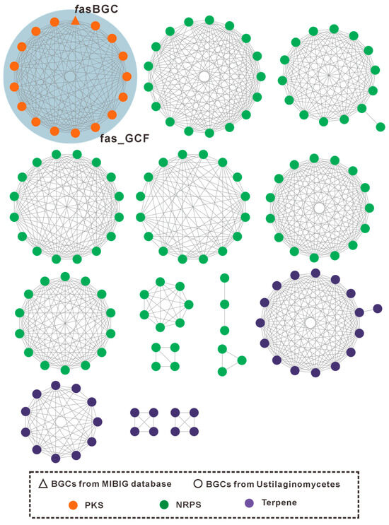
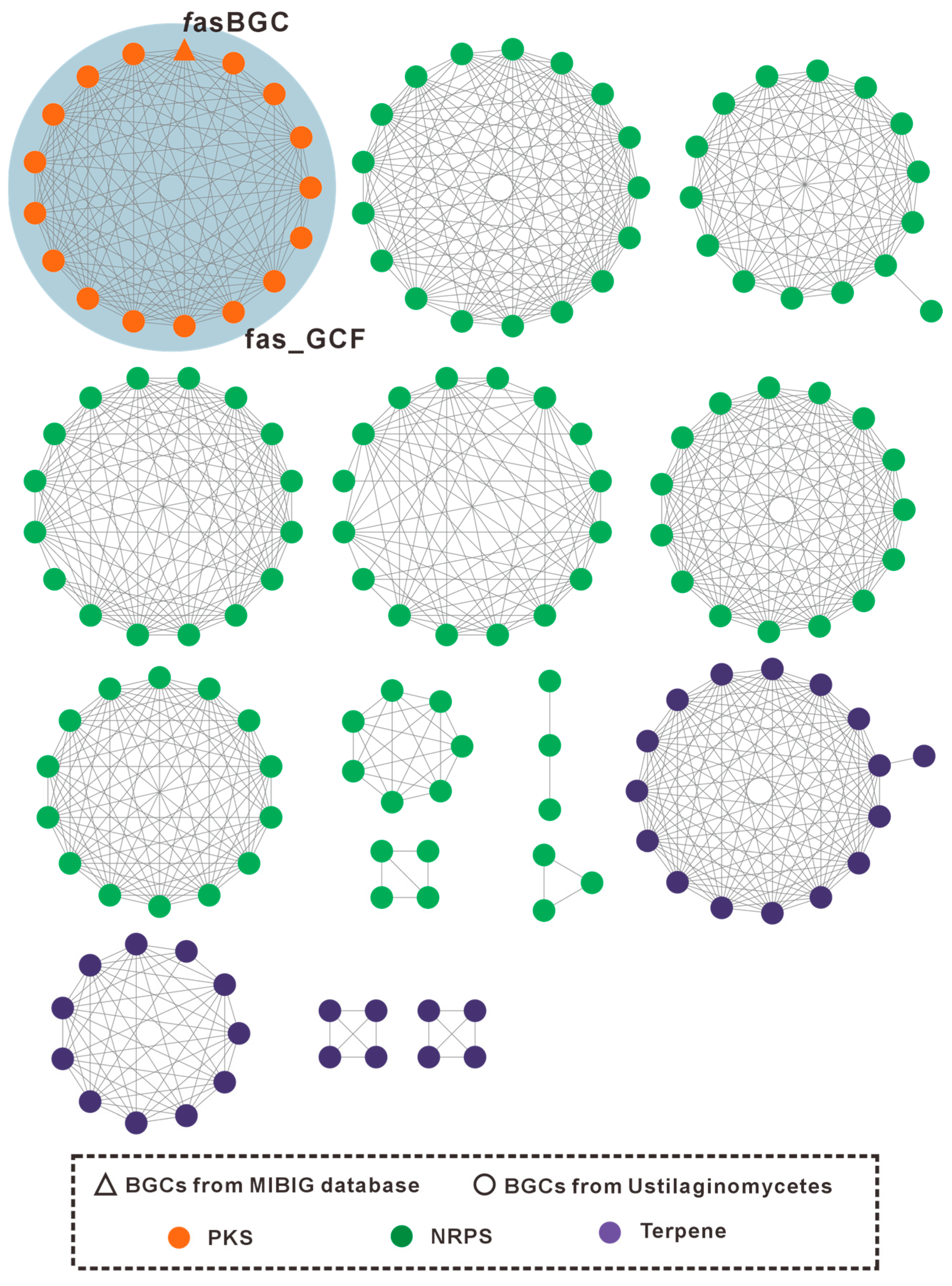
To comprehensively characterize BGCs within the family Ustilaginaceae, antiSMASH 7.0 was employed to predict BGCs across 16 genomes. A total of 181 predicted BGCs were identified (Table S1), distributed relatively evenly among species, with an average of approximately 11–12 BGCs per genome (Figure 1). These findings indicate that Ustilaginaceae species possess substantial biosynthetic potential.
Figure 1.
GCF network of Ustilaginaceae BGCs. Triangles and circles represent reference BGCs from the MIBiG database and BGCs identified from the 16 analyzed Ustilaginaceae genomes, respectively. Distinct colors indicate different BGC classes: green for NRPS, purple for terpene synthases, and orange for PKS.
To further elucidate the characteristics of these BGCs, gene cluster family (GCF) network analysis was performed using BiG-SCAPE. Based on the similarity of Pfam domain compositions within predicted biosynthetic enzymes, BiG-SCAPE classified 181 BGCs derived from the 16 Ustilaginaceae genomes and 1035 reference BGCs from MIBiG 2.1 into 15 GCFs and 1053 singleton clusters (Figure 1, isolated nodes omitted; Figure S1). Within the GCF network, BGCs of distinct chemical classes formed separate subnetworks, including 1 GCF consisting entirely of type I PKS clusters, 10 GCFs comprising exclusively NRPS clusters, and 4 GCFs containing only terpene clusters.
Using antiSMASH and BiG-SCAPE, we first catalogued the overall BGCs composition across the studied genomes. With U. maydis serving as a reference, we then proceeded to examine specific secondary metabolite pathways with established or proposed roles in plant infection, regardless of their BGCs classification. This includes pathways associated with siderophores (NRPS-derived), melanin (PKS-derived), MELs, cellobiose lipids, and itaconic acid. The detailed characterization of these pathways is presented in the following sections.
3.2. Cellobiose-Lipids and Their Biosynthetic Pathways
Ustilagic acids are rare glycolipid biosurfactants composed of a cellobiose disaccharide core glycosidically linked to long-chain hydroxy fatty acids, specifically 15,16-dihydroxypalmitic acid (ustilagic acid A) or 2,15,16-trihydroxypalmitic acid (ustilagic acid B) [19,20,21]. The disaccharide moiety is further acylated with short-chain fatty acids (typically acetate or butyrate) at the 2′- and 6′-positions. Additionally, a novel analogue, ustilagic acid C, bearing distinct acylation patterns, has been characterized in U. maydis [22]. Ustilagic acid represents the first microbial glycolipid discovered with fibroside-like structural features and is regarded as a signature metabolite of the species U. maydis and the genus Ustilago.
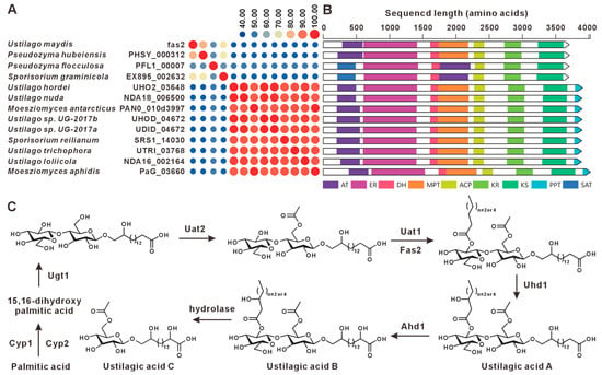
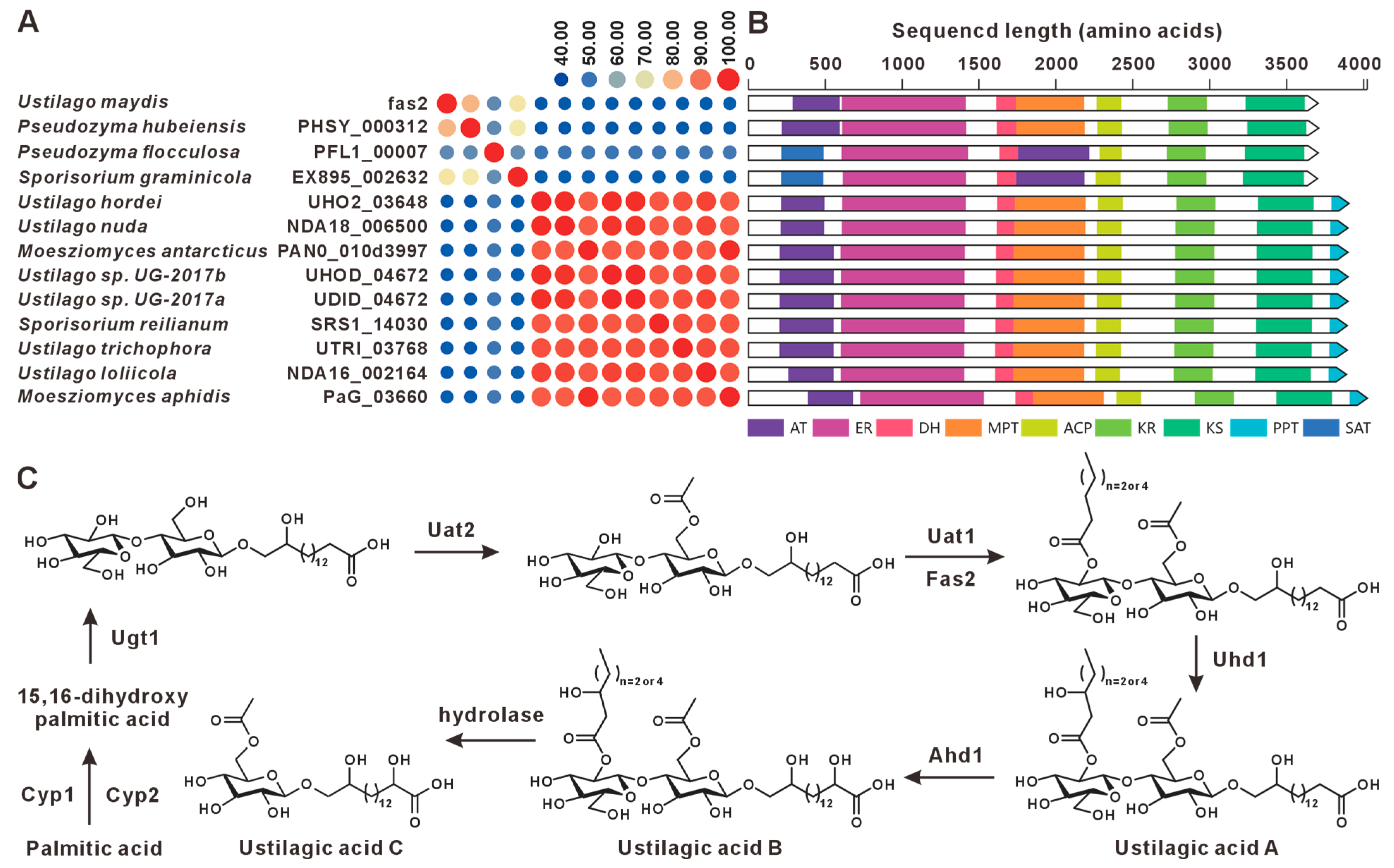
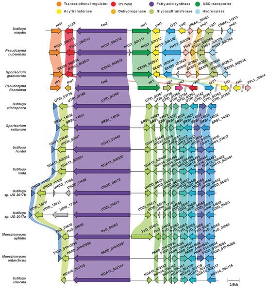
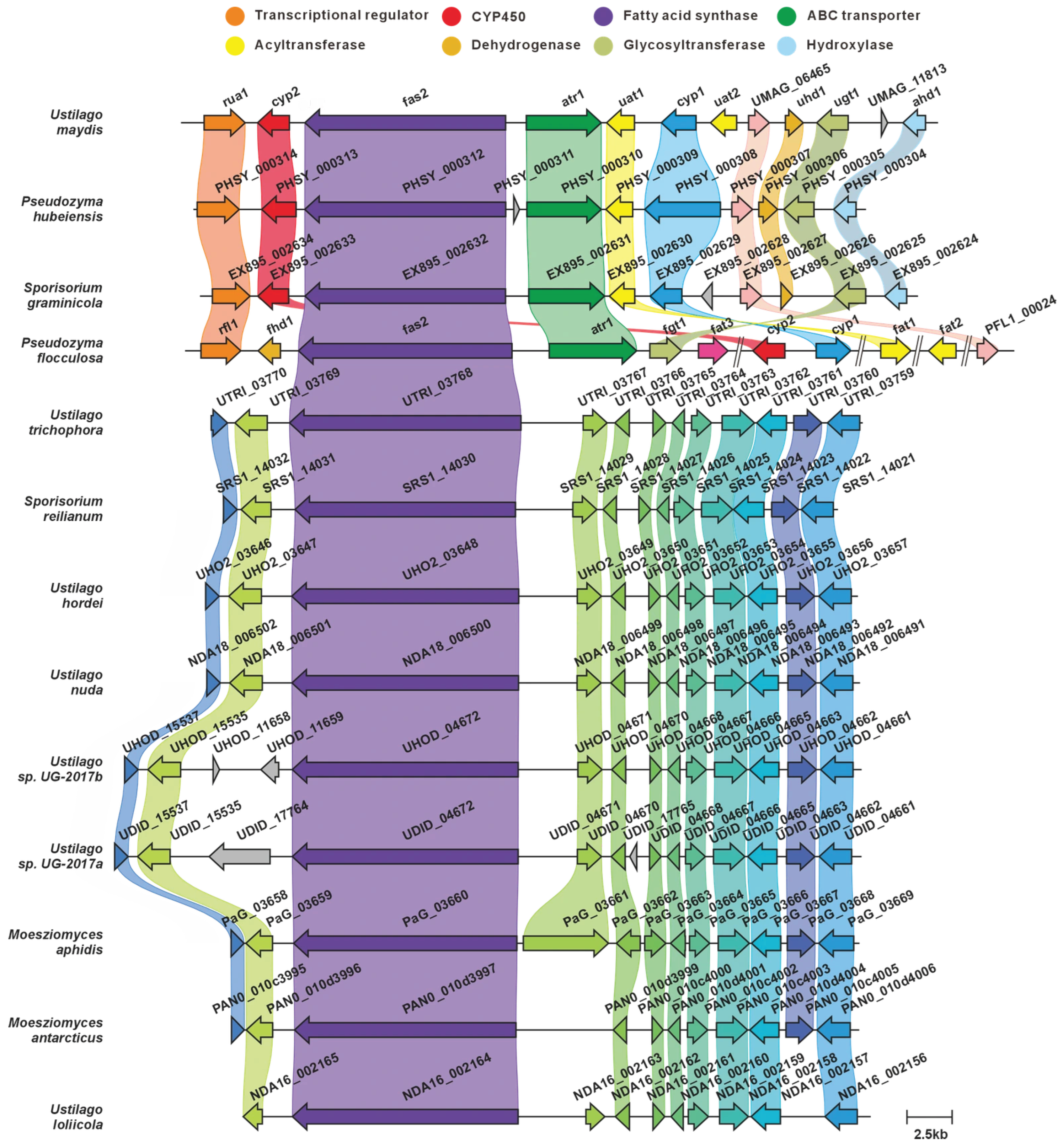
The BGC governing ustilagic acid production in U. maydis comprises nine genes, including one transcriptional regulator (rua1) and nine structural genes (cyp2, fas2, atr1, uat1, cyp1, uat2, uhd1, ugt1, ahd1) [23,24]. This study identified twelve fas2 paralogs across the analyzed genomes, with all fatty acid synthase (fas2) gene products exhibiting >44% pairwise sequence identity (Figure 2A, Table S2). Domain architecture analysis revealed highly conserved domain compositions across these sequences (Figure 2B). Comparative BGC analysis indicated that only the clusters from U. maydis, Pseudozyma hubeiensis, Sporisorium graminicola, and Pseudozyma flocculosa exhibited high synteny and sequence similarity. Unfortunately, we did not identify homologs of the nine structural genes in the vicinity of the fas2 orthologs in any other species examined. Notably, U. maydis and P. flocculosa are responsible for the biosynthesis of ustilagic acid and flocculosin, respectively (Figure 3) [24]. We therefore infer that these four species possess the genetic capacity for ustilagic acid and related glycolipid biosynthesis, whereas the BGCs in the remaining species likely participate in the biosynthesis of alternative fatty acid-derived metabolites. The biosynthetic pathway for ustilagic acid has been previously elucidated, illustrating the functional roles of cluster-encoded genes (Figure 2C) [23].
Figure 2.
Ustilagic acid compound biosynthesis. (A) Comparison of the amino acid sequence identity of fas2 and its homologues. (B) Domain comparison of fas2 and its homologues. The following abbreviations for protein domains are predicted by synthaser: AT, acyltransferase; ER, enoylreductase; DH, dehydratase; MPT, 4′-phosphopantetheinyl transferase; ACP, acyl carrier protein; KR, ketoreductase; KS, ketosynthase; PPT, phosphopantetheinyl transferase; SAT, starter unit acyltransferase. (C) The biosynthetic pathway for ustilagic acid.
Figure 3.
Comparison of the BGC for ustilagic acid and its similar BGCs: homologous genes are connected by a band of the same color.
3.3. MELs and Their Biosynthetic Pathway
MELs are well-characterized biosurfactants originally discovered in 1955 from the fermentation broth of U. maydis and related Ustilaginaceae, exhibiting significant biotechnological potential [25,26]. The molecular scaffold consists of a disaccharide backbone comprising mannose and erythritol, with the C2 and C3 hydroxyl groups of the mannose moiety esterified to short- and medium-chain fatty acids (typically C2–C16) [27]. Based on the acetylation degree at these positions and their differential mobility in thin-layer chromatography, MELs are classified into four structural variants: MEL-A (diacetylated, >70% of total MELs), MEL-B (monoacetylated at C6), MEL-C (monoacetylated at C4), and MEL-D (non-acetylated) [28]. A novel tri-acylated variant termed MEL-E was identified in S. reilianum, in which an additional short-chain fatty acid (C6–C8) replaces the acetyl groups at C4 or C6 of the mannose moiety, resulting in altered physicochemical properties and highlighting the structural diversity of MELs [29].
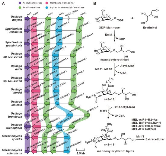
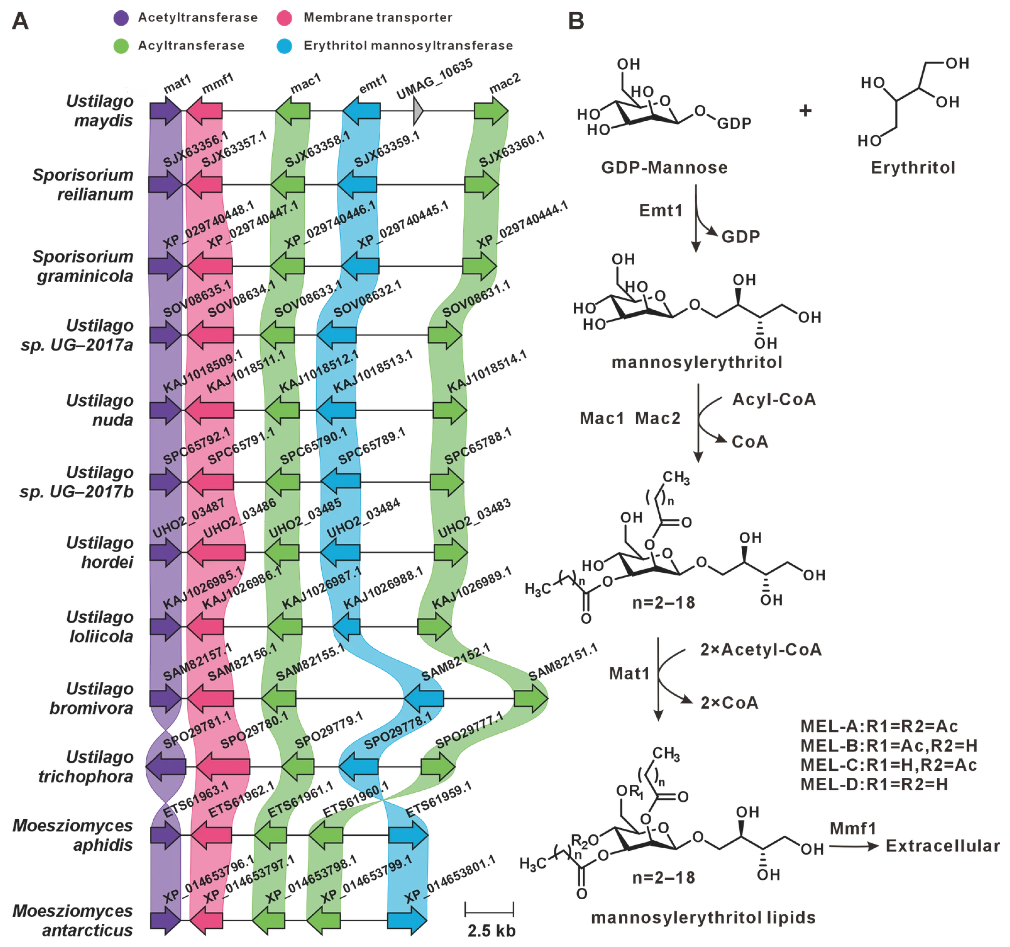
Previous studies have characterized the MELs BGC in U. maydis, which comprises five core genes [30]. In this study, comparative genomic mining across 16 Ustilaginaceae genomes, including U. maydis, revealed that this BGC is widely conserved among 12 species (Figure 4A). The biosynthetic pathway of MELs and the functional roles of individual cluster-encoded genes have been characterized [31,32,33]. We infer that these 12 species possess the genetic capacity for MEL production (Figure 4B).
Figure 4.
MEL biosynthesis. (A) Comparison of the BGC for MELs and its similar BGCs: homologous genes are connected by a band of the same color. (B) The biosynthetic pathway for MELs.
3.4. Siderophores and Non-Ribosomal Peptides
Siderophores constitute a class of low-molecular-weight compounds produced by most microorganisms, exhibiting high affinity for ferric iron acquisition and storage [34]. Assessment of siderophore function in U. maydis pathogenicity has demonstrated that high-affinity iron uptake systems are indispensable for virulence [35]. Ferrichrome and ferrichrome A are two siderophore derivatives produced by U. maydis, each consisting of a cyclic hexapeptide comprising three δ-N-acyl-N-hydroxyornithine residues and three additional amino acids (glycine, serine, and alanine) [36,37]. Structural diversity among siderophores arises from modifications to the ornithine side chains and variations in the three non-ornithine amino acids within the cyclic peptide backbone [38].
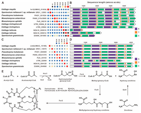
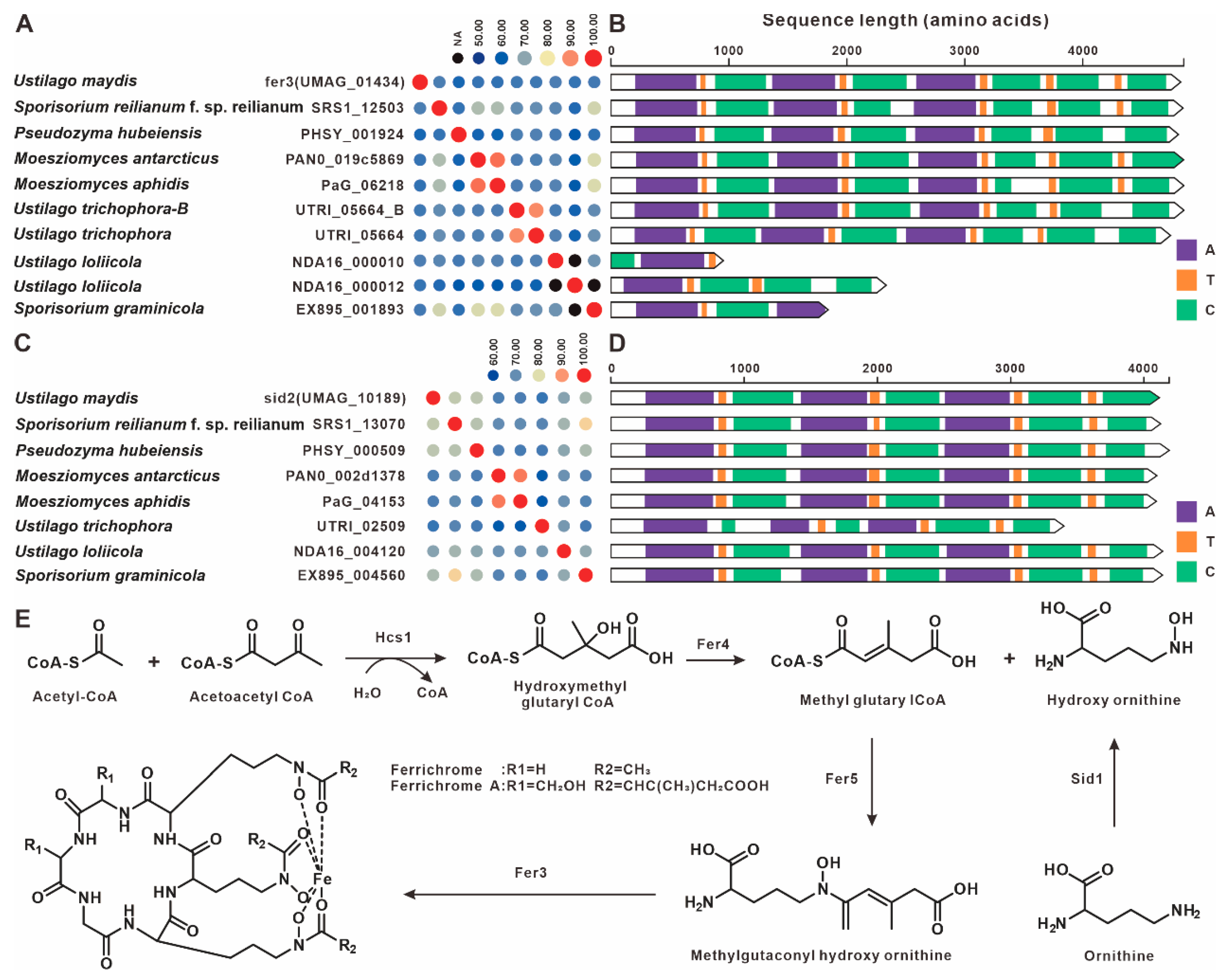
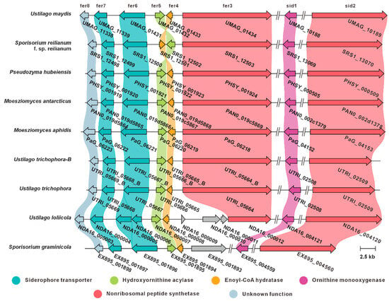
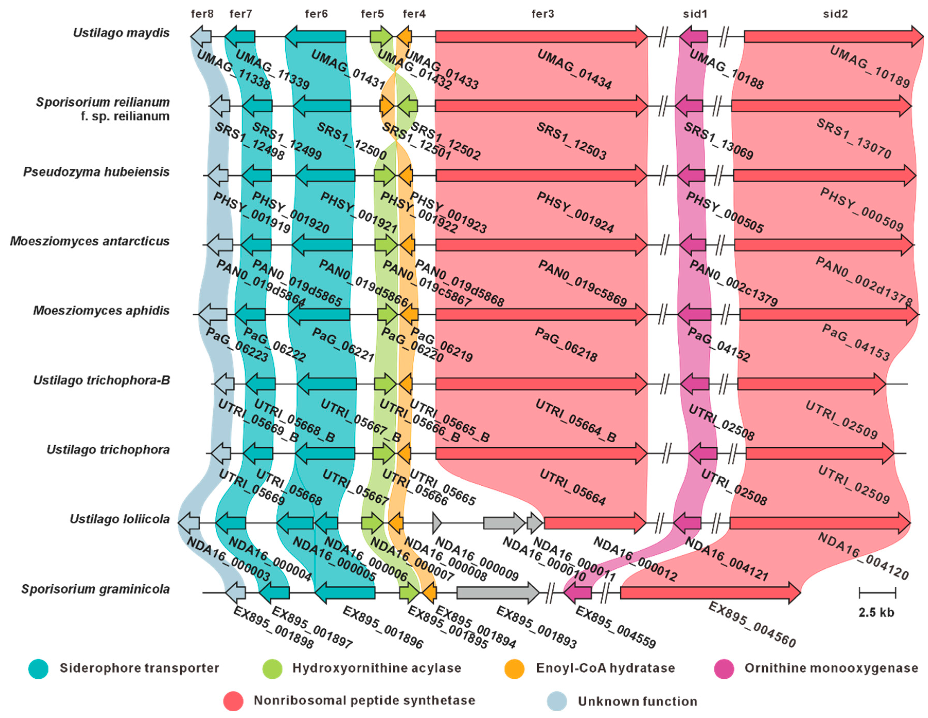
The siderophore BGC in U. maydis comprises six core genes encoding: the NRPS Fer3, enoyl-CoA hydratase Fer4, hydroxyornithine acyltransferase Fer5, two transmembrane transporters (Fer6 and Fer7), and a hypothetical protein Fer8 [39]. In addition, hcs1 in U. maydis catalyzes the formation of hydroxymethyl glutaryl-CoA from acetyl-CoA and acetoacetyl-CoA [39]. In this study, comparative genomic analysis identified eight fer3 paralogs and seven sid2 paralogs across the analyzed genomes. All fer3 gene products exhibited >59% pairwise sequence identity (Figure 5A, Table S3), whereas all sid2 homologs demonstrated >63% identity (Figure 5C, Table S4). In U. maydis, sid2 encodes a multidomain NRPS that acts in concert with the Sid1 L-ornithine-N5-oxygenase to convert precursor amino acids, such as L-ornithine, into the siderophore ferrichrome. Domain architecture analysis revealed highly conserved domain compositions among these paralogs across species (Figure 5B,D). Comparative BGC analysis further indicated that the siderophore BGCs from eight species, including U. maydis, share high synteny and sequence similarity (Figure 6). These results suggest that these species possess the genetic capacity for siderophore biosynthesis. The proposed biosynthetic pathway is illustrated in Figure 5E.
Figure 5.
Siderophore compound biosynthesis. (A) Comparison of the amino acid sequence identity of fer3 and its homologues. (B) Domain comparison of fer3 and its homologues. (C) Comparison of the amino acid sequence identity of sid2 and its homologues. The following abbreviations for protein domains are predicted by Synthaser: A, acyltransferase; T, thiolation domain; C, condensation domain. (D) Domain comparison of sid2 and its homologues. (E) The biosynthetic pathway for Siderophores.
Figure 6.
Comparison of the BGC for Siderophores and its similar BGCs. The fer3 and the sid2 are located on different chromosomes in each species.
3.5. Itaconic Acid and Its Biosynthetic Pathway
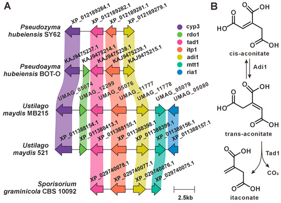
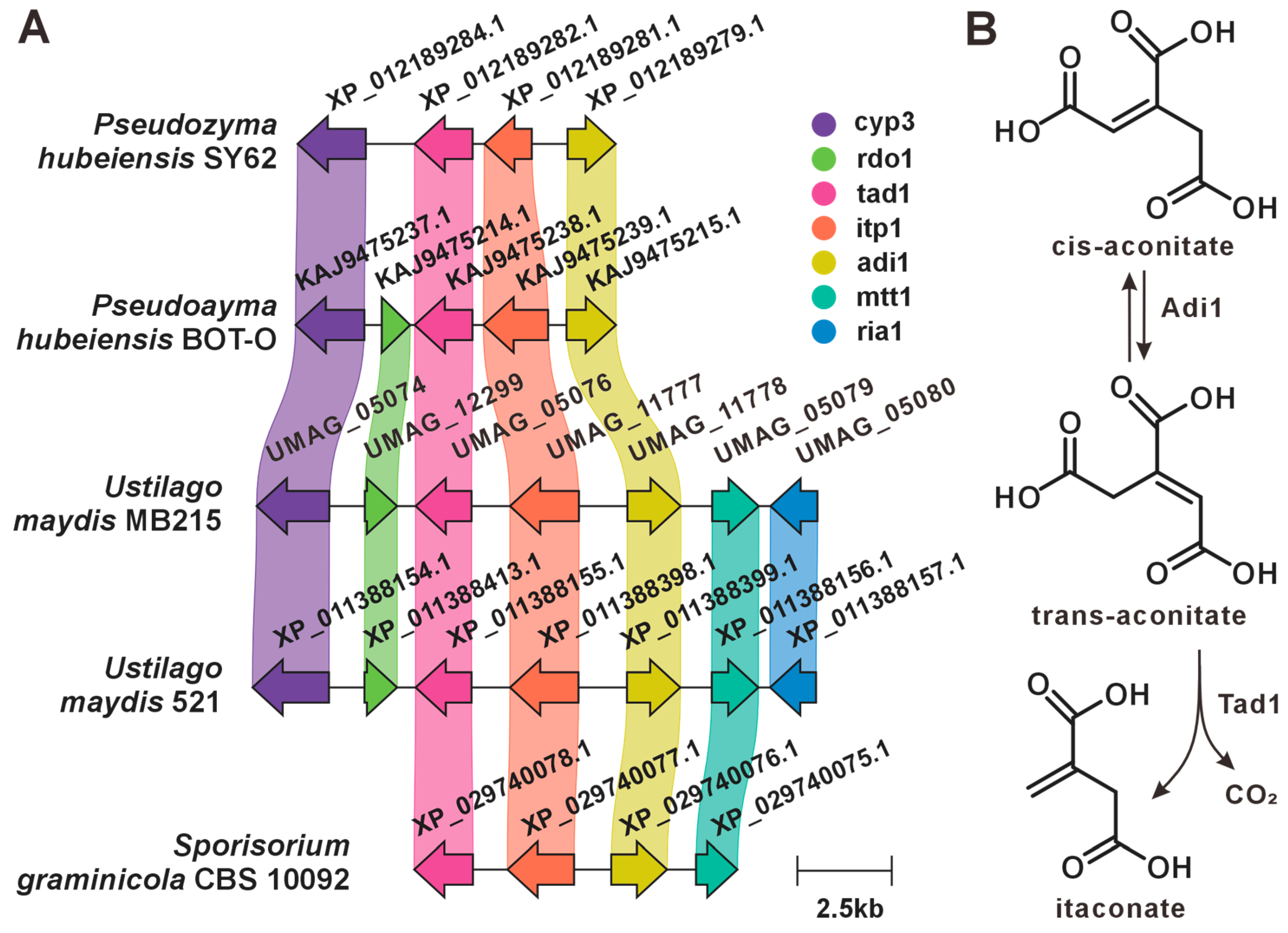
Itaconic acid is an industrially relevant organic compound with diverse biotechnological applications. Historically, itaconic acid was obtained by pyrolytic distillation of citric acid, but current industrial production relies on fermentation by Aspergillus terreus [40]. Genomic analysis indicates that U. maydis lacks homologs of the A. terreus cis-aconitate decarboxylase CadA (a PrpD family protein), suggesting that U. maydis employs an alternative biosynthetic route for itaconate production [41]. Previous studies identified a seven-gene cluster responsible for itaconic acid biosynthesis in the U. maydis strain MB215. The gene product UMAG_05079 (Mtt1) functions as a mitochondrial transporter, actively exporting cis-aconitate from mitochondria to the cytoplasm [42]. Subsequently, cis-aconitate is converted to trans-aconitate by the major facilitator superfamily transporter UMAG_11778 (Adi1), followed by decarboxylation to itaconic acid catalyzed by UMAG_05076 (Tad1). Finally, itaconic acid is secreted across the plasma membrane via the transporter UMAG_11777 (Itp1). This BGC additionally encodes UMAG_05074 (Cyp3), which likely catalyzes itaconic acid modification [product unspecified], the functionally uncharacterized protein UMAG_12299 (Rdo1), and the putative transcriptional regulator UMAG_05080 (Ria1) [43]. In this study, we identified highly similar itaconic acid BGCs in four additional species, inferring their genetic capacity for itaconic acid biosynthesis (Figure 7).
Figure 7.
Itaconic acid biosynthesis. (A) Comparison of the BGC for itaconic acid and its similar BGCs: homologous genes are connected by a band of the same color. (B) The biosynthetic pathway for itaconic acid.
3.6. Melanin and PKS
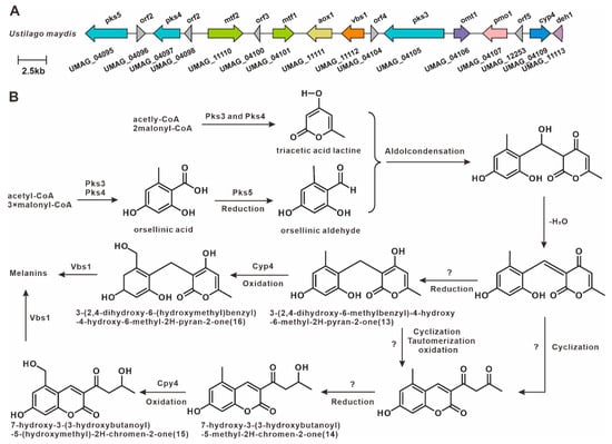
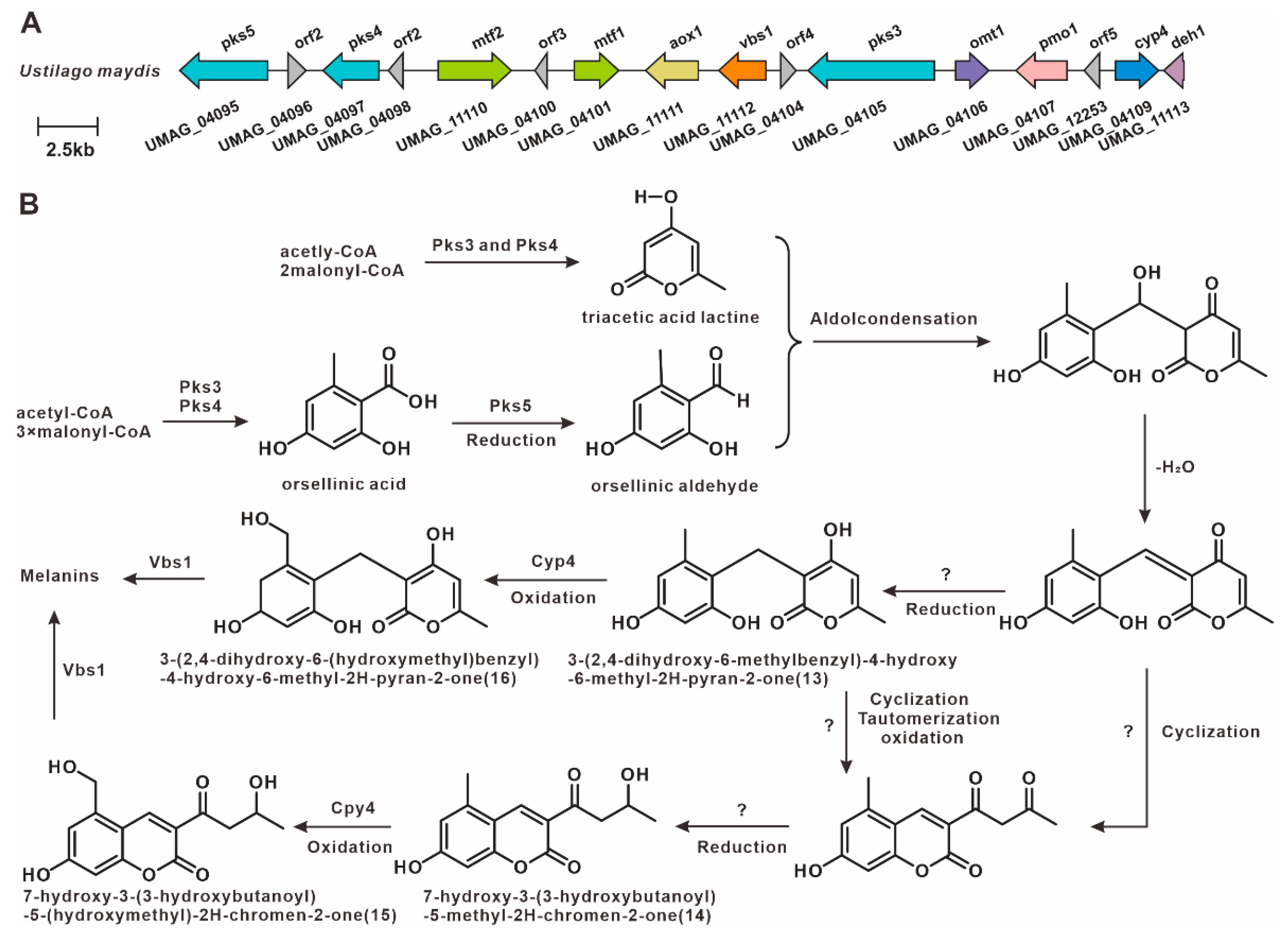
Previous studies have demonstrated that spore formation in plant tumor-like tissues is associated with melanin biosynthesis [44]. In U. maydis, a melanin-associated PKS pathway has been identified. These PKS enzymes synthesize orsellinic acid (OA), triacetic acid lactone (TAL), and related derivatives, which subsequently undergo cyclization and modification to form coumarins and pyran-2-ones [45], ultimately forming this polymer (Figure 8). This study showed that the homologs of the key melanin synthesis genes pks3, pks4, and pks5 were not found in 15 other genomes outside of U. maydis, which suggests that this PKS gene cluster is specific to strain 521 and may be the result of horizontal gene transfer.
Figure 8.
Melanin-associated biosynthesis in U. maydis strain 521 [45]. (A) Strain-specific PKS gene cluster. (B) Proposed melanin biosynthetic pathway.
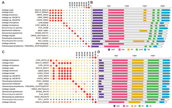
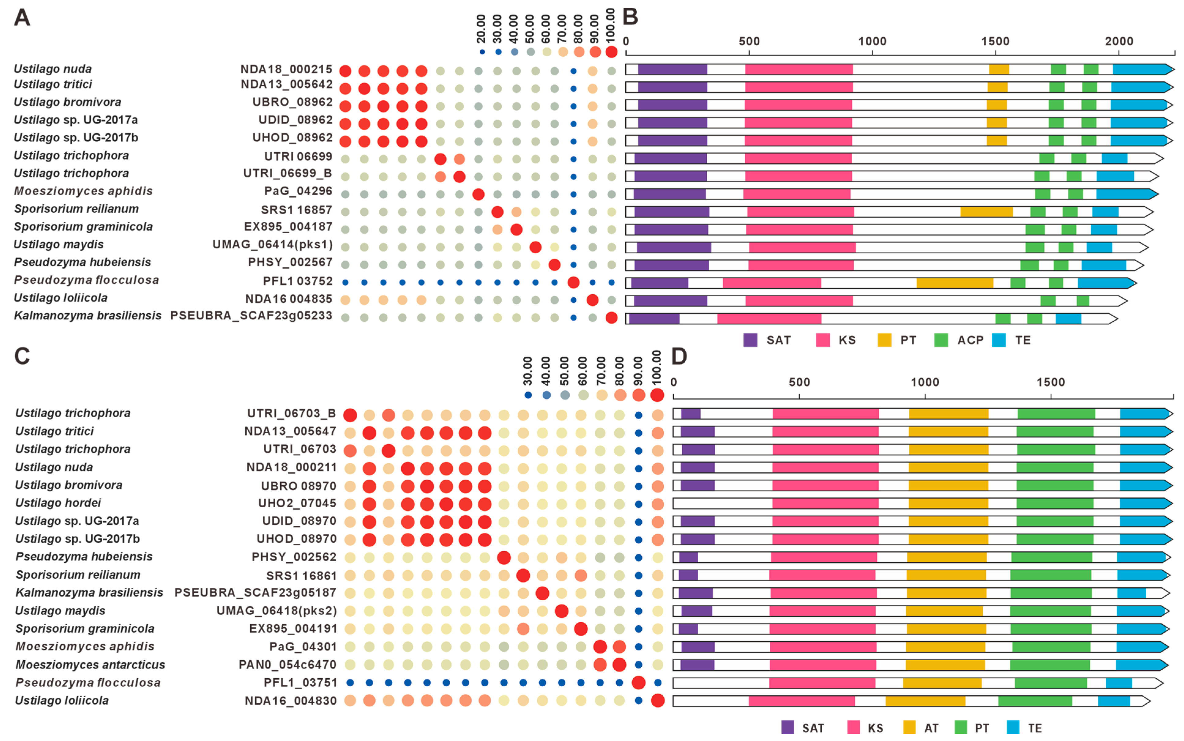
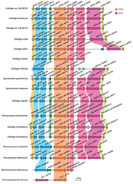
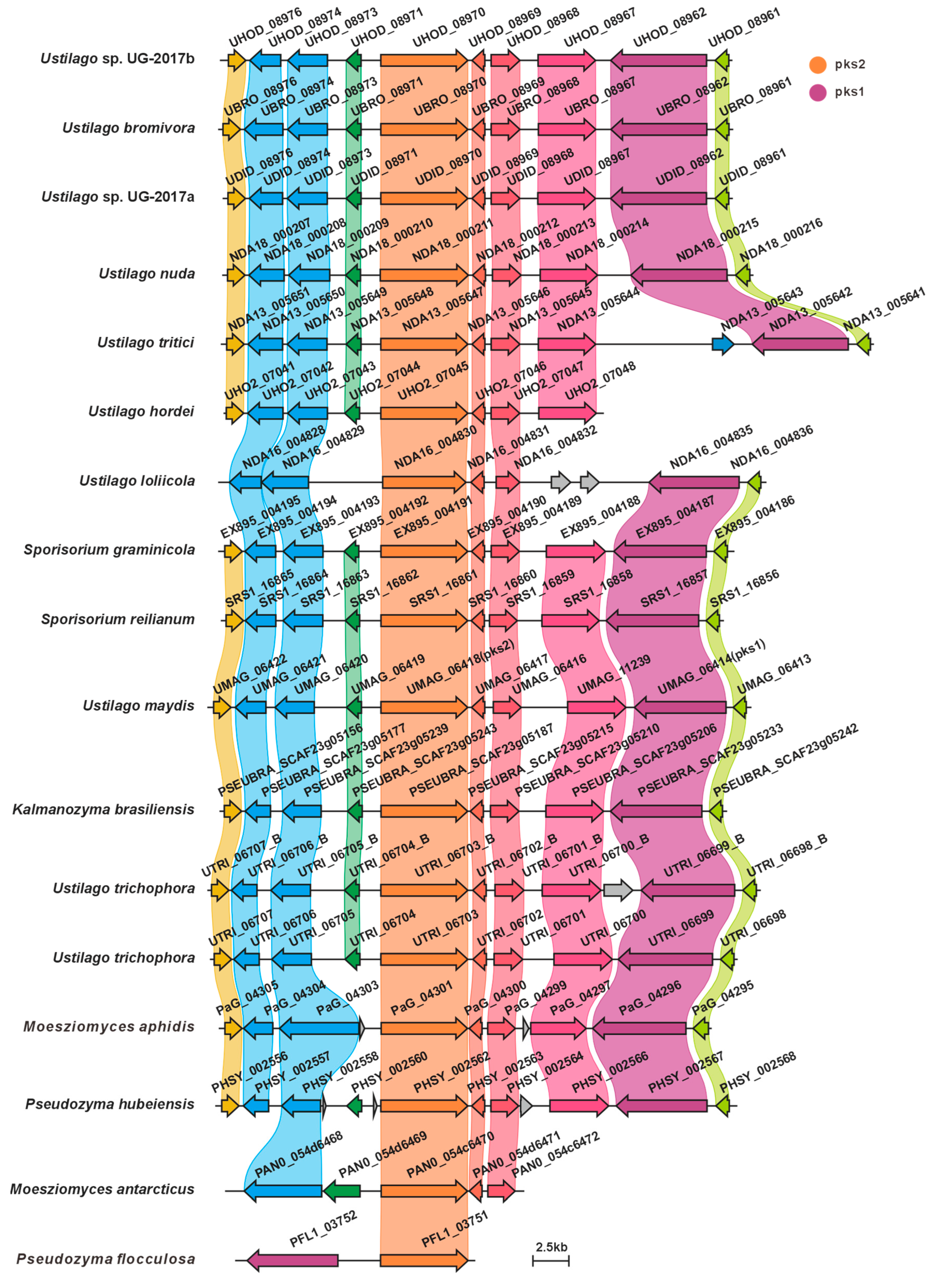
UMAG_06414 (pks1) and UMAG_06418 (pks2) are polyketide synthase genes involved in melanin biosynthesis in U. maydis, and they are located adjacent to each other in the genome. Deletion of pks1 alone is sufficient to completely block melanin biosynthesis, indicating that UMAG_06418 cannot compensate for its function, suggesting that the two may together constitute a functionally complete polyketide synthase system. In this study, we identified homologous gene clusters in 15 species other than U. maydis. Among them, 13 species harbor homologs of pks1, and all 15 species harbor homologs of pks2. Sequence similarity comparisons revealed that, with the exception of P. flocculosa, the pks1 homologs exhibited high sequence similarity (>50%, Figure 9A, Table S5). Similarly, the pks2 homologs, also with the exception of P. flocculosa, showed high sequence similarity (>58.4%, Figure 9C, Table S6). Domain architecture analysis revealed that the domain organizations of these PKSs are highly conserved (Figure 9B,D). Comparative analysis of the BGCs indicated that most of the 16 species display high synteny and sequence similarity. Among them, P. flocculosa showed the lowest similarity compared to the others. In U. hordei and M. antarcticus, only pks2 homologs were present in the gene clusters, suggesting the presence of a putative melanin BGC in these species (Figure 10).
Figure 9.
Comparative analysis of pks1 and pks2 with their homologues. (A) Comparison of the amino acid sequence identity of pks1 and its homologues. (B) Domain comparison of pks1 and its homologues. (C) Comparison of the amino acid sequence identity of pks2 and its homologues. (D) Domain comparison of pks2 and its homologues. The following abbreviations for protein domains are predicted by synthaser: SAT, starter unit acyltransferase; KS, ketosynthase; AT, acyltransferase; PT, pyranone formation (in PKS); ACP, acyl carrier protein; TE, thioesterase.
Figure 10.
Comparison of putative melanin BGCs from 16 Ustilaginaceae species.
4. Discussion
This study systematically analyzed the genomes of 16 species within the family Ustilaginaceae, with particular emphasis on their BGCs. Using antiSMASH 7.0, a total of 181 putative BGCs were identified, averaging approximately 11 BGCs per genome, thereby providing a comprehensive resource for investigating the biosynthetic potential of Ustilaginaceae. BiG-SCAPE network analysis classified these BGCs into 15 gene cluster families (GCFs), revealing distinct distribution patterns of NRPS, PKS, and terpene synthase clusters across the analyzed genomes. Notably, the majority of GCFs comprised BGCs of a single chemical class, suggesting that distinct BGC types may have evolved along relatively independent trajectories. Additionally, network analysis identified potential functional associations among BGCs, offering a framework for subsequent functional characterization and metabolic pathway reconstruction.
The distribution of ustilagic acid BGCs across Ustilaginaceae species was comprehensively analyzed in this study. We identified four species with conserved ustilagic acid BGCs, expanding our understanding of the biosynthetic diversity of this characteristic glycolipid. Previous work had only characterized ustilagic acid production in U. maydis and flocculosin in P. flocculosa [23,24]; our comparative analysis reveals that this BGC is also conserved in S. graminicola and P. hubeiensis, suggesting a broader ecological role among related species. MELs, well-characterized biosurfactants produced by Ustilaginaceae, are known to suppress plant immunity in U. maydis infection; their BGCs were widely distributed across 12 species, suggesting a conserved role in host manipulation. This high level of conservation aligns with the essential function of MELs in pathogenic development reported for U. maydis [31], and indicates that most Ustilaginaceae species likely retain this virulence trait. Siderophores, critical for microbial iron acquisition and virulence, were found to possess highly conserved BGCs across eight species, indicating their essential role in competing for iron in the plant apoplast during infection. This finding underscores the evolutionary conservation of siderophore biosynthesis and its contribution to pathogenicity, consistent with earlier studies [35]. Itaconic acid, an industrially relevant organic compound, has been extensively studied in U. maydis, where it contributes to virulence by inhibiting host aconitase. We observed that the itaconic acid BGC in U. maydis MB215 exhibits high synteny with BGCs from four additional species, suggesting conserved pathogenic roles and their potential utility as novel production strains. This finding expands upon the known itaconic acid pathway [41,43] and indicates that it is not unique to U. maydis but is shared by several other Ustilaginaceae. Melanin-associated PKS pathways, implicated in spore formation within plant tumor-like tissues, have been characterized in U. maydis 521. Melanin enhances fungal resistance to plant defense responses and oxidative stress. The strain-specific distribution of these PKS BGCs offers insights into the evolutionary diversification of melanin biosynthesis mechanisms. Additionally, this study identified homologs of the melanin biosynthesis genes pks1 and pks2 in the remaining 15 Ustilaginaceae species. Both genes are involved in stress tolerance and virulence in U. maydis [45]. Most of these homologs exhibited high sequence similarity, with P. flocculosa showing the lowest similarity compared to the others. The widespread presence of pks1/pks2 homologs suggests that the core melanin pathway is ancestral in Ustilaginaceae, in contrast to the strain-specific PKS cluster (pks3–pks5) found only in U. maydis 521. In U. hordei and M. antarcticus, only pks2 homologs were present in the gene clusters, suggesting that these clusters may have been assembled at the edges of contigs during genome assembly.
Collectively, these results directly address our primary objective: to use U. maydis as a reference for mapping the distribution of pathogenicity-associated BGCs across Ustilaginaceae. We show that BGCs for MELs, siderophores, itaconic acid, and the core melanin pathway (pks1/pks2) are widely conserved among most species, whereas a distinct melanin BGC is unique to U. maydis 521. These findings are consistent with previous functional studies in U. maydis [23,31,35,41,45] and extend them by revealing the phylogenetic distribution of these pathways across the family.
Despite these advances, several limitations should be acknowledged. First, the species selection was constrained by the availability of public genome data at the time of analysis; as more genomes of phytopathogenic Ustilaginaceae become available, future studies should incorporate them to further validate the pathogenicity-associated BGCs identified here. Second, our predictions are purely computational; experimental validation through gene knockouts or heterologous expression is required to confirm the functional roles of the identified BGCs. Third, the presence of a conserved but uncharacterized PKS cluster in 15 species highlights a gap in our understanding of its biological function.
Future research should prioritize the experimental characterization of this uncharacterized PKS cluster using CRISPR-Cas9-mediated genome editing and heterologous expression systems. Such approaches will enable mechanistic dissection of BGC-encoded metabolic pathways and their regulatory networks, ultimately linking genotype to phenotype in plant infection.
In summary, this study provides the first systematic comparative genomic analysis of BGCs across 16 Ustilaginaceae species. Our findings reveal both conserved and species-specific biosynthetic potentials, advancing our understanding of the evolutionary and ecological adaptations within this family. Furthermore, this work establishes a foundation for the discovery and engineering of novel bioactive natural products, as well as for the development of targeted antimicrobial strategies.
5. Conclusions
The dissemination of plant-pathogenic fungi poses a severe threat to global agricultural production, with their virulence intimately linked to secondary metabolite biosynthesis. This study employed comparative genomic and bioinformatic approaches to systematically characterize the distribution and architecture of BGCs across 16 species within the family Ustilaginaceae. Our findings reveal that BGCs governing the production of ustilagic acid, MELs, and siderophores are widely conserved among Ustilaginaceae. Notably, a strain-specific PKS gene cluster associated with melanin biosynthesis was exclusively identified in the U. maydis strain 521. Meanwhile, putative melanin biosynthetic gene clusters were identified in the remaining 15 species of the Ustilaginaceae. This research provides a framework for elucidating pathogenic mechanisms and metabolic regulation in Ustilaginaceae, establishing a theoretical foundation for future synthetic biology applications in antimicrobial development and high-value natural product engineering.
Supplementary Materials
The following supporting information can be downloaded at: https://www.mdpi.com/article/10.3390/jof12050319/s1, Figure S1: GCF network of Ustilaginaceae BGCs. Triangles and circles represent reference BGCs from the MIBiG database and BGCs identified from the 16 analyzed Ustilaginaceae genomes, respectively. Distinct colors indicate different BGC classes: green for NRPS, purple for terpene synthases, and orange for PKS; Table S1: Ustilaginaceae species and genome information used in this study; Table S2: List of predicted biosynthetic gene clusters (BGCs) in Ustilaginaceae strains; Table S3: Sequence identity matrix of fas2 and its orthologs; Table S4: Sequence identity matrix of fer3 and its orthologs; Table S5: Sequence identity matrix of sid2 and its orthologs; Table S6: Sequence identity matrix of pks1 and its orthologs; Table S7: Sequence identity matrix of pks2 and its orthologs.
Author Contributions
Conceptualization, C.L., Z.W. and Y.L.; methodology, C.L., Y.L. and N.Z.; software, C.L., J.Z. and Y.L.; validation, C.L., Y.L. and N.Z.; formal analysis, K.V., L.S., H.W. and M.L.; investigation, C.L., Y.L. and J.Z.; resources, J.Z.; data curation, Z.W.; writing—original draft preparation, C.L., Z.W. and Y.L.; writing—review and editing, M.L. and J.Q.; visualization, C.L., Z.W. and Y.L.; supervision, M.L. and J.Q.; project administration, J.Q.; funding acquisition, M.L. All authors have read and agreed to the published version of the manuscript.
Funding
This research was funded by the National Key Research and Development Program (2021YFD1600401) and Key Core Technology Research and Development in Shaanxi Province Agriculture (2025NYGG010). The APC received no external funding.
Institutional Review Board Statement
Not applicable.
Data Availability Statement
All summary data were provided in the results and Supplemental Files.
Acknowledgments
We thank the High-Performance Computing platform of Northwest A&F University for providing computing resources.
Conflicts of Interest
The authors declare no conflicts of interest.
References
- Gai, Y.; Wang, H. Plant Disease: A Growing Threat to Global Food Security. Agronomy 2024, 14, 1615. [Google Scholar] [CrossRef] [Scilit]
- McDonald, B.A.; Stukenbrock, E.H. Rapid emergence of pathogens in agro-ecosystems: Global threats to agricultural sustainability and food security. Philos. Trans. R. Soc. B Biol. Sci. 2016, 371, 20160026. [Google Scholar] [CrossRef] [Scilit]
- Avery, S.V.; Singleton, I.; Magan, N.; Goldman, G.H. The fungal threat to global food security. Fungal Biol. 2019, 123, 555–557. [Google Scholar] [CrossRef] [Scilit]
- Williamson-Benavides, B.A.; Dhingra, A. Understanding Root Rot Disease in Agricultural Crops. Horticulturae 2021, 7, 33. [Google Scholar] [CrossRef] [Scilit]
- Gonzalez, M.; Pujol, M.; Metraux, J.-P.; Gonzalez-Garcia, V.; Bolton, M.D.; Borrás-Hidalgo, O. Tobacco leaf spot and root rot caused by Rhizoctonia solani Kühn. Mol. Plant Pathol. 2011, 12, 209–216. [Google Scholar] [CrossRef] [Scilit] [PubMed]
- Doehlemann, G.; Ökmen, B.; Zhu, W.; Sharon, A. Plant Pathogenic Fungi. In The Fungal Kingdom; Wiley: Hoboken, NJ, USA, 2017; pp. 701–726. [Google Scholar]
- Lin, C.; Feng, X.-L.; Liu, Y.; Li, Z.-C.; Li, X.-Z.; Qi, J. Bioinformatic Analysis of Secondary Metabolite Biosynthetic Potential in Pathogenic Fusarium. J. Fungi 2023, 9, 850. [Google Scholar] [CrossRef] [Scilit] [PubMed]
- Fisher, M.C.; Henk, D.A.; Briggs, C.J.; Brownstein, J.S.; Madoff, L.C.; McCraw, S.L.; Gurr, S.J. Emerging fungal threats to animal, plant and ecosystem health. Nature 2012, 484, 186–194. [Google Scholar] [CrossRef] [Scilit]
- Faulkner, C.; Robatzek, S. Plants and pathogens: Putting infection strategies and defence mechanisms on the map. Curr. Opin. Plant Biol. 2012, 15, 699–707. [Google Scholar] [CrossRef] [Scilit]
- Djamei, A. Ustilago maydis. Curr. Biol. 2023, 33, R458–R460. [Google Scholar] [CrossRef] [Scilit]
- Ferris, A.C.; Walbot, V. Understanding Ustilago maydis Infection of Multiple Maize Organs. J. Fungi 2021, 7, 8. [Google Scholar] [CrossRef] [Scilit]
- Yu, C.; Qi, J.; Han, H.; Wang, P.; Liu, C. Progress in pathogenesis research of Ustilago maydis, and the metabolites involved along with their biosynthesis. Mol. Plant Pathol. 2023, 24, 495–509. [Google Scholar] [CrossRef] [Scilit]
- Zou, K.; Li, Y.; Zhang, W.; Jia, Y.; Wang, Y.; Ma, Y.; Lv, X.; Xuan, Y.; Du, W. Early infection response of fungal biotroph Ustilago maydis in maize. Front. Plant Sci. 2022, 13, 970897. [Google Scholar] [CrossRef] [Scilit]
- Kretschmer, M.; Damoo, D.; Sun, S.; Lee, C.W.J.; Croll, D.; Brumer, H.; Kronstad, J. Organic acids and glucose prime late-stage fungal biotrophy in maize. Science 2022, 376, 1187–1191. [Google Scholar] [CrossRef] [Scilit] [PubMed]
- Blin, K.; Shaw, S.; Augustijn, H.E.; Reitz, Z.L.; Biermann, F.; Alanjary, M.; Fetter, A.; Terlouw, B.R.; Metcalf, W.W.; Helfrich, E.J.N.; et al. antiSMASH 7.0: New and improved predictions for detection, regulation, chemical structures and visualisation. Nucleic Acids Res. 2023, 51, W46–W50. [Google Scholar] [CrossRef] [Scilit] [PubMed]
- Navarro-Muñoz, J.C.; Selem-Mojica, N.; Mullowney, M.W.; Kautsar, S.A.; Tryon, J.H.; Parkinson, E.I.; De Los Santos, E.L.C.; Yeong, M.; Cruz-Morales, P.; Abubucker, S.; et al. A computational framework to explore large-scale biosynthetic diversity. Nat. Chem. Biol. 2020, 16, 60–68. [Google Scholar] [CrossRef] [Scilit]
- Gilchrist, C.L.M.; Chooi, Y.-H. Synthaser: A CD-Search enabled Python toolkit for analysing domain architecture of fungal secondary metabolite megasynth(et)ases. Fungal Biol. Biotechnol. 2021, 8, 13. [Google Scholar] [CrossRef] [Scilit] [PubMed]
- Gilchrist, C.L.M.; Chooi, Y.-H. Clinker & clustermap.js: Automatic generation of gene cluster comparison figures. Bioinformatics 2021, 37, 2473–2475. [Google Scholar] [CrossRef] [Scilit]
- Haskins, R.H. Biochemistry of the Ustilaginales: I. Preliminary cultural studies of Ustilago zeae. Can. J. Res. 1950, 28, 213–223. [Google Scholar] [CrossRef] [Scilit]
- Lemieux, R.U. The biochemistry of the ustilaginales: III. The degradation products and proof of the chemical heterogeneity of ustilagic acid. Can. J. Chem. 1951, 29, 415–425. [Google Scholar] [CrossRef] [Scilit] [PubMed]
- Lemieux, R.U.; Charanduk, R. Biochemistry of the Ustilaginales: VI: The Acyl Groups of Ustilagic Acid. Can. J. Chem. 1951, 29, 759–766. [Google Scholar] [CrossRef] [Scilit]
- Yang, X.-L.; Awakawa, T.; Wakimoto, T.; Abe, I. Induced production of the novel glycolipid ustilagic acid C in the plant pathogen Ustilago maydis. Tetrahedron Lett. 2013, 54, 3655–3657. [Google Scholar] [CrossRef] [Scilit]
- Teichmann, B.; Linne, U.; Hewald, S.; Marahiel, M.A.; Bölker, M. A biosynthetic gene cluster for a secreted cellobiose lipid with antifungal activity from Ustilago maydis. Mol. Microbiol. 2007, 66, 525–533. [Google Scholar] [CrossRef] [Scilit]
- Teichmann, B.; Labbé, C.; Lefebvre, F.; Bölker, M.; Linne, U.; Bélanger, R.R. Identification of a biosynthesis gene cluster for flocculosin a cellobiose lipid produced by the biocontrol agent Pseudozyma flocculosa. Mol. Microbiol. 2011, 79, 1483–1495. [Google Scholar] [CrossRef] [Scilit]
- Valkenburg, A.D.; Teke, G.M.; Pott, R.W.M.; van Rensburg, E. The fed-batch production of mannosylerythritol lipids by Ustilago maydis DSM 4500 from hydrophilic carbon sources. Bioprocess Biosyst. Eng. 2024, 47, 2043–2054. [Google Scholar] [CrossRef] [Scilit] [PubMed]
- Haskins, R.H.; Thorn, J.A.; Boothroyd, B. Biochemistry of the Ustilaginales: XI. Metabolic products of Ustilago zeae in submerged culture. Can. J. Microbiol. 1955, 1, 749–756. [Google Scholar] [CrossRef] [Scilit] [PubMed]
- Deinzer, H.-T.; Linne, U.; Xie, X.; Bölker, M.; Sandrock, B. Elucidation of substrate specificities of decorating enzymes involved in mannosylerythritol lipid production by cross-species complementation. Fungal Genet. Biol. 2019, 130, 91–97. [Google Scholar] [CrossRef] [Scilit]
- Rau, U.; Nguyen, L.A.; Schulz, S.; Wray, V.; Nimtz, M.; Roeper, H.; Koch, H.; Lang, S. Formation and analysis of mannosylerythritol lipids secreted by Pseudozyma aphidis. Appl. Microbiol. Biotechnol. 2005, 66, 551–559. [Google Scholar] [CrossRef] [Scilit] [PubMed]
- Tiefenbacher, J.; Linne, U.; Freitag, J.; Sandrock, B. Two gene clusters are required for mannosylerythritol lipid biosynthesis in Sporisorium reilianum. mBio 2025, 16, e00899-25. [Google Scholar] [CrossRef] [Scilit]
- Liu, D.; Liu, G.; Liu, S. Promising Application, Efficient Production, and Genetic Basis of Mannosylerythritol Lipids. Biomolecules 2024, 14, 557. [Google Scholar] [CrossRef] [Scilit]
- Hewald, S.; Linne, U.; Scherer, M.; Marahiel, M.A.; Kämper, J.; Bölker, M. Identification of a Gene Cluster for Biosynthesis of Mannosylerythritol Lipids in the Basidiomycetous Fungus Ustilago maydis. Appl. Environ. Microbiol. 2006, 72, 5469–5477. [Google Scholar] [CrossRef] [Scilit]
- Becker, F.; Linne, U.; Xie, X.; Hemer, A.-L.; Bölker, M.; Freitag, J.; Sandrock, B. Import and Export of Mannosylerythritol Lipids by Ustilago maydis. mBio 2022, 13, e02123-22. [Google Scholar] [CrossRef] [Scilit]
- Saika, A.; Koike, H.; Fukuoka, T.; Morita, T. Tailor-made mannosylerythritol lipids: Current state and perspectives. Appl. Microbiol. Biotechnol. 2018, 102, 6877–6884. [Google Scholar] [CrossRef] [Scilit]
- Neilands, J.B. Microbial iron compounds. Annu. Rev. Biochem. 1981, 50, 715–731. [Google Scholar] [CrossRef] [Scilit]
- Mei, B.; Budde, A.D.; Leong, S.A. sid1, a gene initiating siderophore biosynthesis in Ustilago maydis: Molecular characterization, regulation by iron, and role in phytopathogenicity. Proc. Natl. Acad. Sci. USA 1993, 90, 903–907. [Google Scholar] [CrossRef] [Scilit]
- Haas, H.; Eisendle, M.; Turgeon, B.G. Siderophores in fungal physiology and virulence. Annu. Rev. Phytopathol. 2008, 46, 149–187. [Google Scholar] [CrossRef] [Scilit]
- Yuan, W.M.; Gentil, G.D.; Budde, A.D.; Leong, S.A. Characterization of the Ustilago maydis sid2 Gene, Encoding a Multidomain Peptide Synthetase in the Ferrichrome Biosynthetic Gene Cluster. J. Bacteriol. 2001, 183, 4040–4051. [Google Scholar] [CrossRef] [Scilit] [PubMed]
- Winkelmann, G. Ecology of siderophores with special reference to the fungi. BioMetals 2007, 20, 379–392. [Google Scholar] [CrossRef] [Scilit] [PubMed]
- Winterberg, B.; Uhlmann, S.; Linne, U.; Lessing, F.; Marahiel, M.A.; Eichhorn, H.; Kahmann, R.; Schirawski, J. Elucidation of the complete ferrichrome A biosynthetic pathway in Ustilago maydis. Mol. Microbiol. 2010, 75, 1260–1271. [Google Scholar] [CrossRef] [Scilit] [PubMed]
- Fu, J.; Zaghen, S.; Lu, H.; Konzock, O.; Poorinmohammad, N.; Kornberg, A.; Ledesma-Amaro, R.; Koseto, D.; Wentzel, A.; Di Bartolomeo, F.; et al. Reprogramming Yarrowia lipolytica metabolism for efficient synthesis of itaconic acid from flask to semipilot scale. Sci. Adv. 2024, 10, eadn0414. [Google Scholar] [CrossRef] [Scilit] [PubMed]
- Geiser, E.; Przybilla, S.K.; Friedrich, A.; Buckel, W.; Wierckx, N.; Blank, L.M.; Bölker, M. Ustilago maydis produces itaconic acid via the unusual intermediate trans-aconitate. Microb. Biotechnol. 2016, 9, 116–126. [Google Scholar] [CrossRef] [Scilit]
- Ciliberti, C.; Lazar, Z.; Szymański, K.; Yuzbasheva, E.; Yuzbashev, T.; Laptev, I.; Palmieri, L.; Pisano, I.; Agrimi, G. Enhancing selective itaconic acid synthesis in Yarrowia lipolytica through targeted metabolite transport reprogramming. Biotechnol. Biofuels Bioprod. 2025, 18, 65. [Google Scholar] [CrossRef] [Scilit]
- Geiser, E.; Przybilla, S.K.; Engel, M.; Kleineberg, W.; Büttner, L.; Sarikaya, E.; Hartog, T.d.; Klankermayer, J.; Leitner, W.; Bölker, M.; et al. Genetic and biochemical insights into the itaconate pathway of Ustilago maydis enable enhanced production. Metab. Eng. 2016, 38, 427–435. [Google Scholar] [CrossRef] [Scilit] [PubMed]
- Islamovic, E.; García-Pedrajas, M.D.; Chacko, N.; Andrews, D.L.; Covert, S.F.; Gold, S.E. Transcriptome Analysis of a Ustilago maydis ust1 Deletion Mutant Uncovers Involvement of Laccase and Polyketide Synthase Genes in Spore Development. Mol. Plant-Microbe Interact. 2015, 28, 42–54. [Google Scholar] [CrossRef] [Scilit] [PubMed]
- Reyes-Fernández, E.Z.; Shi, Y.-M.; Grün, P.; Bode, H.B.; Bölker, M. An Unconventional Melanin Biosynthesis Pathway in Ustilago maydis. Appl. Environ. Microbiol. 2021, 87, e01510-20. [Google Scholar] [CrossRef] [Scilit] [PubMed]
Disclaimer/Publisher’s Note: The statements, opinions and data contained in all publications are solely those of the individual author(s) and contributor(s) and not of MDPI and/or the editor(s). MDPI and/or the editor(s) disclaim responsibility for any injury to people or property resulting from any ideas, methods, instructions or products referred to in the content. |
© 2026 by the authors. Licensee MDPI, Basel, Switzerland. This article is an open access article distributed under the terms and conditions of the Creative Commons Attribution (CC BY) license.