


Abstract
Nigrospora musae ST1 is a newly identified pathogen responsible for leaf spot disease in Idesia polycarpa. In order to further advance our understanding of this strain and improve management strategies for the leaf spot disease, the PacBio Sequel II platform was used to perform whole-genome sequencing of N. musae ST1. The assembled genome comprised 42 contigs, with a total length of 49,259,803 bp and an average GC content of 56.23%. Functional annotation identified 12,063 protein-coding genes, including 125 Transporter Classification Database (TCDB)-related genes, 3600 pathogen host interaction (PHI) genes, 2503 Virulence Factor Database (DFVF)-related genes, and 722 genes encoding carbohydrate-active enzymes (CAZymes). Integrated analyses of the secretome, PHI, and DFVF databases revealed six secreted carbohydrate-active enzymes implicated in plant pathogenicity, including three glycoside hydrolases, two pectinate lyases, and one cutinase, potentially playing important roles in pathogenicity. A total of 77 secondary metabolite gene clusters were predicted. Comparative genomic analysis between N. musae ST1 and other Nigrospora species revealed differences in genome rearrangements in Nigrospora fungi. In conclusion, this study has clarified the whole-genome structural characteristics and evolutionary relationships of the newly reported pathogenic fungus, N. musae ST1. It provides a theoretical foundation for future investigations into the pathogenic mechanisms of N. musae ST1 infection in I. polycarpa, as well as potential targets for disease control.
1. Introduction
Idesia polycarpa is an important oil-producing woody plant, extensively distributed across the southwest, eastern, and southern China. Due to its high seed oil content and large proportion of unsaturated fatty acids, it has a wide application potential for biodiesel production and the food industry, representing an energy crop and economically valuable tree species with significant development prospects [1,2]. In recent years, I. polycarpa has been designated as a key economically important tree species for the development of specialty industries in Guizhou Province, which is of great significance in promoting the economic development of mountainous regions. However, with the expansion of large-scale cultivation of I. polycarpa, pathogenic fungi could seriously affect its normal growth, thereby reducing its fruit yield. In preliminary investigations, a previously unreported pathogen, N. musae, was isolated and identified as the causal agent of I. polycarpa leaf spot disease [3], posing a new challenge to its cultivation.
N. musae belongs to the kingdom Fungi, phylum Ascomycota, class Sordariomycetes, order Amphisphaeriales, family Apiosporaceae, and genus Nigrospora. Species within this genus comprise beneficial plant endophytes as well as pathogenic species capable of infecting both humans and plants [4]. In particular, in recent years, plant leaf blight caused by fungi of the genus Nigrospora has become increasingly common, affecting a wide range of hosts such as Sesame [5], Ginger [6], Poplar [7], Platycodon grandiflorus [8], Basella alba L [9], I. polycarpa [3], and broad bean [10]. Additional diseases attributed to Nigrospora include southern yew stem blight [11], Akebia trifoliata Fruit Dried-Shrink Disease [12], Siberian Lily (Lilium spp.) Basal Rot [13], Mulberry Shot Hole, and others [14]. It severely affects plant growth and crop yield, and poses a great threat to agricultural production.
Advances in sequencing technologies have enabled whole-genome sequencing to provide comprehensive genomic information on pathogens, including the number of genes, genes encoding secondary metabolites, plant cell wall-degrading enzymes, and so on [15]. Studies have shown that whole-genome sequencing and assembly of pathogenic fungi can help identify the key pathogenic determinants in fungal pathogens. For example, studies on Ustilago crameri [16] and Talaromyces albobiverticillius [17] have shown that secondary metabolites of pathogenic fungi are closely associated with the induction of plant cell death. At present, genomic data for 16 Nigrospora species are available in the NCBI database. However, the genome resources of Nigrospora plant pathogenic fungi are limited, and research on pathogenic genes and their pathogenic mechanisms is still scarce. Whole-genome sequencing thus provides an essential foundation for elucidating the molecular basis of pathogenicity in plant pathogens.
In view of this, we report the genome of the fungus that causes I. polycarpa leaf spot, providing genomic resources for studying the pathogenesis of Nigrospora.
2. Materials and Methods
2.1. Fungal Material and Culture Conditions
The pathogenic fungus was isolated from I. polycarpa leaves exhibiting leaf spot blight symptoms collected from GuiDing (PQ549970.1, PQ559744.1, PQ559741.1), GuiZhou Province, China, and preserved at the College of Biological Sciences, Gui Zhou Education University (Guiyang, China). The strain was cultured in potato dextrose broth at 25 °C for 2 days.
2.2. DNA Extraction
The mycelium of N. musae ST1 was obtained from cultures grown in 200 mL of fresh potato dextrose broth (PDB) at 28 °C for 2 days. Hyphae were aseptically filtered and washed thoroughly with sterile water. Genomic DNA was then extracted using QIAGEN Genomic-tip 10,262 (Qiagen, Inc., Norcross, GA, USA).
2.3. Whole-Genome Sequencing
Whole-genome sequencing of N. musae ST1 was performed by Biomarker Technologies Corporation (Beijing, China). Raw reads were subjected to quality filtering using SMRT Link v5.0.1 (https://www.pacb.com/smrt-link/, accessed on 20 June 2025) to remove low-quality sequences (reads < 500 bp), yielding high-quality clean reads. Genome assembly was conducted de novo using the automatic error correction function of the SMRT portal website [18,19]. Hifiasm v0.12-r304 [20] software was used to assemble the filtered reads, and Pilon v1.22 [21] software was used to further correct the assembly genome to obtain a genome with higher accuracy. Finally, the integrity of the genome assembly was evaluated by using BWA v0.7.17 software [22] and BUSCO v2.0 software [23].
2.4. Transcriptome Sequencing
The experimental samples were sent to Biomarker Technologies Co., Ltd. (Beijing, China) for RNA extraction and cDNA library construction. After passing quality control, libraries were sequenced on the Illumina platform to ensure data quality and accuracy. Clean Reads were accurately aligned to the reference genome using HISAT2 [24] to obtain mapping information of the reads on the genome. The mapped reads were assembled using StringTie v1.2.3 [25]. Gene structure refinement, as well as novel gene prediction, were conducted by comparing the assembled transcripts with the reference genome. The expression levels of genes were calculated using the StringTie [26] tool, with FPKM used as the expression metric.
2.5. Genome Component Prediction
Based on the structural prediction and de novo prediction methods, the genome repeat sequence database was constructed using LTR\U FINDER v1.05 [27], MITE Hunter [28], RepeatScout v1.0.5 [29], and PILER-DF v2.4 [30]. The database was classified with PASTEClassifier [31], and then combined with the Repbase [32] database to generate the final database. RepeatMasker v4.0.6 [33] was used for repeat annotation. Gene structure prediction incorporated de novo prediction, homologous protein prediction, and transcriptome evidence prediction, and then the three prediction results were integrated. Genscan [34], Augustus v2.4 [35], GlimmerHMM v3.0.4 [36], GeneID v1.4 [37], and SNAP (version 2006-07-28) [38] were used for de novo prediction. Homologous protein prediction was performed using GeMoMa v1.3.1 [39]. Hisat2 v2.0.4 [40] and Stringtie v1.2.3 [40] were used for reference transcript-based assembly. TransDecoder v2.0 [41] (Haas) and PASA v2.0.2 [42] were used to predict Unigene sequences based on transcriptome assembly. Finally, EVM v1.1.1 [41] was used to integrate the prediction results obtained by the above three methods, followed by refinement with PASA v2.0.2. tRNA prediction was performed using tRNAscan-SE v1.3.1 [43], and Infernal 1.1 [44] was used to predict rRNAs and other ncRNAs other than tRNAs and rRNAs, based on the Rfam v12.0 [45] database. Circos [46] was used to generate circular genome visualizations. GenBlastA [47] was used to search for homologous gene sequences, and pseudogenes were detected using GeneWise v2.2.0 [48] to identify premature stop codons and frameshift mutations. Secondary metabolite biosynthetic gene clusters were predicted using antiSMASHv6.0.0 [49].
2.6. Gene Function Annotation
The predicted proteins were annotated using blast [50] (e-value: 1 × 10⁻⁵) against Clusters of orthologous groups (KOG, https://ftp.ncbi.nlm.nih.gov/pub/COG/KOG/, accessed on 21 June 2025) [51], Kyoto Encyclopedia of Genes and Genomes (KEGG, https://www.genome.jp/kegg/, accessed on 21 June 2025) [52], Swiss Protein Sequence Database (Swiss-Prot protein knowledgebase, http://www.expasy.org/sprot/, accessed on 21 June 2025) [53], Translation of EMBL (TrEMBL, http://www.ebi.ac.uk/embl/index.html, accessed on 21 June 2025) [54], and Non-Redundant Protein Sequence (Nr) databases (https://ftp.ncbi.nlm.nih.gov/blast/db/FASTA/nr.gz, accessed on 21 June 2025) [55]. Blast2go [56] was used for Gene Ontology (GO) annotation. Hmmer [57] was used for Pfam [58] annotation. Further gene function annotation analysis included COG and KEGG metabolic pathway enrichment analysis and GO functional enrichment analysis. Pathogenicity-related genes were identified by BLAST [54] searches against the TCDB (https://www.tcdb.org/, accessed on 21 June 2025) [59], PHI (www.phi-base.org, accessed on 21 June 2025) [60], CYPED (http://www.cyped.uni-stuttgart.de, accessed on 21 June 2025) [61], and DFVF (http://sysbio.unl.edu/DFVF/, accessed on 21 June 2025) [62] databases. In addition, hmmer [57] was used to perform functional annotation of carbohydrase genes based on the CAZy database (https://www.cazy.org/, accessed on 21 June 2025) [63]. Protein sequences of all predicted genes were analyzed using SignalP 4.0 [64] to identify proteins containing signal peptides. All predicted protein sequences were analyzed using TMHMM 2.0c [65] to identify proteins containing transmembrane helices. Proteins containing both signal peptides and transmembrane helices were excluded to obtain the final secretome. Secreted proteins were further analyzed by EffectorP [66] to predict fungal effector proteins. Proteins longer than 300 amino acids were removed prior to PHI database comparison (http://phi-blast.phi-base.org, accessed 21 June 2025). Functional domain composition was analyzed using NCBI CDD (https://www.ncbi.nlm.nih.gov/cdd, accessed 21 June 2025), and the results were visualized with TBtools v2.056.
2.7. Genome-Level Comparison with Other Nigrospora Species
The available chromosome-level and complete genome assemblies of Nigrospora species (Nigrospora musae Guizhou (accession number: JAQZDL000000000), Nigrospora oryzae GZL1 (accession number: JADKYO000000000), Nigrospora sphaerica ZJJ_C1 (JAGVVL000000000), Nigrospora guilinensis LC3481 (accession number: JAQZDO000000000), Nigrospora hainanensis LC7030 (accession number: JAQZDN000000000), Nigrospora lacticolonia LC3324 (accession number: JAQZDM000000000), Nigrospora osmanthi Z1 (accession number: GCA_029489945.1), Nigrospora pyriformis LC2045 (accession number: JAQZDJ000000000), Nigrospora bambusae LC7114 (accession number: JAQZDR000000000)) were used for comparative analysis. fastANI 1.33 was used for ANI analysis. The online website D-Genies (https://dgenies.toulouse.inra.fr/, accessed 10 November 2025) was used for genome-level collinearity analysis.
2.8. Data Availability
The N. musae ST1 genome sequence was deposited in NCBI under accession number JBSLLA000000000. The genome raw sequencing data and the assembly reported in this paper are available in the NCBI BioProject: PRJNA1367030, and BioSample: SAMN53328914 within GenBank. The SRA accession number is SRR36218717.
3. Results
3.1. Genomic Characteristics
The circular consensus sequencing (CCS) reads of N. musae ST1 were sequenced using the PacBio Sequel II sequencing platform and assembled de novo. The CCS read data were 2.73 G, the complete genome size was 49.25 MB, and the sequencing depth was 55.44×. Hifiasm software was used to assemble CCS reads, and Pilon software was used to further correct the assembled genome using second-generation sequencing data.
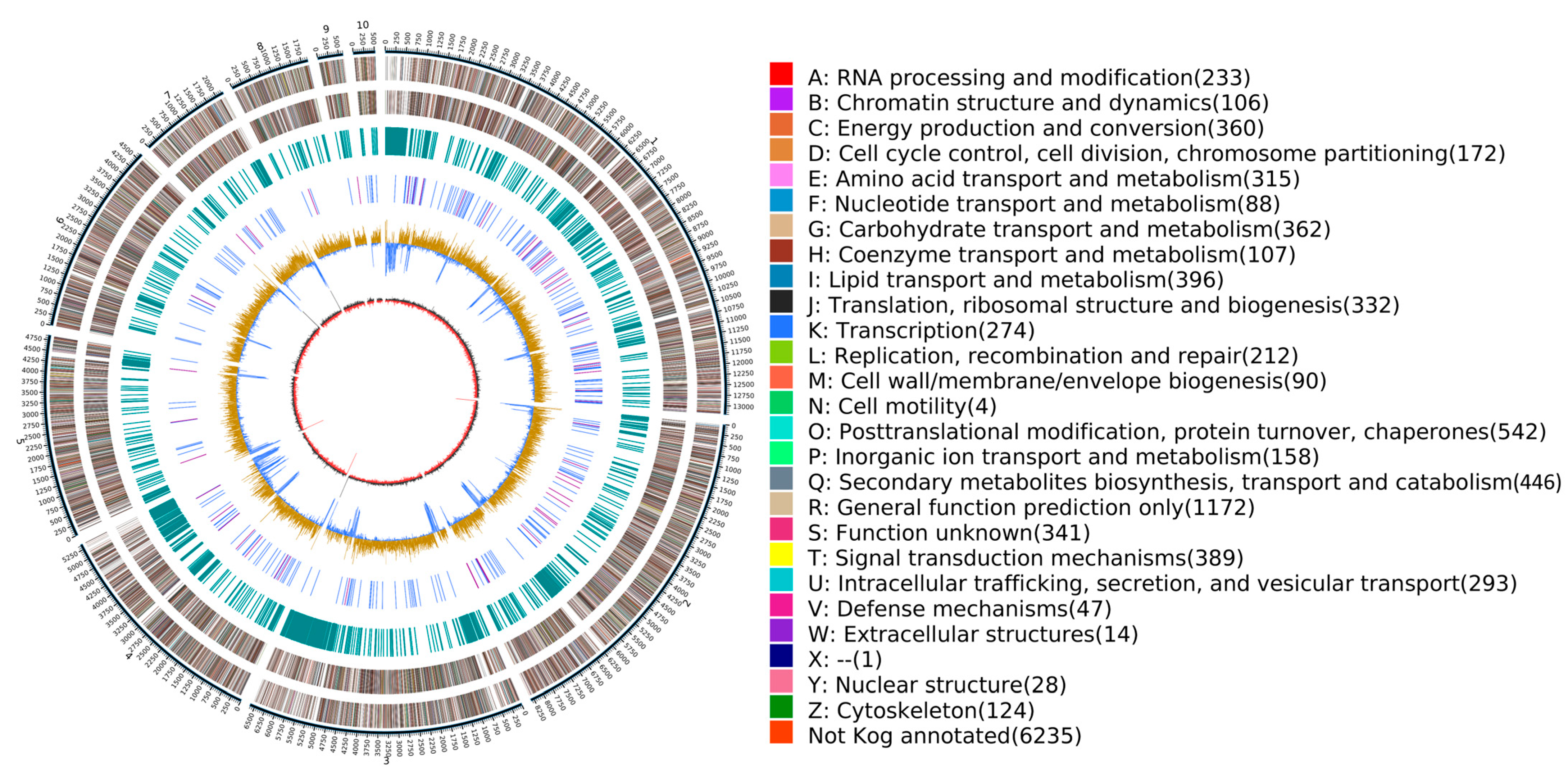
The total length of the final assembled genome was 49,259,803 bp with a GC content of 56.23%. The assembly consisted of 42 contig numbers with no gaps. The N50 length was 6,617,117 bp, and the N90 length was 2,194,971 bp (Table 1). The entire genome atlas of N. musae ST1 was generated using Circos (v 0.69-8) (Figure 1).
Table 1.
The genomic characteristics of N. musae ST1.
Figure 1.
Genome circle diagram of N.musae ST1. The outermost circle represents the location coordinates of the contig 1–10 of the genome sequence.
N. musae ST1 genome completeness was assessed using both next-generation read mapping and BUSCO analysis. An evaluation of next-generation sequence data recovery provided an alignment ratio of 94.56%, indicating 99.97% sequence coverage of the assembled genome and a final sequencing depth of 69.81×. BUSCO analysis revealed 287 complete BUSCO genes, indicating a genome completeness of 98.97%, confirming the high quality of the obtained genome assembly (Table 2).
Table 2.
The assembly evaluation of N. musae ST1.
3.2. Sequence Prediction
3.2.1. Repeat Sequence, Protein-Coding Gene, Non-Coding RNA, and Pseudogene Prediction
Repetitive elements were classified into major repeat categories and subfamilies. The genome contained 2,565,439 bp of repetitive sequences, accounting for 5.21% of the total genome.
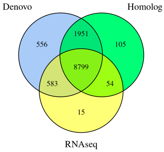
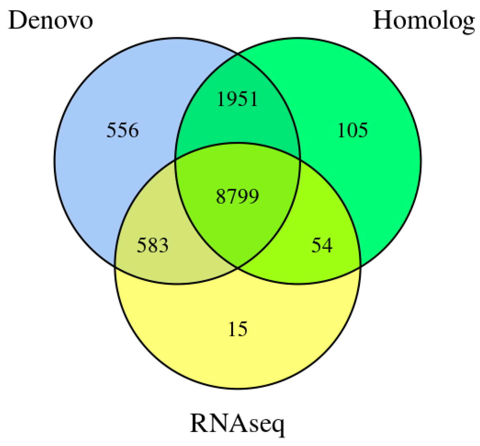
Using a combination of de novo prediction, homologous protein prediction, and transcriptomic evidence, we identified a total of 12,063 protein-coding genes in the genome of N. musae ST1. Among these, 11,507 genes (95.39%) were supported by homology prediction and transcript evidence, indicating high annotation reliability. The total length of the encoded genes was 25,915,843 bp, accounting for 52.61% of the whole genome. The average number of exons per gene was 2.95; the average number of CDSs per gene was 2.88; and the average number of introns per gene was 1.95 (Table A1 and Figure 2).
Figure 2.
Integrated genes of N. musae ST1 derived from three prediction methods.
The results of the genomic data of N. musae ST1 revealed 475 non-coding RNAs, including 78 ribosomal RNAs (rRNAs), 332 transfer RNAs (tRNAs), and 75 other non-coding RNAs. The rRNAs comprised 5 5.8S_rRNAs, 6 28S_rRNAs, 18 18S_rRNAs, and 49 5S_rRNAs. A total of 17 pseudogenes containing premature stop codons or frameshift mutations were detected, with a combined length of 65,191 bp and an average length of 3834.76 bp.
3.2.2. Prediction of Gene Clusters
The analysis of secondary metabolite gene clusters using the AntiSMASH tool identified a total of 77 gene clusters with a combined length of 3,225,987 bp and an average length of 41,895 bp (Table 1). These included 14 terpene, 29 T1PKS, 4 indole, 6 NRPS-like, 6 NRPS, 6 NRPS-Like, 5 NRPS + T1PKS, 4 NRPS-Like + T1PKS, 1 T3PKS, 1 fungal-RiPP, 1 T1PKS + NRPS-like, 1 T1PKS + NRPS, 1 NRPS-like + indole, 1 T1PKS + terpene, 1 NRPS + T1PKS + indole, 1 Betalactone + T1PKS + NRPS-like, and 1 NRPS + NRPS-like clusters. Only 19 BCGs showed homologies with known clusters, which are putatively responsible for the biosynthesis of compounds such as alternapyrone, dimethylcoprogen, serinocyclin, depudecin and betaenone (Table A2). Among them, three BCG showed 100% similarity to known clusters (Table A2). Approximately 96.10% of clusters showed no similarity to characterized BGCs, suggesting that N. musae ST1 may produce numerous previously unknown secondary metabolites.
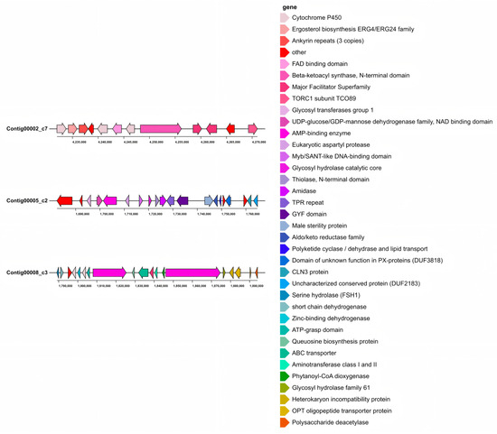
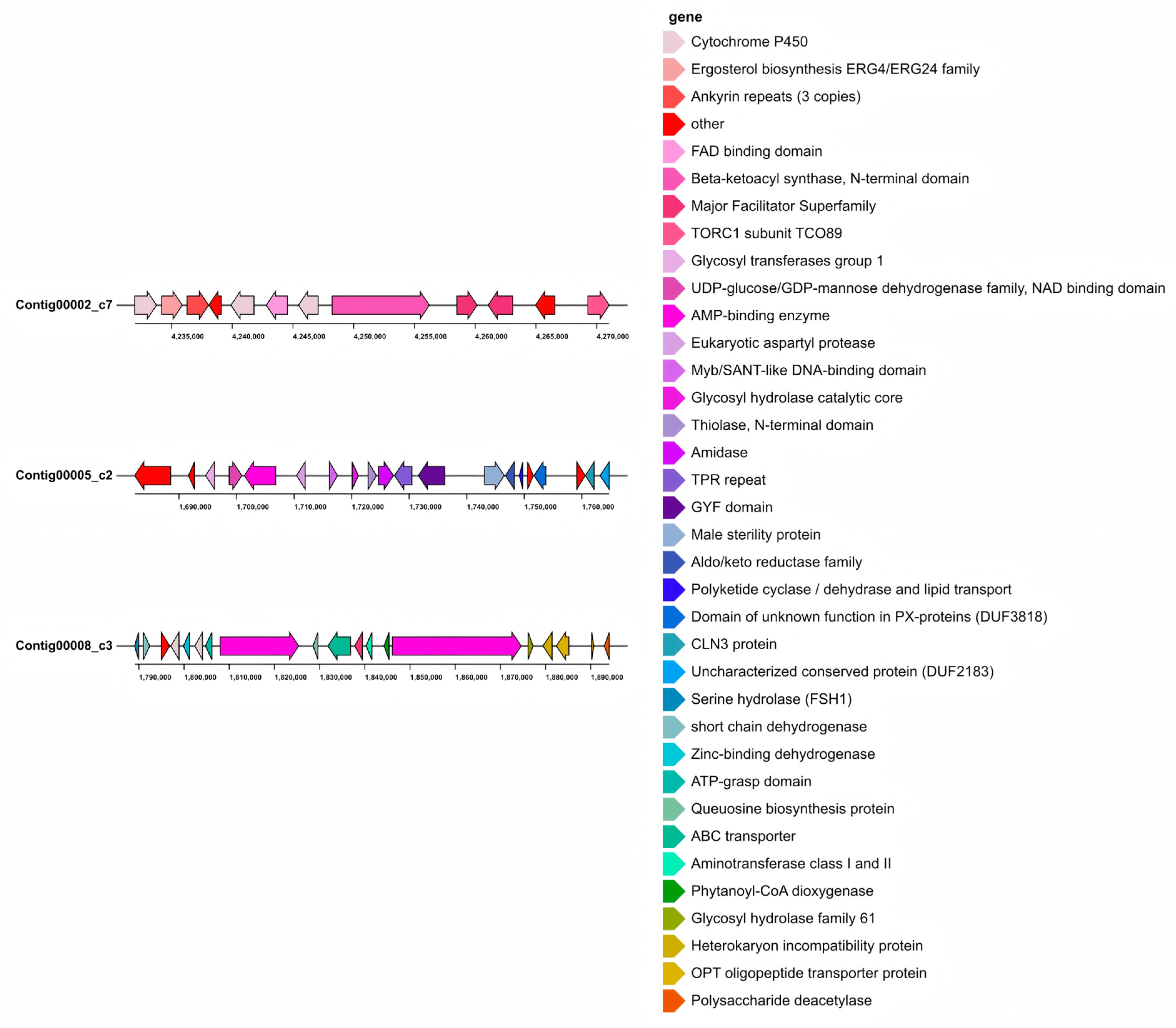
Region 2.7 includes a BGC of alternapyrone and a PKS gene (GenBank: AB120221.1A). Alternapyrone was first discovered in the potato Alternaria solani [67]. As a secondary metabolite of fungi, alternapyrone exhibits cytotoxicity and phytotoxicity (Figure 3) [68,69].
Figure 3.
Three identified BGCs with high identity in N. musae ST1. Region 2.7 had a T1PKS gene cluster; region 5.2 had two NRPSs and an NRPS-like gene cluster; region 8.3 had an NRPS and TIPKS gene cluster.
Region 5.2 exhibits significant similarity with the BGC of Alternaria alternata (GenBank: JQ973666.1). Dimethylcoprogen is an extracellular siderophore complex produced by pathogenic fungi under iron-deficient conditions [70,71]. It can enhance the utilization of iron by fungi to promote fungal growth and control the growth of other microorganisms in the environment (Figure 3) [72].
Region 8.3 contains serinocyclin gene clusters (BGCs) capable of synthesizing two compounds, serinocyclin A and serinocyclin B, classified as a terpene cluster (GenBank: JELW01000052.1). Structurally, serinocyclin A (1) contains three serine units, one hydroxyproline (Hyp), beta alanine (beta Ala), and two uncommon non-proteinogenic amino acids, 1-aminocyclopropane-1-carboxylic acid (Acc) and gamma hydroxylysine (HyLys). The Lys in serinocyclin B (2) replaces the HyLys unit in 1. Notably, serinocyclin is a fungal toxin, and these compounds are toxic to insects (Figure 3) [73].
Comparative analysis of Nigrospora genomes revealed that N. musae ST1 possessed the highest number of BGCs among the species examined, followed by N. musae Guizhou and N. sphaerica. All analyzed genomes were enriched with T1PKS gene clusters and terpene gene clusters. However, substantial differences were observed in the composition and abundance of BGCs among species, indicating differences between the genomes of different plant pathogenic fungi within the genus (Figure A1).
3.3. Functional Annotation of the Genome
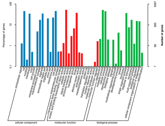
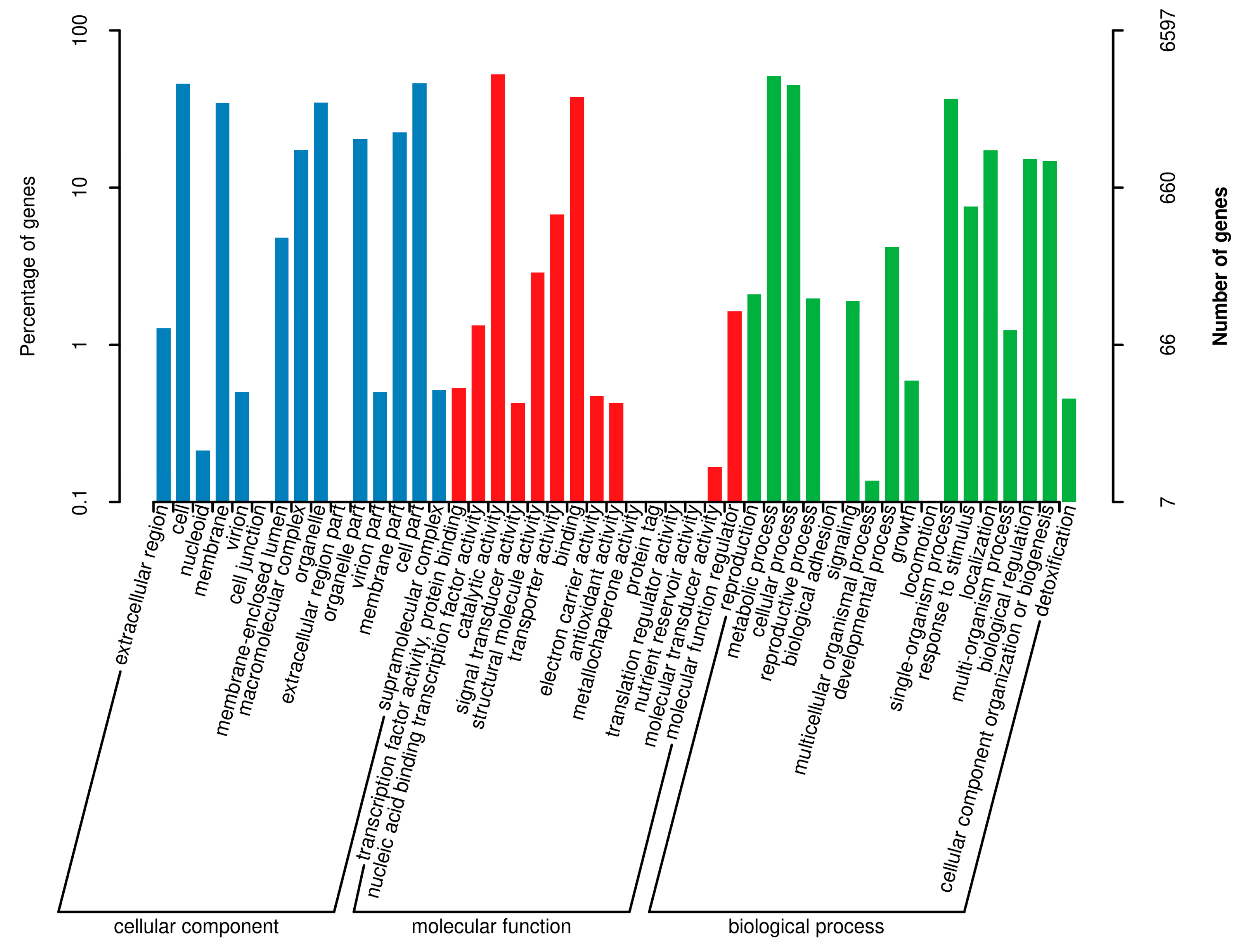
Functional annotation using the GO database (Blast2go v2.5) assigned annotations to 6597 protein-coding genes in the genome of N. musae ST1, corresponding to 47 GO terms, and representing 54.68% of the total predicted genes (Figure 4). Within the Biological Process (BP) category, 17 GO terms were assigned, with the top three terms being “metabolic process”, “cellular process”, and “single-organism process”; within the molecular function (MF) category, 15 GO terms were annotated, with the top three terms being “catalytic activity”, “binding”, and “transporter activity”; within the Biological Process (CC) category, 15 GO terms were annotated, with the top three terms being “cell”, “organelle”, and “cell part”.
Figure 4.
Gene Ontology (GO) functional annotation of the N. musae ST1 genome. The GO classification is presented with horizontal coordinates, while the percentage (left) and numbers (right) of genes are shown on the vertical axis.
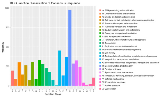
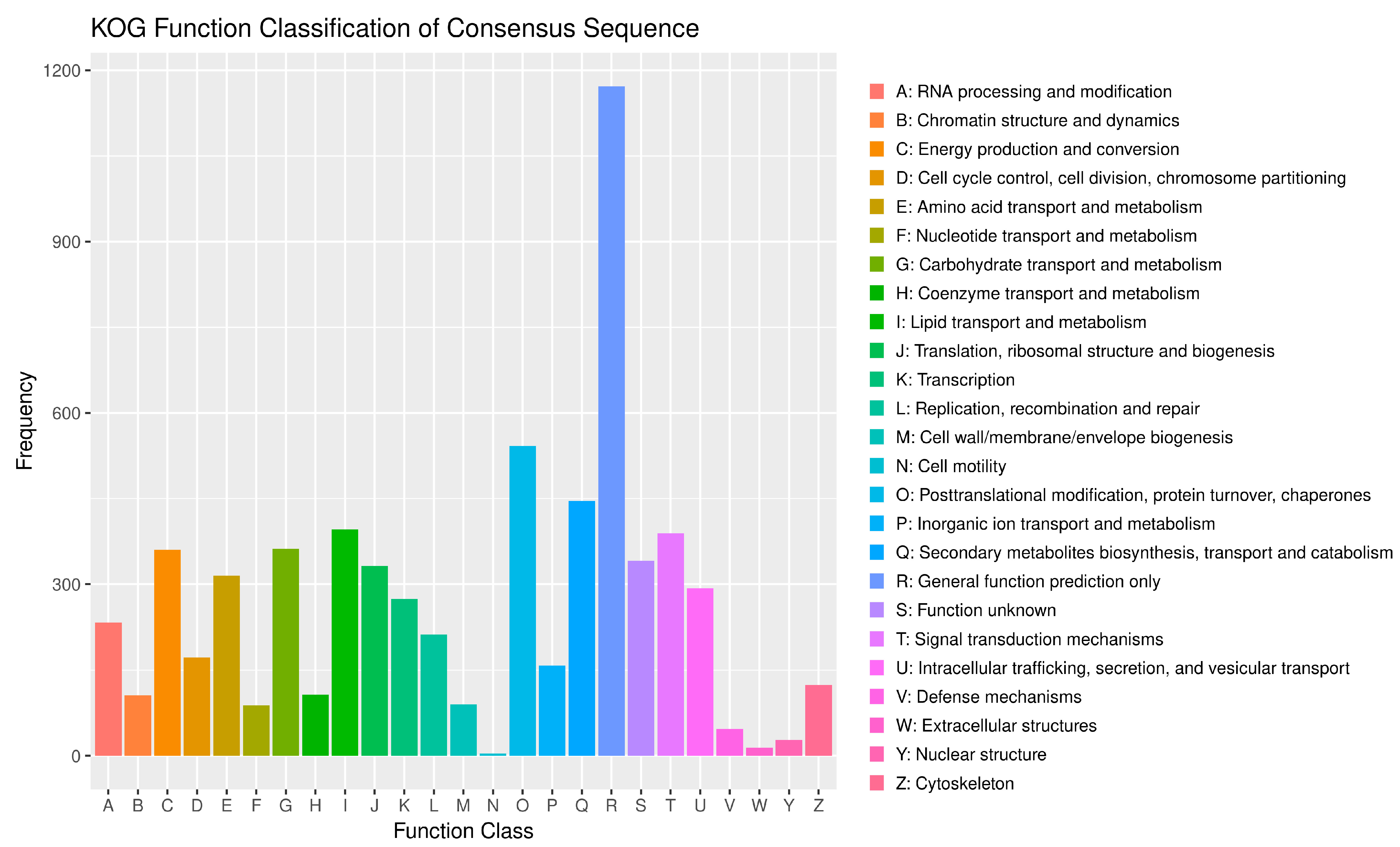
The number of annotated genes for N. musae ST1 in the KOG database was 5828, accounting for 48.31% of all predicted genes (Figure 5). The KOG category with the highest number of annotated genes was “General function prediction only”, with 1172 genes, followed by “Posttranslational modification, protein turnover, chaperones” with 542 genes, “Secondary metabolites biosynthesis, transport and catabolism” with 446 genes, “Lipid transport and metabolism” with 396 genes, “Signal transduction mechanisms” with 389 genes, “Energy production and conversion” with 362 genes, “Carbohydrate transport and metabolism” with 360 genes, and “Function unknown” with 341 genes.
Figure 5.
KOG annotation of N. musae ST1.
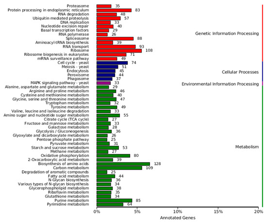
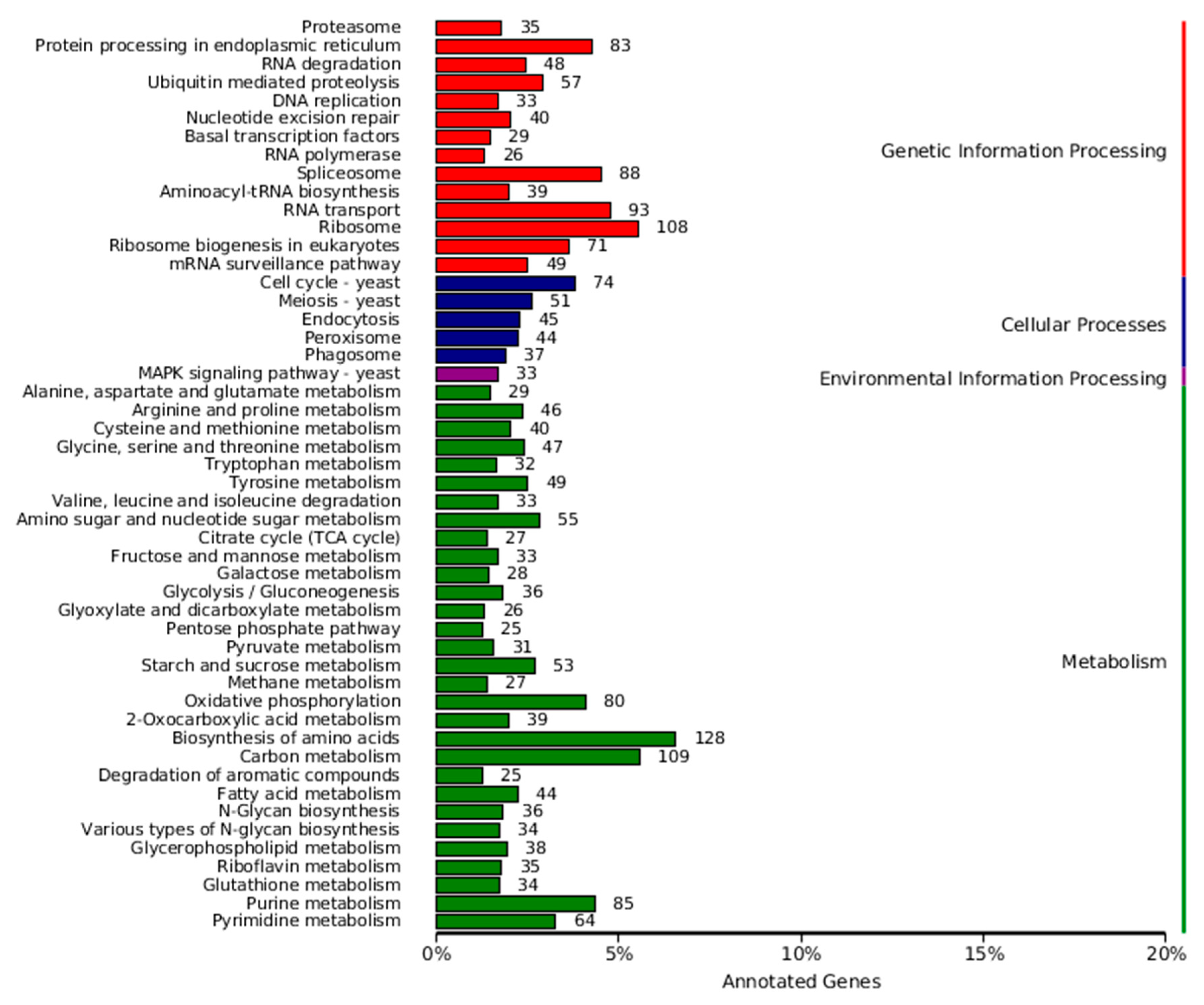
Based on the Kyoto Encyclopedia of Genes and Genomes (KEGG) pathway database, 3365 genes were annotated into 113 KEGG pathways (Figure 6). The six KEGG pathways with the highest number of assigned genes were Biosynthesis of amino acids, Carbon metabolism, Ribosome, RNA transport, Spliceosome, and Purine metabolism.
Figure 6.
KEGG annotation classification statistics of N. musae ST1.
3.4. Carbohydrate-Active Enzymes (CAZymes)
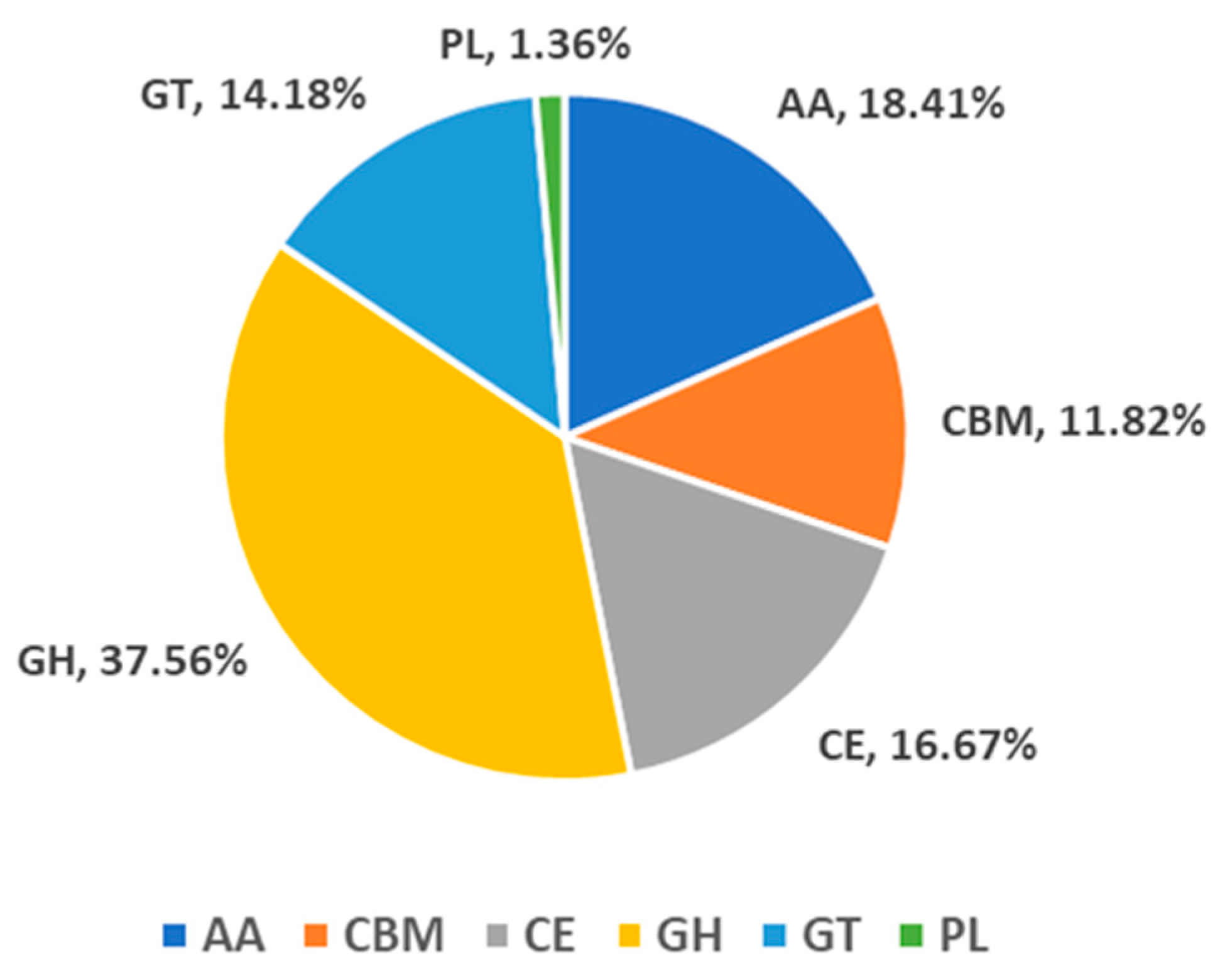
We annotated 722 genes in the CAZy database. Plant pathogenic fungi degrade plant polysaccharides by producing a large number of carbohydrate-active enzymes (CAZymes), which are classified into five categories based on their functions: glycosyl transferases (GTs), glycoside hydrolases (GHs), carbohydrate esterases (CEs), polysaccharide lyases (PLs), and auxiliary activities (AAs). In addition, the database also includes enzymes related to carbohydrate-binding modules (CBMs). In N. musae ST1, the predicted CAZymes comprised 302 GHs, 148 AAs, 134 CEs, 114 GTs, 95 CBMs, and 11 PLs (Figure A2).
3.5. Pathogenicity System Analysis
3.5.1. Analysis of PHI
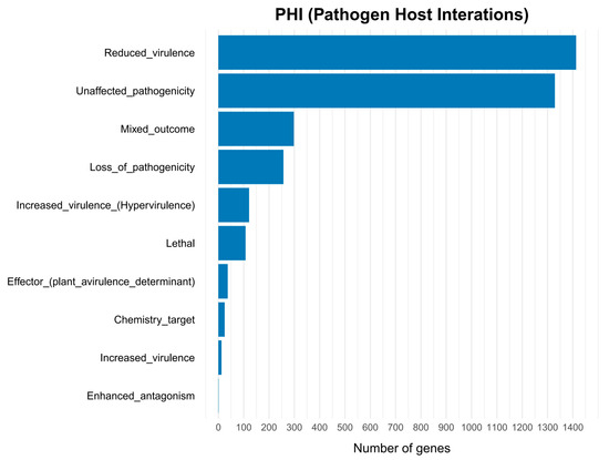
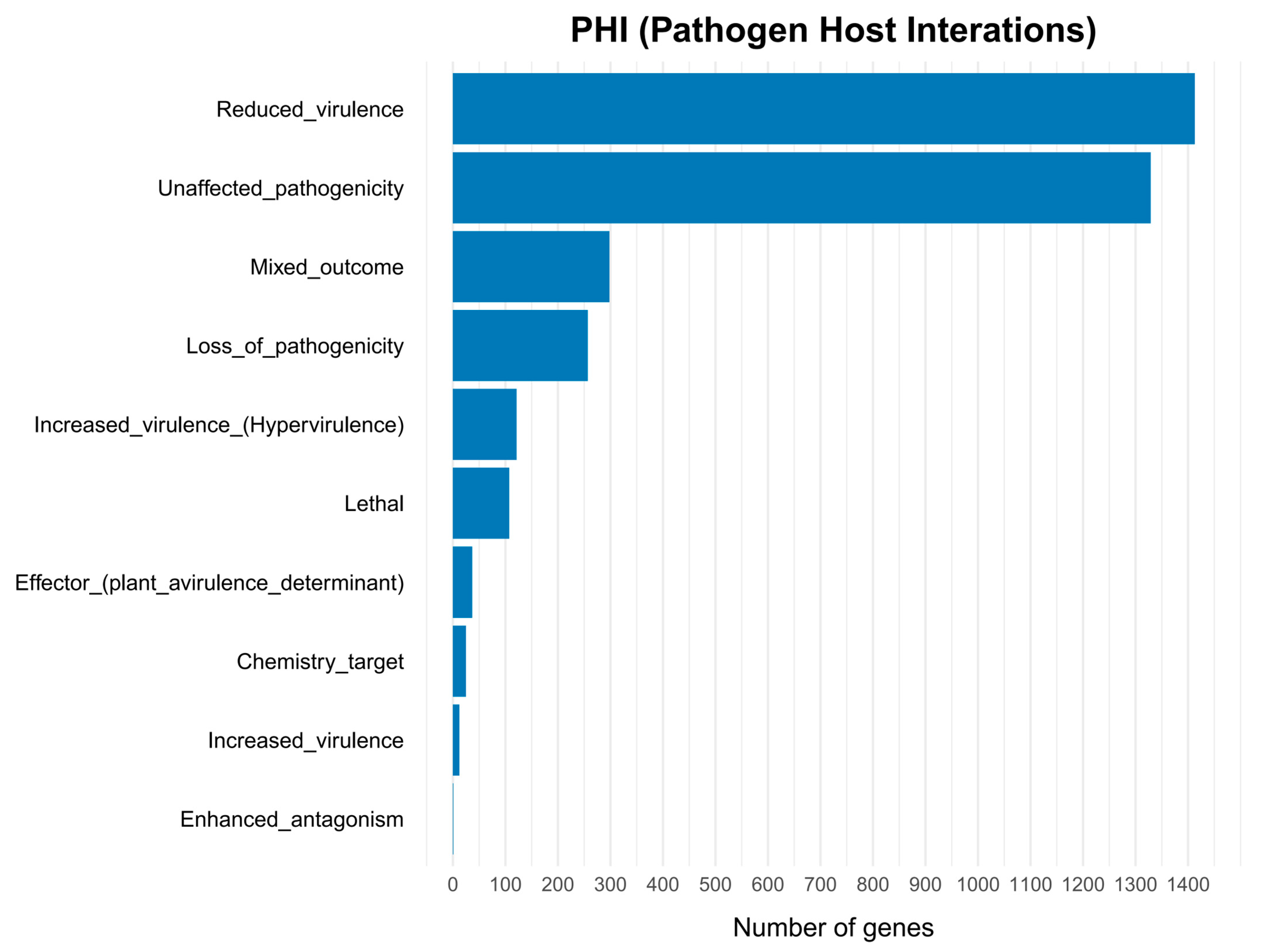
We used the pathogen host interaction (PHI) database to annotate the pathogenicity-related genes in the genome of Nigrospora. A total of 3600 genes were annotated in the PHI database (Table 1), accounting for 29.84% of the predicted genes. Among the genes, 121 genes were associated with hypervirulence, 107 genes were related to lethality, 37 genes were associated with effector function, and 12 genes were associated with increased virulence. The largest group consisted of genes linked to reduced virulence (1413 genes), followed by unaffected pathogenicity with 1329 genes, mixed effects with 298 genes, loss of pathogenicity with 257 genes, chemistry target with 25 genes, and enhanced antagonism with 1 gene (Figure 7). Gibberella zeae (993 genes) and Magnaporthe oryzae (624 genes) had the highest number of homologous genes related to virulence.
Figure 7.
PHI functional classification of proteins encoded in the N. musae ST1 genome.
3.5.2. Database of Fungal Virulence Factor (DFVF) Annotations
Fungal Virulence Factors are widely involved in fungal pathogenicity processes such as host tissue infection, immune evasion, and toxin production. A total of 2503 genes were annotated in the DFVF database, accounting for 20.75% of the predicted genes.
3.5.3. Transporter Classification Database (TCDB) Annotations
Transporters are proteins on the cell membrane that regulate the influx and efflux of substances, thereby participating in nutrient uptake and energy metabolism. In fungi, certain transporters are closely related to fungal resistance and pathogenicity, as well as fungal growth and spore formation [74,75,76,77]. For example, disruption of the ABC1 gene in Magnaporthe grisea reduces its infectivity toward rice and barley [78]. In this study, a total of 125 genes were annotated in the TCDB, representing 1.04% of the predicted genes.
3.5.4. Cytochrome P450 (CYP450) Genes
Cytochrome P450 enzymes constitute a superfamily of structurally and functionally related isoenzymes involved in cell detoxification, xenobiotic degradation, and biosynthesis of secondary metabolites [79]. A total of 585 CYP450 genes were predicted in the N. musae ST1 genome (Table 1).
3.6. Protein Subcellular Localization Analysis
Effector proteins are a class of specialized proteins secreted by pathogens that can promote the infection and colonization process by targeting the host’s immunity signaling or metabolic pathways [80]. Subcellular localization prediction identified a total of 1462 proteins with signal peptides, 2713 transmembrane proteins, and 1071 secreted proteins. Finally, the secreted proteins were further analyzed with EffectorP, and 197 effector proteins were predicted (Table 1).
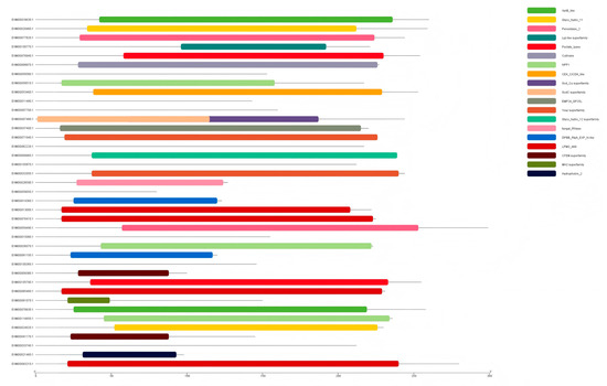
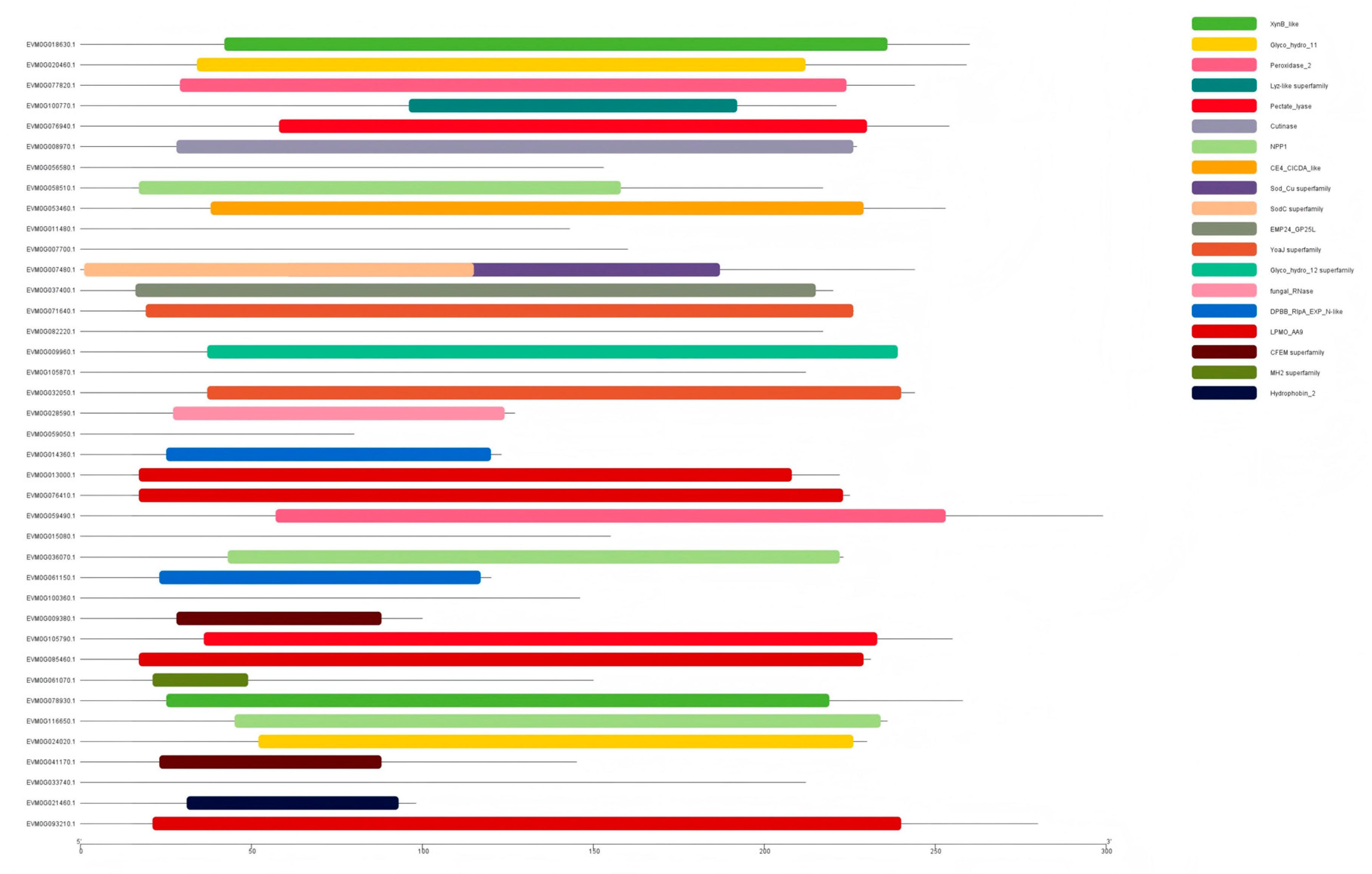
To suppress pattern-triggered immunity (PTI) elicited by plant pathogen-associated molecular patterns (PAMPs), plant pathogens often secrete effectors and enhance pathogenicity by regulating host metabolism and defense response, leading to effector-triggered susceptibility (ETS) [81]. Most known fungal effectors are characterized by signal peptides, low molecular weight, and rich cysteine residues [82]. After filtering the 197 predicted effectors to exclude proteins exceeding 300 amino acids, 39 candidate pathogenicity-related effectors were obtained (Table A3). Among these, 20 were hydroxy proteins, while the remainder included acetylxylan esterase, endoglucanase-like proteins, endo-1,4-beta-xylanase B, and related enzymes. Meanwhile, we analyzed the functional structure of 39 candidate effector proteins, including 30 proteins with domains and 9 proteins lacking domains (Figure 8).
Figure 8.
Distribution of candidate effector domains for N. muse ST1.
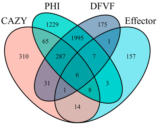
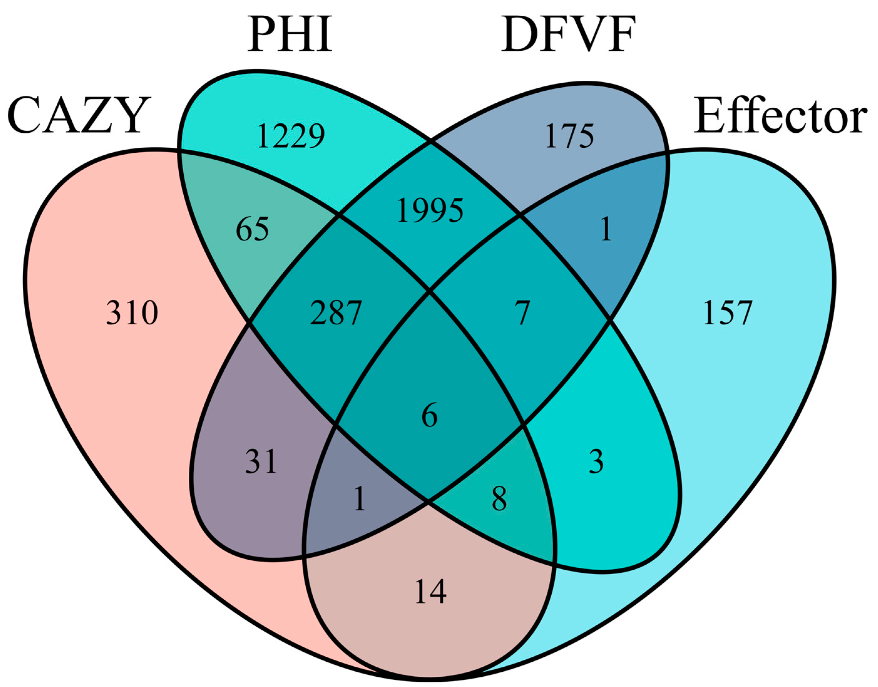
We conducted an integrated analysis of CAZymes, PHI, DFVF pathogenic gene-related databases, and effector proteins. The Venn diagram (Figure 9) showed that six genes were shared by CAZymes, PHI, DFVF, and the secretome. Among the six secretory CAZyme genes, three genes encoded β-1,4-xylanases, two of which were endo-1,4-β-xylanases involved in xylan degradation, two genes encoded pectinate lyases that catalyze the hydrolysis of polygalacturonic acid, and one gene encoded a cutinase that catalyzes the hydrolysis of cutin.
Figure 9.
Venn diagram of genes encoding CAZymes, PHI, DFVF and effector.
3.7. Comparative Genomic Analysis of Other Nigrospora Species
Among the currently available genome-annotation studies for Nigrospora, only three pathogenic species, N. oryzae ZQ1, N. oryzae GZL1, and N. sphaerica ZJJ_C1, were selected. In addition, the genomes of five plant pathogenic fungi of the same genus, N. guilinensis LC3481, N. hainanensis LC7030, N. lacticolonia LC3324, N. osmanthi Z1, and N. pyriformis LC2045, were selected from NCBI for comparison and analysis with the genome of N. musae ST1. Substantial variation was observed among these species in genome size, contig N50, GC content and other genomic features (Table 3).
Table 3.
Comparison of genomic features among different Nigrospora species.
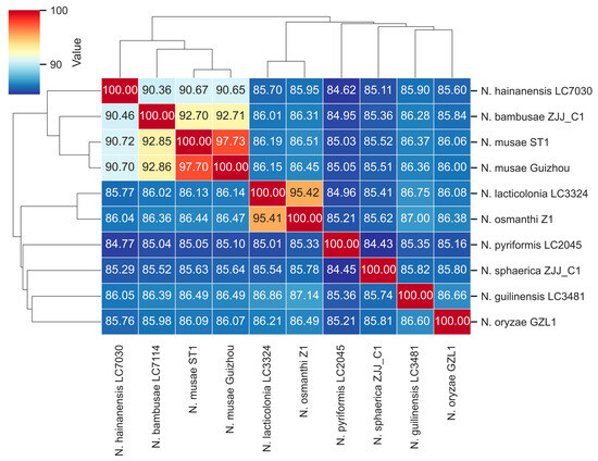
Whole-genome alignment and calculation of the average nucleotide identity (ANI) revealed pairwise similarities among Nigrospora species ranging from 97.73 to 84.43%. As illustrated in Figure 10, N. musae ST1 exhibited low genomic similarity (84.43–86.49%) with N. lacticolonia LC3324, N. osmanthi Z1, N. pyriformis LC2045, N. sphaerica ZJJ_C1, N. guilinensis LC3481, and N. oryzae GZL1. In contrast, N. musae ST1 showed high genomic similarity (90.36–97.73%) to N. hainanensis LC7030, N. bambusae LC7114, and N. musae Guizhou. The similarity between N. musae ST1 and N. musae Guizhou was as high as 97%, indicating that N. musae ST1 and N. musae Guizhou belong to the same species. Collectively, the results above confirmed that N. musae ST1 belongs to the genus Nigrospora according to the current classification.
Figure 10.
The average nucleotide identity (ANI) values based on the fastANI-algorithm-generated matrix for Nigrospora genomes. The clustering was constructed using the Euclidean distance matrix.
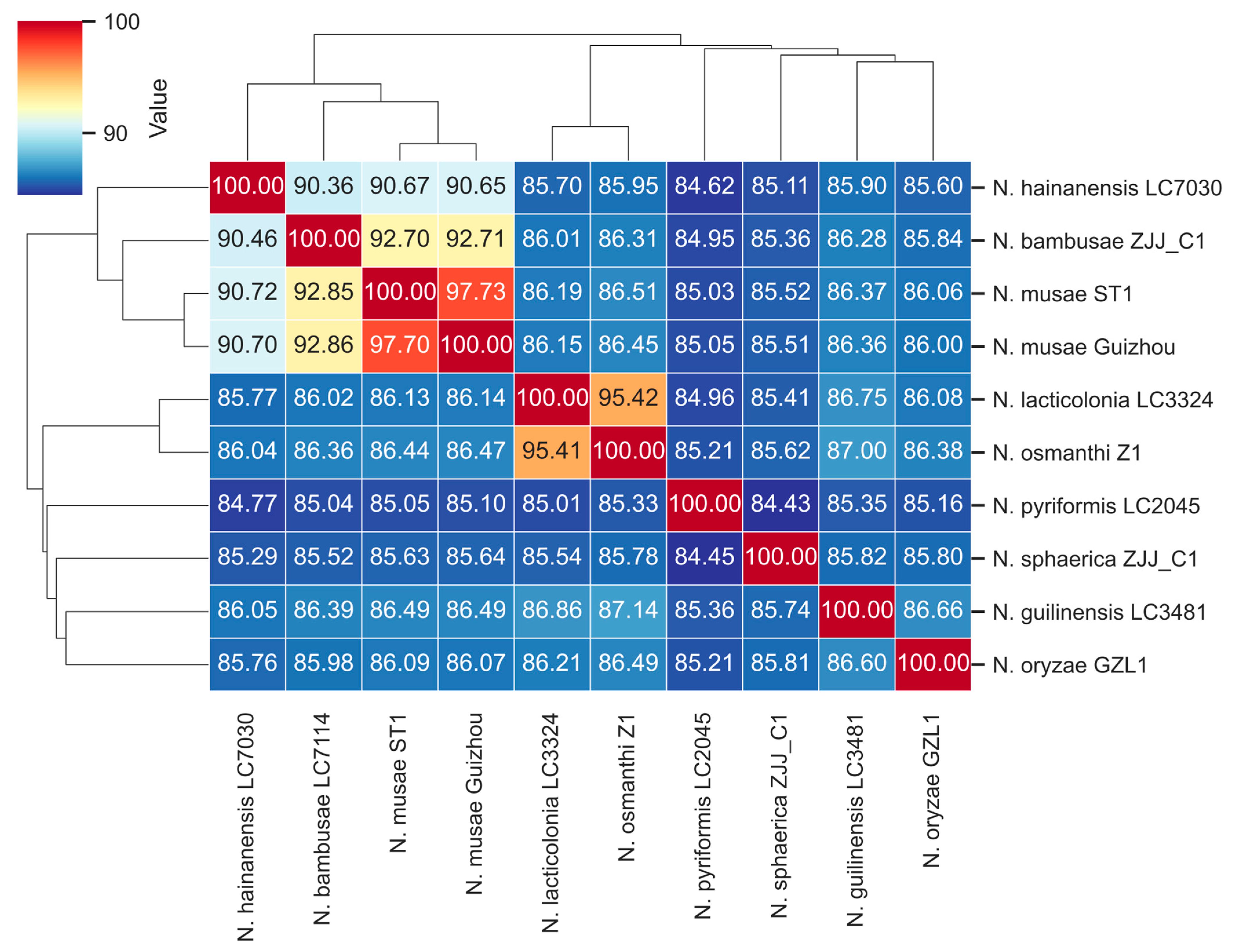
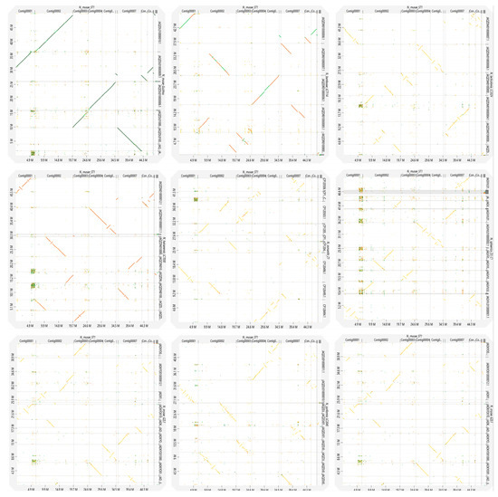
To investigate the genomic synteny and evolutionary relationships within Nigrospora, we performed synteny analysis between N. musae ST1 and nine closely related species, including N. bambusae LC7114, N. guilinensis LC3481, N. hainanensis LC7030, N. lacticolonia LC3324, N. musae Guizhou, N. oryzae GZL1, and N. pyriformis LC2045 (Figure 11). The synteny plots revealed that N. musae ST1 shared the longest and most continuous syntenic blocks with N. musae Guizhou, indicating strong conservation of genome structure between these two isolates. In contrast, syntenic blocks between N. musae ST1 and other species were relatively short and fragmented, suggesting significant genomic structural divergence. These findings provide important genomic-level insights into the evolutionary dynamics and phylogenetic relationships within Nigrospora.
Figure 11.
The visualization of the whole-genome LAST alignment of complete and genome-level Nigrospora species genome assemblies available in the NCBI Genome database (vertical axis) to obtain a complete N. musae ST1 genome assembly (horizontal axis). Orange indicates direct alignments, and blue indicates reverse alignments.
4. Discussion
Genomic information is fundamental for elucidating the pathogenicity of pathogenic fungi. At present, genome assemblies for 16 Nigrospora species are available in NCBI, representing different saprophytes, plant endophytes, plant pathogens, and ecotype-specific strains [85]. Among them, N.oryzae, N.sphaerica and N. musae pose a serious threat to rice, Akebia trifoliata, I. polycarpa, and other cash crops [12,86]. Despite their agricultural importance, genomic resources for Nigrospora remain limited. In this study, the whole genome of the pathogenic fungus N. musae ST1 causing I. polycarpa leaf spot disease was sequenced and assembled. The assembled genome size of N. musae ST1 was 49.26 Mb, and a total of 12,125 coding genes were predicted. There were significant differences in genome size, GC content, gene cluster type, and number compared with other Nigrospora species that have been previously characterized. Notably, even when compared with N. musae Guizhou, another strain of the same species, N. musae ST1, exhibited pronounced differences in the types and number of secondary metabolism gene clusters. Such differences may arise from variations in sequencing technologies or reflect distinct life histories, metabolic potentials and host adaptation strategies due to the different regulatory genes and metabolic pathways in isolates from disparate geographic or ecological origins.
Fungal carbohydrate-active enzymes are closely related to the pathogenicity of pathogens. The plant cell wall is the first barrier for plants to resist pathogens. Pathogens infect plant cells by releasing and secreting a large number of cell wall-degrading enzymes to obtain nutrients [87]. These plant cell wall-degrading enzymes include carbohydrate esterases (CEs), polysaccharide lyases (PLs), and glycoside hydrolases (GHs) [88]. In this study, the number of N. musae ST1 glycoside hydrogenases (GHs) was the largest (302), accounting for 41.8% of the total number of CAZyme genes, significantly higher than the number of polysaccharide lyases (PLs) (11) and carbohydrate esterases (CEs) (134). N. musae ST1 possesses a high number of carbohydrate-active enzymes, indicating its strong capacity for complex carbohydrate degradation and glycosylation. Among them, the abundance of glycoside hydrolases was significantly higher, which is similar to oomycetes such as Phytophthora and Phytopythium. The abundance of carbohydrate-active enzymes contributes to efficient and rapid degradation of carbohydrates, and also enhances pathogenicity and host adaptation [89,90,91]. Additionally, N. musae ST1 exhibited a notably higher number of genes in the GH61 family compared to other glycoside hydrolase families. The proteins encoded by these genes can significantly enhance lignocellulose hydrolysis [92]. Lignocellulose is a complex polymer composed of cellulose, hemicellulose, and lignin, which provides structural reinforcement to the plant cell wall. The GH61 gene family can efficiently degrade lignocellulose in the plant cell wall and promote plant infection by N. musae ST1. For example, members of the GH61 family can serve as key pathogenicity factors. In Pyrenophora graminea, deletion of the PgGH61-2 gene resulted in severely reduced pathogenicity and impaired carbon source utilization [93]. Due to their high catalytic efficiency in depolymerizing recalcitrant substrates, GH61 enzymes can be used for cellulose conversion in biofuel production [94]. N. musae ST1 may overcome physical barriers by efficiently degrading plant cell walls rich in lignocellulose. Notably, the number of polysaccharide lyases identified in the N. musae ST1 strain was extremely low, and PLs primarily target pectin. The pectin degradation function of N. musae ST1 during plant infection may be mediated by members of certain glycoside hydrolase gene families. For instance, GH28 family enzymes encode polygalacturonases, rhamnogalacturonases, and xylogalacturonases that can degrade pectin in plants [95].
We annotated and analyzed genes related to pathogenicity. Genome-wide annotation identified 3600 genes associated with pathogen–host interactions, divided into 10 different categories (Figure 7). This reflects the redundant genetic buffering mechanisms in fungal genomes and the complexity of virulence regulation [96]. A total of 2503 genes were annotated as Fungal Virulence Factors, which play widespread roles in host tissue invasion, immune evasion, and toxin production [62]. A total of 585 CYP450 genes were annotated; these enzymes participate in cellular detoxification, degradation of exogenous substances, and synthesis of secondary metabolites [79]. These genes may regulate toxin synthesis and inhibit host defense responses to enhance pathogenicity. Protein subcellular localization analysis predicted 1462 signal peptides, 2713 transmembrane proteins, and 1071 secreted proteins, from which 197 effector proteins were predicted. Integrated analysis across CAZymes, PHI, DFVF, and the secretome revealed six secreted carbohydrate-active enzymes involved in the degradation of plant structural components and the infection of plants, including cutinases, 1,4-β-xylanases, pectate lyases, and glycoside hydrolase 61. Cutinases can catalyze the hydrolysis of cutin in the plant cuticle, helping pathogenic bacteria break the physical barrier and penetrate the plant epidermis. For example, downregulation of cutinase genes following loss of the transcription factor FvCTF1α in Fusarium verticillioides reduced pathogenicity [97]; Xylan, the main component of plant hemicellulose and cell wall structure, is cleaved at β-1,4-glycosidic bonds by 1,4-β–xylanases; loss of GH11 family genes in Neostagonosporella sichuanensis diminished virulence [98]. Pectinate lyases cleave the α-1,4-glycosidic bond in polygalactonic acid through β elimination reaction, disrupting pectin integrity in the cell wall. The pectate lyase gene of Streptomyces galilaeus is significantly upregulated during pathogenesis, and plant susceptibility is markedly reduced when this gene is disrupted [99]. The pathogenicity of Fusarium graminearum decreased after deletion of different pectate lyase genes [100]. However, in Peronophythora litchii, pectate lyase has dual effects of inducing immunity and inhibiting pathogenesis [101], indicating that the functions of pectate lyases in different pathogenic bacteria are different. This functional divergence may arise from the complex regulatory networks shaped during host–pathogen co-evolution. For instance, while pectate lyases enhance pathogen invasion, they can also be targeted by the host immune recognition system. Although the PlPeL1 protein of Peronophythora litchii is essential for full virulence, it can be bound by the immune regulatory factor LcPIP1 secreted by litchi, thereby attenuating the induction of host susceptibility [101]. Overall, these enzymes likely represent key contributors to N. musae ST1 infection and disease causation, providing valuable candidate genes for future functional validation of pathogenicity factors.
We further analyzed the predicted effector proteins. A total of 39 candidate effectors were identified, comprising 25 apoplastic and 14 non-apoplastic effectors. It is worth noting that in the apoplastic effectors, we identified enzymes related to cell wall degradation, such as glycoside hydrolases, acetylxylan esterases, endoglucanase-like proteins, Endo-1, 4-beta-xylanase B, and putative glycoside hydrolase family 61 proteins. These enzymes are directly involved in the degradation process of plant cell walls. They degrade the physical defense barrier of plant cells by degrading cellulose, hemicellulose, and xylan, facilitating pathogen entry, colonization, and nutrient acquisition [97,98,102]. In addition, a hypersensitive response-inducing protein was identified, capable of triggering host cell death and enhancing plant disease resistance [103]. Although these effector-related genes have been studied in other pathogens, their functions can vary substantially across species. For example, the glucosidase GH12 gene, which is one of the important virulence factors, can induce strong necrosis and resistance in Phytophthora [102]. However, the glucosidase protein in Cytospora chrysosperma of PopulusL can induce plant immunity, but has no effect on pathogenicity [104]. At present, there are few studies on the pathogenicity mechanisms of Nigrospora plant pathogenic fungi, and it is essential to further clarify the roles of N. musae ST1 effectors in virulence, host specificity, or environmental adaptation.
Compared with other Nigrospora species, N. musae ST1 possesses the highest number of secondary metabolite biosynthetic gene clusters, followed by N. musae Guizhou and N. sphaerica ZJJ_C1. Moreover, the genomes of fungi within this genus are all rich in T1PKS and terpene gene clusters (Figure A1). T1PKS gene clusters are responsible for catalyzing the biosynthesis of polyketide compounds, while terpene gene clusters produce terpenoids. Both types of compounds play critical roles in fungal ecological interactions, environmental adaptation, and pathogenicity [105]. For instance, various T1PKS gene clusters identified in the genome of Xanthoria elegans contribute to the synthesis of UV-absorbing compounds that support survival in extreme environments [106]. In the plant pathogenic fungus Cytospora chrysosperma, knockout of the terpene synthase gene CcPtc1 significantly reduced fungal virulence and altered terpenoid metabolite profiles, highlighting the importance of terpene biosynthesis in pathogenicity [107]. Notably, N. musae ST1 and the conspecific strain N. musae Guizhou exhibit significant differences in the types and numbers of gene clusters (Figure A1). Such intraspecific variation in gene clusters is a typical characteristic of fungal genomic plasticity. The expansion and contraction of secondary metabolite gene clusters are major drivers of fungal evolution and can occur through gene duplication, transposon-mediated duplication, and horizontal gene transfer. For example, in the birch pathogen Inonotus obliquus, lineage-specific whole-genome duplication events and subsequent retention of cytochrome P450 genes and small-scale duplications within biosynthetic clusters have greatly diversified terpenoid biosynthesis [108]. Similarly, in the plant pathogen Fusarium, evolutionary analysis of the trichothecene biosynthesis gene cluster further elucidated the molecular mechanism by which new genes are recruited into existing clusters through gene rearrangements, leading to gene cluster expansion [109]. Therefore, the pronounced differences in gene clusters between N. musae ST1 and N. musae Guizhou may reflect genomic differentiation driven by distinct ecological origins, host associations, or microenvironmental pressures. Through such dynamic genomic remodeling, these fungi may have evolved unique chemical repertoires and pathogenic strategies.
There are many types of fungal secondary metabolites, most of which belong to polyketones [110]. These compounds induce cell death by destroying the host cell membrane structure, interfering with mitochondrial function, and inhibiting oxidase activity, thereby enhancing pathogenicity [110,111,112,113,114]. In this study, a total of 77 secondary metabolite biosynthetic gene clusters (bgcs) were predicted in the genome of N. musae ST1. Among them, the number of T1PKS gene clusters was the largest (29). Given their central role in polyketide biosynthesis, these clusters may contribute to the strong pathogenic potential of N. musae ST1. For example, the PKS gene cluster of Cercospora sojina is involved in the early infection process [115], and Alternaria spp fungi can produce a variety of host-specific polyketide toxins [116]. In addition to being involved in fungal pathogenicity, polyketide secondary metabolites can also support fungal survival under adverse conditions, such as DHN-melanin, which protects against environmental stress [117,118]. In addition, the non-ribosomal peptide synthase (NRPS) gene cluster may promote the synthesis of non-ribosomal peptide compounds and affect the pathogenicity and spore formation of pathogens [119,120]. Three gene clusters exhibited 100% similarity with known secondary metabolite gene clusters responsible for producing alternapyrone, dimethylcoprogen, and serinocyclin A/serinocyclin B. Among them, alternapyrone—whose biosynthesis is regulated by a T1PKS gene cluster—exhibits phytotoxicity and inhibits wheat seed germination by targeting early embryonic development [75]. Furthermore, it also demonstrates cytotoxic activity against tumor cells [68], suggesting potential applications in natural product discovery and biomedicine. Notably, 96.10% of BGCs showed no similarity to known gene clusters, indicating that N. musae ST1 may produce a wide array of unknown secondary metabolites. The biological activities of these compounds remain unknown and warrant further investigation. Furthermore, among other species in the same genus, N. musae ST1 possessed the highest number of secondary metabolite clusters, underscoring its extensive metabolic potential.
Based on whole genome sequences, we compared and analyzed the fungi of the genus Nigrospora. The results of ANI analysis showed that the similarity between N. musae ST1 and the strain of the same species, N. musae Guizhou, was as high as 97%, and the similarity with N. hainanensis LC7030 and N. bambusae LC7114 was 90.36–92.71%. In contrast, similarity with N. lacticolonia LC3324, N. osmanthi Z1, N. pyriformis LC2045, N. sphaerica ZJJ_C1, N. guilinensis LC3481, and N. oryzae GZL1 was only 84.43–86.49%, which further confirmed the classification status of N. musae ST1 as a distinct species within the genus. Collinearity analysis showed that there were extensive genome rearrangements among different species of Nigrospora. Even between the strains N. musae ST1 and N. musae Guizhou, genome structures were not completely conserved, and there were segment inversions and translocation variations. Research on fungal species diversity and host adaptation indicates that fungi often evolve from broad-spectrum to specialized hosts, with genomic plasticity serving as a key mechanism driving this adaptive process [121]. Furthermore, in closely related fungal groups, variations in genomic synteny have been confirmed to be closely associated with host adaptation and the differentiation of pathogenic strategies. Comparative genomic studies of Colletotrichum fungi have revealed significant syntenic rearrangements at the genome assembly level among strains of the same species, correlated with the distribution of secondary metabolite gene clusters and differences in gene expression. This indicates that genomic structural variation is an important evolutionary driver for fungi to adapt to different ecological niches [122]. Such structural variation likely represents an important evolutionary strategy for Nigrospora fungi to adapt to different hosts and ecological environments.
5. Conclusions
In this study, we performed de novo whole-genome sequencing of N. musae ST1 using the PacBio Sequel II platform. In order to gain a better understanding of N. musae ST1 genome structure and function, we further predicted CAZymes and annotated PHI, DFVF, TCDB, and P450-encoding genes. These results provide a basis for the study of the pathogenicity genes of the fungus. In addition, based on the large number of secondary metabolite biosynthetic gene clusters identified in this study, we can further explore the role of secondary metabolite gene clusters on the pathogenicity of Nigrospora.
Author Contributions
Methodology, Y.-Z.C.; investigation, Y.-Z.C.; validation, Y.-Z.C.; resources, Y.-Z.C.; data analysis, Y.C., Y.-Z.C. and J.Y.; writing—original draft preparation, Y.-Z.C. and Y.C.; writing—review and editing, Y.-Z.C. and J.Y.; supervision, Y.-Z.C.; funding acquisition, Y.-Z.C. All authors have read and agreed to the published version of the manuscript.
Funding
This study was supported by funds from Guizhou Provincial Science and Technology Projects (QKHJC [2024]youth041), the Guizhou Provincial Science and Technology Department (No. QKHJC-MS [2026] 484), Guizhou Forestry Research Project (QLKH [2024]21), and Scientific Research Fund Project of Guizhou Education University (2025GCC011).
Institutional Review Board Statement
Not applicable.
Informed Consent Statement
Not applicable.
Data Availability Statement
The read data of Pacbio Sequel II sequencing have been submitted to the NCBI Sequence Read Archive with accession number PRJNA1367030. This Whole Genome Shotgun project has been deposited at GenBank under accession number JBSLLA000000000. The version described in this paper is version JBSLLA000000000.
Conflicts of Interest
The authors declare no conflicts of interest.
Appendix A
Table A1.
Basic information statistics of the predicted genes.
Table A2.
Putative biosynthetic gene clusters of N. musae ST1 showing similarity to a minimum number of known gene clusters in the biosynthetic gene cluster database.
Table A3.
N. muse ST1 candidate effectors.
Appendix B
Figure A1.
Gene cluster analysis of Nigrospora pathogenic fungi.
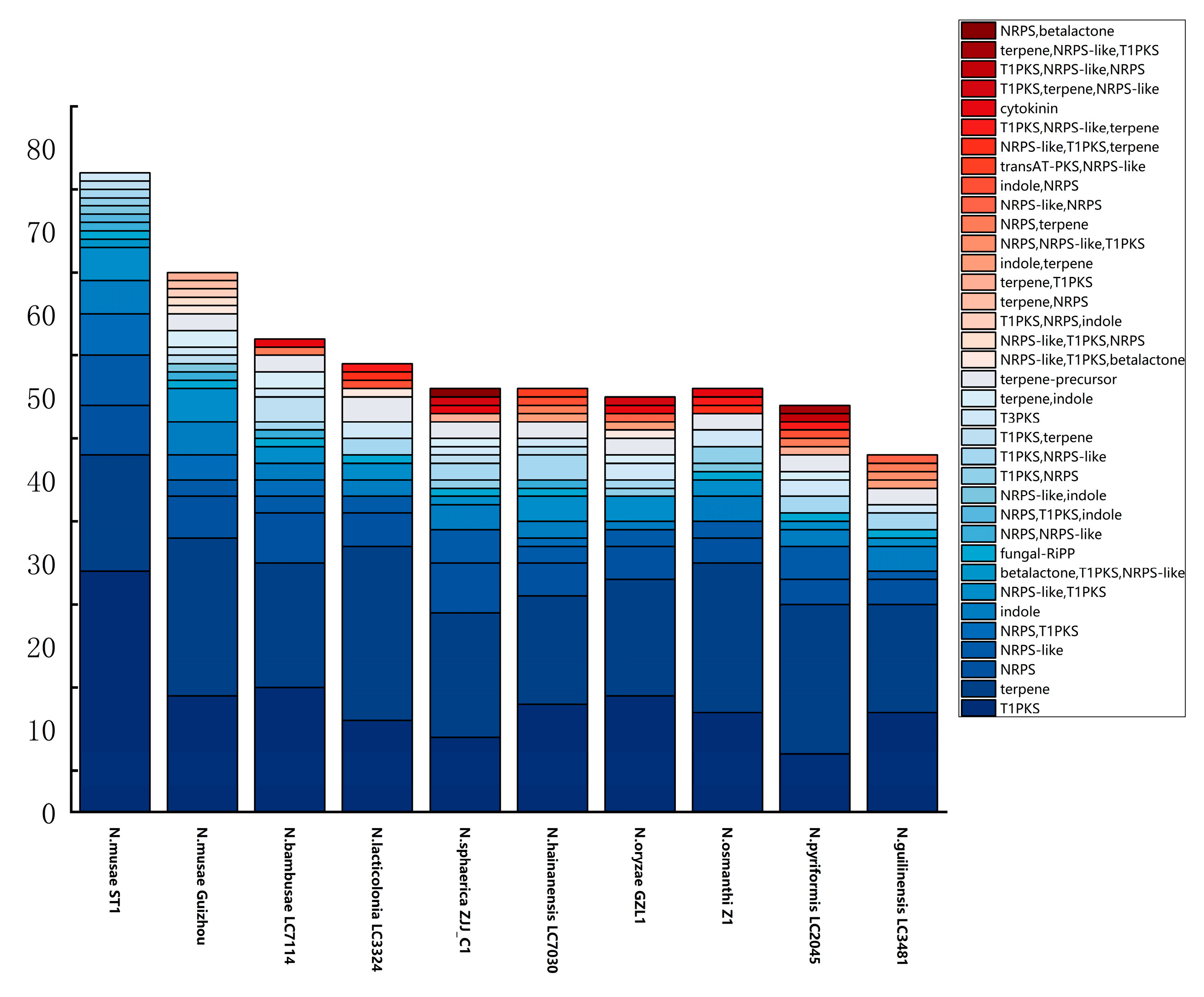
Figure A2.
Predicted carbohydrate-active enZYmes of N. musae ST1. AA, auxiliary activity; GH, glycoside hydrolase; GT, glycosyl transferase; PL, polysaccharide lyase; CE, carbohydrate esterase; CBM, carbohydrate-binding module.
References
- Yang, F.X.; Su, Y.Q.; Li, X.H.; Zhang, Q.; Sun, R.C. Preparation of biodiesel from Idesia polycarpa var. vestita fruit oil. Ind. Crops Prod. 2008, 29, 622–628. [Google Scholar] [CrossRef] [Scilit]
- Luo, T.T.; Xu, J.Q.; Zhen, Z.Q.; Pan, X.; Feng, L.; Qin, L.K.; Ren, T.Y. Quality analysis of Idesia polycarpa fruit oil samples from cultivars with different phenotypes. Food Chem. X 2025, 25, 102127. [Google Scholar] [CrossRef] [Scilit]
- Chen, Y.; Wang, Z.; Song, M.; Yang, Y. First report of Nigrospora musae causing Idesia polycarpa leaf spot in China. Plant Dis. 2025, 109, 1995. [Google Scholar] [CrossRef] [Scilit]
- Wang, M.; Liu, F.; Crous, P.W.; Cai, L. Phylogenetic reassessment of Nigrospora: Ubiquitous endophytes, plant and human pathogens. Persoonia Mol. Phylogeny Evol. Fungi 2017, 39, 118–142. [Google Scholar] [CrossRef] [Scilit]
- Rehman, A.; Alam, M.W.; Saira, M.; Naz, S.; Mushtaq, R.; Chohan, T.A.; Din, S.U.; Noureen, A.; Gilani, K.; Hussain, D. Nigrospora sphaerica Causing Leaf Blight Disease on Sesame in Pakistan. Plant Dis. 2021, 106, 317. [Google Scholar] [CrossRef] [Scilit] [PubMed]
- Liu, Z.; Zhou, S.; Qi, L.; Wang, X.; Song, J.; Li, D.; Chen, T.; Wang, Q. First Report of Nigrospora oryzae Causing Leaf Spot on Ginger in China. Plant Dis. 2021, 106, 316. [Google Scholar] [CrossRef] [Scilit]
- Zhang, H.F.; Kong, N.; Ji, S.D.; Liu, B.; Tian, Z.; Qi, J.Y.; Liu, Z.H. First Report of Leaf Blight Caused by Nigrospora oryzae on Poplar in China. Plant Dis. 2021, 106, 1063. [Google Scholar] [CrossRef] [Scilit]
- Chen, Y.; Yan, D.K.; Han, K.L.; Ma, C.; Gao, Z.L.; Bao, X.X.; Wang, F. First Report of Necrosis of Balloon Flower (Platycodon grandiflorus) Caused by Nigrospora sphaerica in China. Plant Dis. 2022, 106, 1297. [Google Scholar] [CrossRef] [Scilit]
- Liao, K.; Jiang, Y.L.; Xiao, T.; Peng, Y.Q.; Hao, Y.L.; Jin, C.Z.; Guo, K.F. First report of Nigrospora musae causing white leaf spot of Basella alba L. in China. Plant Dis. 2023, 107, 2261. [Google Scholar] [CrossRef] [Scilit]
- Jia, W.Y.; Luo, M.Y.; Wei, T.P.; Zhang, H.; Zeng, Y.; Jiang, Y.L. Nigrospora musae and N. oryzae as new causal agents of broad bean leaf spot disease in China. Crop Prot. 2024, 177, 106567. [Google Scholar] [CrossRef] [Scilit]
- Chen, X.Y.; Xu, X.; Yuan, H.; Luo, T.T.; Zhu, T.H.; Li, S.J.; Liu, Y.G.; Lin, T.T.; Qiao, T.M.; Yang, C.L.; et al. Nigrospora musae Causing Taxus chinensis var. mairei stem blight in Sichuan, China. Plant Dis. 2023, 107, 4032. [Google Scholar] [CrossRef] [Scilit]
- Hong, X.J.; Huang, S.X.; Liu, Y.S. A Genome Sequence Resource of Nigrospora sphaerica Causing Fruit Dried-Shrink Disease in Akebia trifoliata. Plant Dis. 2022, 106, 745–747. [Google Scholar] [CrossRef] [Scilit]
- Zhang, R.Q.; Bai, Y.G.; Hu, X.Y.; Wang, W.D.; Zhou, L.H.; Li, X.Y.; Yang, Y.D. Nigrospora oryzae Causing Basal Rot on Siberian Lily (Lilium spp.) Ornamental Hybrids in China. Plant Dis. 2025, 109, 1987. [Google Scholar] [CrossRef] [Scilit]
- Arunakumar, G.S.; Gnanesh, B.N.; Supriya, M.; Sivaprasad, V. First Report of Nigrospora sphaerica Causing Shot Hole Disease on Mulberry in India. Plant Dis. 2019, 103, 1783–1784. [Google Scholar] [CrossRef] [Scilit]
- Chen, Y.J.; Liu, Q.; Zhang, Y.J.; Jiang, Z.L.; Fu, H.L.; Wu, H.; Liu, M.J.; Jiang, J.H.; Li, L.D. Whole-genome sequence of Sclerotium delphinii, a pathogenic fungus of Dendrobium officinale southern blight. Genomic 2024, 116, 110932. [Google Scholar] [CrossRef] [Scilit]
- Liang, J.; Yin, D.; Shu, X.; Xiang, T.; Zhang, C.; Li, H.; Wang, A. Integrated Genome Sequencing and Transcriptome Analysis Identifies Candidate Pathogenicity Genes from Ustilago crameri. J. Fungi 2024, 10, 82. [Google Scholar] [CrossRef] [Scilit]
- Wang, T.; Chen, S.C.; Niu, Q.H.; Xu, G.L.; Lu, C.X.; Zhang, J. Genomic Sequence Resource of Talaromyces albobiverticillius, the Causative Pathogen of Pomegranate Pulp Rot Disease. J. Fungi 2023, 9, 909. [Google Scholar] [CrossRef] [Scilit]
- Luo, R.; Liu, B.; Xie, Y.; Li, Z.; Huang, W.; Yuan, J.; He, G.; Chen, Y.; Pan, Q.; Liu, Y.; et al. SOAPdenovo2: An empirically improved memory-efficient short-read de novo assembler. GigaScience 2012, 1, 18. [Google Scholar] [CrossRef] [Scilit] [PubMed]
- Li, R.; Li, Y.; Kristiansen, K.; Wang, J. SOAP: Short oligonucleotide alignment program. Bioinformatics 2008, 24, 713–714. [Google Scholar] [CrossRef] [Scilit]
- Cheng, H.Y.; Concepcion, G.T.; Feng, X.W.; Zhang, H.W.; Li, H. Haplotype-resolved de novo assembly using phased assembly graphs with hifiasm. Nat. Methods 2021, 18, 170–175. [Google Scholar] [CrossRef] [Scilit]
- Walker, B.J.; Abeel, T.; Shea, T.; Priest, M.; Abouelliel, A.; Sakthikumar, S.; Cuomo, C.A.; Zeng, Q.; Wortman, J.; Young, S.K.; et al. Pilon: An integrated tool for comprehensive microbial variant detection and genome assembly improvement. PLoS ONE 2014, 9, e112963. [Google Scholar] [CrossRef] [Scilit]
- Li, H.; Durbin, R. Fast and accurate short read alignment with Burrows-Wheeler Transform. Bioinformatics 2009, 25, 1754–1760. [Google Scholar] [CrossRef] [Scilit]
- Simao, F.A.; Waterhouse, R.M.; Ioannidis, P.; Kriventseva, E.V.; Zdobnov, E.M. BUSCO: Assessing genome assembly and annotation completeness with single-copy orthologs. Bioinformatics 2015, 31, 3210–3212. [Google Scholar] [CrossRef] [Scilit]
- Kim, D.; Langmead, B.; Salzberg, S.L. HISAT: A fast spliced aligner with low memory requirements. Nat. Methods 2015, 12, 357–360. [Google Scholar] [CrossRef] [Scilit] [PubMed]
- Pertea, M.; Pertea, G.M.; Antonescu, C.M.; Chang, T.C.; Mendell, J.T.; Salzberg, S.L. StringTie enables improved reconstruction of a transcriptome from RNA-seq reads. Nat. Biotechnol. 2015, 33, 290–295. [Google Scholar] [CrossRef] [Scilit]
- Djebali, S.; Davis, C.A.; Merkel, A.; Dobin, A.; Lassmann, T.; Mortazavi, A.; Tanzer, A.; Lagarde, J.; Lin, W.; Schlesinger, F.; et al. Landscape of transcription in human cells. Nature 2012, 489, 101–108. [Google Scholar] [CrossRef] [Scilit] [PubMed]
- Xu, Z.; Wang, H. LTR_FINDER: An efficient tool for the prediction of full-length LTR retrotransposons. Nucleic Acids Res. 2007, 35, W265–W268. [Google Scholar] [CrossRef] [Scilit] [PubMed]
- Han, Y.J.; Wessler, S.R. MITE-Hunter: A program for discovering miniature inverted-repeat transposable elements from genomic sequences. Nucleic Acids Res. 2010, 38, e199. [Google Scholar] [CrossRef] [Scilit]
- Price, A.L.; Jones, N.C.; Pevzner, P.A. De novo identification of repeat families in large genomes. Bioinformatics 2005, 21, i351–i358. [Google Scholar] [CrossRef] [Scilit]
- Edgar, R.C.; Myers, E.W. Identification and classification of genomic repeats. Bioinformatics 2005, 21, i152–i158. [Google Scholar] [CrossRef] [Scilit]
- Wicker, T.; Sabot, F.; Hua-Van, A.; Bennetzen, J.L.; Capy, P.; Chalhoub, B.; Flavell, A.; Leroy, P.; Morgante, M.; Panaud, O.; et al. A unified classification system for eukaryotic transposable elements. Nat. Rev. Genet. 2007, 8, 973–982. [Google Scholar] [CrossRef] [Scilit]
- Jurka, J.; Kapitonov, V.V.; Pavlicek, A.; Klonowski, P.; Kohany, O.; Walichiewicz, J. Repbase Update, a database of eukaryotic repetitive elements. Cytogenet. Genome Res. 2005, 110, 462–467. [Google Scholar] [CrossRef] [Scilit]
- Chen, N. Using RepeatMasker to identify repetitive elements in genomic sequences. Curr. Protoc. Bioinform. 2004, 5, 4–10. [Google Scholar] [CrossRef] [Scilit]
- Burge, C.; Karlin, S. Prediction of complete gene structures in human genomic DNA. J. Mol. Biol. 1997, 268, 78–94. [Google Scholar] [CrossRef] [Scilit] [PubMed]
- Stanke, M.; Waack, S. Gene prediction with a hidden Markov model and a new intron submodel. Bioinformatics 2003, 19, 215–225. [Google Scholar] [CrossRef] [Scilit] [PubMed]
- Majoros, W.H.; Pertea, M.; Salzberg, S.L. TigrScan and GlimmerHMM: Two open source ab initio eukaryotic gene-finders. Bioinformatics 2004, 20, 2878–2879. [Google Scholar] [CrossRef] [Scilit]
- Blanco, E.; Parra, G.; Guigó, R. Using Geneid to identify genes. Curr. Protoc. Bioinform. 2007, 18, 4.3.1–4.3.28. [Google Scholar] [CrossRef] [Scilit]
- Korf, I. Gene finding in novel genomes. BMC Bioinform. 2004, 5, 59. [Google Scholar] [CrossRef] [Scilit]
- Keilwagen, J.; Wenk, M.; Erickson, J.L.; Schattat, M.H.; Grau, J.; Hartung, F. Using intron position conservation for homology-based gene prediction. Nucleic Acids Res. 2016, 44, e89. [Google Scholar] [CrossRef] [Scilit] [PubMed]
- Pertea, M.; Kim, D.; Pertea, G.M.; Leek, J.T.; Salzberg, S.L. Transcript-level expression analysis of RNA-seq experiments with HISAT, StringTie and Ballgown. Nat. Protoc. 2016, 11, 1650–1667. [Google Scholar] [CrossRef] [Scilit]
- Haas, B.J.; Salzberg, S.L.; Zhu, W.; Pertea, M.; Allen, J.E.; Orvis, J.; White, O.; Buell, C.R.; Wortman, J.R. Automated eukaryotic gene structure annotation using EVidenceModeler and the Program to Assemble Spliced Alignments. Genome Biol. 2008, 9, R7. [Google Scholar] [CrossRef] [Scilit]
- Campbell, M.A.; Haas, B.J.; Hamilton, J.P.; Mount, S.M.; Buell, C.R. Comprehensive analysis of alternative splicing in rice and comparative analyses with Arabidopsis. BMC Genom. 2006, 7, 327. [Google Scholar] [CrossRef] [Scilit]
- Lowe, T.M.; Eddy, S.R. tRNAscan-SE: A program for improved detection of transfer RNA genes in genomic sequence. Nucleic Acids Res. 1997, 25, 955–964. [Google Scholar] [CrossRef] [PubMed]
- Nawrocki, E.P.; Eddy, S.R. Infernal 1.1: 100-fold faster RNA homology searches. Bioinformatics 2013, 29, 2933–2935. [Google Scholar] [CrossRef] [Scilit] [PubMed]
- Nawrocki, E.P.; Burge, S.W.; Bateman, A.; Daub, J.; Eberhardt, R.Y.; Eddy, S.R.; Floden, E.W.; Gardner, P.P.; Jones, T.A.; Tate, J.; et al. Rfam 12.0: Updates to the RNA families database. Nucleic Acids Res. 2015, 43, D130–D137. [Google Scholar] [CrossRef] [Scilit] [PubMed]
- Krzywinski, M.; Schein, J.; Birol, I.; Connors, J.; Gascoyne, R.; Horsman, D.; Jones, S.J.; Marra, M.A. Circos: An information aesthetic for comparative genomics. Genome Res. 2009, 19, 1639–1645. [Google Scholar] [CrossRef] [Scilit]
- She, R.; Chu, J.S.C.; Wang, K.; Pei, J.; Chen, N.S. GenBlastA: Enabling BLAST to identify homologous gene sequences. Genome Res. 2009, 19, 143–149. [Google Scholar] [CrossRef] [Scilit]
- Birney, E.; Clamp, M.; Durbin, R. GeneWise and genomewise. Genome Res. 2004, 14, 988–995. [Google Scholar] [CrossRef] [Scilit]
- Blin, K.; Shaw, S.; Kloosterman, A.M.; Charlop-Powers, Z.; van Wezel, G.P.; Medema, M.H.; Weber, T. antiSMASH 6.0: Improving cluster detection and comparison capabilities. Nucleic Acids Res. 2021, 49, W29–W35. [Google Scholar] [CrossRef] [Scilit]
- Tatusov, R.L.; Galperin, M.Y.; Natale, D.A.; Koonin, E.V. The COG database: A tool for genome-scale analysis of protein functions and evolution. Nucleic Acids Res. 2000, 28, 33–36. [Google Scholar] [CrossRef] [Scilit]
- Kanehisa, M.; Goto, S.; Kawashima, S.; Okuno, Y.; Hattori, M. The KEGG resource for deciphering the genome. Nucleic Acids Res. 2004, 32, D277–D280. [Google Scholar] [CrossRef] [Scilit]
- Boeckmann, B.; Bairoch, A.; Apweiler, R.; Blatter, M.C.; Estreicher, A.; Gasteiger, E.; Martin, M.J.; Michoud, K.; O’Donovan, C.; Phan, I.; et al. The SWISS-PROT protein knowledgebase and its supplement TrEMBL in 2003. Nucleic Acids Res. 2003, 31, 365–370. [Google Scholar] [CrossRef] [Scilit]
- Deng, Y.Y.; Li, J.Q.; Wu, S.F.; Zhu, Y.; Chen, Y.; He, F.C. Integrated nr database in protein annotation system and its localization. Comput. Eng. 2006, 32, 71–74. [Google Scholar]
- Altschul, S.F.; Madden, T.L.; Schaffer, A.A.; Zhang, J.; Zhang, Z.; Miller, W.; Lipman, D.J. Gapped BLAST and PSI-BLAST: A new generation of protein database search programs. Nucleic Acids Res. 1997, 25, 3389–3402. [Google Scholar] [CrossRef] [Scilit] [PubMed]
- Conesa, A.; Götz, S.; García-Gómez, J.M.; Terol, J.; Talón, M.; Robles, M. Blast2GO: A universal tool for annotation, visualization and analysis in functional genomics research. Bioinformatics 2005, 21, 3674–3676. [Google Scholar] [CrossRef] [Scilit]
- Ashburner, M.; Ball, C.A.; Blake, J.A.; Botstein, D.; Butler, H.; Cherry, J.M.; Davis, A.P.; Dolinski, K.; Dwight, S.S.; Eppig, J.T.; et al. Gene Ontology: Tool for the unification of biology. Nat. Genet. 2000, 25, 25–29. [Google Scholar] [CrossRef] [Scilit] [PubMed]
- Eddy, S.R. Profile hidden Markov models. Bioinformatics 1998, 14, 755–763. [Google Scholar] [CrossRef] [Scilit] [PubMed]
- Finn, R.D.; Coggill, P.; Eberhardt, R.Y.; Eddy, S.R.; Mistry, J.; Mitchell, A.L.; Potter, S.C.; Punta, M.; Qureshi, M.; Sangrador-Vegas, A.; et al. The Pfam protein families database: Towards a more sustainable future. Nucleic Acids Res. 2016, 44, D279–D285. [Google Scholar] [CrossRef] [Scilit]
- Saier, M.H., Jr.; Tran, C.V.; Barabote, R.D. TCDB: The Transporter Classification Database for membrane transport protein analyses and information. Nucleic Acids Res. 2006, 34, D181–D186. [Google Scholar] [CrossRef] [Scilit]
- Winnenburg, R.; Baldwin, T.K.; Urban, M.; Rawlings, C.; Köhler, J.; Hammond-Kosack, K.E. PHI-base: A new database for pathogen host interactions. Nucleic Acids Res. 2006, 34, D459–D464. [Google Scholar] [CrossRef] [Scilit]
- Fischer, M.; Knoll, M.; Sirim, D.; Wagner, F.; Funke, S.; Pleiss, J. The Cytochrome P450 Engineering Database: A navigation and prediction tool for the cytochrome P450 protein family. Bioinformatics 2007, 23, 2015–2017. [Google Scholar] [CrossRef] [Scilit]
- Lu, T.; Yao, B.; Zhang, C. DFVF: Database of fungal virulence factors. Database 2012, 2012, bas032. [Google Scholar] [CrossRef] [Scilit]
- Cantarel, B.L.; Coutinho, P.M.; Rancurel, C.; Bernard, T.; Lombard, V.; Henrissat, B. The Carbohydrate-Active EnZymes database (CAZy): An expert resource for glycogenomics. Nucleic Acids Res. 2009, 37, D233–D238. [Google Scholar] [CrossRef] [Scilit] [PubMed]
- Petersen, T.N.; Brunak, S.; von Heijne, G.; Nielsen, H. SignalP 4.0: Discriminating signal peptides from transmembrane regions. Nat. Methods 2011, 8, 785–786. [Google Scholar] [CrossRef] [Scilit]
- Krogh, A.; Larsson, B.; von Heijne, G.; Sonnhammer, E.L. Predicting transmembrane protein topology with a hidden Markov model: Application to complete genomes. J. Mol. Biol. 2001, 305, 567–580. [Google Scholar] [CrossRef] [Scilit]
- Sperschneider, J.; Gardiner, D.M.; Dodds, P.N.; Tini, F.; Covarelli, L.; Singh, K.B.; Manners, J.M.; Taylor, J.M. EffectorP: Predicting Fungal Effector Proteins from Secretomes Using Machine Learning. New Phytol. 2016, 210, 743–761. [Google Scholar] [CrossRef] [Scilit]
- Fujii, I.; Yoshida, N.; Shimomaki, S.; Oikawa, H.; Ebizuka, Y. An Iterative Type I Polyketide Synthase PKSN Catalyzes Synthesis of the Decaketide Alternapyrone with Regio-Specific Octa-Methylation. Chem. Biol. 2005, 12, 1301–1309. [Google Scholar] [CrossRef] [Scilit]
- Zhang, H.; Feng, J.; Wang, D.; Tang, B.; Xu, C.; Ye, T. Total Synthesis and Stereochemical Assignment of Alternapyrone. Molecules 2025, 30, 1597. [Google Scholar] [CrossRef] [Scilit]
- Li, H.; Hu, J.; Wei, H.; Solomon, P.S.; Vuong, D.; Lacey, E.; Stubbs, K.A.; Piggott, A.M.; Chooi, Y.H. Chemical Ecogenomics-Guided Discovery of Phytotoxic α-Pyrones from the Fungal Wheat Pathogen Parastagonospora nodorum. Org. Lett. 2018, 20, 6148–6152. [Google Scholar] [CrossRef] [Scilit] [PubMed]
- Gsaller, F.; Blatzer, M.; Abt, B.; Schrettl, M.; Lindner, H.; Haas, H. The first promoter for conditional gene expression in Acremonium chrysogenum: Iron starvation-inducible mir1 P. J. Biotechnol. 2013, 163, 77–80. [Google Scholar] [CrossRef] [Scilit] [PubMed]
- Krasnoff, S.B.; Keresztes, I.; Donzelli, B.G.; Gibson, D.M. Metachelins, mannosylated and N-oxidized coprogen-type siderophores from Metarhizium robertsii. J. Nat. Prod. 2014, 77, 1685–1692. [Google Scholar] [CrossRef] [Scilit]
- Renshaw, J.C.; Robson, G.D.; Trinci, A.P.J.; Wiebe, M.G.; Livens, F.R.; Collison, D.; Taylor, R.J. Fungal siderophores: Structures, functions and applications. Mycol. Res. 2002, 106, 1123–1142. [Google Scholar] [CrossRef] [Scilit]
- Krasnoff, S.B.; Keresztes, I.; Gillilan, R.E.; Szebenyi, D.M.E.; Donzelli, B.G.; Churchill, A.C.; Gibson, D.M. Serinocyclins A and B, cyclic heptapeptides from Metarhizium anisopliae. J. Nat. Prod. 2007, 70, 919–924. [Google Scholar] [CrossRef] [Scilit]
- Jackson-Hayes, L.; Hill, T.W.; Loprete, D.M.; Fay, L.M.; Gordon, B.S.; Nkashama, S.A.; Patel, R.K.; Sartain, C.V. Two GDP-mannose transporters contribute to hyphal form and cell wall integrity in Aspergillus nidulans. Microbiology 2008, 154, 2037–2047. [Google Scholar] [CrossRef] [Scilit] [PubMed]
- Kadry, A.A.; El-Ganiny, A.M.; Mosbah, R.A.; Kaminskyj, S.G.W. Deletion of Aspergillus nidulans GDP-mannose transporters affects hyphal morphometry, cell wall architecture, spore surface character, cell adhesion, and biofilm formation. Med. Mycol. 2018, 56, 621–630. [Google Scholar] [CrossRef] [Scilit] [PubMed]
- Hayashi, K.; Schoonbeek, H.J.; De Waard, M.A. Expression of the ABC transporter BcatrD from Botrytis cinerea reduces sensitivity to sterol demethylation inhibitor fungicides. Pestic. Biochem. Physiol. 2002, 73, 110–121. [Google Scholar] [CrossRef] [Scilit]
- Hayashi, K.; Schoonbeek, H.J.; Sugiura, H.; Waard, M.A.D. Multidrug resistance in Botrytis cinerea with decreased accumulation of the azole fungicide oxpoconazole and increased transcription of the ABC transporter gene BcatrD. Pestic. Biochem. Physiol. 2001, 70, 168–179. [Google Scholar] [CrossRef] [Scilit]
- Urban, M.; Bhargava, T.; Hamer, J.E. An ATP–driven efflux pump is a novel pathogenicity factor in rice blast disease. EMBO J. 1999, 18, 512–521. [Google Scholar] [CrossRef] [Scilit]
- Bhattacharyya, S.; Sinha, K.; Sil, P.C. CytochromeP450s: Mechanisms and biological implications in drug metabolism and its interaction with oxidative stress. Curr. Drug Metab. 2014, 15, 719–742. [Google Scholar] [CrossRef] [Scilit]
- Jones, J.D.G.; Dangl, J.L. The plant immune system. Nature 2006, 444, 323–329. [Google Scholar] [CrossRef] [Scilit] [PubMed]
- Liu, D.; Lun, Z.Q.; Liu, N.; Yuan, G.; Wang, X.; Li, S.; Peng, Y.L.; Lu, X. Identification and characterization of novel candidate effector proteins from Magnaporthe oryzae. J. Fungi 2023, 9, 574. [Google Scholar] [CrossRef] [Scilit]
- Jones, D.A.B.; Bertazzoni, S.; Turo, C.J.; Syme, R.A.; Hane, J.K. Bioinformatic prediction of plant–pathogenicity effector proteins of fungi. Curr. Opin. Microbiol. 2018, 46, 49. [Google Scholar] [CrossRef] [Scilit]
- Zhao, Q.; Zhang, L.Y.; Wu, J.Z. Genome Sequencing and Analysis of Nigrospora oryzae, a Rice Leaf Disease Fungus. J. Fungi 2024, 10, 100. [Google Scholar] [CrossRef] [Scilit]
- Wang, Y.F.; Yao, J.A.; Li, Z.Q.; Huo, J.F.; Zhou, S.Q.; Liu, W.D.; Wu, H.X. Genome Sequence Resource for Nigrospora oryzae, an important pathogenic fungus threatening crop production. Mol. Plant Microbe Interact. 2021, 34, 835–838. [Google Scholar] [CrossRef] [Scilit] [PubMed]
- Zhang, Y.Y.; Zhang, T.; Li, H.Y.; Zheng, R.; Ren, J.; Yang, Q.; Jiang, N. Nigrospora humicola (Apiosporaceae, Amphisphaeriales), a New Fungus from Soil in China. Diversity 2024, 16, 118. [Google Scholar] [CrossRef] [Scilit]
- Liu, Y.; An, J.H.; Safdar, A.; Shen, Y.; Sun, Y.; Shu, W.H.; Tan, X.J.; Zhu, B.; Xiao, J.X.; Schirawski, J.; et al. Identification and Characterization of Nigrospora Species and a Novel Species, Nigrospora anhuiensis, Causing Black Leaf Spot on Rice and Wild Rice in the Anhui Province of China. J. Fungi 2024, 10, 156. [Google Scholar] [CrossRef] [Scilit]
- Yang, C.; Liu, R.; Pang, J.H.; Ren, B.; Zhou, H.B.; Wang, G.; Wang, E.T. Poaceae-specific cell wall-derived oligosaccharides activate plant immunity via OsCERK1 during Magnaporthe oryzae infection in rice. Nat. Commun. 2021, 12, 2178. [Google Scholar] [CrossRef] [Scilit] [PubMed]
- Ospina-Giraldo, M.D.; Griffith, J.G.; Laird, E.W.; Mingora, C. The CAZyome of Phytophthora spp.: A comprehensive analysis of the gene complement coding for carbohydrate-active enzymes in species of the genus Phytophthora. BMC Genom. 2010, 11, 525. [Google Scholar] [CrossRef] [Scilit]
- Villanueva, O.; Nguyen, H.D.T.; Ellouze, W. Profiling the Genomes and Secreted Effector Proteins in Phytopythium vexans Global Strains. J. Fungi 2025, 11, 477. [Google Scholar] [CrossRef] [Scilit]
- Lamour, K.H.; Mudge, J.; Gobena, D.; Hurtado-Gonzales, O.P.; Schmutz, J.; Kuo, A.; Miller, N.A.; Rice, B.J.; Raffaele, S.; Cano, L.M.; et al. Genome sequencing and mapping reveal loss of heterozygosity as a mechanism for rapid adaptation in the vegetable pathogen Phytophthora capsici. Mol. Plant Microb. Interact 2012, 25, 1350–1360. [Google Scholar] [CrossRef] [Scilit]
- Stam, R.; Jupe, J.; Howden, A.J.; Morris, J.A.; Boevink, P.C.; Hedley, P.E.; Huitema, E. Identification and characterisation CRN effectors in Phytophthora capsici shows modularity and functional diversity. PLoS ONE 2013, 8, e59517. [Google Scholar] [CrossRef] [Scilit]
- Harris, P.V.; Welner, D.; McFarland, K.C.; Re, E.; Poulsen, J.C.N.; Brown, K.; Salbo, R.; Ding, H.S.; Vlasenko, E.; Merino, S.; et al. Stimulation of Lignocellulosic Biomass Hydrolysis by Proteins of Glycoside Hydrolase Family 61: Structure and Function of a Large, Enigmatic Family. Biochemistry 2010, 49, 3305–3316. [Google Scholar] [CrossRef] [Scilit]
- Horn, S.J.; Vaaje-Kolstad, G.; Westereng, B.; Eijsink, V. Novel enzymes for the degradation of cellulose. Iotechnol. Biofuels 2012, 5, 45. [Google Scholar] [CrossRef] [Scilit]
- Guo, M.; Si, E.; Wang, Y.; Wang, J.; Yao, L.; Meng, Y.; Ma, X.; Li, B.; Zhang, H.; Yang, K.; et al. The glycoside hydrolase 61 member PgGH61–2 of Pyrenophora graminea is required for full virulence on barley (Hordeum vulgare). Int. J. Biol. Macromol. 2025, 3, 147577. [Google Scholar] [CrossRef] [Scilit]
- Gacura, M.D.; Sprockett, D.D.; Heidenreich, B.; Blackwood, C.B. Comparison of pectin-degrading fungal communities in temperate forests using glycosyl hydrolase family 28 pectinase primers targeting Ascomycete fungi. J. Microbiol. Methods 2016, 123, 108–113. [Google Scholar] [CrossRef] [Scilit]
- Urban, M.; Cuzick, A.; Seager, J.; Wood, V.; Rutherford, K.; Venkatesh, S.Y.; De Silva, N.; Martinez, M.C.; Pedro, H.; Yates, A.D.; et al. PHI-base: The pathogen–host interactions database. Nucleic Acids Res. 2020, 48, 613–620. [Google Scholar] [CrossRef] [Scilit] [PubMed]
- Peng, M.; Wang, J.; Lu, X.; Wang, M.; Wen, G.; Wu, C.; Lu, G.; Wang, Z.; Shim, W.B.; Yu, W.Y. Functional analysis of cutinase transcription factors in Fusarium verticillioides. Phytopathol. Res. 2024, 6, 48. [Google Scholar] [CrossRef] [Scilit]
- Liu, L.; Li, C.; Liang, F.; Han, S.; Li, S.; Yang, C.; Liu, Y. Global characterization of GH11 family xylanases genes in Neostagonosporella sichuanensis and functional analysis of Nsxyn1 and Nsxyn2. Front. Microbiol. 2024, 15, 1507998. [Google Scholar] [CrossRef] [Scilit]
- Zhang, C.; Yang, C.; Jin, M.; Feng, Z.; Osei, R.; Cai, F.; Ma, T.; Wang, Y. A PL1 family pectate lyase CP966RS08110 gene was the pathogenic factor of Streptomyces galilaeus 5T-1 causing potato common scab. Front. Microbiol. 2024, 15, 1469709. [Google Scholar] [CrossRef] [Scilit]
- Cai, L.; Xu, X.; Dong, Y.; Jin, Y.; Rashad, Y.M.; Ma, D.; Gu, A. Roles of Three FgPel Genes in the Development and Pathogenicity Regulation of Fusarium graminearum. J. Fungi 2024, 10, 666. [Google Scholar] [CrossRef] [Scilit]
- Li, W.; Li, P.; Deng, Y.; Situ, J.; He, Z.; Zhou, W.; Li, M.; Xi, P.; Liang, X.; Kong, G.; et al. A plant cell death-inducing protein from litchi interacts with Peronophythora litchii pectate lyase and enhances plant resistance. Nat. Commun. 2024, 15, 22. [Google Scholar] [CrossRef] [Scilit]
- Zhang, Q.; Chen, X.; You, H.; Chen, B.; Jia, L.; Li, S.; Zhang, X.; Ma, J.; Wu, X.; Wang, K.; et al. Specific selection on XEG1 and XLP1 genes correlates with host range and adaptability in Phytophthora. Nat. Commun. 2025, 16, 3638. [Google Scholar] [CrossRef] [Scilit]
- Liu, H.L.; Chen, S.S.; Gao, X.; Qian, H.J.; Wang, Y.H.; Zhang, D.Y.; Xu, L.S.; Huang, L.L. The secreted elicitor protein VmHrp1 from Valsa mali activates plant immunity through RLP26 to enhance disease resistance. Plant J. 2025, 123, e70393. [Google Scholar] [CrossRef] [Scilit]
- Xu, Z.; Luo, Z.; Xiong, D.; Gao, M.; Tian, C. A glycoside hydrolase 12 protein from Cytospora chrysosperma triggers plant immunity but is not essential to virulence. Phytopathol. Res. 2023, 5, 32. [Google Scholar] [CrossRef] [Scilit]
- Keller, N.P.; Turner, G.; Bennett, J.W. Fungal secondary metabolism—From biochemistry to genomics. Nat. Rev. Microbiol. 2005, 3, 937–947. [Google Scholar] [CrossRef] [Scilit]
- Yuan, X.; Li, Y.; Luo, T.; Bi, W.; Yu, J.; Wang, Y. Genomic Analysis of the Xanthoria elegans and Polyketide Synthase Gene Mining Based on the Whole Genome. Mycobiology 2023, 51, 36–48. [Google Scholar] [CrossRef] [Scilit]
- Yang, Y.; Yu, L.; Qiu, X.; Xiong, D.; Tian, C. A putative terpene cyclase gene (CcPtc1) is required for fungal development and virulence in Cytospora chrysosperma. Front. Microbiol. 2023, 14, 1084828. [Google Scholar] [CrossRef] [Scilit] [PubMed]
- Safronov, O.; Bal, G.L.; Sipari, N.; Wilkens, M. Independent evolution of betulin biosynthesis in Inonotus obliquus. Sci. Rep. 2025, 15, 21319. [Google Scholar] [CrossRef] [Scilit] [PubMed]
- Proctor, R.H.; McCormick, S.P.; Alexander, N.J.; Desjardins, A.E. Evidence that a secondary metabolic biosynthetic gene cluster has grown by gene relocation during evolution of the filamentous fungus Fusarium. Mol. Microbiol. 2009, 74, 1128–1142. [Google Scholar] [CrossRef] [Scilit] [PubMed]
- Xu, D.; Xue, M.Y.; Shen, Z.; Jia, X.W.; Hou, X.W.; Lai, D.W.; Zhou, L.G. Phytotoxic secondary metabolites from fungi. Toxins 2021, 13, 261. [Google Scholar] [CrossRef] [Scilit]
- Baker, S.E.; Kroken, S.; Inderbitzin, P.; Asvarak, T.; Li, B.Y.; Shi, L.; Yoder, O.C.; Turgeon, B.G. Synthase-encoding genes are required for biosynthesis of the polyketide virulence factor, T-toxin, by Cochliobolus heterostrophus. Mol. Plant Microbe Interact. 2006, 19, 139–149. [Google Scholar] [CrossRef] [Scilit]
- Braun, H.; Woitsch, L.; Hetzer, B.; Geisen, R.; Zange, B.; Schmidt-Heydt, M. Trichoderma harzianum: Inhibition of mycotoxin producing fungi and toxin biosynthesis. Int. J. Food Microbiol. 2018, 280, 10–16. [Google Scholar] [CrossRef] [Scilit] [PubMed]
- Langevin, F.O.; Eudes, F.O.; Comeau, A. Effect of Trichothecenes Produced by Fusarium graminearum during Fusarium Head Blight Development in Six Cereal Species. Eur. J. Plant Pathol. 2004, 110, 735–746. [Google Scholar] [CrossRef] [Scilit]
- Izumi, Y.; Ohtani, K.; Miyamoto, Y.; Masunaka, A.; Fukumoto, T.; Gomi, K.; Tada, Y.; Ichimura, K.; Peever, T.L.; Akimitsu, K. A Polyketide Synthase Gene, ACRTS2, Is Responsible for Biosynthesis of Host-Selective ACR-Toxin in the Rough Lemon Pathotype of Alternaria alternata. Mol. Plant Microbe Interact 2012, 25, 1419–1429. [Google Scholar] [CrossRef] [Scilit] [PubMed]
- Luo, X.M.; Cao, J.D.; Huang, J.K.; Wang, Z.Y.; Guo, Z.Y.; Chen, Y.H.; Ma, S.M.; Liu, J. Genome sequencing and comparative genomics reveal the potential pathogenic mechanism of Cercospora sojina Hara on soybean. DNA Res. 2018, 25, 25–37. [Google Scholar] [CrossRef] [Scilit] [PubMed]
- Hatta, R.; Ito, K.; Hosaki, Y.; Tanaka, T.; Tanaka, A.; Yamamoto, M.; Akimitsu, K.; Tsuge, T. A conditionally dispensable chromosome controls host-specific pathogenicity in the fungal plant pathogen Alternaria alternata. Genetics 2002, 161, 59–70. [Google Scholar] [CrossRef] [Scilit]
- Malz, S.; Grell, M.N.; Thrane, C.; Maier, F.J.; Rosager, P.; Felk, A.; Albertsen, K.S.; Salomon, S.; Bohn, L.; Schäfer, W.; et al. Identification of a gene cluster responsible for the biosynthesis of aurofusarin in the Fusarium graminearum species complex. Fungal Genet. Biol. 2005, 42, 420–433. [Google Scholar] [CrossRef] [Scilit]
- Palonen, E.K.; Raina, S.; Brandt, A.; Meriluoto, J.; Keshavarz, T.; Soini, J.T. Melanisation of Aspergillus terreus—Is Butyrolactone I Involved in the Regulation of Both DOPA and DHN Types of Pigments in Submerged Culture? Microorganisms 2017, 5, 22. [Google Scholar] [CrossRef] [Scilit]
- Feng, Y.Q.; Yin, Z.Y.; Wu, Y.X.; Xu, L.S.; Du, H.X.; Wang, N.N.; Huang, L.L. LaeA controls virulence and secondary metabolism in apple canker pathogen Valsa mali. Front. Microbiol. 2020, 11, 581203. [Google Scholar] [CrossRef] [Scilit]
- Meng, L.L.; Sun, C.C.; Gao, L.Y.; Saleem, M.; Li, B.H.; Wang, C.X. Hydroxybenzoate hydroxylase genes underlying protocatechuic acid production in Valsa mali are required for full pathogenicity in apple trees. Mol. Plant Pathol. 2021, 22, 1370–1382. [Google Scholar] [CrossRef] [Scilit]
- Silva-Valderrama, I.; Úrbez-Torres, J.R.; Davies, T.J. Co-phylogenetic constraints on host breadth within an emerging fungal pathogen complex of global concern. Ecol. Monogr. 2025, 95, e70027. [Google Scholar] [CrossRef] [Scilit]
- Baroncelli, R.; Cobo-Díaz, J.F.; Benocci, T.; Peng, M.; Battaglia, E.; Haridas, S.; Andreopoulos, W.; LaButti, K.; Pangilinan, J.; Lipzen, A.; et al. Genome evolution and transcriptome plasticity is associated with adaptation to monocot and dicot plants in Colletotrichum fungi. GigaScience 2024, 13, giae036. [Google Scholar] [CrossRef] [Scilit] [PubMed]

Disclaimer/Publisher’s Note: The statements, opinions and data contained in all publications are solely those of the individual author(s) and contributor(s) and not of MDPI and/or the editor(s). MDPI and/or the editor(s) disclaim responsibility for any injury to people or property resulting from any ideas, methods, instructions or products referred to in the content. |
© 2026 by the authors. Licensee MDPI, Basel, Switzerland. This article is an open access article distributed under the terms and conditions of the Creative Commons Attribution (CC BY) license.