


Abstract
Cucurbitaceous plants comprise a wide range of economically important vegetable and fruit crops. In this study, Alternaria species associated with Cucurbitaceae were investigated using an integrative approach combining multilocus phylogenetic analyses and morphological characterization. Two novel species, Alternaria charantiicola sp. nov. and A. cucumicola sp. nov., were identified from Momordica charantia and Cucumis sp., respectively. In addition, five Alternaria species, namely A. zeae, A. lycopersici, A. sanguisorbae, A. pulvinifungicola, and A. solanicola, are reported for the first time from cucurbitaceous hosts. These findings expand the known species diversity of Alternaria associated with Cucurbitaceae in China and provide a taxonomic basis for the accurate diagnosis of Alternaria-related diseases in cucurbit crops.
1. Introduction
Cucurbitaceae is one of the most important families of food plants and is commonly referred to as the cucurbits or the gourd family [1]. In terms of agricultural importance, it ranks after Poaceae, Fabaceae, and Solanaceae [2]. The family comprises approximately 115 genera and 960 species, which are mainly distributed in tropical and subtropical regions, with a few species extending into temperate areas [3]. In China, Cucurbitaceae includes about 32 genera and 154 species, predominantly distributed in southern and southwestern regions, with some species also occurring in northern China [4]. Cucurbitaceous plants encompass a wide range of economically important vegetable and fruit crops, including cucumber (Cucumis sativus), pumpkin (Cucurbita pepo, C. moschata, C. argyrosperma), wax gourd (Benincasa hispida), and bitter gourd (Momordica charantia), as well as fruit crops such as watermelon (Citrullus lanatus), melon (C. melo), and monk fruit (Siraitia grosvenorii) [1,5]. Among them, M. charantia and S. grosvenorii are notable for their combined culinary and medicinal value and have traditionally been used for cough relief, heat alleviation, regulation of intestinal function, and strengthening of the spleen and stomach [5]. However, the frequent occurrence and rapid spread of diseases have become a major bottleneck constraining the sustainable development of Cucurbitaceae crop production. In particular, fungal diseases cause severe yield losses in cucurbit crops, including powdery mildew caused by Erysiphe cichoracearum, downy mildew caused by Pseudoperonospora cubensis, Fusarium wilt caused by Fusarium oxysporum, and root rot caused by F. solani [5]. Notably, the genus Alternaria has been widely reported from Cucurbitaceae, including A. alternata [6], A. baoshanensis [7], A. brassicae [8], A. caudata [8], A. cucumericola [8], A. cucumerina [6], A. gaisen [8], A. granulosa [8], A. hydrangea [9], A. jingzhouensis [10], A. infectoria [8], A. loofahae [8], A. momordicae [10], A. nigrescens [8], A. peponicola [8], A. peponis [8], and A. tenuissima [6].
The genus Alternaria was established by Nees in 1816 and typified by A. tenuis [11]. It ranks among the top ten most cited fungal genera and is associated with more than 4000 host plant species worldwide [7,12,13,14]. To date, over 400 species have been recognized within Alternaria, encompassing saprophytic, endophytic, and phytopathogenic lifestyles [12,13,15,16]. Historically, species delimitation in Alternaria relied primarily on morphological characteristics, including colony features, conidial morphology, and sporulation patterns [8]. Based on conidial size, the genus was traditionally divided into large-spored taxa (60–100 μm) and small-spored taxa (<60 μm). However, the taxonomy of small-spored Alternaria has long been problematic owing to their highly conserved and overlapping morphological traits [8]. In recent decades, DNA-based molecular approaches have greatly improved the resolution of phylogenetic relationships within Alternaria [7,13,14]. Early multilocus analyses tended to cluster many small-spored taxa in section Alternaria, containing 11 species and one species complex [14]. With the continued discovery and description of new small-spored species, taxonomic boundaries within section Alternaria have become increasingly refined, and species delimitation has gradually become clearer. To date, more than 30 phylogenetically distinct species have been accepted within this section, highlighting the previously underestimated diversity and complexity of this group [7,10,15,16,17,18]. Recent studies have shown that many taxa within section Alternaria form species complexes characterized by subtle morphological differentiation yet distinct multilocus phylogenetic lineages [7,15]. The integration of multilocus datasets with emerging phylogenomic evidence has progressively refined species boundaries, uncovering extensive cryptic diversity among small-spored Alternaria [14,19].
In this study, Alternaria species associated with Cucurbitaceae in China were examined through a comprehensive taxonomic investigation. An integrative approach combining multilocus phylogenetic analyses with morphological characterization was applied, leading to the recognition of two novel species and five newly recorded species from symptomatic cucurbitaceous leaves. This study establishes a taxonomic baseline for understanding the diversity of Alternaria associated with Cucurbitaceae.
2. Materials and Methods
2.1. Sampling and Isolation
Plants exhibiting symptoms resembling Alternaria leaf spot or blight were collected from three Cucurbitaceae genera (Citrullus, Cucumis, and Momordica) across six provinces in China (Hainan, Heilongjiang, Hubei, Guangxi, Jilin, and Liaoning) during 2016–2024. Symptomatic tissues were excised into small fragments (approximately 2 × 2 mm) using sterile blades and placed on moist filter paper in Petri dishes, followed by incubation at 25 °C in the dark. Single conidia were examined under a stereomicroscope and aseptically transferred onto potato dextrose agar (PDA: Difco, Montreal, QC, Canada) to obtain pure cultures. All plant specimens and fungal isolates were deposited in the Fungal Herbarium of Yangtze University (Jingzhou, China). Dried cultures of isolates were preserved in the same collection for long-term storage.
2.2. Morphological Characteristics
Colony characteristics were examined on 90 mm PDA plates incubated at 25 °C in the dark for 7 days. Conidial morphology, including size, shape, and sporulation patterns, was assessed on potato carrot agar (PCA) and V8 juice agar (V8A) following incubation at 22 °C under an 8 h light/16 h dark photoperiod. After 7 days, conidia and sporulation structures were observed and photographed using a Nikon Eclipse Ni-U microscope (Nikon, Tokyo, Japan). Conidial dimensions were measured from 50 randomly selected conidia per isolate.
2.3. DNA Extraction, PCR Amplification, and Sequencing
Fresh mycelia of each isolate were harvested from 5–7-day-old colonies grown on PDA. Genomic DNA was extracted using a modified CTAB method [20]. Seven gene fragments, including the internal transcribed spacer (ITS) region of rDNA, glyceraldehyde-3-phosphate dehydrogenase (GAPDH), translation elongation factor 1-alpha (TEF1), RNA polymerase II second largest subunit (RPB2), Alternaria major allergen gene (Alt a 1), endopolygalacturonase (EndoPG) gene, and an anonymous gene region (OPA10-2), were amplified using the primer pairs ITS5/ITS4 [21], gpd1/gpd2 [22], EF1-728F/EF1-986R [23], RPB2-5F/RPB2-7cR [24], Alt-for/Alt-rev [25], PG3/PG2b [26], and OPA10-2L/OPA10-2R [26], respectively. PCR amplifications were performed in a Bio-Rad T100™ Thermal Cycler (Bio-Rad, Hercules, CA, USA) using a total reaction volume of 25 μL, which consisted of 21 μL of 1.1× Taq PCR Star Mix (TSINGKE Co., Ltd., Beijing, China), 1 μL of each primer (10 μM), and 2 μL of template DNA. The PCR cycling conditions followed those described previously [18]. PCR products were verified by electrophoresis on 1% agarose gels and sequenced commercially by TSINGKE (Beijing, China). All newly generated sequences were deposited in GenBank (https://www.ncbi.nlm.nih.gov/), with accession numbers provided in Table 1.
Table 1.
GenBank accession numbers of Alternaria spp. used for phylogenetic analysis.
2.4. Phylogenetic Analysis
Nucleotide sequences generated in this study were first subjected to BLASTn searches against the NCBI database (https://www.ncbi.nlm.nih.gov/; accessed on 23 January 2026) to assess sequence similarity. Reference sequences of Alternaria species used for phylogenetic analyses were retrieved from GenBank (Table 1). Phylogenetic analyses were conducted using the One-click Fungal Phylogenetic Tool (OFTP: v1.9.0) [27]. Sequence alignments were performed with MAFFT v7.520 [28] and subsequently trimmed using TrimAl v1.2 [29]. ModelFinder v1.6.12 [30] was used to evaluate and select the best-fitting nucleotide substitution models for each dataset based on the Bayesian information criterion (BIC). Phylogenetic trees were inferred using maximum likelihood (ML) and Bayesian inference (BI) methods based on concatenated, partitioned datasets. ML analyses were performed in IQ-TREE v1.6.12 [31] with 1000 bootstrap replicates to assess branch support. BI analyses were carried out in MrBayes v3.2.7 [32] using four Markov chain Monte Carlo (MCMC) chains run for 50,000,000 generations, sampling every 100 generations. In addition, for the BI analyses, the first 25% of trees were discarded as burn-in, and posterior probabilities were calculated from the remaining trees. The BI consensus tree was generated once the average standard deviation of split frequencies dropped below 0.01.
3. Results
3.1. Fungal Isolates
In total, 79 Alternaria isolates were obtained, and 13 representative isolates were selected for further study based on observed morphological diversity and preliminary ITS sequence comparisons.
3.2. Phylogenetic Analysis
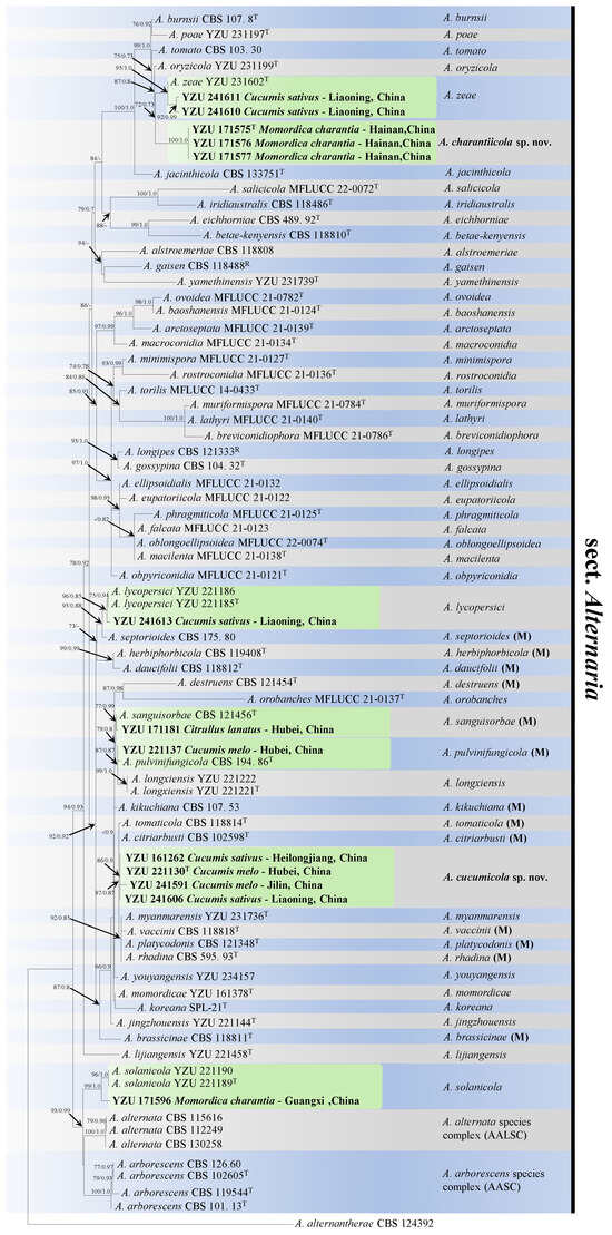
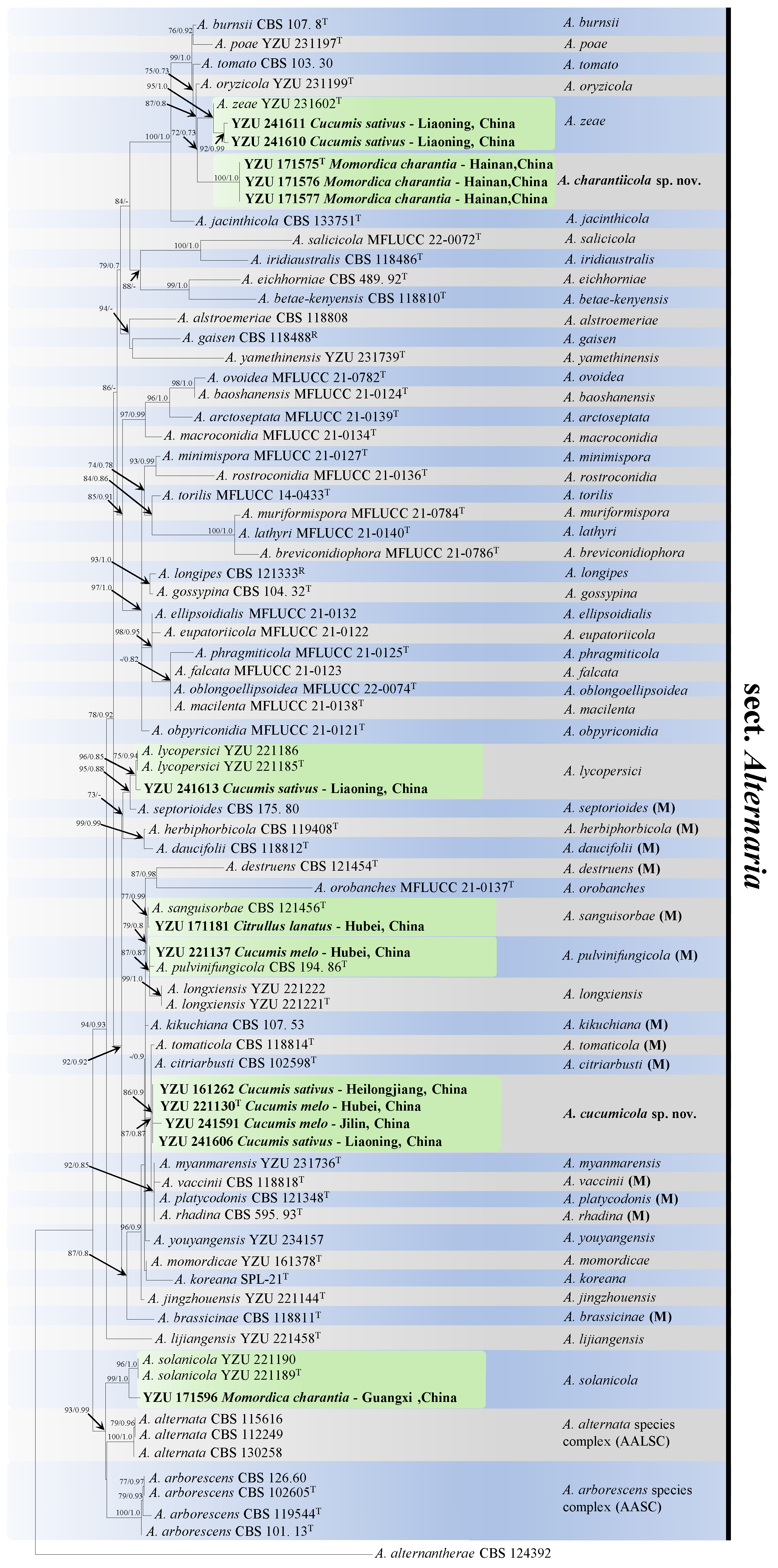
Based on BLASTn searches, all isolates obtained in this study were assigned to Alternaria section Alternaria. In total, 84 strains were included in the phylogenetic analyses, including 13 representative small-spored isolates obtained in this study, with A. alternantherae CBS 124392 selected as the outgroup. The concatenated dataset comprised seven loci (ITS, GAPDH, TEF1, RPB2, Alt a 1, EndoPG, and OPA10-2), yielding an alignment of 3399 characters, of which 9.26% were missing. The individual gene regions contributed 513 bp (ITS), 576 bp (GAPDH), 210 bp (TEF1), 555 bp (RPB2), 471 bp (Alt a 1), 444 bp (EndoPG), and 630 bp (OPA10-2). The best-fit substitution models were K2P (ITS), TNe + G4 (GAPDH), K2P (TEF1), TNe + I (RPB2), K2P + G4 (Alt a 1), K3P + I (EndoPG), and K2P + G4 (OPA10-2). Maximum likelihood (ML) and Bayesian inference (BI) analyses produced congruent topologies; the ML tree is shown in Figure 1. Phylogenetic reconstruction resolved the examined isolates into seven distinct clades within section Alternaria. Isolates YZU 241610 and YZU 241611, obtained from C. sativus, clustered with A. zeae YZU 231602 with strong support (BS = 95%, PP = 1.00). In contrast, isolates YZU 171575, YZU 171576, and YZU 171577 from M. charantia formed a well-supported (BS = 72%, PP = 0.73), independent lineage closely related to A. zeae, representing a distinct novel clade. Isolate YZU 241613 (C. sativus) grouped with A. lycopersici (BS = 96%, PP = 0.85). Meanwhile, isolates YZU 171181 from C. lanatus and YZU 221137 from C. melo clustered within the same clade (BS = 79%, PP = 0.8), forming sister relationships with A. sanguisorbae CBS 121456 (BS = 77%, PP = 0.99) and A. pulvinifungicola CBS 194.86, respectively. Notably, isolates YZU 161262 (C. sativus), YZU 221130 (C. melo), YZU 241591 (C. melo), and YZU 241606 (C. sativus) formed a strongly supported monophyletic group (BS = 86%, PP = 0.90), clearly separated from all previously described species (BS = 87%, PP = 0.87), representing another novel phylogenetic lineage. Isolate YZU 171596 clustered with A. solanicola with strong support (BS = 99%, PP = 1.00). In summary, all isolates obtained in this study belong to section Alternaria, comprising two novel phylogenetic clades and five clades corresponding to previously described species (Figure 1).
Figure 1.
Maximum likelihood phylogenetic tree inferred from concatenated sequences of ITS, GAPDH, RPB2, TEF1, Alt a 1, EndoPG, and OPA10-2 for Alternaria spp. Bootstrap support values (BS) and Bayesian posterior probabilities (PP) are shown at the nodes (BS/PP). Strains obtained in this study are indicated in bold. Ex-type strains are marked with “T”, representative strains with “R”, and morphospecies with “(M)”. Alternaria alternantherae CBS 124392 was used as the outgroup.
3.3. Taxonomy
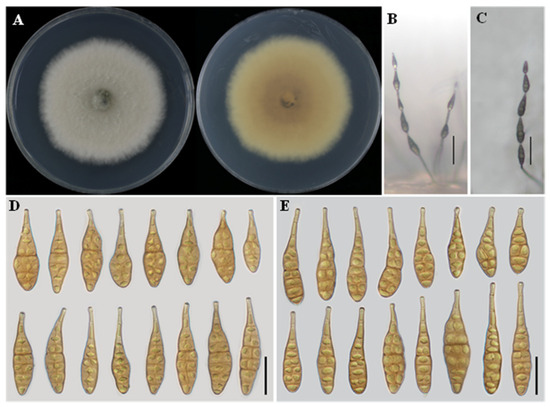
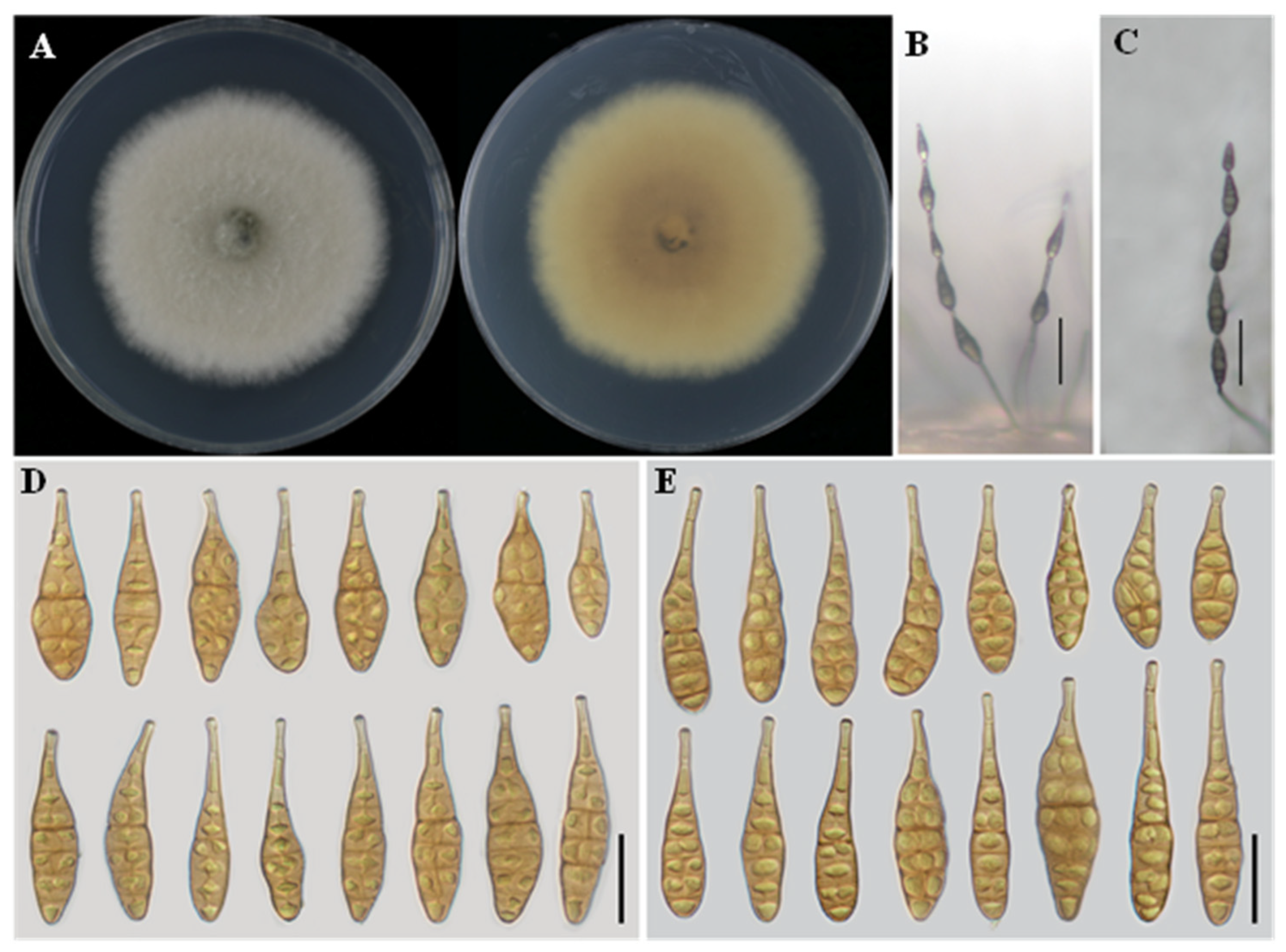
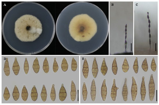
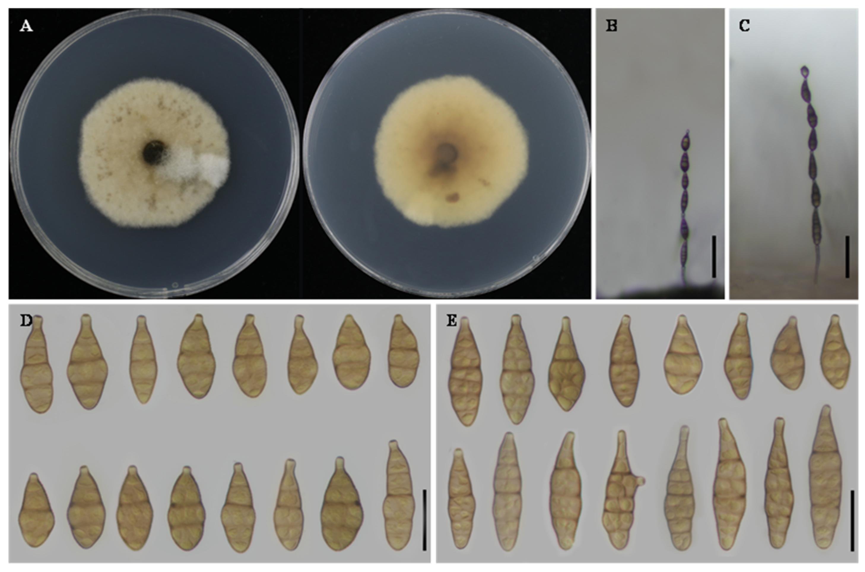
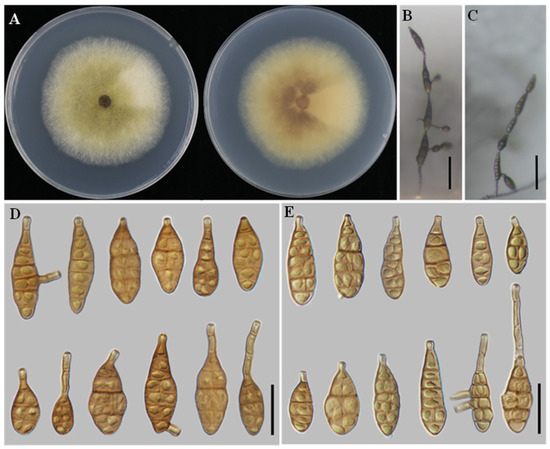
Alternaria charantiicola L. He, S.L.L. Aung and J.X. Deng, sp. nov. (Figure 2).
Figure 2.
Morphology of Alternaria charantiicola sp. nov. (YZU 171577). (A) Colony on PDA for 7 days at 25 °C; (B) sporulation on PCA; (C) sporulation on V8A; (D) Conidia on PCA; (E) Conidia on V8A. Scale bars: 50 μm (B,C); 25 μm (D,E).
MycoBank No.: 862358.
Etymology: The specific epithet charantiicola refers to the host plant Momordica charantia, from which the fungus was isolated.
Typification: China, Hainan Province, Haikou City; diseased leaves of M. charantia, 2017, J.X. Deng; Holotype: YZU-H-2017075; Ex-type culture: YZU 171575.
Description: Colonies on PDA cottony, white with filamentous margins, reverse rosy buff to vinaceous buff at the center, 56–57 mm in diameter. On PCA, conidiophores solitary, arising from the substrate, simple, straight or flexuous, 19–85 × 2–4 μm, with 2–3 septa. Conidiogenous cells terminal, determinate, cylindrical, apically doliiform, 6–15 × 2–4 μm, with 1–2 conidiogenous loci. Conidia in chains of 4–6 units, arising from the apex or near the apex of conidiophores or terminal hyphae, muriform, ellipsoidal to flask-shaped, rostrate, body 36–54 × 11–15 μm, with 2–5 transverse septa; apical beak 3–9 × 2–3 μm. On V8A, conidiophores simple, straight or flexuous, 39–55 × 2–5 μm. Conidiogenous cells terminal, determinate, cylindrical, apically doliiform, 5–14 × 3–4 μm, with a single conidiogenous locus, sometimes slightly swollen near the locus. Conidia in chains of 3–5 units, unbranched, muriform, ellipsoidal to flask-shaped, rostrate, body 39–55 × 11–18 μm, with 2–5 transverse septa; apical beak 7–21 × 2–3 μm.
Notes: Phylogenetic analyses based on a concatenated dataset of seven loci unambiguously placed A. charantiicola within a clade comprising A. zeae YZU 231602 and A. oryzicola YZU 231199. Nucleotide sequence comparisons showed that strain YZU 171575 differed from the representative strain of A. zeae by 2 bp in Alt a 1, 22 bp in OPA10-2, 1 bp in RPB2, and 10 bp in TEF1. In comparison with A. oryzicola, a total of 33 bp differences were detected, including 2 bp in Alt a 1, 19 bp in OPA10-2, 2 bp in RPB2, and 10 bp in TEF1. Morphologically, isolate YZU 171575 is further distinguished from A. zeae and A. oryzicola by its larger conidial body size (Table 2). Taken together, the concordant molecular and morphological evidence supports the recognition of isolate YZU 171575 as a distinct species, herein described as A. charantiicola sp. nov.
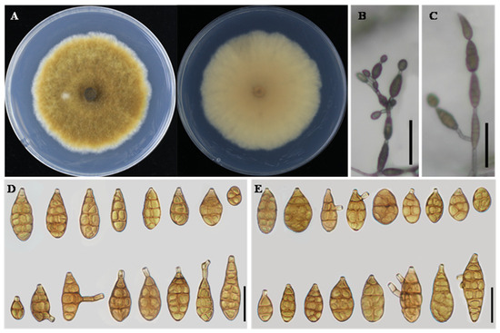
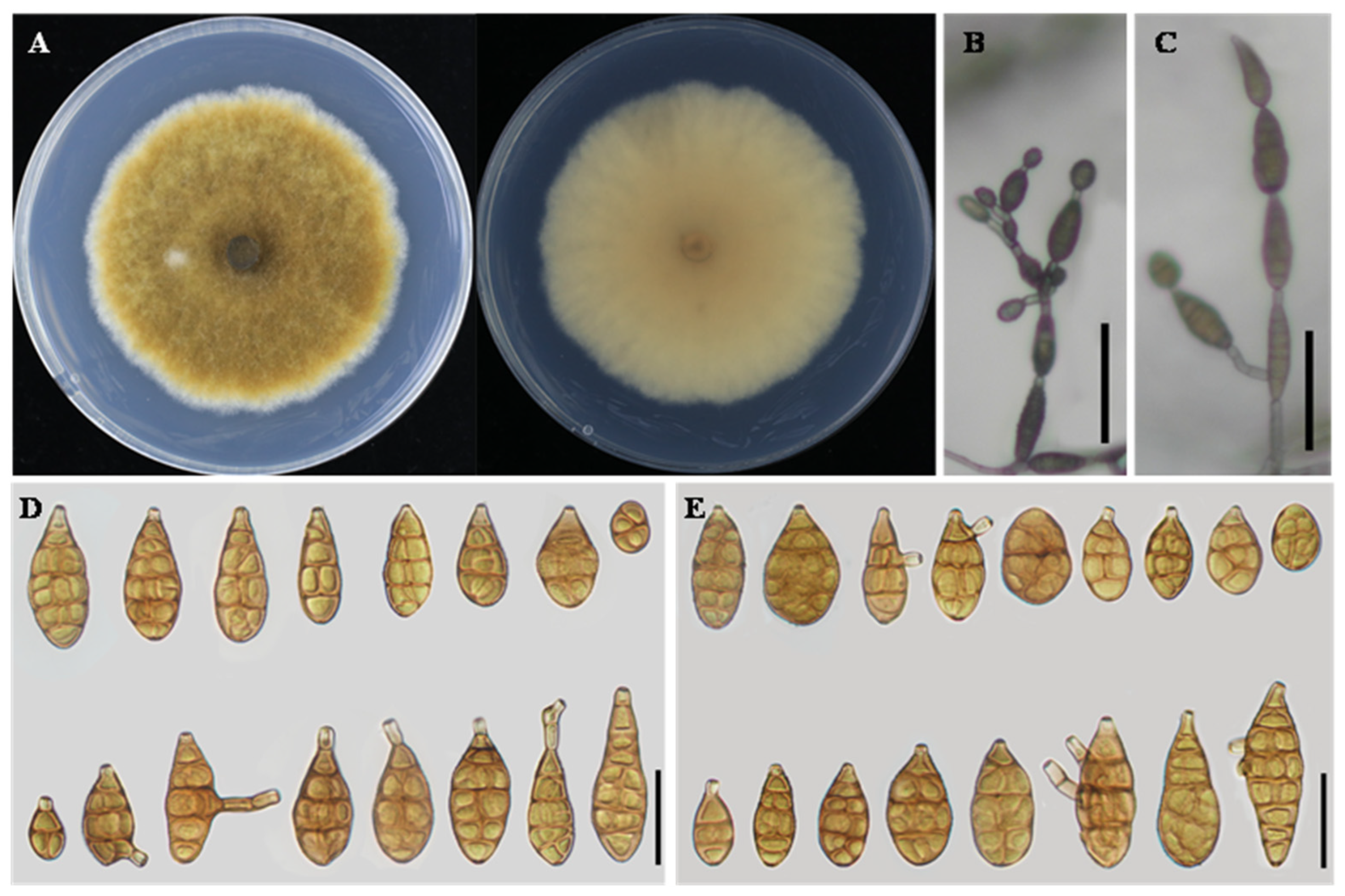
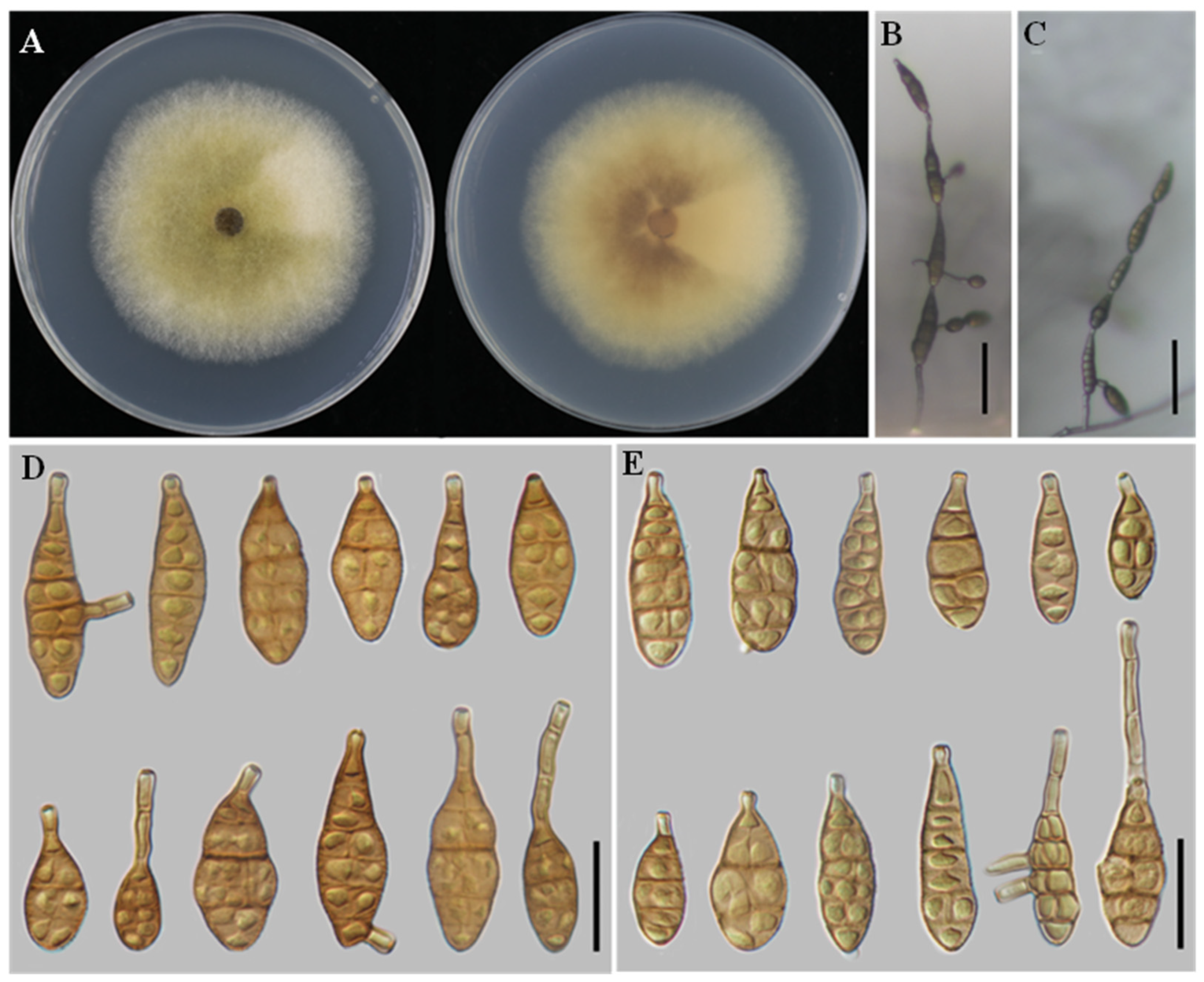
Alternaria cucumicola L. He, S.L.L. Aung and J.X. Deng, sp. nov. (Figure 3).
Figure 3.
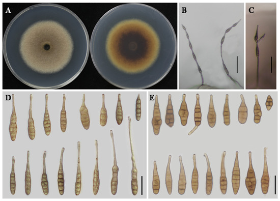
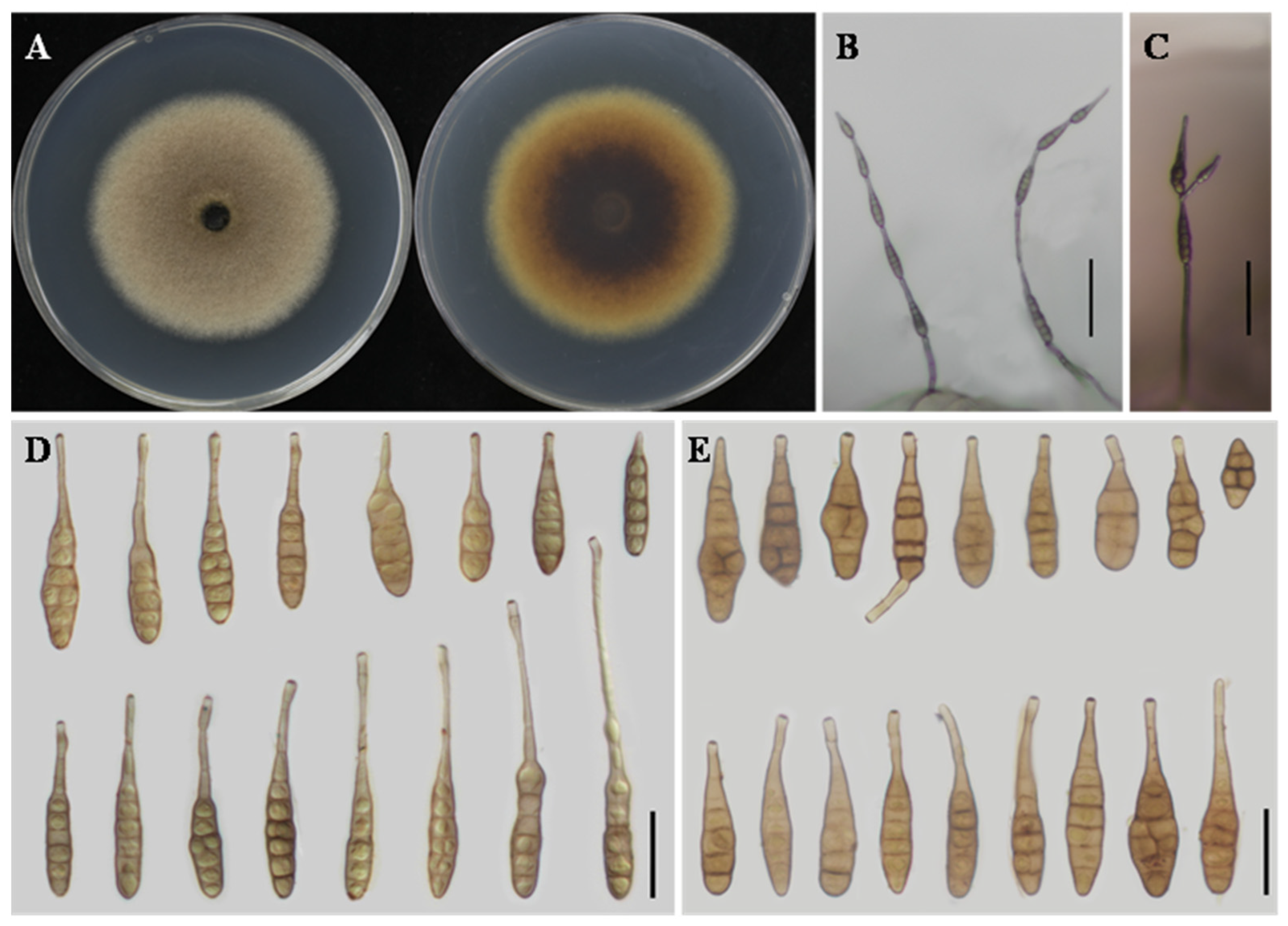
Morphology of Alternaria cucumicola sp. nov. (YZU 221130). (A) Colony on PDA for 7 days at 25 °C; (B) sporulation on PCA; (C) sporulation on V8A; (D) Conidia on PCA; (E) Conidia on V8A. Scale bars: 50 μm (B,C); 25 μm (D,E).
MycoBank No.: 862357.
Etymology: The specific epithet cucumicola refers to the host genus Cucumis (C. sativus and C. melo), from which the fungus was isolated.
Typification: China, Hubei Province, Wuxue City; diseased leaves of C. melo, 2022, J.X. Deng; Holotype: YZU-H-2022029; Ex-type culture: YZU 221130.
Additional specimens examined (China): Heilongjiang Province, on C. sativus, YZU 161262, 2016; Jilin Province, Changchun City, on C. melo, YZU 241591, 2024; Liaoning Province, Jinzhou City, on C. sativus, YZU 241606, 2024.
Description: Colonies on PDA circular, velvety to cottony, pale brown to light olivaceous brown, darker at the center, with a narrow whitish margin; reverse pale yellow to light brown, 60–65 mm in diameter. On PCA, conidiophores arising from the substrate or from aerial hyphae, erect to slightly flexuous, mostly unbranched, occasionally geniculate near the apex, 30–80 × 3–5 μm. Conidiogenous cells integrated, terminal, apically doliiform, with a single conidiogenous locus. Conidiation predominantly acrogenous, with conidia produced successively from the apical beak of the terminal conidium; the apical beak may develop multiple conidiogenous loci, giving rise to short conidial chains. Conidia in chains of 2–6 units, ellipsoid to narrow-obclavate or obclavate, 18–42 × 9–16 μm, with 2–5 transverse septa. Apical beak absent to short, usually 3–15 μm long. On V8A, conidiophores similar in morphology to those on PCA, 25–65 × 3–5 μm. Conidiation both acrogenous and intercalary; in addition to apical conidiation, lateral conidia were frequently observed arising from the beak or body of intercalary conidia within the chain, resulting in branched or irregular conidial chains. Conidia solitary or in chains of 3–6 units, ellipsoid to obclavate, 20–45 × 10–18 μm, with 2–5 transverse septa. Apical beak short or absent, 2–12 μm long.
Notes: Phylogenetic analyses based on the combined multilocus dataset placed A. cucumicola within a clade comprising A. citriarbusti CBS 102598, A. platycodonis CBS 121348, A. rhadina CBS 595.93, A. tomaticola CBS 118814, A. vaccinii CBS 118818, and A. myanmarensis YZU 231736. Within this clade, A. cucumicola was resolved as a clearly independent lineage, strongly supported (BS = 99%, PP = 1.00). Phylogenetically, A. platycodonis, A. rhadina, and A. myanmarensis were identical. A. cucumicola differs from these taxa at two loci, with 1 bp in GAPDH and 3 bp in EndoPG (including two gaps). It also differs from A. citriarbusti by 1 bp in RPB2 and 2 bp in EndoPG, and from A. tomaticola by 2 bp in Alt a 1 and 1 bp in EndoPG. Morphologically, isolate YZU 221130 is distinguished from closely related taxa by its conidial body size, shorter conidial chains, and the absence of a thin, elongated apical beak (Table 2). Collectively, the evidence supports the delimitation of isolate YZU 221130 as a distinct species.
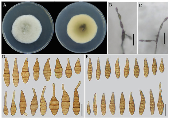
Alternaria zeae H.F. Liu and J.X. Deng, MycoKeys 116: 167. 2025 (Figure 4).
Figure 4.
Morphology of Alternaria zeae (YZU 241610). (A) Colony on PDA for 7 days at 25 °C; (B) sporulation on PCA; (C) sporulation on V8A; (D) Conidia on PCA; (E) Conidia on V8A. Scale bars: 50 μm (B,C); 25 μm (D,E).
Additional isolates examined: China, Liaoning Province, Jinzhou City, Linghe District; diseased leaves of C. sativus; collected in 2024 by J.X. Deng. Living culture deposited as YZU 241610 and YZU 241611.
Description: Colonies on PDA cottony, pale vinaceous grey, with filamentous margins; reverse vinaceous grey with buff filamentous margins, 61–62 mm in diameter. On PCA, conidiophores solitary, arising directly from the substrate, simple, flexuous, 24–149 × 2–3 μm, with 2–6 septa. Conidiogenous cells terminal, determinate, cylindrical, smooth, thin-walled, apically doliiform, 2–5 × 2–4 μm, with a single conidiogenous locus, cicatrized at conidial secession. Conidia borne in unbranched chains of 4–6 units, arising from the apex or near the apex of conidiophores or terminal hyphae, muriform, ovate, ellipsoid to flask-shaped, rostrate; conidial body 28–42 × 9–17 μm, with 1–6 transverse septa; apical beak 4–26 (–50) × 2–3 μm. On V8A, conidiophores simple, straight to flexuous, 19–65 × 2–4 μm, with 2–3 septa. Conidiogenous cells terminal, determinate, cylindrical, smooth, thin-walled, apically doliiform, 5–12 × 2–4 μm, with a single conidiogenous locus, sometimes slightly swollen near the conidiogenous locus. Conidia borne in chains of 4–6 units, arising from the apex or near the apex of conidiophores or terminal hyphae, muriform, ellipsoid to flask-shaped, rostrate; conidial body 27–45 × 8–16 μm, with 1–6 transverse septa; apical beak 3–16 × 2–3 μm, occasionally elongating to form a secondary conidiophore; chains unbranched.
Notes: A. zeae was originally described from Zea mays [16]. In the present study, multilocus phylogenetic analyses revealed that isolates YZU 241610 and YZU 241611 clustered with A. zeae with strong support (BS = 95%, PP = 1.00) (Figure 1). Based on both phylogenetic evidence and morphological characteristics, these isolates are identified as A. zeae. This study represents a new host record of A. zeae on C. sativus.
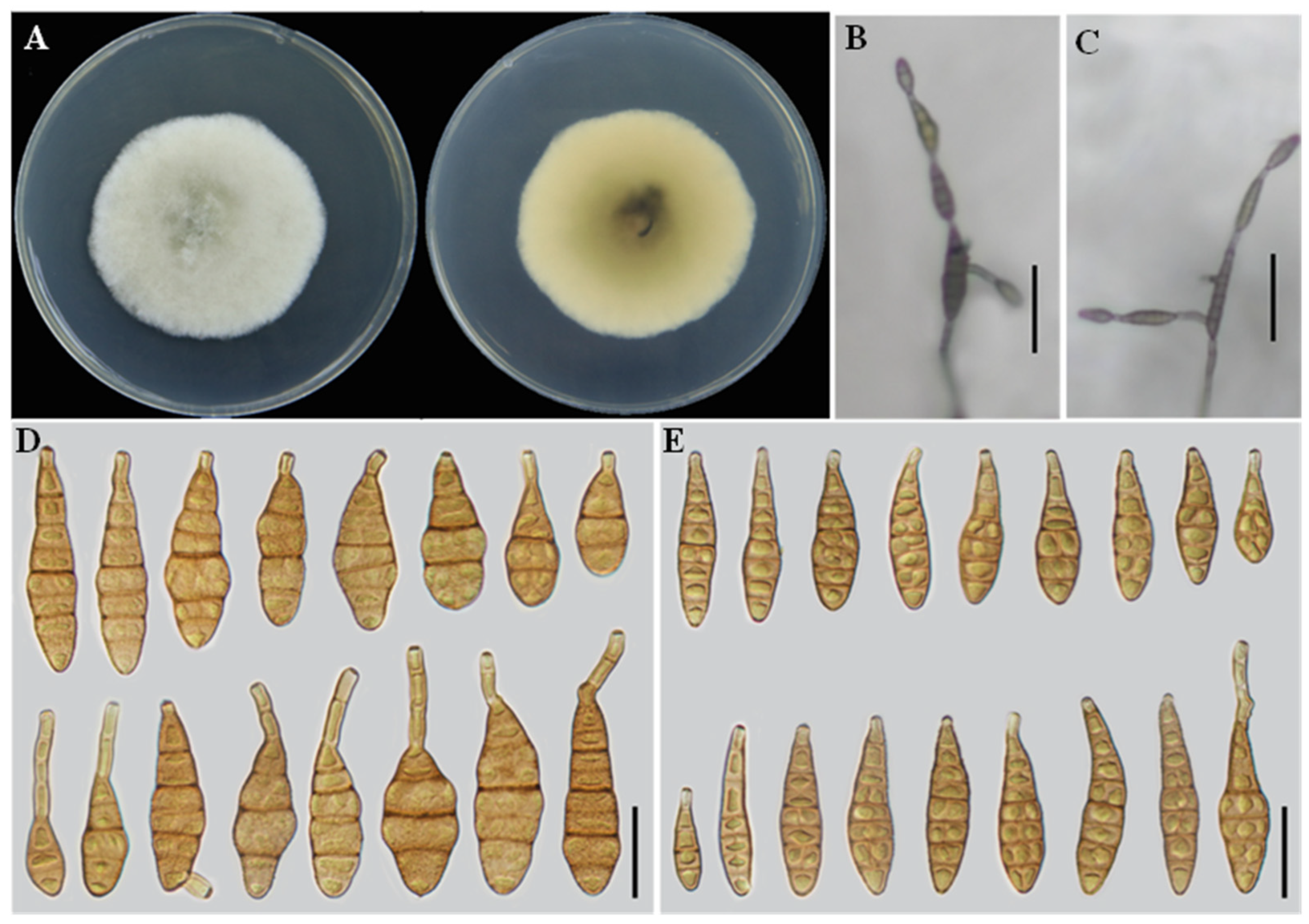
Alternaria lycopersici Y.N. Gou and J.X. Deng, J. Fungi. 9: 800. 2023 (Figure 5).
Figure 5.
Morphology of Alternaria lycopersici (YZU 241613). (A) Colony on PDA for 7 days at 25 °C; (B) sporulation on PCA; (C) sporulation on V8A; (D) Conidia on PCA; (E) Conidia on V8A. Scale bars: 50 μm (B,C); 25 μm (D,E).
Additional isolates examined: China, Liaoning Province, Jinzhou City, Linghe District; diseased leaves of C. sativus; collected in 2024 by J.X. Deng. Living culture deposited as YZU 241613.
Description: Colonies on PDA cottony, light cottony, and buff in the center, reverse villiform with white at the edge, 42–44 mm in diameter. On PCA, conidiophores solitary, arising from the substrate, simple, flexuous, 42–81 × 3–5 μm, with 2–5 septa. Conidiogenous cells terminal, apically doliiform, 5–10 × 2–5 μm, with a single conidiogenous locus. Conidia borne in chains of 5–7 units, arising from the apex or near the apex of conidiophores or terminal hyphae, muriform, clavate, ovoid, or long ellipsoid to flask-shaped, rostrate; conidial body 25–42 × 11–17 μm, with 1–4 transverse septa. On V8A, conidiophores solitary, arising from the substrate, simple, flexuous, 50–67 × 2–5 μm, with 2–5 septa. Conidiogenous cells terminal, apically doliiform, 5–10 × 2–5 μm, with a single conidiogenous locus. Conidia borne in chains of 5–7 units, arising from the apex or near the apex of conidiophores or terminal hyphae, muriform, clavate, long ellipsoid or ovoid, rostrate; conidial body 16–53 × 10–15 μm, with 1–4 transverse septa; chains unbranched.
Notes: A. lycopersici was previously known only from Solanum lycopersicum [17]. In the present study, isolate YZU 241613 was recovered from diseased leaves of C. sativus. Phylogenetic inference based on seven concatenated loci placed this isolate within the A. lycopersici clade with strong statistical support (BS = 96%, PP = 0.85). Its morphological features were also in agreement with the original description of A. lycopersici [17]. Accordingly, C. sativus is herein reported as a new host for A. lycopersici.
Alternaria sanguisorbae M.X. Gao and T.Y. Zhang, Mycosystema 19: 456. 2000 (Figure 6).
Figure 6.
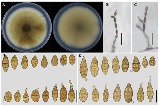
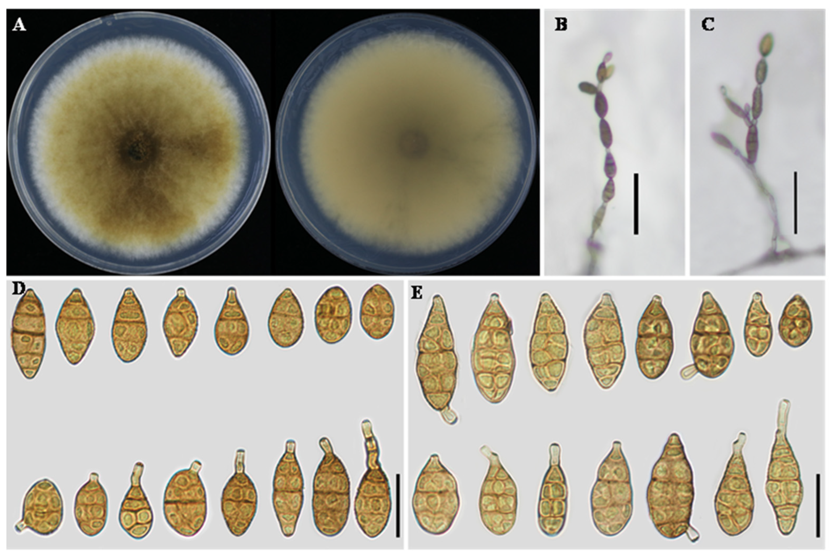
Morphology of Alternaria sanguisorbae (YZU 171181). (A) Colony on PDA for 7 days at 25 °C; (B) sporulation on PCA; (C) sporulation on V8A; (D) Conidia on PCA; (E) Conidia on V8A. Scale bars: 50 μm (B,C); 25 μm (D,E).
Additional isolates examined: China, Hubei Province, Wuxue City; diseased leaves of C. lanatus; collected in 2017 by J.X. Deng. Living culture deposited as YZU 171181.
Description: Colonies on PDA honey-colored, with white filamentous margins on the surface; reverse buff with filamentous margins, 55–60 mm in diameter. On PCA, conidiophores solitary, arising directly from the substrate, simple, flexuous, 45–80 × 3–5 μm, with 4–6 septa. Conidiogenous cells terminal, 3–4 × 3–5 μm, with a single conidiogenous locus, cicatrized after conidial secession. Conidiation predominantly acrogenous: conidia produced successively from the apical beak of terminal conidia; the beak may develop multiple conidiogenous loci, resulting in short conidial chains. Conidia in chains of 3–7 units, unbranched to slightly branched, ovoid to ellipsoid; conidial body 20–35 × 9–14 μm, with 4–6 transverse septa; apical beak 5–21 × 2–3 μm. On V8A, conidiophores solitary, 35–82 × 3–5 μm, with 4–5 septa. Conidiogenous cells 3–4 × 3–5 μm, with a single conidiogenous locus, cicatrized after conidial secession. Conidiation both acrogenous and intercalary; in addition to apical conidiation, lateral conidia frequently arise from the beak or body of intercalary conidia within the chain, giving rise to branched or irregular conidial chains. Conidia in chains of 3–7 units, ovoid to ellipsoid; conidial body 18–45 × 9–13 μm, with 4–6 transverse septa; apical beak 5–20 × 2–3 μm.
Notes: A. sanguisorbae was previously redefined as A. alternata by Woudenberg et al. [14], and was originally described from Sanguisorba officinalis [33]. In the present study, isolate YZU 171181 from C. lanatus clustered with A. sanguisorbae in the multilocus phylogeny. Accordingly, this isolate is reassigned to A. sanguisorbae, representing a new host record.
Alternaria pulvinifungicola E.G. Simmons, CBS Biodiversity Ser. (Utrecht) 6: 514. 2007 (Figure 7).
Figure 7.
Morphology of Alternaria pulvinifungicola (YZU 221137). (A) Colony on PDA for 7 days at 25 °C; (B) sporulation on PCA; (C) sporulation on V8A; (D) Conidia on PCA; (E) Conidia on V8A. Scale bars: 50 μm (B,C); 25 μm (D,E).
Additional isolates examined: China, Hubei Province, Jingzhou City; diseased leaves of C. melo; collected in 2022 by J.X. Deng. Living culture deposited as YZU 221137.
Description: Colonies on PDA olivaceous buff, with white sectoring and filamentous margins on the surface; reverse vinaceous buff with filamentous margins, 55–56 mm in diameter. On PCA, conidiophores solitary, arising from the substrate, simple, flexuous, 45–92 × 3–5 μm, with 4–6 septa. Conidiogenous cells terminal, apically doliiform, 3–32 × 3–5 μm, with 1–2 conidiogenous loci, cicatrized at conidial secession. Conidia borne in chains of 2–6 units, arising from the apex or near the apex of conidiophores or terminal hyphae, ovoid to ellipsoid, or obclavate; conidial body 25–51 × 7–17 μm, with 1–4 transverse septa; apical beak 20–95 × 3–5 μm; chains occasionally branched, bearing 2–4 conidia. On V8A, conidiophores solitary, 19–26 × 3–5 μm, with 1–4 septa. Conidiogenous cells terminal, 3–14 × 3–5 μm, with 1–2 conidiogenous loci. Conidia borne in chains of 2–6 units, arising from the apex or near the apex of conidiophores or terminal hyphae, ovoid to ellipsoid; conidial body 25–40 × 9–13 μm, with 1–6 transverse septa; apical beak 4–48 × 2–3 μm, occasionally developing into secondary conidiophores; chains branched.
Notes: A. pulvinifungicola was originally described from Quercus sp. [8], and was later treated as a synonym of A. alternata [14]. In the present study, isolate YZU 221137, obtained from C. melo, clustered with A. pulvinifungicola in the multilocus phylogenetic analysis. Based on this phylogenetic placement, the isolate is reassigned to A. pulvinifungicola, representing a new host record.
Alternaria solanicola Y.N. Gou and J.X. Deng, J. Fungi. 9: 800. 2023 (Figure 8).
Figure 8.
Morphology of Alternaria solanicola (YZU 171596). (A) Colony on PDA for 7 days at 25 °C; (B) sporulation on PCA; (C) sporulation on V8A; (D) Conidia on PCA; (E) Conidia on V8A. Scale bars: 50 μm (B,C); 25 μm (D,E).
Additional isolates examined: China, Guangxi Province, Nanning City; diseased leaves of M. charantia; collected in 2017 by J.X. Deng. Living culture deposited as YZU 171596.
Description: Colonies on PDA circular, cottony to floccose, white on the surface with a faint greyish-green center; reverse pale yellow to buff, 55–57 mm in diameter. On PCA, conidiophores solitary, arising from the substrate, straight to slightly flexuous, 25–70 × 3–5 μm. Conidia borne in chains of 2–4 units, short to long ovoid or ellipsoid; conidial body 20–50 × 7–14 μm, with 1–4 transverse septa and 0–2 longitudinal septa; beak absent or present, apical, 6–26 μm long. On V8A, conidiophores straight to slightly curved, septate, 18–65 × 3–5 μm. Conidia similar to those on PCA, produced in chains of 2–4 units, short to long ovoid or ellipsoid, 22–55 × 6–14 μm, with 1–4 transverse septa and 0–2 longitudinal septa.
Notes: A. solanicola was originally described from S. lycopersicum [17]. In the present study, isolate YZU 171596 recovered from M. charantia was placed within the A. solanicola clade in the multilocus phylogeny. Its morphological characteristics also correspond to those reported for this species. On this basis, YZU 171596 is regarded as A. solanicola, representing a new host record on M. charantia.
Table 2.
Conidial morphology of Alternaria spp. from this study and previous publications.
4. Discussion
In this study, Alternaria species associated with Cucurbitaceae were investigated across six provinces and three host genera in China. Using an integrative approach combining multilocus phylogenetic analyses with morphological characterization, two novel species, A. charantiicola sp. nov. and A. cucumicola sp. nov., together with five newly recorded species, were identified. Among these taxa, two previously described morphospecies, A. sanguisorbae and A. pulvinifungicola, were re-evaluated and taxonomically reassigned based on molecular phylogenetic evidence. Notably, the five newly recorded species had previously been reported only from non-cucurbit hosts, and their detection in this study extends their known host range, suggesting that some small-spored Alternaria species may exhibit relatively broad host associations or opportunistic colonization on cucurbit plants. Furthermore, A. cucumicola sp. nov. was consistently isolated from different cucurbit hosts across multiple geographic locations, indicating a potentially broad host association within Cucurbitaceae rather than strict host specificity. Although the isolates were obtained from symptomatic leaves, pathogenicity tests were not conducted; therefore, the taxa reported here should be regarded as disease-associated rather than confirmed pathogens, and their pathogenic roles require further verification. Overall, these findings expand current knowledge of host diversity within section Alternaria and provide a strengthened taxonomic framework for future systematic studies of Alternaria associated with cucurbit hosts.
All isolates obtained in the present study belong to the small-spored group within Alternaria section Alternaria. As noted above, the taxonomy of small-spored Alternaria has long been controversial owing to highly similar and overlapping morphological characteristics [15]. Based solely on morphological features, Simmons [8] recognized 128 small-spored species. However, subsequent molecular phylogenetic studies demonstrated that many of these taxa are phylogenetically indistinguishable and have largely been treated as A. alternata within section Alternaria, with the 35 morphospecies described by Simmons [8] subsequently subsumed into this complex [13,14]. This reclassification represented a pragmatic taxonomic solution at the time, given the limited molecular data available and the widespread reliance on morphology-based taxonomy. By consolidating poorly resolved morphospecies under A. alternata, it helped stabilize the taxonomy of section Alternaria and established A. alternata as a central reference taxon within the section. Nevertheless, with the increasing availability of multilocus phylogenetic data, the taxonomic status of several morphologically defined taxa within section Alternaria is being re-evaluated, as these analyses continue to reveal previously unrecognized lineages within the A. alternata species complex [7,15].
Two additional novel small-spored Alternaria species were identified in this study through an integrative analysis combining multilocus phylogeny and morphological characterization. One species, A. charantiicola sp. nov., formed a well-supported and clearly distinct lineage in the phylogenetic analyses, clustering with but phylogenetically separated from A. zeae and A. oryzicola. Nucleotide sequence comparisons revealed that A. charantiicola sp. nov. differs from A. zeae by 30 bp across four loci (Alt a 1, OPA10-2, RPB2, and TEF1) and from A. oryzicola by 33 bp across the four same loci. Morphologically, A. charantiicola sp. nov. is readily distinguished from these closely related taxa by its noticeably larger conidial body size (Table 2), further supporting its recognition as an independent species. The second novel species, A. cucumicola sp. nov., was resolved within a clade comprising six taxa, including five morphospecies (A. tomaticola, A. citriarbusti, A. vaccinii, A. platycodonis, and A. rhadina) that were previously synonymized under A. alternata [14], as well as the recently described species A. myanmarensis [18]. Phylogenetic analyses showed that, despite relatively limited nucleotide differences between A. cucumicola sp. nov. and its closest relatives, the species was consistently resolved as an independent lineage (Figure 1). In contrast, A. platycodonis, A. rhadina, and A. myanmarensis showed identical nucleotide sequences across the analyzed loci, suggesting that they may represent potential synonyms. Morphological characters further support the distinct status of A. cucumicola sp. nov. Its conidia are larger than those of A. myanmarensis but smaller than those of A. citriarbusti (Table 2). In addition, A. cucumicola produces markedly shorter conidial chains (2–6 conidia per chain) than A. tomaticola (10–15), A. vaccinii (8–10), and A. rhadina (9–15). The absence of a thin, elongated apical beak further distinguishes A. cucumicola sp. nov. from A. platycodonis. Taken together, these concordant phylogenetic and morphological differences support the recognition of A. cucumicola as a novel species. These results highlight that, in taxonomic classification of section Alternaria, morphospecies concepts should not be overlooked, as morphological distinctions remain essential for accurate species delimitation.
In addition to the two new species, five small-spored Alternaria species are reported here as new records from Cucurbitaceae hosts, namely A. zeae, A. lycopersici, A. sanguisorbae, A. pulvinifungicola, and A. solanicola. Multilocus phylogenetic analyses showed that the examined isolates clustered with reference strains of the corresponding taxa in well-supported clades, and their morphological characteristics were consistent with previously published descriptions. In particular, two isolates identified as A. sanguisorbae and A. pulvinifungicola, respectively, formed distinct phylogenetic lineages together with their reference strains. Although these species were previously treated as synonyms of A. alternata [14], our results support their interpretation as distinct phylogenetic lineages within section Alternaria. Therefore, we retain the morphospecies names A. sanguisorbae and A. pulvinifungicola to distinguish these taxa from the broader A. alternata species complex. These findings further suggest that morphospecies concepts remain useful in the taxonomy of section Alternaria when interpreted in combination with multilocus phylogenetic evidence. Moreover, the continued discovery and description of new taxa indicates that the taxonomy of small-spored Alternaria remains dynamic and may benefit from comprehensive reassessment as additional molecular datasets become available.
5. Conclusions
In conclusion, the present study examined Alternaria species associated with Cucurbitaceae using an integrative approach combining multilocus phylogenetic analyses and morphological characterization. Two novel species and five newly recorded species were identified and documented. These findings enhance current understanding of species diversity and host associations of Alternaria in Cucurbitaceae and provide a robust taxonomic basis for future systematic and diversity studies of Alternaria associated with cucurbit hosts.
Author Contributions
Conceptualization, L.H. and J.D.; methodology, L.H. and S.L.L.A.; software, P.S. and Z.L.; validation, L.H., Z.L. and S.L.L.A.; formal analysis, P.S. and Z.L.; investigation, S.L.L.A., P.S. and Z.L.; resources, J.D. and S.L.L.A.; data curation, L.H. and S.L.L.A.; writing—original draft preparation, L.H. and S.L.L.A.; writing—review and editing, J.D. and S.L.L.A.; visualization, L.H.; supervision, J.D. and S.L.L.A.; project administration, J.D.; funding acquisition, J.D. All authors have read and agreed to the published version of the manuscript.
Funding
This research was funded by the National Natural Science Foundation of China (No. 32270022); Major Scientific and Technological Project in the Inner Mongolia Autonomous Region (2025YFHH0275).
Institutional Review Board Statement
Not applicable.
Informed Consent Statement
Not applicable.
Data Availability Statement
The nucleotide sequences generated in this study were deposited in GenBank database.
Acknowledgments
Thanks to all members of the research team for their support of this project.
Conflicts of Interest
The authors declare no conflicts of interest.
References
- Xu, Z.; Chang, L. Cucurbitaceae. In Identification and Control of Common Weeds; Springer: Singapore, 2017; Volume 3, pp. 417–432. [Google Scholar]
- Zhang, X.J.; Yi, S.Q.; Wu, P.K. Research progress on disease-resistant genes in Cucurbitaceae crops. J. Zhejiang Univ. Sci. 2025, 51, 851–864. [Google Scholar] [CrossRef]
- Renner, S.S.; Schaefer, H. Phylogeny and evolution of the Cucurbitaceae. In Genetics and Genomics of Cucurbitaceae; Springer: Berlin/Heidelberg, Germany, 2016; pp. 13–23. [Google Scholar]
- Lu, A.M.; Chen, S.K. Flora of China; Science Press: Beijing, China, 1986; Volume 73. [Google Scholar]
- Mondal, B.; Mondal, C.K.; Mondal, P. Deaseas of cucurbits and their management. In Stresses of Cucurbits: Current Status and Management; Springer: Berlin/Heidelberg, Germany, 2020; pp. 115–222. [Google Scholar]
- Ma, G.P.; Bao, S.W.; Zhao, J.; Sui, Y.; Wu, X.H. Morphological and molecular characterization of Alternaria species causing leaf blight on watermelon in China. Plant Dis. 2021, 105, 60–70. [Google Scholar] [CrossRef] [Scilit]
- Li, J.F.; Jiang, H.B.; Jeewon, R.; Hongsanan, S.; Bhat, D.J.; Tang, S.M.; Lumyong, S.; Mortimer, P.E.; Xu, J.-C.; Camporesi, E. Alternaria: Update on species limits, evolution, multi-locus phylogeny, and classification. Stud. Fungi 2022, 8, 1. [Google Scholar] [CrossRef] [Scilit]
- Simmons, E.G. Alternaria: An Identification Manual: Fully Illustrated and with Catalogue Raisonné 1796–2007; CBS Fungal Biodiversity Centre: Utrecht, The Netherlands, 2007. [Google Scholar]
- Liu, F.Y.; Gou, Y.N.; Liu, T.Y.; Huang, C.X.; Deng, J.X. Cucumis sativus and Ipomoea purpurea: New hosts for Alternaria hydrangeae in China. J. Phytopathol. 2023, 171, 48–56. [Google Scholar] [CrossRef] [Scilit]
- Aung, S.L.L.; Liu, F.Y.; Gou, Y.N.; Nwe, Z.M.; Yu, Z.H.; Deng, J.X. Morphological and phylogenetic analyses reveal two new Alternaria species (Pleosporales, Pleosporaceae) in Alternaria section from Cucurbitaceae plants in China. MycoKeys 2024, 107, 125. [Google Scholar] [CrossRef] [Scilit] [PubMed]
- Lawrence, D.P.; Rotondo, F.; Gannibal, P.B. Biodiversity and taxonomy of the pleomorphic genus Alternaria. Mycol. Prog. 2016, 15, 3. [Google Scholar] [CrossRef] [Scilit]
- Lawrence, D.P.; Gannibal, P.B.; Peever, T.L.; Pryor, B.M. The sections of Alternaria: Formalizing species-group concepts. Mycologia 2013, 105, 530–546. [Google Scholar] [CrossRef] [Scilit]
- Woudenberg, J.; Groenewald, J.; Binder, M.; Crous, P.W. Alternaria redefined. Stud. Mycol. 2013, 75, 171–212. [Google Scholar] [CrossRef] [Scilit] [PubMed]
- Woudenberg, J.; Seidl, M.; Groenewald, J.Z.; De Vries, M.; Stielow, J.; Thomma, B.P.; Crous, P.W. Alternaria section Alternaria: Species, formae speciales or pathotypes? Stud. Mycol. 2015, 82, 1–21. [Google Scholar] [CrossRef] [Scilit]
- He, J.; Li, D.-W.; Cui, W.-L.; Huang, L. Seven new species of Alternaria (Pleosporales, Pleosporaceae) associated with Chinese fir, based on morphological and molecular evidence. MycoKeys 2024, 101, 1–44. [Google Scholar] [CrossRef] [Scilit]
- Liu, H.F.; Liu, F.Y.; Ke, H.Y.; Shi, Q.X.; Deng, J.X.; Sang, H. Three novel species of Alternaria (Pleosporales, Pleosporaceae) from cereal crops (Poaceae) in China. MycoKeys 2025, 116, 167. [Google Scholar] [CrossRef] [Scilit]
- Gou, Y.; Aung, S.L.L.; Guo, Z.; Li, Z.; Shen, S.; Deng, J. Four new species of small-spored Alternaria isolated from Solanum tuberosum and S. lycopersicum in China. J. Fungi 2023, 9, 880. [Google Scholar] [CrossRef] [Scilit]
- Nwe, Z.M.; Htut, K.N.; Aung, S.L.L.; Gou, Y.-N.; Huang, C.-X.; Deng, J.-X. Two novel species and a new host record of Alternaria (Pleosporales, Pleosporaceae) from sunflower (Compositae) in Myanmar. MycoKeys 2024, 105, 337. [Google Scholar] [CrossRef] [Scilit]
- Dettman, J.R.; Eggertson, Q. Phylogenomic analyses of Alternaria section Alternaria: A high-resolution, genome-wide study of lineage sorting and gene tree discordance. Mycologia 2021, 113, 1218–1232. [Google Scholar] [CrossRef] [Scilit]
- Cubero, O.F.; Crespo, A.; Fatehi, J.; Bridge, P.D. DNA extraction and PCR amplification method suitable for fresh, herbarium-stored, lichenized, and other fungi. Plant Syst. Evol. 1999, 216, 243–249. [Google Scholar] [CrossRef] [Scilit]
- White, T.J.; Bruns, T.; Lee, S.; Taylor, J. Amplification and Direct Sequencing of Fungal Ribosomal RNA Genes for Phylogenetics; Elsevier: Amsterdam, The Netherlands, 1990; Volume 18, pp. 315–322. [Google Scholar]
- Berbee, M.L.; Pirseyedi, M.; Hubbard, S. Cochliobolus phylogenetics and the origin of known, highly virulent pathogens, inferred from ITS and glyceraldehyde-3-phosphate dehydrogenase gene sequences. Mycologia 1999, 91, 964–977. [Google Scholar] [CrossRef] [Scilit] [PubMed]
- Carbone, I.; Kohn, L.M. A method for designing primer sets for speciation studies in filamentous ascomycetes. Mycologia 1999, 91, 553–556. [Google Scholar] [CrossRef] [Scilit]
- Liu, Y.J.; Whelen, S.; Hall, B.D. Phylogenetic relationships among ascomycetes: Evidence from an RNA polymerse II subunit. Mol. Biol. Evol. 1999, 16, 1799–1808. [Google Scholar] [CrossRef] [Scilit] [PubMed]
- Hong, S.G.; Cramer, R.A.; Lawrence, C.B.; Pryor, B.M. Alt a 1 allergen homologs from Alternaria and related taxa: Analysis of phylogenetic content and secondary structure. Fungal Genet. Biol. 2005, 42, 119–129. [Google Scholar] [CrossRef] [Scilit]
- Andrew, M.; Peever, T.; Pryor, B. An expanded multilocus phylogeny does not resolve morphological species within the small-spored Alternaria species complex. Mycologia 2009, 101, 95–109. [Google Scholar] [CrossRef] [Scilit]
- Zeng, X.Y.; Tan, T.J.; Tian, F.H.; Wang, Y.; Wen, T.C. OFPT: A one-stop software for fungal phylogeny. Mycosphere 2023, 14, 1730–1741. [Google Scholar] [CrossRef] [Scilit]
- Katoh, K.; Standley, D.M. MAFFT multiple sequence alignment software version 7: Improvements in performance and usability. Mol. Biol. Evol. 2013, 30, 772–780. [Google Scholar] [CrossRef] [Scilit]
- Capella-Gutiérrez, S.; Silla-Martínez, J.M.; Gabaldón, T. trimAl: A tool for automated alignment trimming in large-scale phylogenetic analyses. Bioinformatics 2009, 25, 1972–1973. [Google Scholar] [CrossRef] [Scilit]
- Kalyaanamoorthy, S.; Minh, B.Q.; Wong, T.K.; Von Haeseler, A.; Jermiin, L.S. ModelFinder: Fast model selection for accurate phylogenetic estimates. Nat. Methods 2017, 14, 587–589. [Google Scholar] [CrossRef] [Scilit]
- Nguyen, L.-T.; Schmidt, H.A.; Von Haeseler, A.; Minh, B.Q. IQ-TREE: A fast and effective stochastic algorithm for estimating maximum-likelihood phylogenies. Mol. Biol. Evol. 2015, 32, 268–274. [Google Scholar] [CrossRef] [Scilit]
- Ronquist, F.; Teslenko, M.; Van Der Mark, P.; Ayres, D.L.; Darling, A.; Höhna, S.; Larget, B.; Liu, L.; Suchard, M.A.; Huelsenbeck, J.P. MrBayes 3.2: Efficient Bayesian phylogenetic inference and model choice across a large model space. Syst. Biol. 2012, 61, 539–542. [Google Scholar] [CrossRef] [Scilit] [PubMed]
- Zhang, T.Y. Alternaria. Flora Fungorum Sinicorum; Science Press: Beijing, China, 2003; Volume 16. [Google Scholar]
- Simmons, E.G. Alternaria themes and variations (226–235): Classification of citrus pathogens. Mycotaxon 1999, 70, 263–323. [Google Scholar] [CrossRef] [Scilit]
- Simmons, E. Alternaria themes and variations (63–72). Mycotaxon 1993, 48, 91–107. [Google Scholar] [CrossRef] [Scilit]

Disclaimer/Publisher’s Note: The statements, opinions and data contained in all publications are solely those of the individual author(s) and contributor(s) and not of MDPI and/or the editor(s). MDPI and/or the editor(s) disclaim responsibility for any injury to people or property resulting from any ideas, methods, instructions or products referred to in the content. |
© 2026 by the authors. Licensee MDPI, Basel, Switzerland. This article is an open access article distributed under the terms and conditions of the Creative Commons Attribution (CC BY) license.