


Abstract
Background: The mid-term prognosis after transcatheter tricuspid valve replacement (TTVR) is poorly defined, and echocardiographic predictors remain uncertain. Objectives: To describe mid-term all-cause mortality, 30-day major adverse events (MAEs), and echocardiographic risk markers of TTVR. Methods: Consecutive patients undergoing TRAVEL (Transcatheter right atrial-ventricular valve replacement With LuX-Valve) at three centers were retrospectively analyzed. Firth Cox regression was applied for mortality, and Firth logistic regression was used for exploratory MAE analyses. Results: A total of 62 patients (median age 66.5 years [61.0–72.8], 83.8% female) were included. Over a mean follow-up of 46.2 months, seven deaths (11.2%) occurred, corresponding to mid-term survival of 88.8%. Within 30 days, 14 patients (22.6%) experienced MAEs, including three deaths (4.8%), major bleeding in eight (12.9%), surgical re-exploration in seven (11.3%), repeat open-heart tricuspid valve replacement in one (1.6%), and permanent pacemaker implantation in six (9.7%). TR was reduced to mild or less in 95.2% at 1 year. Conclusions: In this LuX-Valve TTVR cohort, 30-day MAEs mainly reflected perioperative or device-related complications rather than a direct signal of baseline RV dysfunction or remodeling. LuX-Valve implantation showed favorable mid-term survival and clinically meaningful procedural effectiveness; RV-centered echocardiographic markers should be considered hypothesis-generating and require validation in larger cohorts.
1. Introduction
Severe tricuspid regurgitation (TR) is associated with reduced survival and impaired quality of life [1]. While conventional surgical repair or replacement is an option, isolated tricuspid valve surgery carries a high perioperative risk, with in-hospital mortality rates ranging from 8% to 10%, particularly in patients with prior left-sided valve surgery, advanced heart failure, or end-organ dysfunction [2,3]. Conventional surgical repair or replacement carries high perioperative risk, and transcatheter tricuspid valve replacement (TTVR) has therefore emerged as a promising alternative for high-risk patients [4,5,6,7]. The therapeutic landscape now includes edge-to-edge repair systems, such as the TriClip device, which has demonstrated safety and efficacy in reducing TR [8], and various orthotopic replacement systems like TRICVALVE, designed to mimic surgical valve replacement via a percutaneous approach [9]. Although early feasibility studies have demonstrated procedural success and symptomatic improvement, data on mid- to long-term outcomes after TTVR remain limited. Transthoracic echocardiography (TTE) represents the cornerstone imaging modality for evaluating eligibility for TTVR and remains the primary tool for follow-up [10,11,12]. However, the prognostic value of echocardiographic parameters in patients undergoing TTVR has not been well validated.
In this study, we analyzed echocardiographic data and follow-up outcomes from three of the eight centers participating in the TRAVEL (Transcatheter Right Atrial-Ventricular Valve Replacement With LuX-Valve) trial, which investigated the LuX-Valve system in patients with severe TR [7]. The objective was to describe mid-term mortality, 30-day MAE characteristics, perioperative complications, procedural effectiveness, and TR reduction associated with LuX-Valve implantation, while exploring RV-focused echocardiographic markers in a hypothesis-generating manner.
2. Materials and Methods
2.1. Study Population
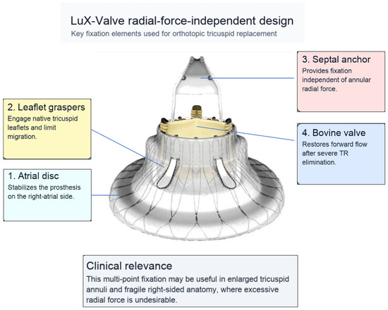
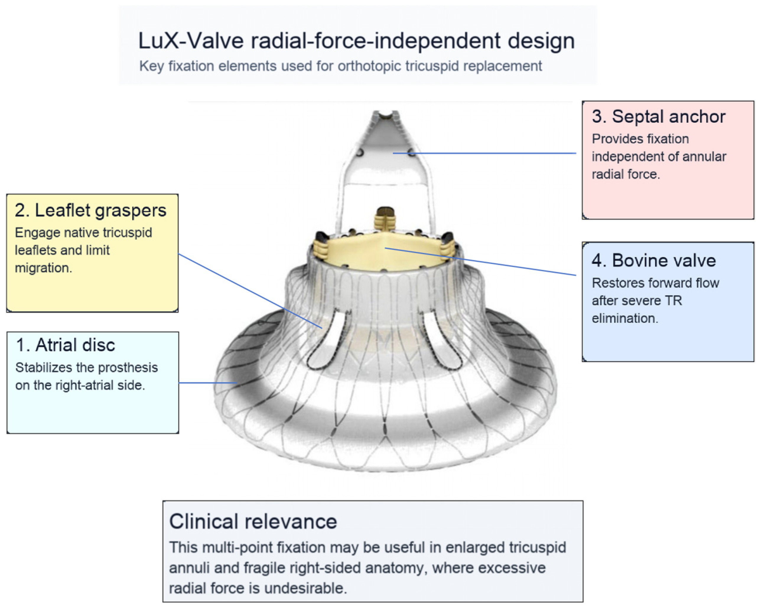
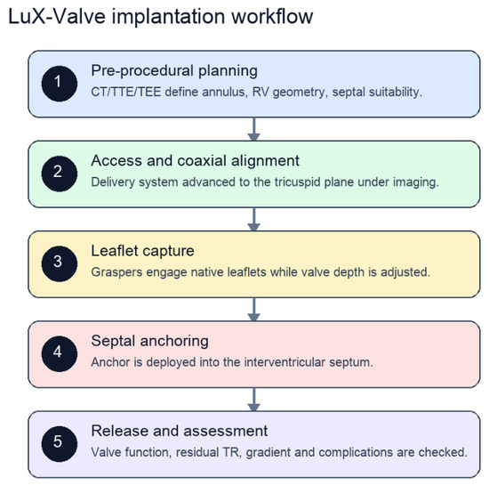
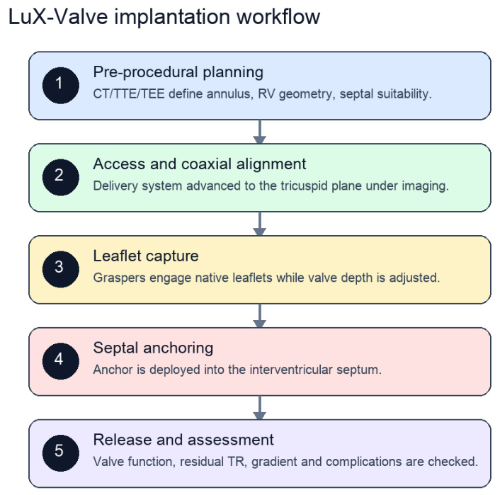
We retrospectively evaluated consecutive patients with severe tricuspid regurgitation who underwent TTVR with the LuX-Valve system and achieved complete follow-up at three participating centers in the TRAVEL trial [7]. No patients were excluded due to missing data. Patients were included if they had symptomatic TR (grade ≥ 4) [13,14] despite optimal medical therapy and were considered at high or prohibitive risk for conventional surgery by a multidisciplinary heart team. The LuX-Valve system is a novel radial force-independent orthotopic transcatheter tricuspid valve (Figure 1 and Figure 2). It consists of a self-expanding nitinol alloy stent frame with two expandable polytetrafluoroethylene-coated discs and a tri-leaflet bovine pericardial tissue valve. A detailed description and illustration of the device and procedural workflow have been previously published [7]. This study has been approved by the Ethics Review Committee of Fuwai Hospital.
Figure 1.
LuX-Valve device design. The radial force-independent system uses an atrial disc, tricuspid leaflet graspers, a bovine pericardial valve, and an interventricular septal anchor to achieve orthotopic fixation without relying primarily on annular radial force.
Figure 2.
Key steps for LuX-Valve implantation. Pre-procedural imaging defines eligibility and geometry; implantation proceeds through access and coaxial alignment, leaflet capture, septal anchoring, release, and immediate assessment of residual TR, gradients, and complications.
2.2. Echocardiography
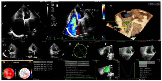
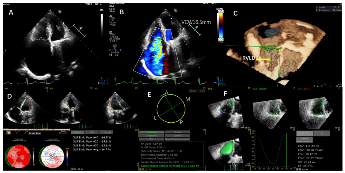
Baseline two-dimensional and three-dimensional transthoracic echocardiography (TTE) was performed within one week prior to TTVR in accordance with current guideline recommendations [13]. Examinations were conducted using Epiq 7c or CvX systems (Philips, Amsterdam, The Netherlands) or the E95 system (GE Vingmed Ultrasound AS, Horten, Norway). All echocardiographic analyses were conducted offline in the core laboratory by an experienced echocardiographer blinded to clinical outcomes, utilizing a CPA 2.5 workstation (TomTec ARENA, Unterschleissheim, Germany). Values were averaged over five consecutive cardiac cycles. Interobserver and intraobserver reproducibility was evaluated in a random subset of 20 patients by calculating intraclass correlation coefficients (ICC). RVFWS was derived from three segments of the RV free wall. RV end-systolic volume was quantified using three-dimensional TTE and indexed to body surface area (RVESVI) (Figure 3). Additional RV-focused parameters included TR grade [14], right atrial volume, RVLD, TVAPD, tricuspid annular plane systolic excursion (TAPSE), RV fractional area change (RV-FAC), tissue Doppler systolic velocity (S-TDI), systolic pulmonary artery pressure (sPAP), and RV-pulmonary artery coupling assessed by TAPSE/sPAP [15,16]. Left-sided parameters were retained only for baseline characterization.
Figure 3.
Two-dimensional and three-dimensional echocardiographic measurements. (A). Apical four-chamber view of a patient 21 years after mitral valve replacement; (B). Tricuspid regurgitation graded as 5, with a vena contracta width (VCW) of 16.5 mm; (C). Right ventricular longitudinal diameter (RVLD) measured using three-dimensional echocardiography; (D). Strain analysis used for ventricular functional assessment; (E). Tricuspid valve anteroposterior diameter (TVAPD) measured using a three-dimensional software package; (F). Right ventricular end-systolic volume index (RVESVI) measured using a 3D automated RV analysis software package.
2.3. Endpoints
The primary endpoint was 30-day MAE, defined as a composite of cardiovascular death and device-related complications, including significant intraoperative or postoperative hemorrhage, surgical re-exploration via sternotomy, unplanned reintervention, and implantation of a permanent pacemaker for newly developed atrioventricular conduction block. The secondary endpoint was all-cause mortality assessed through 5 years after the index procedure. Given the heterogeneity of the composite MAE endpoint and the limited number of events, individual components were summarized descriptively rather than modeled separately.
2.4. Statistical Analysis
Continuous variables are presented as mean ±standard deviation or median [interquartile range], and categorical variables as counts and percentages. Variables for multivariable models were pre-specified based on clinical importance and significance in univariable analysis (p < 0.10), with emphasis on RV parameters including RVESVI, RVLD, TVAPD, RVFWS, TAPSE, RV-FAC, S-TDI, sPAP, and TAPSE/sPAP. To minimize overfitting given the small event count, only a maximum of two covariates were included in any multivariable model. Given the limited number of events, Firth’s penalized likelihood method was applied to reduce small-sample bias. Long-term all-cause mortality was analyzed using Firth Cox proportional hazards regression, with results expressed as hazard ratios (HR) and 95% confidence intervals (CI). The proportional hazards assumption was verified using Schoenfeld residuals. In addition, 30-day MAEs were evaluated using Firth logistic regression, with odds ratios (OR) and 95% CI reported. Model performance was assessed by discrimination and calibration. Discrimination was evaluated using time-dependent receiver operating characteristic curves and Harrell’s concordance index. For MAE models, area under the ROC curve was compared using the DeLong test, and calibration was examined graphically with calibration plots. Risk stratification thresholds were prespecified based on clinical relevance and ROC analysis: RVESVI > 42 mL/m2 and sPAP > 35, >45, and >50 mmHg. Kaplan–Meier curves were constructed for these subgroups, and differences were compared using the log-rank test. All statistical analyses were performed using R software (version 4.3.2; R Foundation for Statistical Computing, Vienna, Austria). A two-sided p value < 0.05 was considered statistically significant. Reviewer-requested exploratory threshold analyses were also performed for TAPSE, RVFWS, 3D RVEF, RV-FAC, and RV basal diameter; tissue Doppler S’ thresholds were not modeled because S’ values were not available in the analyzable dataset.
3. Results
3.1. Baseline Characteristics
A total of 62 patients with TR grade ≥ 4 were included. The median age was 66.5 years (IQR, 61.0–72.8), and 52 patients (83.8%) were female. All patients were symptomatic, classified as New York Heart Association (NYHA) functional class III (80.6%) or IV (19.4%). Atrial fibrillation was present in 58 patients (93.6%), hypertension in 16 (25.8%), and ascites in five (8.1%). Twenty-six patients (41.2%) had a history of mitral valve replacement, 13 (21.0%) had undergone combined aortic and mitral valve replacement, and six (9.6%) had received permanent pacemaker implantation. The remaining patients were diagnosed with heart failure with preserved ejection fraction (HFpEF, n = 12). A detailed breakdown of prior cardiac surgery types is provided in the footnote of Table 1.
Table 1.
Baseline characteristics and univariate COX analysis.
Baseline TTE demonstrated preserved RV-FAC and RVFWS, whereas both left atrial and right atrial volumes were markedly enlarged. Detailed baseline characteristics and results of univariable Cox regression analyses for mortality are summarized in Table 1, and comprehensive echocardiographic measurements are presented in Table 2. Among RV-centered echocardiographic parameters, greater TVAPD (HR, 1.14; 95% CI, 1.01–1.28; p = 0.030) and increased RVLD (HR, 1.11; 95% CI, 1.01–1.21; p = 0.019) were significantly associated with all-cause mortality; NYHA class IV also conferred higher clinical risk (HR, 6.50; 95% CI, 1.40–38.90; p = 0.015).
Table 2.
Baseline echocardiographic measurements and univariate COX analysis.
3.2. Day Major Adverse Events and Exploratory Imaging Models
Within 30 days after TTVR, 14 patients (22.6%), including three deaths, experienced MAEs. Significant intraoperative or postoperative hemorrhage occurred in eight patients, seven of whom required surgical re-exploration via sternotomy. In addition, one patient underwent repeat open-heart tricuspid valve replacement, and six patients required permanent pacemaker implantation for newly developed atrioventricular conduction block. Because this composite endpoint was dominated by perioperative or device-related complications, RVFWS, RVLD, RVESVI, and TVAPD were retained only as exploratory imaging variables in subsequent models (Supplemental Table S1).
Firth logistic regression analyses are summarized in Table 3. Although larger RVESVI, RVLD, and TVAPD showed associations in RV-focused exploratory models, the 30-day MAE endpoint was dominated by perioperative or device-related complications and estimates were imprecise because of the small number of events. Accordingly, these models should be interpreted as hypothesis-generating rather than as evidence that baseline RV dysfunction or remodeling directly caused early MAEs.
Table 3.
Exploratory Firth logistic regression models for RV-focused variables and 30-day complications after TTVR.
Exploratory sPAP threshold analyses using >35, >45, and >50 mmHg cutoffs were incorporated into the revised analysis plan because pulmonary pressure and RV-pulmonary arterial coupling are directly relevant to RV reserve in severe TR. These thresholds should be interpreted with caution and validated with patient-level data in larger cohorts.
Reviewer-requested exploratory threshold analyses for 30-day MAEs are presented in Table 3. TAPSE < 15 mm showed a signal in this exploratory analysis (OR, 3.53; 95% CI, 1.02–12.24; p = 0.047), whereas TAPSE < 17 mm showed a nonsignificant trend (OR, 2.36; 95% CI, 0.63–8.89; p = 0.203). The corresponding exploratory estimates were as follows: RVFWS < 19% (OR, 0.99; 95% CI, 0.28–3.55; p = 0.992), RVFWS < 16% (OR, 1.92; 95% CI, 0.51–7.24; p = 0.336), 3D RVEF < 45% (OR, 2.60; 95% CI, 0.46–14.81; p = 0.282), 3D RVEF < 35% (OR, 1.09; 95% CI, 0.04–28.28; p = 0.958), RV-FAC < 35% (OR, 3.96; 95% CI, 0.79–19.93; p = 0.095), RV-FAC < 25% (OR, 3.34; 95% CI, 0.06–176.10; p = 0.550), RV basal diameter > 50 mm (OR, 0.48; 95% CI, 0.11–2.11; p = 0.329), and RV basal diameter > 55 mm (OR, 0.61; 95% CI, 0.10–3.94; p = 0.608). These estimates should be interpreted cautiously because several thresholds produced sparse cells and the early endpoint was dominated by procedural complications.
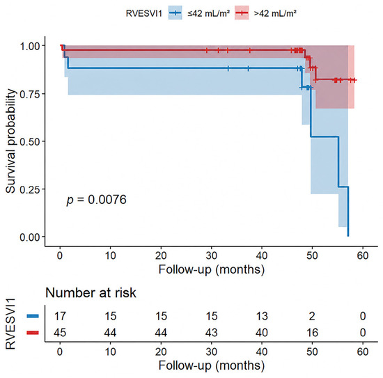
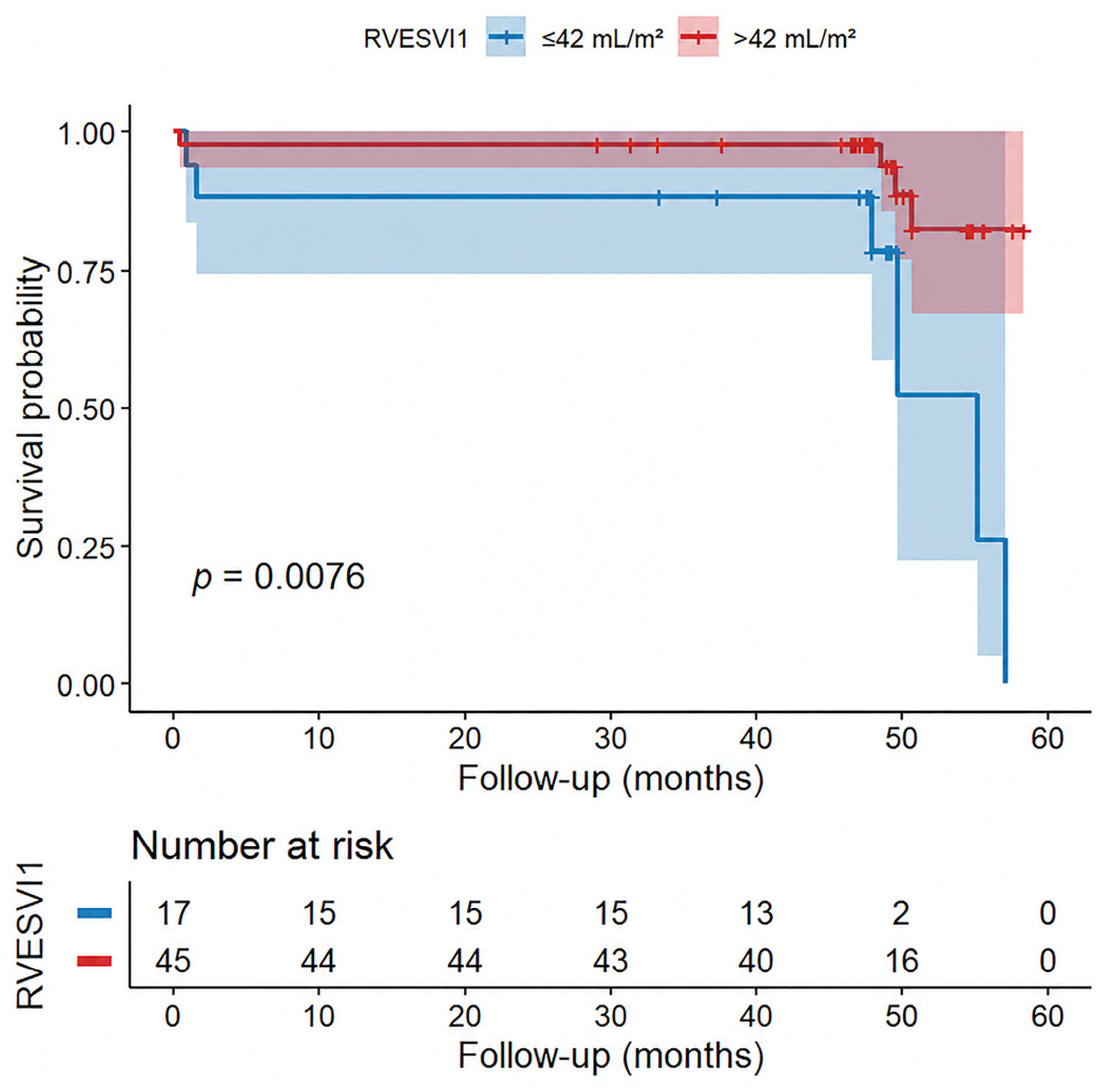
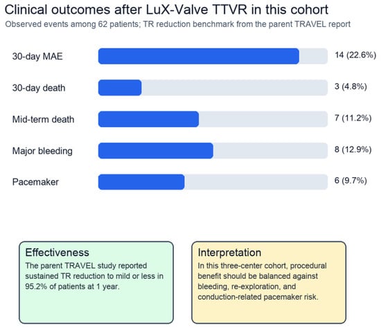
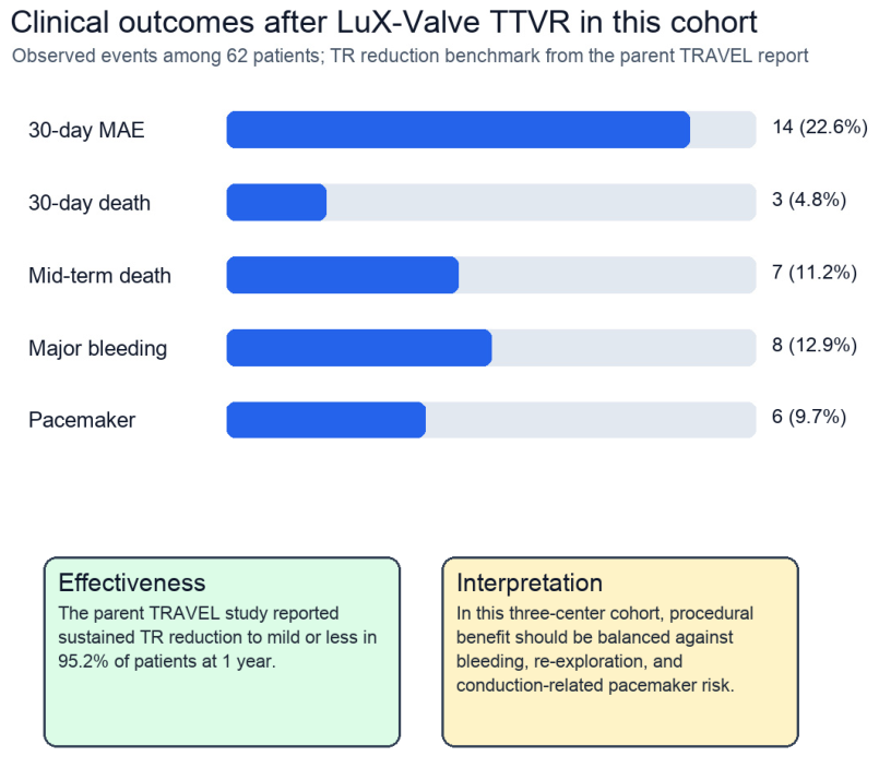
The RVESVI-based risk stratification is presented as a hypothesis-generating analysis and should be considered alongside the procedural composition of 30-day MAEs. Kaplan–Meier analysis stratified by RVESVI > 42.0 mL/m2 showed significantly higher event rates (log-rank p = 0.007) (Figure 4). Procedural effectiveness and complications of LuX-Valve TTVR are summarized in Table 4 and Figure 5.
Figure 4.
Kaplan–Meier curves stratified by RVESVI > 42 mL/m2.
Table 4.
LuX-Valve procedural outcomes, effectiveness, and early safety profile.
Figure 5.
Outcomes and early safety profile after LuX-Valve TTVR in the present cohort, including 30-day MAEs, mortality, bleeding, and pacemaker implantation, with contextual TR reduction from the parent TRAVEL study.
3.3. LuX-Valve Procedural Outcomes and Valve-Specific Effectiveness (Table 5)
Table 5.
LuX-Valve device characteristics, implantation technique, and potential benefits compared with other transcatheter tricuspid approaches.
All patients underwent orthotopic TTVR with the LuX-Valve system. The device is designed as a radial force-independent tricuspid prosthesis, using an atrial disc, leaflet graspers, and an interventricular septal anchor for fixation. In this cohort, the mean follow-up was 46.2 months, with seven all-cause deaths (11.2%) and three deaths within 30 days (4.8%). The principal early complications were bleeding requiring intervention, surgical re-exploration, repeat tricuspid valve replacement, and new permanent pacemaker implantation. These outcomes highlight the need to report procedural benefit together with safety events when applying LuX-Valve TTVR in high-risk severe TR.
The effectiveness endpoint was clinically centered on elimination or durable reduction of severe TR after valve replacement. In the parent TRAVEL report, LuX-Valve implantation achieved 100% device success, 98.4% procedural success, and sustained reduction of TR to mild or less in 95.2% of patients at 1 year [7]. The present three-center analysis should therefore be interpreted as a mid-term prognostic extension of a procedure with established short-term TR reduction, while acknowledging that center-specific follow-up TR grades should be reported when available.
3.4. Reproducibility
The interobserver and intraobserver ICC were 0.88 and 0.91, respectively, for RVESVI. Reproducibility for RVFWS and other RV functional indices should be reported in future analyses to support broader clinical implementation.
4. Discussion
In this multicenter study of 62 patients undergoing TTVR with the LuX-Valve system, we summarized mid-term outcomes, perioperative safety, procedural effectiveness, and exploratory RV-centered imaging signals. The principal findings were as follows: (1) LuX-Valve TTVR achieved favorable mid-term survival and established procedural effectiveness in the TRAVEL context; (2) 30-day MAEs occurred in 14 patients (22.6%) and were mainly composed of perioperative bleeding, surgical re-exploration, repeat intervention, and conduction-related pacemaker implantation; and (3) RV remodeling and functional markers showed exploratory associations but should not be interpreted as demonstrating that baseline RV dysfunction or remodeling directly drove 30-day MAEs. These results support reporting early safety and procedural effectiveness separately from hypothesis-generating imaging risk stratification.
4.1. Mid-Term Outcome of TTVR
For patients with severe TR, TTVR has been shown to be superior to optimal medical therapy alone in reducing cardiac mortality and heart failure hospitalization at 1 year, as demonstrated in recent trials such as TRAVEL and TRISCEND [4,5]. In pairwise comparisons, patients undergoing valve replacement consistently achieved more favorable outcomes than controls, including a lower incidence of all-cause mortality [4,5]. Compared with optimal medical therapy alone, treatment with TTVR also resulted in substantial improvements in symptoms, functional capacity, and quality of life [6]. With the rapid evolution of the TTVR field and an expanding evidence base, the first device has already received CE Mark and U.S. Food and Drug Administration approval, with commercial implantation now underway [17].
At this stage, longer-term follow-up is particularly important to determine the durability and prognostic impact of TTVR in patients with severe TR. In the present study, we analyzed the cohort from three high-volume centers that participated in the TRAVEL study initiated in 2020 [5], with a mean follow-up of 3.8 years. The overall mortality rate was 11.2%, consistent with previously reported 1-year mortality rates, thereby providing the first evidence that mid-term outcomes after TTVR are favorable. Notably, the 1-year mortality in our cohort was only 4.8%, substantially lower than the 10.3% previously reported in the overall TRAVEL cohort of 126 patients across eight centers. This difference likely reflects the inclusion of more experienced, higher-volume centers in the current analysis, underscoring that outcomes after TTVR are not uniform and may be strongly influenced by institutional expertise and operator experience. This selection bias limits the generalizability of our findings to lower-volume centers or non-trial populations.
4.2. Prognostic Stratification
Although the feasibility, safety, and symptomatic benefits of TTVR in patients with severe TR have been demonstrated in multicenter studies, prognostic stratification beyond procedural success has remained limited. Imaging-based markers to guide patient selection and postoperative monitoring have not been well established. Our findings extend this evidence by emphasizing RV-centered markers, especially RVESVI, RVLD, TVAPD, RVFWS, TAPSE, RV-FAC, S-TDI, sPAP, and TAPSE/sPAP, rather than left-sided parameters. For 30-day MAEs, however, these imaging models should be interpreted cautiously because the endpoint was largely procedural or device-related. RVESVI > 42.0 mL/m2 emerged as a hypothesis-generating cutoff that may be incorporated into routine preoperative echocardiographic assessment for further investigation in larger cohorts. The additional sPAP thresholds of >35, >45, and >50 mmHg were added because pulmonary vascular load is central to RV reserve and post-procedural adaptation. Additional reviewer-requested RV functional thresholds were incorporated to improve clinical interpretability, but only TAPSE < 15 mm reached statistical significance in this exploratory threshold analysis.
4.3. Pathophysiological Insights
The prognostic value of RV remodeling in this study is biologically plausible. In severe TR, chronic volume overload promotes progressive RV dilation, annular enlargement, leaflet malcoaptation, and impaired contractile reserve. RVESVI reflects the burden of maladaptive remodeling at end systole, whereas RVLD and TVAPD capture chamber and annular geometry that may influence both procedural complexity and post-procedural adaptation. RVFWS, TAPSE, RV-FAC, S-TDI, and TAPSE/sPAP provide complementary information about RV systolic function and RV-pulmonary arterial coupling.
Successful TR reduction abruptly changes RV loading conditions. Patients with advanced RV dilation, impaired contractile reserve, or elevated sPAP may be vulnerable to afterload mismatch, low cardiac output, and bleeding or hemodynamic instability after valve implantation. For this reason, RV-pulmonary interaction is more clinically actionable than left-sided strain alone in patients selected for TTVR. The revised analysis therefore prioritizes RV parameters and pulmonary pressure thresholds when assessing worse outcomes. The LuX-Valve system may be particularly relevant in this anatomy because its fixation strategy is radial force-independent. The combination of an atrial disc, leaflet graspers, and an interventricular septal anchor is intended to stabilize the prosthesis in a dilated tricuspid annulus without excessive radial force. This design may broaden treatment options for high-risk patients with severe TR, but it also requires careful imaging guidance, septal assessment, and post-release evaluation for residual TR, valve gradient, bleeding, and conduction disturbance.
In contrast to analyses centered on LV-GLS, RVESVI directly reflects chronic volume overload and maladaptive RV remodeling resulting from longstanding severe TR. Persistent regurgitant flow leads to progressive dilation, increased wall stress, and impaired contractile efficiency. An enlarged RVESVI indicates reduced ventricular reserve and heightened vulnerability to abrupt hemodynamic shifts once regurgitation is eliminated by TTVR [18,19]. The sudden reduction in RV preload, coupled with increased effective forward afterload, may precipitate acute afterload mismatch, presenting as low cardiac output and RV failure. Marked RV enlargement may also increase the technical difficulty of anchoring the LuX-Valve device and heighten the risk of bleeding.
5. Limitations
This study has several limitations. First, the sample size was relatively small, with only 62 patients across three centers, and the number of clinical events was limited (seven deaths and 14 30-day MAEs). This significantly limits statistical power and introduces a high risk of model overfitting, even with the use of Firth’s penalized regression; the wide confidence intervals for certain predictors reflect this inherent statistical uncertainty. Second, this is a post hoc analysis of a single-arm clinical trial enrolling a highly selected patient group from three high-volume centers, which introduces selection bias and limits generalizability to broader clinical practice. Third, echocardiographic examinations were performed at each participating center, and although analyses were conducted by an experienced observer, inter-institutional variability cannot be completely excluded. Fourth, RV remodeling, RV functional indices, sPAP, and TR reduction were assessed using available baseline and follow-up data; longitudinal imaging follow-up with standardized core-laboratory adjudication would be necessary to more accurately capture reverse remodeling after TTVR. Fifth, the small number of events precluded internal validation techniques and robust sensitivity analyses of individual components of the composite MAE endpoint, including sPAP threshold analyses. Future prospective, large-scale multicenter studies with standardized imaging protocols, longer follow-up, and external validation are warranted to confirm RV-focused predictors and to establish validated risk scores for clinical practice. The newly added RV functional threshold estimates were exploratory and were limited by sparse event counts for several cutoffs.
6. Conclusions
LuX-Valve TTVR showed favorable mid-term survival and clinically meaningful procedural effectiveness, with 30-day MAEs mainly reflecting perioperative or device-related complications. RV-centered echocardiographic parameters, including RVESVI, RVLD, TVAPD, RV functional indices, sPAP thresholds, and RV-pulmonary arterial coupling, remain hypothesis-generating markers for risk stratification and require validation in larger cohorts. TAPSE, RVFWS, 3D RVEF, RV-FAC, and RV basal diameter threshold findings also require validation in larger cohorts.
Supplementary Materials
The following supporting information can be downloaded at: https://www.mdpi.com/article/10.3390/jcdd13080381/s1, Table S1: Univariable analyses for predictors of 30-day complications after TTVR.
Author Contributions
X.Z. and H.S. contributed equally to this work. Conceptualization, K.P.; methodology, X.Z., H.S. and Y.P.; validation, X.Z. and Y.J.; formal analysis, F.L. and X.M.; investigation, X.M.; resources, X.H.; data curation, K.P.; writing—original draft preparation, X.Z., H.S. and Y.P.; writing—review and editing, J.Y.; visualization, Y.P.; supervision, K.P.; project administration, J.Y.; funding acquisition, K.P. All authors have read and agreed to the published version of the manuscript.
Funding
This work was supported by the Development and Transformation of New Technology and Construction of Precision Diagnosis and Treatment System for Transcatheter Interventional Diagnosis and Treatment of Structural Heart Diseases (2022YFC2503400), the National Key R&D Program of China (2020YFC2008100), Research on Key Techniques of Minimally Invasive Treatment for Valvular Heart Diseases (2023-YBSF-105) and Noncommunicable Chronic Diseases-National Science and Technology Major Project (No. 2024ZD0527000 and No. 2024ZD0527002).
Institutional Review Board Statement
This study is a retrospective analysis, and according to the policy of our institution, this type of study does not require separate ethical approval.
Informed Consent Statement
Patient consent was waived due to the retrospective nature of this study, which involved the analysis of existing, de-identified medical records and ultrasound imaging data.
Data Availability Statement
The data presented in this study are available on request from the corresponding authors. The data are not publicly available due to restrictions concerning patient privacy and confidentiality.
Acknowledgments
The authors gratefully acknowledge the administrative support provided by the Department of Ultrasound at Fuwai Hospital. We also extend our sincere thanks to the clinical research coordinators for their invaluable assistance in data collection and management. The authors are solely responsible for the content of this publication.
Conflicts of Interest
The authors declare no conflicts of interest. The funders had no role in the design of the study; in the collection, analyses, or interpretation of data; in the writing of the manuscript; or in the decision to publish the results.
Correction Statement
This article has been republished with a minor correction to the Institutional Review Board Statement. This change does not affect the scientific content of the article.
Abbreviations
The following abbreviations are used in this manuscript:
| CI | confidence interval |
| OR | odds ratio |
| HR | hazard ratio |
| MAE | major adverse event |
| NYHA | New York Heart Association |
| HFpEF | heart failure with preserved ejection fraction |
| RV | right ventricle/ventricular |
| LV-GLS | left ventricular global longitudinal strain |
| RV-GLS | right ventricular global longitudinal strain |
| RVESVI | right ventricular end-systolic volume index |
| RVLD | right ventricular longitudinal dimension |
| TAPSE | tricuspid annular plane systolic excursion |
| TTE | transthoracic echocardiography |
| TVAPD | tricuspid annular anterior–posterior diameter |
| TTVR | transcatheter tricuspid valve replacement |
| TR | tricuspid regurgitation |
| TRAVEL | Transcatheter Right Atrial-Ventricular Valve Replacement With LuX-Valve |
| ICC | intraclass correlation coefficients |
References
- Hahn, R.T. Tricuspid Regurgitation. N. Engl. J. Med. 2023, 388, 1876–1891. [Google Scholar] [CrossRef] [Scilit] [PubMed]
- Zack, C.J.; Fender, E.A.; Chandrashekar, P.; Reddy, Y.N.; Bennett, C.E.; Stulak, J.M.; Miller, V.M.; Nishimura, R.A. National Trends and Outcomes in Isolated Tricuspid Valve Surgery. J. Am. Coll. Cardiol. 2017, 70, 2953–2960. [Google Scholar] [CrossRef] [Scilit] [PubMed]
- Kawsara, A.; Alqahtani, F.; Nkomo, V.T.; Eleid, M.F.; Pislaru, S.V.; Rihal, C.S.; Nishimura, R.A.; Schaff, H.V.; Crestanello, J.A.; Alkhouli, M. Determinants of Morbidity and Mortality Associated with Isolated Tricuspid Valve Surgery. J. Am. Heart Assoc. 2021, 10, e018417. [Google Scholar] [CrossRef] [Scilit] [PubMed]
- Hausleiter, J.; Stolz, L.; Lurz, P.; Rudolph, V.; Hahn, R.; Estévez-Loureiro, R.; Davidson, C.; Zahr, F.; Kodali, S.; Makkar, R.; et al. Transcatheter Tricuspid Valve Replacement. J. Am. Coll. Cardiol. 2025, 85, 265–291. [Google Scholar] [CrossRef] [Scilit] [PubMed]
- Hahn, R.T.; Makkar, R.; Thourani, V.H.; Makar, M.; Sharma, R.P.; Haeffele, C.; Davidson, C.J.; Narang, A.; O’Neill, B.; Lee, J.; et al. Transcatheter Valve Replacement in Severe Tricuspid Regurgitation. N. Engl. J. Med. 2025, 392, 115–126. [Google Scholar] [CrossRef] [Scilit] [PubMed]
- Arnold, S.V.; Hahn, R.T.; Thourani, V.H.; Makkar, R.; Makar, M.; Sharma, R.P.; Haeffele, C.; Davidson, C.J.; Narang, A.; O’Neill, B.; et al. TRISCEND II Pivotal Trial Investigators. Quality of Life After Transcatheter Tricuspid Valve Replacement: 1-Year Results From TRISCEND II Pivotal Trial. J. Am. Coll. Cardiol. 2025, 85, 206–216. [Google Scholar] [CrossRef] [Scilit] [PubMed]
- Pan, X.; Lu, F.; Wang, Y.; Guo, Y.; Chen, M.; Meng, X.; Zhang, H.; Dong, N.; Shang, X.; Yang, J.; et al. Transcatheter Tricuspid Valve Replacement With the Novel System: 1-Year Outcomes From the TRAVEL Study. JACC Cardiovasc. Interv. 2025, 18, 1276–1285. [Google Scholar] [CrossRef] [Scilit] [PubMed]
- Lang, R.M.; Addetia, K.; Narang, A.; Mor-Avi, V. 3-Dimensional echocardiography. J. Am. Coll. Cardiol. Imaging 2018, 11, 1854. [Google Scholar] [CrossRef] [Scilit] [PubMed]
- Hahn, R.T. State-of-the-Art Review of Echocardiographic Imaging in the Evaluation and Treatment of Functional Tricuspid Regurgitation. Circ. Cardiovasc. Imaging 2016, 9, e005332. [Google Scholar] [CrossRef] [Scilit] [PubMed]
- Hahn, R.T.; Chandrashekhar, Y. Tricuspid Regurgitation: A Voyage of Discovery. JACC Cardiovasc. Imaging 2019, 12, 572–575. [Google Scholar] [CrossRef] [Scilit] [PubMed]
- Lang, R.M.; Badano, L.P.; Mor-Avi, V.; Afilalo, J.; Armstrong, A.; Ernande, L.; Flachskampf, F.A.; Foster, E.; Goldstein, S.A.; Kuznetsova, T.; et al. Recommendations for cardiac chamber quantification by echocardiography in adults: An update from the American Society of Echocardiography and the European Association of Cardiovascular Imaging. Eur. Heart J. Cardiovasc. Imaging 2015, 16, 233–271. [Google Scholar] [CrossRef] [Scilit] [PubMed]
- Hahn, R.T.; Zamorano, J.L. The need for a new tricuspid regurgitation grading scheme. Eur. Heart J. Cardiovasc. Imaging 2017, 18, 1342–1343. [Google Scholar] [CrossRef] [Scilit] [PubMed]
- Brener, M.I.; Lurz, P.; Hausleiter, J.; Rodés-Cabau, J.; Fam, N.; Kodali, S.K.; Rommel, K.P.; Muntané-Carol, G.; Gavazzoni, M.; Nazif, T.M.; et al. Right Ventricular-Pulmonary Arterial Coupling and Afterload Reserve in Patients Undergoing Transcatheter Tricuspid Valve Repair. J. Am. Coll. Cardiol. 2022, 79, 448–461. [Google Scholar] [CrossRef] [Scilit] [PubMed]
- Redfield, M.M.; Borlaug, B.A. Heart Failure with Preserved Ejection Fraction: A Review. JAMA 2023, 329, 827–838. [Google Scholar] [CrossRef] [Scilit] [PubMed]
- Furman, D.; Whisenant, B. Transcatheter Tricuspid Valve Clinical Trials: Incomplete Data and FDA-Approved Devices. Struct. Heart 2024, 8, 100335. [Google Scholar] [CrossRef] [Scilit] [PubMed]
- Hahn, R.T.; Saric, M.; Faletra, F.F.; Garg, R.; Gillam, L.D.; Horton, K.; Khalique, O.K.; Little, S.H.; Mackensen, G.B.; Oh, J.; et al. Recommended standards for the performance of transesophageal echocardiographic screening for structural heart intervention: From the american society of echocardiography. J. Am. Soc. Echocardiogr. 2022, 35, 1–76. [Google Scholar] [CrossRef] [Scilit] [PubMed]
- Potter, E.L.; Ramkumar, S.; Wright, L.; Marwick, T.H. Incremental Prognostic Value of Global Longitudinal Strain Over Ejection Fraction in Patients With Heart Failure. JACC Cardiovasc. Imaging 2025, 18, 431–441. [Google Scholar]
- Fortmeier, V.; Lachmann, M.; Körber, M.I.; Unterhuber, M.; Schöber, A.R.; Stolz, L.; Stocker, T.J.; Kassar, M.; Gerçek, M.; Rudolph, T.K.; et al. Sex-Related Differences in Clinical Characteristics and Outcome of Patients Undergoing Transcatheter Tricuspid Valve Intervention. J. Am. Coll. Cardiol. 2024, 84, 1123–1136. [Google Scholar]
- Pieske, B.; Tschöpe, C.; de Boer, R.A.; Fraser, A.G.; Anker, S.D.; Donal, E.; Edelmann, F.; Fu, M.; Guazzi, M.; Lam, C.S.; et al. How to diagnose heart failure with preserved ejection fraction: The HFA-PEFF diagnostic algorithm: A consensus recommendation from the Heart Failure Association (HFA) of the European Society of Cardiology (ESC). Eur. Heart J. 2019, 40, 3297–3317. [Google Scholar] [CrossRef] [Scilit] [PubMed]

Disclaimer/Publisher’s Note: The statements, opinions and data contained in all publications are solely those of the individual author(s) and contributor(s) and not of MDPI and/or the editor(s). MDPI and/or the editor(s) disclaim responsibility for any injury to people or property resulting from any ideas, methods, instructions or products referred to in the content. |
© 2026 by the authors. Licensee MDPI, Basel, Switzerland. This article is an open access article distributed under the terms and conditions of the Creative Commons Attribution (CC BY) license.