


Abstract
Background: Systemic lupus erythematosus-associated pulmonary arterial hypertension (SLE-PAH) is a severe complication requiring early recognition and risk stratification. However, factors associated with prevalent disease, non-invasive detection, incident PAH, and prognosis after diagnosis are frequently conflated. Methods: PubMed, Web of Science, Scopus, and the Cochrane Library were initially searched through 23 September 2026, and searches of all four databases were updated on 1 November 2026. Observational evidence was evaluated using a structured clinical-pathway framework and the Newcastle–Ottawa Scale, and findings were synthesized narratively because of clinical and methodological heterogeneity and potentially overlapping cohorts. Results: Thirty-five studies were included in the qualitative synthesis. Serositis, interstitial lung disease, pericardial effusion, anti-U1 RNP and anti-Ro/SSA antibodies, reduced diffusing capacity, and microvascular abnormalities were mainly associated with prevalent or concomitant disease. Non-invasive detection was supported by a multimodal approach incorporating symptoms, biomarkers, pulmonary function, and echocardiography, with right heart catheterization required for PAH confirmation. Evidence on incident, catheterization-confirmed PAH prediction was limited. After diagnosis, functional class, exercise capacity, pulmonary vascular load, right-heart function, and serial risk status were recurrent prognostic indicators. Conclusions: Evidence is more extensive for prognosis after established SLE-PAH than for incident prediction, highlighting the need for externally validated SLE-specific screening and risk-stratification tools.
1. Introduction
Systemic lupus erythematosus (SLE) is a chronic autoimmune disease characterized by heterogeneous clinical manifestations and multisystem involvement [1,2]. Despite substantial advances in diagnosis and treatment, cardiovascular and pulmonary complications remain important contributors to disease-related morbidity and mortality. Among these complications, pulmonary arterial hypertension (PAH) is particularly severe because it may lead to progressive right ventricular dysfunction, impaired functional capacity, reduced quality of life, and premature death [3,4].
SLE-associated pulmonary arterial hypertension (SLE-PAH) is classified as Group 1 pulmonary hypertension (PH); however, PH in patients with SLE may arise through several distinct pathophysiological mechanisms. According to the 2022 European Society of Cardiology/European Respiratory Society (ESC/ERS) guidelines, precapillary PH is defined by a mean pulmonary arterial pressure (mPAP) > 20 mmHg, pulmonary arterial wedge pressure (PAWP) ≤ 15 mmHg, and pulmonary vascular resistance (PVR) > 2 Wood units, as assessed by right heart catheterization (RHC) [5]. Importantly, precapillary haemodynamics alone do not establish a diagnosis of Group 1 SLE-PAH. Alternative mechanisms must be considered, including PH associated with left-heart disease (Group 2), interstitial lung disease (ILD) or hypoxia (Group 3), and chronic thromboembolic pulmonary hypertension (CTEPH; Group 4). The latter is particularly relevant in patients with antiphospholipid antibodies or previous thromboembolic events. Consequently, the diagnostic evaluation should consider competing causes of PH before attributing pulmonary vascular disease to SLE-associated PAH. Differences in diagnostic workup, haemodynamic definitions, screening strategies, and reliance on echocardiography without confirmatory RHC may contribute to the variability in reported PH and PAH prevalence among patients with SLE [5,6,7,8]. Echocardiography remains essential for estimating the probability of PH and identifying candidates for invasive evaluation, but it cannot independently establish the haemodynamic or aetiological diagnosis. These distinctions are fundamental when interpreting evidence related to disease susceptibility, detection, incident prediction, and prognosis.
The pathogenesis of SLE-PAH is complex and involves immune dysregulation, endothelial dysfunction, inflammation, vasculopathy, thrombosis, and pulmonary vascular remodelling [4,9]. Numerous clinical and serological characteristics have been associated with SLE-PAH, including Raynaud phenomenon, serositis, pericardial effusion, ILD, antiphospholipid antibodies, and anti-U1 ribonucleoprotein antibodies [10,11,12]. Functional impairment, invasive haemodynamic abnormalities, and measures of right ventricular adaptation have also been associated with disease severity and adverse outcomes among patients with established SLE-PAH [13,14,15,16]. More recent studies have expanded this evidence by evaluating genetic susceptibility, revised haemodynamic definitions, right ventricular–pulmonary arterial (RV-PA) coupling, right ventricular dyssynchrony, and right atrial strain [17,18,19,20,21].
Several previous evidence syntheses have assessed risk factors, biomarkers, survival, and treatment in SLE-PAH. Wang et al. examined serological biomarkers associated with SLE-PAH, while subsequent meta-analyses evaluated broader groups of clinical and laboratory risk factors [11,22,23]. Atsumi et al. combined a systematic review with expert consensus to identify factors potentially relevant to PAH screening in patients with SLE [12]. Other reviews have addressed survival, prognostic factors, or the effects of immunosuppressive treatment [24,25]. Collectively, these publications have identified several potentially relevant variables, but they have not consistently distinguished the different clinical and temporal roles of the reported findings.
This distinction is methodologically and clinically important. A characteristic found more frequently in patients with established PAH than in patients with SLE without PAH represents an association with prevalent disease but does not necessarily predict the future development of PAH. Similarly, symptoms, biomarkers, pulmonary function variables, and echocardiographic findings may facilitate non-invasive detection and selection for RHC without representing predictors of incident, RHC-confirmed PAH. Conversely, functional capacity, invasive haemodynamic variables, and measures of right ventricular adaptation assessed in patients with established SLE-PAH primarily provide information about disease severity and prognosis. Referring to all these variables as “predictors” may therefore obscure their distinct clinical applications and overstate the temporal evidence provided by cross-sectional or case–control studies.
Accordingly, this systematic review aimed to evaluate and classify clinical, functional, serological, genetic, echocardiographic, imaging, and invasive haemodynamic evidence according to its position within the SLE-PAH clinical pathway: (1) factors associated with prevalent SLE-PAH; (2) indicators relevant to non-invasive screening or detection; (3) predictors of incident PAH identified in longitudinal studies; and (4) prognostic factors evaluated after PAH diagnosis. We additionally examined whether the available evidence supports prospectively validated predictors of incident PAH or an integrated SLE-specific screening or prognostic model.
2. Materials and Methods
2.1. Protocol and Registration
This systematic review was conducted in accordance with the Preferred Reporting Items for Systematic Reviews and Meta-Analyses (PRISMA) 2020 statement [26]. The review protocol was prospectively registered in the International Prospective Register of Systematic Reviews (PROSPERO; CRD420261417960).
2.2. Eligibility Criteria
Studies were eligible if they included adults with systemic lupus erythematosus (SLE), diagnosed according to established classification criteria, and evaluated factors related to PAH susceptibility, detection, severity, or prognosis. Eligible variables included clinical characteristics, serological biomarkers, functional measures, echocardiographic and other imaging parameters, and invasive haemodynamic variables.
For analyses of disease susceptibility, severity, and prognosis, SLE-associated PAH (SLE-PAH) was required to be confirmed by RHC, using the haemodynamic definition applicable at the time of the study. Eligible studies compared patients with SLE-PAH with patients with SLE without PAH, longitudinally evaluated the development of PAH in an initially PAH-free SLE population, or examined clinically relevant outcomes among patients with established SLE-PAH.
Studies evaluating non-invasive screening or detection were eligible when they assessed indicators of increased PH probability or the need for RHC. Studies relying on echocardiographic estimates without systematic RHC confirmation were classified exclusively as evidence of suspected or echocardiographically defined PH, regardless of the terminology used in the original publications. Such findings were not interpreted as confirmed Group 1 SLE-PAH, predictors of incident PAH, or prognostic factors after PAH diagnosis. Historical RHC-based haemodynamic definitions were interpreted according to the diagnostic criteria applicable at the time of each study. Cross-sectional associations and baseline disease-severity measures were not considered evidence of incident prediction or longitudinal prognosis. No lower publication-year limit was applied.
Evidence was classified according to its clinical role as: (1) factors associated with prevalent SLE-PAH; (2) indicators relevant to non-invasive screening or detection; (3) predictors of incident PAH identified in longitudinal studies; and (4) prognostic factors associated with mortality, clinical worsening, hospitalisation, treatment response, or functional deterioration after PAH diagnosis.
Prospective and retrospective cohort studies, case–control studies, cross-sectional studies, and registry-based observational studies were eligible. Studies of mixed connective tissue disease populations were included only when SLE-specific data could be extracted separately.
Case reports, small case series, conference abstracts without sufficient data, reviews, meta-analyses, editorials, guidelines, expert opinions, and animal or in vitro studies were excluded. Studies were also excluded when the PH classification or diagnostic method was insufficiently described, when SLE-specific results could not be extracted, or when none of the prespecified susceptibility, detection, incident prediction, or prognostic outcomes was evaluated.
2.3. Information Sources and Search Strategy
An initial systematic literature search was conducted in PubMed, Web of Science, Scopus, and the Cochrane Library from database inception to 23 September 2026. The original search strategy combined terms related to systemic lupus erythematosus (SLE), pulmonary hypertension (PH) or pulmonary arterial hypertension (PAH), potential predictors and biomarkers, and right heart catheterization (RHC).
To improve search sensitivity and address the restrictive nature of the initial strategy, an updated literature search was conducted on 1 November 2026 in PubMed, Web of Science, Scopus, and the Cochrane Library. The search approach was refined to enhance the identification of relevant studies on systemic lupus erythematosus and pulmonary hypertension, with search expressions adapted to the characteristics, indexing systems, and search functionalities of each database.
No language restrictions or lower publication-date limits were applied. Reference lists of relevant systematic reviews and eligible publications were additionally screened to identify potentially relevant studies. Embase was not searched, representing a limitation of the search coverage.
The original and updated database-specific search strategies, search dates, and retrieval counts are presented in Supplementary Tables S3A and S3B, respectively.
2.4. Study Selection
Available bibliographic records exported from the expanded searches in PubMed, Web of Science, and Scopus were combined and deduplicated using DOI and normalized-title-based matching. The original search results were reported separately from the expanded search to avoid double-counting across search rounds. The repeated Cochrane Library search identified one additional record, which was included in the PRISMA identification count and documented in Supplementary Table S3B. Two reviewers (TB and PŠP) independently screened titles and abstracts according to the predefined eligibility criteria. Potentially eligible reports were sought in full text and assessed independently.
When multiple publications originated from the same cohort, each report was assessed separately and linked to the underlying cohort. Multiple reports were retained when they evaluated different exposures, outcomes, follow-up periods, or clinically distinct research questions; however, overlapping participants were not counted as independent populations in the interpretation of the evidence.
Disagreements were resolved through discussion and consensus. Reasons for exclusion were recorded at the full-text stage, and the complete selection process was documented using a PRISMA 2020 flow diagram.
2.5. Data Extraction
Two reviewers (TB and PŠP) independently extracted data from the included studies using a standardized data extraction form. Disagreements were resolved through discussion and consensus. Extracted information included the first author, publication year, country, study design, recruitment setting, sample size, participant characteristics, SLE classification criteria, method and criteria used to diagnose PAH, follow-up duration, evaluated variables, outcome definitions, statistical methods, effect estimates, and principal findings.
The evaluated variables included clinical characteristics, serological and genetic markers, functional measures, echocardiographic and other imaging parameters, and invasive haemodynamic variables. When available, adjusted effect estimates, including odds ratios, hazard ratios, confidence intervals, and corresponding p-values, were preferentially extracted.
Each variable was classified according to its clinical role as: (1) a factor associated with prevalent SLE-PAH; (2) an indicator relevant to non-invasive screening or detection; (3) a predictor of incident PAH identified in a longitudinal study; or (4) a prognostic factor associated with outcomes after PAH diagnosis.
Potentially overlapping publications from the same registry or institution were identified by comparing study centres, recruitment periods, participant characteristics, and sample sizes. Multiple reports from the same or partially overlapping cohort were retained when they addressed different exposures, outcomes, follow-up periods, or research questions. However, overlapping populations were not treated as independent evidence for the same association.
2.6. Methodological Quality and Risk-of-Bias Assessment
The methodological quality of the included observational studies was independently appraised by two reviewers (TB and PŠP) using the Newcastle–Ottawa Scale (NOS) [27]. Potential design-specific sources of bias were additionally examined qualitatively, without assigning formal risk-of-bias ratings using specialized assessment tools.
Cohort and case–control studies were evaluated across three domains: selection of participants, comparability of study groups, and ascertainment of exposure or outcome. Cross-sectional studies were evaluated using an adapted version of the NOS appropriate for cross-sectional designs.
Particular attention was given to the method used to diagnose PAH, representativeness of the study population, adequacy of comparator-group selection, adjustment for relevant confounding variables, completeness and duration of follow-up, outcome ascertainment, and selective reporting. For studies evaluating non-invasive screening or detection, the presence and application of an appropriate reference standard were also considered.
NOS scores were retained as descriptive indicators of methodological quality, with scores of 7–9, 4–6, and <4 representing higher, intermediate, and lower NOS-based methodological quality, respectively. Importantly, these categories were not considered equivalent to definitive low-, moderate-, or high-risk-of-bias judgments. Given the heterogeneity of study designs and research objectives, particular attention was also directed toward design-specific methodological limitations, including incomplete application of the reference standard and potential verification bias in screening studies; missing data, overfitting, and lack of external validation in prediction-model studies; and confounding, participant attrition, outcome ascertainment, and statistical adjustment in prognostic studies. Therefore, overall NOS scores were interpreted cautiously and in conjunction with individual sources of potential bias. Assessments were performed independently by two reviewers, and disagreements were resolved through discussion and consensus. Detailed study-level NOS assessments are provided in Supplementary Table S1.
2.7. Evidence Classification and Data Synthesis
Because of substantial clinical and methodological heterogeneity among the included studies, findings were synthesized qualitatively. Heterogeneity was assessed by comparing study populations, study designs, PAH definitions, evaluated variables, timing of measurement, follow-up periods, outcome definitions, and reported effect measures.
To avoid conflating association, detection, prediction, and prognosis, findings were organized according to their position within the SLE-PAH clinical pathway: (1) factors associated with prevalent SLE-PAH; (2) indicators relevant to non-invasive screening or detection; (3) predictors of incident PAH; and (4) prognostic factors evaluated after PAH diagnosis.
Within these categories, the evidence was further grouped into clinical and functional, serological and genetic, echocardiographic and imaging, and invasive haemodynamic domains. Greater interpretative weight was assigned to longitudinal studies, RHC-confirmed PAH populations, multivariable analyses, and findings reproduced across independent cohorts. Associations identified exclusively in cross-sectional or case–control comparisons were not interpreted as evidence of temporal prediction.
Where multiple publications originated from the same or partially overlapping cohort, their findings were presented according to the distinct research question addressed by each publication. Such reports were not considered independent replications of the same association.
2.8. Assessment of Feasibility for Quantitative Synthesis
The feasibility of quantitative synthesis was assessed separately for each exposure–outcome combination reported in at least two studies. Before statistical pooling was considered, studies were evaluated for comparability of population characteristics, PAH diagnostic criteria, exposure definitions, timing of predictor assessment, outcome definitions, follow-up duration, effect measures, covariate adjustment, and independence of the underlying cohorts.
Particular attention was given to publications derived from the Chinese Systemic Lupus Erythematosus Treatment and Research Group (CSTAR) and its PAH-focused cohorts (CSTAR-PAH) because of potential participant overlap.
Studies evaluating non-invasive detection of PH were considered separately from studies of RHC-confirmed SLE-PAH. Factors associated with prevalent disease were not combined with predictors of incident PAH or prognostic factors evaluated after PAH diagnosis.
Although several clinical and serological variables were reported in more than one publication, statistical pooling was not considered methodologically appropriate because the relevant studies differed in PAH definitions, timing of variable assessment, outcome definitions, effect measures, and covariate adjustment. Furthermore, several publications originated from the same or partially overlapping CSTAR-PAH populations and could not be treated as independent datasets.
Echocardiographic, imaging, and haemodynamic studies generally evaluated different parameters and clinical outcomes, with an insufficient number of independent studies available for any single parameter–outcome combination. Consequently, no meta-analysis was performed, and the evidence was synthesized narratively according to the predefined clinical-pathway categories.
A detailed assessment of the feasibility of quantitative synthesis is provided in Supplementary Table S2.
2.9. Certainty of Evidence Assessment
The certainty of evidence was evaluated using a structured, GRADE-informed approach adapted to the observational nature and methodological heterogeneity of the included studies. Evidence was assessed according to the four predefined clinical-pathway categories: factors associated with prevalent SLE-PAH, non-invasive screening and detection indicators, predictors of incident PAH, and prognostic factors after PAH diagnosis. Cross-sectional assessments of contemporaneous disease severity and acute physiological responses were evaluated separately as contextual evidence and were not classified as longitudinal prognostic evidence.
The assessment considered study limitations and potential risk of bias, inconsistency, indirectness, imprecision, publication bias, and the independence of underlying patient cohorts. For prognostic-factor evidence, particular attention was given to the phase of investigation, longitudinal study design, multivariable adjustment, external validation, and reproducibility across independent populations.
Certainty was categorized as high, moderate, low, or very low, with explicit consideration of limitations arising from single-study evidence, small sample sizes, overlapping cohorts, heterogeneous outcome definitions, and incomplete reporting. Newcastle–Ottawa Scale scores were not considered sufficient to establish certainty of evidence independently.
The resulting certainty judgments and their methodological justifications are summarized in Supplementary Table S6.
3. Results
3.1. Study Selection
The initial systematic literature search, conducted from database inception to 23 September 2026, identified 339 records across PubMed, Web of Science, Scopus, and the Cochrane Library. To improve search sensitivity and ensure comprehensive identification of relevant studies, an expanded search was performed on 1 November 2026 using broader search strategies without lower publication-date restrictions.
The expanded search identified 5354 records (PubMed, n = 1057; Web of Science, n = 2696; Scopus, n = 1600; and Cochrane Library, n = 1). After removal of 1667 duplicate records, 3687 records underwent title and abstract screening, of which 3622 were excluded. Subsequently, 65 reports were sought for retrieval, with one report unavailable for full-text assessment. Of the 64 reports assessed for eligibility, 29 were excluded: reviews, meta-analyses, editorials, conference abstracts, or case reports (n = 9); populations not restricted to SLE or without extractable SLE-specific results (n = 2); insufficiently described PAH diagnosis or classification (n = 7); absence of relevant susceptibility, detection, severity, or prognostic outcomes (n = 6); insufficient or non-extractable data (n = 4); and absence of systematic RHC confirmation for prognostic analysis (n = 1).
A total of 35 studies met the eligibility criteria and were included in the qualitative synthesis. These studies provided evidence on factors associated with prevalent SLE-associated PAH, non-invasive screening and detection of PH, incident PAH prediction, disease severity, and prognosis after PAH diagnosis. No quantitative meta-analysis was performed because of substantial clinical and methodological heterogeneity. The study selection process is presented in Figure 1.
Figure 1.
PRISMA 2020 flow diagram illustrating the identification, screening, eligibility assessment, and inclusion of studies in the systematic review. A total of 5354 records were identified across PubMed, Web of Science, Scopus, and the Cochrane Library, of which 35 studies were included in the qualitative synthesis.
3.2. Characteristics of Included Studies
Thirty-five studies published between 2009 and 2026 were included in the qualitative synthesis (Table 1). The evidence base comprised prospective and retrospective cohort studies, registry-based analyses, case–control and cross-sectional studies, prognostic investigations, and genetic-association studies. These studies addressed different stages of the SLE-PAH clinical pathway, including factors associated with prevalent disease, non-invasive screening and detection of pulmonary hypertension, prediction of incident PAH, haemodynamic disease severity, and prognosis after PAH diagnosis. A substantial proportion of the publications originated from Chinese research cohorts, with additional evidence from European and other Asian populations. Cohort provenance and potential participant overlap are summarized in Supplementary Table S5.
Study populations varied substantially in size, from small single-centre case–control samples to a large multicentre SLE cohort comprising 3624 participants. The included publications evaluated patients at different stages of the SLE-PAH clinical pathway. Some compared patients with established SLE-PAH with patients with SLE without PAH and therefore provided evidence on factors associated with prevalent disease. Other studies assessed variables relevant to non-invasive detection or screening, while prognostic studies evaluated mortality, clinical worsening, hospitalisation, functional deterioration, or composite clinical outcomes among patients with established SLE-PAH.
Diagnostic confirmation and haemodynamic definitions varied across the included studies. Studies investigating established SLE-associated PAH, disease severity, or prognosis generally relied on RHC confirmation, using haemodynamic criteria applicable at the time of recruitment. In contrast, several observational studies evaluating non-invasive screening and detection relied on echocardiographically estimated pulmonary artery systolic pressure without systematic RHC confirmation. Earlier studies applied different echocardiographic thresholds, including sPAP > 30 mmHg in Prabu et al. (2009) [7] and PASP ≥ 40 mmHg in Li et al. (2014) [8]. These findings were classified as evidence of non-invasive PH detection rather than confirmed Group 1 SLE-associated PAH. More recent studies incorporated the revised 2022 ESC/ERS haemodynamic thresholds (mPAP > 20 mmHg, PAWP ≤ 15 mmHg, and PVR > 2 Wood units) [5]. Differences in diagnostic criteria and confirmation methods were considered when interpreting the findings across the clinical-pathway categories.
The extent of confirmatory diagnostic assessment varied considerably across studies evaluating non-invasive PH detection. Pan et al. (2022) defined concomitant PH using repeated echocardiographic estimates of sPAP ≥ 40 mmHg without RHC confirmation [28]. Ruiz-Irastorza et al. (2013) evaluated a structured screening strategy in 245 patients with SLE, identifying 12 cases of PH, all attributed to secondary causes rather than SLE-associated Group 1 PAH [29]. In Cefle et al. (2011), echocardiography was performed selectively in patients with clinical or radiological suspicion of PH, without RHC confirmation [30]. Similarly, in the prospective screening study by Álvarez Troncoso et al. (2024), RHC was performed selectively, confirming PAH in four patients [6]. These differences in screening thresholds, patient selection, diagnostic verification, and exclusion of alternative PH mechanisms introduce potential verification bias and diagnostic heterogeneity. Accordingly, findings from echocardiography-based studies were interpreted as evidence of non-invasive PH detection and were not considered equivalent to RHC-confirmed SLE-associated PAH.
The extent of diagnostic evaluation varied among the included studies. Qian et al. (2019) excluded patients with marked restrictive pulmonary impairment (TLC < 60% predicted) and pulmonary thromboembolism identified by ventilation/perfusion scintigraphy or computed tomography pulmonary angiography [13]. Guo et al. (2020) applied additional exclusion criteria, including significant left-heart disease, antiphospholipid syndrome, marked pulmonary restriction, moderate-to-severe interstitial fibrosis, and chronic thromboembolic pulmonary hypertension [14]. Although these approaches reduced the likelihood of including patients with alternative PH mechanisms, less extensive pulmonary involvement could still coexist with Group 1 PAH. Furthermore, incomplete reporting of diagnostic investigations in other studies limits the assessment of potential PH-subtype misclassification.
The assessment of antiphospholipid antibodies (aPL), antiphospholipid syndrome (APS), and thromboembolic disease varied across the included studies. Huang et al. (2016) assessed anticardiolipin antibodies, anti-β2-glycoprotein I antibodies, and lupus anticoagulant but did not identify a significant association between aPL positivity and prevalent RHC-confirmed SLE-PAH [10]. Importantly, seven patients with evidence of possible pulmonary thromboembolism on ventilation/perfusion scintigraphy or computed tomography pulmonary angiography were excluded from the initial catheterized population. In the prospective CSTAR-PAH cohort, Qian et al. (2019) evaluated aPL positivity as a prognostic variable, but it was not significantly associated with mortality in univariable Cox regression (HR 0.69, 95% CI 0.27–1.79; p = 0.449) [13]. Guo et al. (2020) explicitly excluded patients with APS and chronic thromboembolic pulmonary hypertension [14]. However, aPL seropositivity, clinically defined APS, and previous thromboembolic events were not assessed or reported consistently across the included publications. Consequently, the available evidence does not permit reliable conclusions regarding the independent contribution of these factors to Group 1 SLE-PAH susceptibility or prognosis, particularly when alternative thromboembolic mechanisms of pulmonary hypertension cannot be fully excluded.
The investigated variables included clinical manifestations and functional measures, serological and genetic markers, invasive haemodynamic parameters, and echocardiographic or other imaging indices. Several publications were derived from the CSTAR or CSTAR-PAH research network and included overlapping or potentially overlapping participant populations. These reports were retained because they addressed different exposures, clinical outcomes, follow-up periods, or research questions; however, they were not interpreted as independent replications of the same association.
Several reports originated from the same or potentially overlapping Chinese SLE-PAH research populations. In particular, Qian et al. (2019) and Wang et al. (2022) evaluated the same underlying CSTAR-PAH cohort of 310 patients recruited from 14 referral centres between November 2006 and May 2016, although they addressed different prognostic questions [13,16]. Zhang et al. (2019) and Qian et al. (2023) also used CSTAR-derived populations, but the extent of individual participant overlap could not be established from the published information [17,31]. Qu et al. (2021) investigated a broader longitudinal SLE cohort initially free of PAH, addressing a distinct incident-disease prediction question [32]. Furthermore, Guo et al. (2020) and Guo et al. (2021) recruited patients at the same institution during partially overlapping periods, while Li et al. (2024) included patients from Peking Union Medical College Hospital over a broader period [14,15,19]. The echocardiographic studies by Lan, Tan, and Hu also originated from the same institution, making participant overlap possible [18,20,21]. Therefore, repeated findings from these related publications were not regarded as independent external replication. A study-level comparison of source populations, recruitment periods, participating centres, and potential overlap is presented in Supplementary Table S5.
Following the expanded literature search, the Registry to Evaluate Early and Long-Term Pulmonary Arterial Hypertension Disease Management (REVEAL) validation study by Qu et al. (2021) and the prognostic model developed by Qu et al. (2023) were also identified as analyses derived from the same underlying CSTAR-PAH cohort as Qian et al. (2019) and Wang et al. (2022) [13,16,33,34].
These publications were therefore considered complementary analyses rather than independent patient populations. Potential participant overlap was additionally considered for the PUMCH-based studies by Qian et al. (2023, GDF-15) and Guo et al. (2025), whereas Marasco et al. (2025) provided evidence from a geographically distinct European cohort [35,36,37].
The principal characteristics of the included studies are summarized in Table 1.
Table 1.
Characteristics of Included Studies.
3.3. Methodological Quality and Risk-of-Bias Assessment
All 35 included studies were assessed using the Newcastle–Ottawa Scale (NOS), adapted where appropriate for cross-sectional study designs. Twenty studies scored 7–9 points, indicating higher NOS-based methodological quality; 13 studies scored 4–6 points, indicating intermediate methodological quality; and two studies scored below four points, indicating lower methodological quality. These categories were interpreted as descriptive indicators of methodological quality rather than definitive risk-of-bias judgments.
The principal methodological strengths included clearly defined study populations, established SLE classification criteria, appropriate comparator groups in several studies, RHC confirmation in studies specifically investigating established SLE-PAH, and the use of multivariable statistical analyses. However, methodological limitations varied considerably across the included studies. Several earlier investigations relied on echocardiographically estimated pulmonary artery pressure without systematic RHC confirmation, while others performed echocardiography selectively in patients with suspected PH. These approaches introduced potential selection and verification bias and limited the ability to distinguish Group 1 SLE-associated PAH from other causes of PH.
Additional limitations included small sample sizes, cross-sectional or retrospective study designs, incomplete adjustment for potential confounders, heterogeneous diagnostic criteria, and limited external validation of screening and prognostic models. Several publications originated from the same or potentially overlapping CSTAR and CSTAR-PAH cohorts and were therefore not interpreted as independent replications of the same association. The detailed study-level NOS assessments and methodological considerations are presented in Table 2 and Supplementary Table S1.
Table 2.
Newcastle–Ottawa Scale (NOS) assessment of the methodological quality of the included studies.
3.4. Factors Associated with Prevalent SLE-PAH
Eight studies contributed evidence on factors associated with prevalent RHC-confirmed or historically haemodynamically defined SLE-associated PAH, encompassing clinical and serological characteristics, autoantibody profiles, microvascular abnormalities, genetic susceptibility, and circulating biomarkers. These included Lian et al. (2012), Guo et al. (2015), Huang et al. (2016), Zhang et al. (2019), Donnarumma et al. (2019), Qian et al. (2023; HLA association), Qian et al. (2023; GDF-15), and Marasco et al. (2025) [10,17,31,35,37,39,42,47]. Differences in historical haemodynamic definitions and diagnostic procedures were considered when interpreting the findings. Because the reported associations were primarily derived from cross-sectional or case–control comparisons involving patients with established disease, they were interpreted as associations with prevalent SLE-PAH rather than validated predictors of incident PAH.
Lian et al. (2012) conducted a retrospective case–control study comparing 41 patients with SLE-associated PAH and 106 patients with SLE without PAH [39]. Raynaud′s phenomenon, anticardiolipin antibodies, and anti-U1 RNP antibodies were independently associated with prevalent PAH in multivariable analyses. Serositis and higher SLE disease activity were also associated with PAH in the overall comparisons. The study applied historical haemodynamic criteria, including exercise-based thresholds, which limits direct comparability with populations defined according to contemporary resting haemodynamic criteria. These findings represent associations with established disease rather than longitudinal predictors of incident PAH.
Guo et al. (2015) investigated autoantibodies against endothelin receptor type A (ETRA) in an exploratory cohort of 76 patients with SLE-associated PAH and an independent RHC-confirmed validation cohort of 82 patients, alongside SLE controls [42]. ETRA autoantibodies were more frequently detected in patients with SLE-associated PAH than in controls (41.5% versus 17.1%). Autoantibody titres showed a weak but statistically significant correlation with pulmonary artery systolic pressure measured by RHC (r = 0.2159, p = 0.0257). These findings support an association between ETRA autoantibodies and established SLE-associated PAH, but do not establish their prospective predictive value for incident PAH.
Huang et al. (2016) [10] compared 111 patients with RHC-confirmed SLE-PAH with 444 patients with SLE without PAH. In multivariable logistic regression, interstitial lung disease (adjusted OR 17.027, 95% CI 3.624–79.990), pericardial effusion (adjusted OR 21.290, 95% CI 4.097–110.621), anti-RNP antibody positivity (adjusted OR 12.399, 95% CI 3.581–42.934), and anti-Ro/SSA antibody positivity (adjusted OR 4.836, 95% CI 1.675–13.958) were independently associated with prevalent SLE-PAH. Other significant associations included longer SLE duration, absence of acute rash, lower SLEDAI and erythrocyte sedimentation rate, and higher serum uric acid. The wide confidence intervals for several estimates indicate considerable uncertainty regarding their magnitude, while the cross-sectional comparison does not establish prediction of incident PAH.
In the multicentre CSTAR-PAH study, Zhang et al. (2019) evaluated 292 patients with RHC-confirmed SLE-PAH and compared them with SLE patients without PAH [31]. Multivariable logistic regression identified serositis (adjusted OR 5.524, 95% CI 3.605–8.465), anti-RNP antibody positivity (adjusted OR 13.332, 95% CI 9.500–18.710), and DLCO < 70% predicted (adjusted OR 10.018, 95% CI 6.619–15.162) as independently associated with prevalent SLE-PAH (all p < 0.001). Although these associations were statistically significant, the cross-sectional design does not establish whether these characteristics preceded PAH development or independently predict incident disease.
Donnarumma et al. (2019) conducted a retrospective case–control study including 21 patients with RHC-confirmed SLE-PAH and 44 patients with SLE without PAH [47]. Nailfold capillaroscopy was available in 16 patients with SLE-PAH and all 44 controls. A scleroderma pattern was observed in 56.3% (9/16) of patients with SLE-PAH compared with 15.9% (7/44) of controls (p = 0.002). In multivariable logistic regression, the scleroderma capillaroscopic pattern remained independently associated with prevalent SLE-PAH (adjusted OR 6.393, 95% CI 1.530–26.716; p = 0.011). Although these findings suggest an association between peripheral microvascular abnormalities and pulmonary vascular disease, the small sample size, wide confidence interval, and retrospective case–control design limit the precision of the estimate and preclude conclusions regarding prediction of incident PAH.
Genetic susceptibility was evaluated by Qian et al. (2023), who identified the human leukocyte antigen (HLA) class II allele HLA-DQA1*03:02 as being associated with increased susceptibility to SLE-PAH [17]. The same allele was also associated with less favourable outcomes among patients with established SLE-PAH and is therefore considered separately in the prognostic synthesis.
Qian et al. (2023) investigated the association between growth differentiation factor-15 (GDF-15) and prevalent SLE-PAH in a cohort including 65 patients with RHC-confirmed SLE-PAH, 51 patients with SLE without PAH, and 32 healthy controls [35]. Serum GDF-15 concentrations were significantly higher in patients with SLE-PAH. A threshold of ≥733 pg/mL demonstrated an area under the receiver operating characteristic curve of 0.84 and was associated with prevalent SLE-PAH in univariable logistic regression (OR 13.42, 95% CI 5.00–35.92; p < 0.001). Although these findings suggest potential diagnostic utility, the threshold was derived from the same study population and has not been independently validated. The prognostic associations of GDF-15 were considered separately.
Marasco et al. (2025) conducted a retrospective multicentre study comparing 20 patients with RHC-confirmed Group 1 SLE-PAH and 87 patients with SLE without PAH [37]. In multivariable logistic regression, Raynaud′s phenomenon was independently associated with prevalent PAH (adjusted OR 10.52, 95% CI 2.8–38.9; p = 0.0004), whereas anti-RNP antibody positivity showed a borderline association (adjusted OR 3.82, 95% CI 0.9–14.8; p = 0.0529). A scleroderma pattern on nailfold capillaroscopy was also more frequent among patients with PAH, although capillaroscopic data were available for only nine PAH cases. These findings support an association between selected clinical, serological, and microvascular characteristics and prevalent disease but do not establish their predictive value for incident PAH.
3.5. Non-Invasive Screening and Detection Indicators
Nine studies published before 2016 contributed evidence relevant to non-invasive detection of PH in patients with SLE, predominantly using echocardiographic rather than systematically RHC-confirmed definitions [7,8,29,30,38,40,43,44,45]. Prabu et al. (2009) identified elevated pulmonary artery pressure in 12 of 283 patients, with lupus anticoagulant positivity associated with this finding [7]. In the CSTAR cohort, Li et al. (2014) identified 74 cases of probable PH among 1934 patients, with pericarditis (OR 4.248), pleuritis (OR 3.061), and anti-RNP antibody positivity (OR 2.559) associated with echocardiographically defined PH [8]. Foïs et al. (2010), Cefle et al. (2011), Lee and Cho (2014), and two studies by Kim et al. (2015) reported associations involving clinical manifestations, autoantibodies, arterial stiffness, serum uric acid, and anemia-related variables [30,38,40,43,44].
Ruiz-Irastorza et al. (2013) identified 12 cases of PH among 245 patients undergoing screening, all attributed to secondary cardiopulmonary causes rather than Group 1 SLE-associated PAH [29]. Min et al. (2015) reported an association between echocardiographically suspected PH and poorer survival in patients with SLE, although RHC confirmation was available in only four patients [45]. Differences in echocardiographic thresholds, patient selection, and incomplete invasive confirmation limit the interpretation of these findings as evidence specific to Group 1 SLE-associated PAH. Detailed study-level findings are presented in Table 1 and Supplementary Table S4.
Álvarez Troncoso et al. (2024) prospectively evaluated 201 patients with SLE, of whom 16 (8.0%) had an intermediate or high echocardiographic probability of PH [6]. RHC was performed in six patients, confirming PH in all six, including four with PAH. Clinical manifestations, anti-Ro/SSA antibody positivity, cardiac biomarkers, and pulmonary function abnormalities were associated with increased echocardiographic PH probability. The TAPSE/sPAP ratio demonstrated good discrimination (AUC 0.89; threshold < 0.55 mm/mmHg), although selective RHC confirmation limited its validation for detecting Group 1 SLE-associated PAH.
Pan et al. (2022) retrospectively evaluated 408 patients with newly diagnosed SLE, of whom 34 (8.3%) had concomitant echocardiographically defined PH [28]. Interstitial pneumonitis (adjusted OR 3.35), myocardial damage (adjusted OR 16.70), and elevated IgG levels (adjusted OR 1.09) were associated with PH, whereas anti-dsDNA and anti-rRNP antibody positivity showed inverse associations. Because PH was identified using echocardiography without RHC confirmation, these findings represent associations with prevalent PH rather than predictors of incident or confirmed Group 1 SLE-associated PAH.
Overall, transthoracic echocardiography remains the principal non-invasive method for identifying patients who require further evaluation, but it cannot confirm PAH or reliably distinguish among PH subtypes. The available evidence supports a multimodal detection strategy incorporating symptoms, serological findings, cardiac biomarkers, pulmonary function tests, and echocardiographic assessment. Nevertheless, RHC remains necessary for definitive diagnosis, and no externally validated SLE-specific screening algorithm has yet been established.
3.6. Predictors of Incident PAH
Only one included study prospectively evaluated predictors of incident, RHC-confirmed PAH among patients with SLE who did not have PAH at baseline (Qu et al., 2021) [32]. This multicentre longitudinal cohort included 3624 consecutively evaluated patients with SLE. During a median follow-up of 4.84 years, 92 patients (2.54%) developed PAH.
The final prediction model incorporated five clinical variables—acute or subacute cutaneous lupus, arthritis, renal involvement, thrombocytopenia, and ILD—and three autoantibodies: anti-U1 RNP, anti-Ro/SSA, and anti-La/SSB. These variables were integrated into a nomogram estimating the individual probability of developing PAH within 10 years.
The model demonstrated satisfactory discrimination and calibration on internal validation, with a Harrell concordance index of 0.78 and a Brier score of 0.03. The authors proposed annual screening for patients whose predicted 10-year PAH probability exceeded 4.62%.
This study provides the strongest available evidence for the longitudinal prediction of incident SLE-PAH because PAH was absent at baseline, developed during follow-up, and was confirmed by RHC. However, the prediction model was developed and internally validated within a Chinese SLE cohort and has not yet undergone independent external validation. Therefore, its generalizability to other populations and its clinical utility as a basis for routine risk-stratified screening remain to be established.
3.7. Disease Severity and Prognostic Factors After PAH Diagnosis
Cross-sectional evidence on disease severity was distinguished from longitudinal prognostic evidence. Yang et al. (2019) evaluated one-minute heart-rate recovery (HRR1) in 21 women with RHC-confirmed SLE-PAH and 42 matched healthy controls [48]. Among patients with SLE-PAH, impaired HRR1 was associated with poorer functional capacity and more adverse contemporaneous haemodynamic characteristics. Similarly, Tian et al. (2014) investigated haemodynamic disease severity and acute physiological responses to inhaled iloprost in 54 patients with SLE-associated PAH [41]. Both studies provided information on disease severity rather than longitudinal prediction of clinical outcomes.
Fifteen of the 35 included studies evaluated longitudinal post-diagnosis outcomes in invasively assessed cohorts, including patients with established SLE-PAH and haemodynamically reclassified pulmonary vascular disease. These outcomes encompassed mortality, treatment-goal achievement, clinical worsening, hospitalisation, lung transplantation, and composite clinical endpoints. Cross-sectional severity studies and investigations relying on echocardiographically defined PH without systematic RHC confirmation were not counted as longitudinal prognostic evidence for confirmed SLE-PAH (Table 1 and Supplementary Table S4).
The prognostic findings were interpreted according to the specific clinical endpoints evaluated. Studies reporting overall survival or mortality-related outcomes included Hachulla et al. (2018), Qian et al. (2019), Wang et al. (2022), and the exploratory survival analysis by Qian et al. (2023) [13,16,17,46]. These findings were distinguished from treatment goal achievement, which was evaluated by Qian et al. (2019) and Qian et al. (2023) [13,17], and from composite clinical endpoints combining mortality with clinical worsening, hospitalisation, treatment escalation, or other adverse events, as assessed in the studies by Guo et al. (2020, 2021), Lan et al. (2024), Tan et al. (2024), Hu et al. (2024), Li et al. (2024), and Kang et al. (2026) [14,15,18,19,20,21,49]. These endpoint categories were not mutually exclusive because some studies evaluated more than one outcome. Importantly, the components of composite endpoints differed across studies, and treatment escalation, particularly in Li et al. (2024) and Kang et al. (2026), may reflect clinical decision-making rather than objective disease progression alone. Furthermore, baseline prognostic characteristics were distinguished from variables reassessed during follow-up. In Wang et al. (2022), changes in risk category provided additional prognostic information, but their interpretation requires consideration of treatment exposure, survival to follow-up assessment, and missing follow-up data [16]. Consequently, associations with treatment goal achievement, clinical worsening, or post-treatment risk status should not automatically be interpreted as independent predictors of all-cause mortality.
Qian et al. (2019) conducted a prospective multicentre study involving 310 patients with RHC-confirmed SLE-PAH recruited from 14 centres [13]. Survival status was available for 282 patients, among whom 42 deaths (14.9%) occurred. Estimated survival rates at one, three, and five years were 92.1%, 84.8%, and 72.9%, respectively. In multivariable Cox regression adjusted for age and sex, a baseline cardiac index < 2.5 L/min/m2 independently predicted mortality (adjusted HR 2.62, 95% CI 1.33–5.18; p = 0.006). In the secondary treatment goal achievement (TGA) analysis involving 263 patients, baseline serositis (adjusted HR 1.99, 95% CI 1.27–3.11), 6MWD > 380 m (adjusted HR 2.12, 95% CI 1.22–3.68), and cardiac index ≥ 2.5 L/min/m2 (adjusted HR 2.14, 95% CI 1.26–3.62) were independently associated with achieving treatment goals. TGA was also associated with lower mortality in a univariable time-dependent Cox analysis (HR 0.18, 95% CI 0.05–0.59; p = 0.005). These findings support the prognostic relevance of haemodynamic impairment and treatment response, although incomplete follow-up and observational treatment exposure limit causal interpretation.
Higher World Health Organization (WHO) or New York Heart Association (NYHA) functional class and reduced 6MWD were repeatedly associated with mortality or clinical worsening [13,16,46,49].
Increased pulmonary vascular resistance (PVR), reduced cardiac index (CI), increased right atrial pressure, and other markers of impaired right ventricular adaptation were associated with adverse outcomes [13,14,16,49].
Guo et al. (2020) prospectively evaluated 120 patients with RHC-confirmed SLE-PAH, of whom 49 (40.8%) experienced the composite endpoint of all-cause mortality or clinical worsening during a median follow-up of 48 months [14]. Patients with pulmonary arterial compliance (PAC) < 1.39 mL/mmHg had a 3.09-fold higher risk of the composite endpoint (95% CI 1.54–6.20; p = 0.001) compared with those with preserved PAC. When right ventricular afterload was stratified using both PAC and pulmonary vascular resistance (PVR), the combined afterload category remained independently associated with adverse outcomes in multivariable Cox regression (adjusted HR 2.009, 95% CI 1.390–2.904; p < 0.001). The highest-risk group was characterized by PAC < 1.39 mL/mmHg and PVR ≥ 10.3 Wood units. However, these thresholds were derived within the study cohort and require independent external validation.
Guo et al. (2021) prospectively evaluated 112 patients with RHC-confirmed SLE-PAH, of whom 47 (42.0%) experienced the composite endpoint of all-cause mortality or clinical worsening [15]. In multivariable Cox regression, the TAPSE/sPAP ratio (HR 0.004; p = 0.017) and six-minute walk distance (6MWD; HR 0.997; p = 0.036) were independently associated with the composite endpoint. Patients with both impaired RV–pulmonary arterial coupling (TAPSE/sPAP < 0.184 mm/mmHg) and reduced exercise capacity (6MWD < 395 m) had the highest risk of adverse outcomes (HR 4.62, 95% CI 2.27–9.41; p < 0.001). However, these cut-off values and the combined risk classification were derived within the study cohort and require independent external validation.
Lan et al. (2024) prospectively evaluated 46 patients with RHC-confirmed SLE-PAH and 30 matched healthy controls [18]. During a median follow-up of 24 months (IQR 18–31), 16 patients (34.8%) experienced the composite endpoint of all-cause mortality or adverse clinical events. The three-dimensional echocardiography-derived right ventricular stroke volume to end-systolic volume ratio (RVSV/RVESV), used as a surrogate of RV–pulmonary arterial coupling, was substantially lower in patients experiencing adverse events than in those without events (0.64 [0.59–0.67] versus 1.30 [1.09–1.46]; p < 0.001). A data-derived RVSV/RVESV threshold < 0.712 identified patients at increased risk of adverse outcomes in ROC and Kaplan–Meier analyses. However, the small number of clinical events and absence of independent external validation limit the generalizability of this threshold.
Tan et al. (2024) evaluated 53 patients with RHC-confirmed SLE-PAH using two-dimensional speckle-tracking echocardiography to assess right ventricular mechanical dyssynchrony [20]. The RV-SD6 parameter was associated with adverse clinical events in Cox regression analysis (HR 1.03, 95% CI 1.00–1.06; p < 0.05). RV-SD6 demonstrated an area under the ROC curve of 0.764, with an optimal cut-off of 51.10 ms, sensitivity of 83.3%, and specificity of 68.3%. RV-SD6 was also inversely correlated with right ventricular free-wall longitudinal strain (r = −0.788; p < 0.01). However, the modest sample size, internally derived threshold, and lack of independent external validation limit the clinical applicability of this prognostic marker. Hu et al. (2024) evaluated right atrial function using two-dimensional speckle-tracking echocardiography in 66 patients with SLE-PAH and 33 healthy controls [21]. During a mean follow-up of 17.2 ± 9.9 months, 23 patients (34.8%) experienced the composite endpoint of all-cause mortality, right heart failure, or rehospitalisation due to disease progression. Right atrial reservoir strain (RASr) was significantly reduced in patients experiencing adverse clinical events. Patients with RASr < 33.45% had significantly worse event-free survival than those with RASr ≥ 33.45% (log-rank p < 0.0001). RASr remained independently associated with adverse clinical outcomes in multivariable analysis (p = 0.010). However, the relatively small cohort, limited number of events, data-derived threshold, and absence of independent external validation warrant caution when interpreting its prognostic utility.
In the French Pulmonary Hypertension Registry, Hachulla et al. (2018) analysed 51 patients with RHC-confirmed SLE-PAH among 69 initially identified cases [46]. The estimated overall survival rates were 89.4% (95% CI 76.2–96.5%) at three years and 83.9% (95% CI 68.8–92.1%) at five years. Anti-U1 RNP antibody positivity was associated with more favourable survival (p = 0.04). However, the relatively small sample size and observational design warrant caution when interpreting this association, which illustrates that a marker of PAH susceptibility may have a different relationship with prognosis after diagnosis.
Qian et al. (2023) investigated HLA-DQA1*03:02 in a genetic association study comprising 172 patients with RHC-confirmed SLE-PAH, 1303 patients with SLE without PAH, and 9906 healthy controls, with an additional replication cohort of 61 patients with SLE-PAH [17]. The HLA-DQA1*03:02 allele was associated with prevalent SLE-PAH compared with SLE without PAH (sex-adjusted OR 2.04, 95% CI 1.54–2.71; p = 6.19 × 10⁻⁷). Among patients with established SLE-PAH, carriers of this allele had lower rates of treatment goal achievement (HR 0.58, 95% CI 0.40–0.85; p = 0.005) and poorer survival (HR 2.82, 95% CI 1.03–7.71; p = 0.04). The association with treatment goal achievement remained significant after adjustment for sex, age, baseline serositis, and 6MWD > 380 m (adjusted HR 0.51, 95% CI 0.31–0.83; p = 0.007). However, the survival analysis was exploratory and limited by the small number of deaths, while the genetic association with prevalent disease does not establish prediction of incident PAH. Independent prognostic validation and replication in other ancestral populations are required. Qian et al. (2023) also evaluated GDF-15 in 65 patients with RHC-confirmed SLE-PAH [35]. Elevated GDF-15 was associated with mortality and treatment-goal achievement; however, only seven deaths occurred, limiting the reliability of its prognostic value.
Wang et al. (2022) prospectively evaluated risk stratification in 310 patients with RHC-confirmed SLE-PAH from 14 centres [16]. Of 305 patients included in the survival analysis, 67 (22.0%) died during a median follow-up of 59.2 months. Overall survival at one, three, and five years was 92.1%, 84.9%, and 79.8%, respectively. Using the four-strata risk classification at baseline, five-year survival ranged from 93.3% in the low-risk group to 58.4% in the high-risk group (log-rank p < 0.001). Follow-up risk assessment was available for 245 patients; using the four-strata approach, five-year survival ranged from 90.1% in the low-risk group to 21.4% in the high-risk group (log-rank p < 0.001). Improvement in risk category was associated with more favourable survival. However, 28 patients died before the first follow-up assessment, 32 lacked follow-up evaluations, and repeat RHC was performed in only 18 patients, introducing potential survivor and selection bias. Moreover, this CSTAR-PAH cohort may overlap with other included publications. Qu et al. (2021) externally validated the REVEAL prognostic models in 306 patients with SLE-PAH, while Qu et al. (2023) developed an eight-variable mortality model with an optimism-corrected C-index of 0.77 [33,34]. Both analyses originated from the CSTAR-PAH cohort and were not considered independent replications.
Li et al. (2024) retrospectively evaluated 236 patients from the CSTAR-PAH cohort, including 22 patients with mild SLE-PAH according to the revised 2022 ESC/ERS haemodynamic criteria, 14 with unclassified SLE-PH, and 200 with conventional SLE-PAH [19]. During follow-up, disease progression occurred in 5/22 (22.7%), 1/14 (7.1%), and 102/200 (51.0%) patients, respectively. The mild SLE-PAH and unclassified SLE-PH groups demonstrated significantly longer progression-free survival than the conventional SLE-PAH group in Kaplan–Meier analyses. However, progression was defined using a composite endpoint that included treatment escalation, functional deterioration, hospitalisation, and death. Notably, all five progression events in the mild SLE-PAH group involved the initiation of additional PAH-specific treatment. Moreover, 57 of 293 initially identified patients were excluded because of insufficient follow-up, introducing potential selection bias. The small reclassified subgroups, non-randomized treatment decisions, and possible overlap with other CSTAR-PAH publications limit causal interpretation. Guo et al. (2025) evaluated 285 patients with RHC-confirmed SLE-PAH, reporting 58 deaths or lung transplantations [36]. The pulmonary-to-systemic vascular resistance ratio (Rp) was associated with adverse outcomes (adjusted HR 24.72, 95% CI 5.59–109.29), while its combination with 6MWD yielded a C-index of 0.75 (95% CI 0.68–0.82). These findings require independent external validation.
Kang et al. (2026) retrospectively evaluated 68 patients with RHC-confirmed SLE-PAH from eight Korean tertiary centres [49]. During follow-up, 35 patients (51.5%) experienced the composite endpoint, comprising treatment escalation due to worsening PAH symptoms (n = 21), lung transplantation (n = 5), death (n = 4), or hospitalisation (n = 5). In multivariable logistic regression, higher mean pulmonary arterial pressure (mPAP; adjusted OR 5.401, 95% CI 1.129–25.837; p = 0.035), higher SLE Disease Activity Index (SLEDAI; adjusted OR 5.495, 95% CI 1.041–29.012; p = 0.045), and combination PAH therapy (adjusted OR 1.695, 95% CI 1.015–3.267; p = 0.017) were associated with adverse composite outcomes. However, the association with combination therapy may reflect confounding by indication, particularly because treatment escalation was itself part of the composite endpoint. Furthermore, the use of logistic regression without accounting for time to event, the small number of clinical events, and the retrospective design limit the interpretation of these findings.
Overall, functional impairment, reduced exercise capacity, increased pulmonary vascular load, impaired right-heart function, and an unfavourable or persistently elevated risk category were the most consistent indicators of adverse outcomes. Advanced echocardiographic and genetic markers showed additional potential but remain insufficiently validated. Because several reports were derived from the same or potentially overlapping CSTAR and CSTAR-PAH cohorts, they were interpreted as complementary analyses rather than independent confirmation of the same prognostic associations.
A detailed study-level summary of the reported effect estimates, confidence intervals, clinical outcomes, follow-up characteristics, and statistical approaches is provided in Supplementary Table S4.
3.8. Feasibility of Quantitative Synthesis
A quantitative meta-analysis was considered inappropriate because of substantial clinical and methodological heterogeneity across the included studies. The studies differed in design, patient selection, PAH definitions, diagnostic methods, evaluated variables, follow-up duration, and outcome definitions. Most individual factors were assessed in only one or a small number of studies, frequently using different measurement methods and statistical models.
Several publications were derived from the same or potentially overlapping CSTAR and CSTAR-PAH cohorts, precluding their treatment as independent datasets. In addition, studies evaluating prevalent SLE-PAH, non-invasive detection, incident PAH, and prognosis after PAH diagnosis addressed distinct clinical questions and could not be meaningfully pooled. Therefore, the findings were synthesized narratively according to their position within the SLE-PAH clinical pathway.
The decision not to perform a meta-analysis was based on the comparability of specific exposure–outcome combinations and the independence of the underlying cohorts, rather than on statistical or clinical heterogeneity alone. For example, Huang et al. (2016) and Zhang et al. (2019) both reported adjusted associations between anti-RNP antibody positivity and prevalent RHC-confirmed SLE-PAH [10,31]. However, differences in comparator selection, recruitment settings, and covariate adjustment, together with uncertainty regarding participant overlap between the underlying Chinese research populations, prevented these estimates from being considered sufficiently independent and methodologically comparable for reliable pooling. Similarly, serositis was evaluated in relation to prevalent RHC-confirmed SLE-PAH by Zhang et al. (2019), whereas Pan et al. (2022) examined concomitant echocardiographically defined PH without RHC confirmation; combining these estimates would therefore conflate different diagnostic outcomes [28,31]. Among prognostic studies, pulmonary vascular resistance, cardiac index, and right atrial pressure were evaluated using different parameter definitions, thresholds, statistical models, follow-up periods, and clinical endpoints. Moreover, several prognostic publications originated from identical or potentially overlapping CSTAR-PAH populations, including Qian et al. (2019) and Wang et al. (2022) [13,16]. Pulmonary arterial compliance was directly evaluated as a prognostic exposure in only one included study [14], while investigations of RV–pulmonary arterial coupling used non-equivalent echocardiographic indices [15,18]. Consequently, no repeatedly evaluated exposure–outcome combination was supported by a sufficient set of clearly independent studies reporting appropriately comparable estimates. Supplementary Table S2 presents the study-level reasons for not pooling each relevant association.
3.9. Certainty of Evidence
The structured GRADE-informed assessment identified 15 distinct bodies of evidence encompassing prevalent disease associations, non-invasive PH detection, incident PAH prediction, disease severity, and prognosis after PAH diagnosis. Across the included evidence, certainty was rated as low or very low, with no evidence body reaching moderate or high certainty (Supplementary Table S6).
Evidence concerning clinical and serological associations with prevalent RHC-confirmed SLE-PAH was generally rated as low certainty, whereas evidence on microvascular abnormalities and selected circulating biomarkers was rated as very low certainty. Non-invasive PH detection findings were predominantly supported by very-low-certainty evidence because of incomplete RHC verification, heterogeneous echocardiographic definitions, and limited independent validation.
Longitudinal prediction of incident RHC-confirmed PAH was supported by low-certainty evidence from a single model-development cohort without independent external validation. Cross-sectional disease severity and acute physiological responses were supported by very-low-certainty evidence and were evaluated separately from longitudinal prognosis. For established SLE-PAH, conventional clinical, functional, and haemodynamic prognostic factors and established risk-stratification approaches were supported by low-certainty evidence, whereas emerging biomarkers, advanced right-heart imaging parameters, and selected composite outcomes were predominantly supported by very-low-certainty evidence.
The principal limitations affecting certainty included observational study designs, residual confounding, imprecision, heterogeneous diagnostic and outcome definitions, potential cohort overlap, and limited external validation. Although the prognostic literature was more extensive than the evidence on incident PAH prediction, this greater volume of research did not translate into higher certainty of evidence.
4. Discussion
4.1. Principal Findings
This systematic review evaluated evidence across the entire clinical pathway of SLE-associated PAH, distinguishing factors associated with prevalent disease, non-invasive indicators supporting detection, predictors of incident PAH, and prognostic factors assessed after PAH diagnosis. This distinction represents the central interpretative finding of the review. Although previous studies and meta-analyses have frequently grouped these variables under the common term “predictors”, they address fundamentally different clinical questions and should not be interpreted interchangeably.
Among the 35 included studies, the available evidence was distributed unevenly across the SLE-PAH clinical pathway. Earlier investigations predominantly contributed evidence on echocardiographically detected PH, whereas studies involving RHC-confirmed SLE-PAH mainly addressed prevalent disease associations, haemodynamic severity, and prognosis after diagnosis. Only Qu et al. (2021) prospectively investigated incident RHC-confirmed PAH in patients with SLE initially free of PAH [32]. Although the literature on prognosis after established PAH diagnosis was more extensive than that on incident prediction, the certainty of evidence remained low or very low because of methodological limitations, heterogeneous outcomes, and potentially overlapping cohorts.
This structure provides a clinically more meaningful interpretation than combining all reported variables into a single list of risk factors. A marker associated with prevalent PAH may help identify patients requiring further evaluation but does not necessarily predict future disease. Similarly, a variable associated with mortality after PAH diagnosis is a prognostic factor and should not automatically be used to select unaffected patients for screening.
4.2. Factors Associated with Prevalent SLE-PAH
Clinical, serological, and microvascular characteristics associated with prevalent SLE-PAH should not be interpreted as established predictors of future disease development. Earlier investigations and more recent RHC-confirmed studies identified associations involving Raynaud′s phenomenon, autoantibody profiles, serositis, pulmonary function abnormalities, and microvascular alterations [10,31,37,39,42,47]. However, the predominantly cross-sectional and case–control designs preclude conclusions regarding temporal relationships or causality. Associations involving lower SLE disease activity, reduced inflammatory markers, or elevated uric acid may additionally reflect disease duration, previous treatment, renal dysfunction, or haemodynamic consequences of established pulmonary vascular disease [10].
Diagnostic heterogeneity represents another important limitation. Pulmonary hypertension in patients with SLE may result from left-heart disease, pulmonary involvement, or chronic thromboembolic disease rather than Group 1 PAH. Antiphospholipid antibody positivity should be distinguished from clinically defined antiphospholipid syndrome, and neither finding independently establishes chronic thromboembolic pulmonary hypertension. Moreover, RHC confirms haemodynamic abnormalities but does not independently determine PH aetiology. Consequently, appropriate evaluation for alternative mechanisms, including thromboembolic disease, remains essential when interpreting associations attributed to SLE-associated PAH [5,10,13,14].
Emerging microvascular, circulating biomarker, and genetic findings offer potential complementary approaches to disease characterisation. Nevertheless, nailfold capillaroscopy patterns, endothelin receptor autoantibodies, and genetic susceptibility markers have not been established as independently validated screening tools for incident SLE-PAH [17,42,47]. Their clinical utility requires confirmation in independent populations using standardized diagnostic criteria and longitudinal study designs.
4.3. Non-Invasive Detection and Screening
A validated screening algorithm for PAH in asymptomatic patients with SLE has not been established. The available evidence supports an individualized, multimodal approach incorporating clinical manifestations, serological findings, pulmonary function testing, and echocardiography [1,2,6]. Earlier studies, including those by Prabu et al. (2009) and Li et al. (2014), identified characteristics associated with echocardiographically detected PH, but their findings cannot be directly extrapolated to RHC-confirmed Group 1 SLE-associated PAH [7,8].
The prospective study by Álvarez Troncoso et al. (2024) further supports the potential value of combining non-invasive parameters, although selective RHC verification limits conclusions regarding diagnostic accuracy and the clinical applicability of proposed thresholds [6]. Similarly, reduced DLCO was associated with prevalent RHC-confirmed PAH in Zhang et al. (2019), but its diagnostic specificity is limited by the potential contribution of interstitial lung disease and other pulmonary abnormalities [31]. DLCO should therefore be interpreted alongside spirometry, lung volumes, including FVC and TLC, thoracic imaging, and clinical findings when distinguishing pulmonary vascular disease from alternative causes of PH.
Non-invasive findings may help select patients for further evaluation but cannot independently establish Group 1 SLE-PAH. RHC confirmation and exclusion of alternative PH mechanisms remain essential for accurate diagnosis [5].
4.4. Prediction of Incident PAH
Evidence supporting the prediction of incident SLE-associated PAH remains limited. Qu et al. (2021) developed the only longitudinal prediction model in patients with SLE initially free of PAH, with incident disease confirmed by RHC [32]. Despite promising internal performance, the limited number of events, potential overfitting, uncertainty regarding missing-data handling, and extrapolation beyond the median observation period restrict its clinical applicability. Both the model and its internally derived screening threshold require independent external validation before implementation in routine clinical practice.
4.5. Prognosis After PAH Diagnosis
After SLE-PAH diagnosis, functional class, exercise capacity, pulmonary vascular load, and right-heart function remain central to prognostic assessment. These parameters should be integrated into established multiparametric risk assessment frameworks rather than interpreted as independent SLE-specific prognostic tools [13,14,16,46,49,50,51,52,53,54,55,56].
Emerging parameters, including pulmonary arterial compliance, the TAPSE/sPAP ratio, three-dimensional RV–pulmonary arterial coupling, right ventricular mechanical dyssynchrony, and right atrial strain, have demonstrated associations with adverse clinical outcomes [14,15,18,20,21]. However, small study populations, internally derived thresholds, potential cohort overlap, and limited external validation restrict conclusions regarding their incremental prognostic value beyond established risk models.
Serological and genetic findings require similarly cautious interpretation. Anti-U1 RNP antibody positivity has been associated with prevalent SLE-PAH but was linked to more favourable survival after diagnosis in one cohort [46]. Conversely, HLA-DQA1*03:02 was associated with both susceptibility and less favourable outcomes in an exploratory investigation [17]. These observations highlight the distinction between disease susceptibility and prognosis and require confirmation in independent populations.
4.6. Risk Stratification and Clinical Implications
The available evidence supports serial, multidimensional risk assessment after SLE-PAH diagnosis. Changes in risk category during follow-up may provide important prognostic information beyond baseline assessment, as demonstrated by Wang et al. (2022) [16]. Established ESC/ERS and REVEAL risk frameworks incorporate complementary clinical, functional, biomarker, and haemodynamic parameters. Novel imaging indices, pulmonary arterial compliance, and selected genetic markers should therefore be considered potential adjuncts rather than replacements for established risk assessment until their incremental predictive value and clinical utility have been demonstrated in independent cohorts [5,14,15,17,18,20,21].
Although newer studies have expanded SLE-specific prognostic research, important uncertainties remain. The model developed by Qu et al. (2023) requires external validation before routine clinical application [34]. Similarly, the prognostic implications of haemodynamic reclassification under updated PH definitions warrant further investigation, particularly regarding appropriate surveillance and management strategies [19]. The multicentre findings of Kang et al. (2026) broaden the available evidence beyond the major Chinese cohorts but do not eliminate the need for independent validation of individual prognostic markers [49].
From a clinical perspective, evaluation of suspected existing PH, prediction of incident PAH, and prognostic reassessment after confirmed PAH diagnosis represent distinct processes. Associations with prevalent disease should not be used as validated predictors of future PAH, while the incident-risk model proposed by Qu et al. (2021) remains investigational pending external validation [32]. Treatment decisions should continue to follow contemporary PAH and SLE recommendations because the present review was not designed to compare therapeutic strategies [2,5].
Interpretation of prognostic associations also requires consideration of SLE disease activity, systemic organ involvement, baseline haemodynamic severity, and treatment exposure. Observations from Qian et al. (2019) and Kang et al. (2026) suggest that inflammatory manifestations and therapeutic management may influence clinical outcomes [13,49]. However, associations between intensified treatment and adverse outcomes may reflect confounding by indication rather than treatment-related harm, particularly when treatment escalation contributes to composite endpoints. Prospective studies incorporating standardized disease activity measurements and longitudinal treatment data are needed to distinguish these effects and clarify potential differences between inflammatory and established pulmonary vasculopathic phenotypes.
4.7. Relationship to Previous Reviews
Previous meta-analyses primarily pooled factors associated with the presence of SLE-PAH or evaluated prognosis after diagnosis [11,22,23,24]. Those studies provided valuable estimates of association but generally did not classify findings according to their temporal and clinical role. The present review does not contradict those meta-analyses. Instead, it provides a different interpretation of the evidence by demonstrating that prevalent associations, detection indicators, predictors of incident disease, and prognostic factors should be considered separately. This framework prevents cross-sectional associations from being presented as validated longitudinal predictors and prevents prognostic markers measured after PAH diagnosis from being interpreted as screening variables. The review also incorporates more recent evidence on structured echocardiographic detection, pulmonary arterial compliance, RV-PA coupling, right ventricular dyssynchrony, right atrial strain, genetic susceptibility, revised haemodynamic definitions, and multivariable prognostic modelling. These newer domains were not fully represented in earlier meta-analyses.
4.8. Strengths and Limitations
The principal strength of this systematic review is the comprehensive synthesis of 35 studies addressing distinct aspects of the SLE-PAH clinical pathway. By separating factors associated with prevalent disease, non-invasive detection, incident PAH prediction, and prognosis after diagnosis, while distinguishing cross-sectional disease severity from longitudinal outcomes, this approach reduces the risk of interpreting associations as validated predictive relationships. The review also incorporates historical studies and recent evidence on genetic susceptibility, advanced right-heart imaging, and prognostic modelling.
Several limitations should be acknowledged. All included studies were observational, with substantial heterogeneity in study populations, diagnostic criteria, outcome definitions, follow-up duration, and treatment exposure. Some studies relied on echocardiographic PH definitions without systematic RHC confirmation, preventing direct extrapolation to Group 1 SLE-associated PAH. Potentially overlapping CSTAR and CSTAR-PAH cohorts further limit the independence of reported associations, while the predominance of Asian study populations may restrict generalizability. These factors, together with differences in statistical approaches, precluded reliable quantitative synthesis.
Although the literature search was expanded across multiple databases, the absence of Embase and limitations in record retrieval may have reduced search comprehensiveness. Methodological appraisal was based on the Newcastle–Ottawa Scale, which does not replace design-specific risk-of-bias instruments such as PROBAST, QUADAS-2, or QUIPS. Potential overfitting, verification bias, residual confounding, and incomplete outcome ascertainment therefore remain important considerations.
The GRADE-informed assessment consistently indicated low or very low certainty of evidence (Supplementary Table S6). The greater volume of prognostic research did not establish greater certainty, particularly because of limited independent validation, imprecision, and potential cohort overlap. Consequently, the reported associations should be interpreted cautiously until confirmed in independent, prospectively designed studies.
4.9. Future Research
Future research should distinguish between the detection of prevalent PH, prediction of incident PAH, assessment of disease severity, and prognosis after confirmed PAH diagnosis. Prospective multicentre studies involving patients with SLE who are free of PAH at baseline are needed to establish reliable predictors of incident disease. Such studies should incorporate standardized diagnostic criteria, systematic longitudinal follow-up, and predefined indications for RHC.
The incident PAH prediction model developed by Qu et al. (2021) requires independent external validation before clinical implementation [32]. Future investigations should also evaluate whether cardiac biomarkers, pulmonary function parameters, capillaroscopy findings, genetic markers, and advanced echocardiographic measures provide incremental predictive value beyond established clinical information.
For patients with confirmed SLE-PAH, future studies should assess the incremental prognostic value of emerging markers against established PAH risk-stratification tools. Particular priorities include serial risk assessment, RV–pulmonary arterial coupling, right atrial function, and the influence of SLE disease activity and treatment exposure. Standardized outcome definitions, transparent reporting of cohort overlap, and independent validation will be essential.
Overall, the evidence on prognosis after established SLE-PAH diagnosis is more extensive than that concerning prediction of incident disease. However, the certainty of evidence remains low or very low across the evaluated clinical domains. The principal research priority is therefore the development and external validation of clinically useful approaches to early detection, incident PAH prediction, and prognostic assessment.
5. Conclusions
This systematic review of 35 studies demonstrates that evidence concerning systemic lupus erythematosus-associated pulmonary arterial hypertension (SLE-PAH) addresses distinct clinical questions that should not be interpreted interchangeably. Clinical, serological, pulmonary function, and microvascular findings have primarily been associated with prevalent disease or non-invasive detection of pulmonary hypertension. Cross-sectional assessments of disease severity do not establish longitudinal prognostic value.
Evidence supporting the prediction of incident RHC-confirmed PAH remains particularly limited, with only one longitudinal prediction study identified and no independent external validation of its proposed risk model. In contrast, research addressing prognosis after established SLE-PAH diagnosis is more extensive and highlights the relevance of functional status, pulmonary vascular haemodynamics, right-heart function, and serial risk assessment. Nevertheless, the certainty of evidence across the evaluated clinical domains remains low or very low, while emerging imaging, genetic, and biomarker-based approaches require further validation.
Future prospective multicentre studies should apply standardized diagnostic criteria, account for potentially overlapping cohorts, and independently validate strategies for early detection, incident PAH prediction, and prognostic assessment. Until such evidence becomes available, RHC remains essential for haemodynamic confirmation when clinically indicated, and management should rely on comprehensive, multidimensional clinical assessment and established recommendations.
Supplementary Materials
The following supporting information can be downloaded at https://www.mdpi.com/article/10.3390/jcdd13110612/s1, PRISMA 2020 Checklist; Table S1: Detailed study-level methodological quality assessment using the Newcastle–Ottawa Scale (NOS); Table S2: Assessment of the feasibility of quantitative synthesis for repeatedly evaluated factors and outcomes; Table S3A: Original Database Search Strategies and Results (23 September 2026); Table S3B: Expanded Database Search Strategies and Results (1 November 2026); Table S4: Selected numerical results, outcome definitions, and analytic characteristics of the 35 included studies; Table S5: Source cohorts and potential participant overlap across the 35 included studies; Table S6: GRADE-informed certainty of evidence by clinical-pathway question and outcome. Table S7. Reports Excluded Following Full-Text Assessment and Reasons for Exclusion.
Author Contributions
Conceptualization, M.R. and T.B.; methodology, M.R., T.B. and P.Š.P.; literature search, T.B. and P.Š.P.; study selection, T.B. and P.Š.P.; data extraction, T.B. and P.Š.P.; formal analysis and interpretation, M.R., T.B. and P.Š.P.; writing—original draft preparation, T.B.; writing—review and editing, M.R., T.B., P.Š.P., H.Đ., D.P., A.H., D.F. and J.R.; supervision, M.R. All authors have read and agreed to the published version of the manuscript.
Funding
This research received no external funding.
Institutional Review Board Statement
Ethical review and approval were waived because this study is a systematic review of previously published data and did not involve the collection of new data from human participants.
Informed Consent Statement
Not applicable.
Data Availability Statement
All data generated or analysed during this systematic review are included in the article and its Supplementary Materials.
Conflicts of Interest
The authors declare no conflicts of interest.
Abbreviations
The following abbreviations are used in this manuscript:
| 6MWD | Six-minute walk distance |
| BNP | B-type natriuretic peptide |
| CI | Cardiac index |
| CSTAR | Chinese Systemic Lupus Erythematosus Treatment and Research Group |
| DLCO | Diffusing capacity of the lungs for carbon monoxide |
| HLA | Human leukocyte antigen |
| ILD | Interstitial lung disease |
| NOS | Newcastle–Ottawa Scale |
| NT-proBNP | N-terminal pro-b-type natriuretic peptide |
| NYHA | New York Heart Association |
| PAH | Pulmonary arterial hypertension |
| PH | Pulmonary hypertension |
| PVR | Pulmonary vascular resistance |
| REVEAL | Registry to Evaluate Early and Long-Term Pulmonary Arterial Hypertension Disease Management |
| RHC | Right heart catheterization |
| RV–PA | Right ventricular–pulmonary arterial |
| SLE | Systemic lupus erythematosus |
| sPAP | Systolic pulmonary artery pressure |
| TTE | Transthoracic echocardiography |
| WHO | World Health Organization |
References
- Aringer, M.; Costenbader, K.; Daikh, D.; Brinks, R.; Mosca, M.; Ramsey-Goldman, R.; Smolen, J.S.; Wofsy, D.; Boumpas, D.T.; Kamen, D.L.; et al. 2019 European League Against Rheumatism/American College of Rheumatology Classification Criteria for Systemic Lupus Erythematosus. Arthritis Rheumatol. 2019, 71, 1400–1412. [Google Scholar] [CrossRef] [Scilit]
- Fanouriakis, A.; Kostopoulou, M.; Andersen, J.; Aringer, M.; Arnaud, L.; Bae, S.-C.; Boletis, J.; Bruce, I.N.; Cervera, R.; Doria, A.; et al. EULAR Recommendations for the Management of Systemic Lupus Erythematosus: 2023 Update. Ann. Rheum. Dis. 2024, 83, 15–29. [Google Scholar] [CrossRef] [Scilit]
- Johnson, S.R.; Granton, J.T. Pulmonary Hypertension in Systemic Sclerosis and Systemic Lupus Erythematosus. Eur. Respir. Rev. 2011, 20, 277–286. [Google Scholar] [CrossRef] [Scilit]
- Tselios, K.; Gladman, D.D.; Urowitz, M.B. Systemic Lupus Erythematosus and Pulmonary Arterial Hypertension: Links, Risks, and Management Strategies. Open Access Rheumatol. 2017, 9, 1–9. [Google Scholar] [CrossRef] [Scilit]
- Humbert, M.; Kovacs, G.; Hoeper, M.M.; Badagliacca, R.; Berger, R.M.F.; Brida, M.; Carlsen, J.; Coats, A.J.S.; Escribano-Subias, P.; Ferrari, P.; et al. 2022 ESC/ERS Guidelines for the Diagnosis and Treatment of Pulmonary Hypertension. Eur. Respir. J. 2023, 61, 2200879. [Google Scholar] [CrossRef] [Scilit]
- Álvarez Troncoso, J.; Soto Abánades, C.; Robles-Marhuenda, Á.; Alcolea Batres, S.; Fernández Velilla Peña, M.; Jiménez Valero, S.; Sorriguieta Torre, R.; Rios-Blanco, J.J. Prevalence, Risk Factors and Echocardiographic Predictors of Pulmonary Hypertension in Systemic Lupus Erythematosus: Towards a Screening Protocol. RMD Open 2024, 10, e003674. [Google Scholar] [CrossRef] [Scilit]
- Prabu, A.; Patel, K.; Yee, C.-S.; Nightingale, P.; Situnayake, R.D.; Thickett, D.R.; Townend, J.N.; Gordon, C. Prevalence and Risk Factors for Pulmonary Arterial Hypertension in Patients with Lupus. Rheumatology 2009, 48, 1506–1511. [Google Scholar] [CrossRef] [Scilit]
- Li, M.; Wang, Q.; Zhao, J.; Li, Z.; Ye, Z.; Li, C.; Li, X.; Zhu, P.; Wang, Z.; Zheng, Y.; et al. Chinese SLE Treatment and Research Group (CSTAR) Registry: II. Prevalence and Risk Factors of Pulmonary Arterial Hypertension in Chinese Patients with Systemic Lupus Erythematosus. Lupus 2014, 23, 1085–1091. [Google Scholar] [CrossRef] [Scilit]
- Dhala, A. Pulmonary Arterial Hypertension in Systemic Lupus Erythematosus: Current Status and Future Direction. Clin. Dev. Immunol. 2012, 2012, 854941. [Google Scholar] [CrossRef] [Scilit]
- Huang, C.; Li, M.; Liu, Y.; Wang, Q.; Guo, X.; Zhao, J.; Lai, J.; Tian, Z.; Zhao, Y.; Zeng, X. Baseline Characteristics and Risk Factors of Pulmonary Arterial Hypertension in Systemic Lupus Erythematosus Patients. Medicine 2016, 95, e2761. [Google Scholar] [CrossRef] [Scilit]
- Wang, J.; Qian, J.; Wang, Y.; Zhao, J.; Wang, Q.; Tian, Z.; Li, M.; Zeng, X. Serological Biomarkers as Risk Factors of SLE-Associated Pulmonary Arterial Hypertension: A Systematic Review and Meta-Analysis. Lupus 2017, 26, 1390–1400. [Google Scholar] [CrossRef] [Scilit]
- Atsumi, T.; Bae, S.-C.; Gu, H.; Huang, W.-N.; Li, M.; Nikpour, M.; Okada, M.; Prior, D.; Atanasov, P.; Jiang, X.; et al. Risk Factors for Pulmonary Arterial Hypertension in Patients With Systemic Lupus Erythematosus: A Systematic Review and Expert Consensus. ACR Open Rheumatol. 2023, 5, 663–676. [Google Scholar] [CrossRef] [Scilit]
- Qian, J.; Li, M.; Zhang, X.; Wang, Q.; Zhao, J.; Tian, Z.; Wei, W.; Zuo, X.; Zhang, M.; Zhu, P.; et al. Long-Term Prognosis of Patients with Systemic Lupus Erythematosus-Associated Pulmonary Arterial Hypertension: CSTAR-PAH Cohort Study. Eur. Respir. J. 2019, 53, 1800081. [Google Scholar] [CrossRef] [Scilit]
- Guo, X.; Lai, J.; Wang, H.; Tian, Z.; Zhao, J.; Li, M.; Fang, Q.; Fang, L.; Liu, Y.; Zeng, X. Predictive Value of Pulmonary Arterial Compliance in Systemic Lupus Erythematosus Patients With Pulmonary Arterial Hypertension. Hypertension 2020, 76, 1161–1168. [Google Scholar] [CrossRef] [Scilit]
- Guo, X.; Lai, J.; Wang, H.; Tian, Z.; Wang, Q.; Zhao, J.; Li, M.; Fang, Q.; Fang, L.; Liu, Y.; et al. Predictive Value of Non-Invasive Right Ventricle to Pulmonary Circulation Coupling in Systemic Lupus Erythematosus Patients with Pulmonary Arterial Hypertension. Eur. Heart J. Cardiovasc. Imaging 2021, 22, 111–118. [Google Scholar] [CrossRef] [Scilit]
- Wang, Q.; Qian, J.; Li, M.; Zhang, X.; Wei, W.; Zuo, X.; Zhu, P.; Ye, S.; Zhang, W.; Zheng, Y.; et al. Risk Assessment in Systemic Lupus Erythematosus-Associated Pulmonary Arterial Hypertension: CSTAR-PAH Cohort Study. Ther. Adv. Chronic Dis. 2022, 13, 20406223221112528. [Google Scholar] [CrossRef] [Scilit]
- Qian, J.; Chen, Y.; Yang, X.; Wang, Q.; Zhao, J.; Deng, X.; Ding, Y.; Li, S.; Liu, Y.; Tian, Z.; et al. Association Study Identified HLA-DQA1 as a Novel Genetic Risk of Systemic Lupus Erythematosus-Associated Pulmonary Arterial Hypertension. Arthritis Rheumatol. 2023, 75, 2207–2215. [Google Scholar] [CrossRef] [Scilit]
- Lan, W.-F.; Deng, Y.; Dai, P.; Wu, D.-D.; Hu, J.; Liao, J.; Meng, H. Right Ventricular-Pulmonary Arterial Coupling Ratio Derived from 3-Dimensional Echocardiography Predicts Outcomes in Systemic Lupus Erythematosus-Associated Pulmonary Arterial Hypertension Patients. Lupus 2024, 33, 155–165. [Google Scholar] [CrossRef] [Scilit]
- Li, Y.; Qian, J.; Dong, X.; Zhao, J.; Wang, Q.; Wang, Y.; Zeng, X.; Tian, Z.; Li, M. The Prognosis and Management of Reclassified Systemic Lupus Erythematosus Associated Pulmonary Arterial Hypertension According to 2022 ESC/ERS Guidelines. Arthritis Res. Ther. 2024, 26, 109. [Google Scholar] [CrossRef] [Scilit]
- Tan, X.-L.; Deng, Y.; Lan, W.-F.; Dai, P.; Hu, J.; Lan, J. Right Ventricular Dyssynchrony for the Prediction of Prognosis in Patients with Systemic Lupus Erythematosus-Associated Pulmonary Arterial Hypertension: A Study with Two-Dimensional Speckle Tracking. Int. J. Cardiovasc. Imaging 2024, 40, 967–979. [Google Scholar] [CrossRef] [Scilit]
- Hu, J.; Deng, Y.; Dai, P.; Xie, D.-W.; Lan, W.-F.; Tan, X.-L. Prognostic Impact of Right Atrial Strain in Systemic Lupus Erythematosus with Pulmonary Arterial Hypertension. Echocardiography 2024, 41, e15921. [Google Scholar] [CrossRef] [Scilit]
- Liu, Y.; Cheng, Z.; Zha, B.; Chen, X.; Gong, Z.; Ji, L.; Wei, L. Risk factors of pulmonary arterial hypertension in patients with systemic lupus erythematosus: A meta-analysis. Lupus 2023, 32, 1310–1319. [Google Scholar] [CrossRef] [Scilit]
- Lun, X.; Yang, J.; Liu, Y.; Zhao, F.; Wei, Z.; Sun, Y.; Zhou, X. Risk Factors of Systemic Lupus Erythematosus Patients with Pulmonary Arterial Hypertension: A Systematic Review and Meta-Analysis. Medicine 2023, 102, e36654. [Google Scholar] [CrossRef] [Scilit]
- Qian, J.; Wang, Y.; Huang, C.; Yang, X.; Zhao, J.; Wang, Q.; Tian, Z.; Li, M.; Zeng, X. Survival and Prognostic Factors of Systemic Lupus Erythematosus-Associated Pulmonary Arterial Hypertension: A PRISMA-Compliant Systematic Review and Meta-Analysis. Autoimmun. Rev. 2016, 15, 250–257. [Google Scholar] [CrossRef] [Scilit]
- Bruera, S.; Sreedhar, A.; Agarwal, S.K.; Muthukumar, V.; Geng, Y.; Lopez-Olivo, M.A. Immunosuppression for the Treatment of Pulmonary Hypertension in Patients with Systemic Lupus Erythematosus: A Systematic Review. Int. J. Rheum. Dis. 2023, 26, 1022–1028. [Google Scholar] [CrossRef] [Scilit]
- Page, M.J.; McKenzie, J.E.; Bossuyt, P.M.; Boutron, I.; Hoffmann, T.C.; Mulrow, C.D.; Shamseer, L.; Tetzlaff, J.M.; Akl, E.A.; Brennan, S.E.; et al. The PRISMA 2020 Statement: An Updated Guideline for Reporting Systematic Reviews. BMJ 2021, 372, n71. [Google Scholar] [CrossRef] [Scilit]
- Wells, G.A.; Shea, B.; O’Connell, D.; Peterson, J.; Welch, V.; Losos, M.; Tugwell, P. The Newcastle–Ottawa Scale (NOS) for Assessing the Quality of Nonrandomised Studies in Meta-Analyses; Ottawa Hospital Research Institute: Ottawa, ON, Canada, 2000; Available online: https://ohri.ca/en/who-we-are/core-facilities-and-platforms/ottawa-methods-centre/newcastle-ottawa-scale (accessed on 23 September 2026).
- Pan, Y.; Sun, Y.; He, L. Predictive Factors for Concomitant Pulmonary Arterial Hypertension at Diagnosis of Systemic Lupus Erythematosus in a Chinese Population. Int. J. Rheum. Dis. 2022, 25, 76–82. [Google Scholar] [CrossRef] [Scilit]
- Ruiz-Irastorza, G.; Garmendia, M.; Villar, I.; Egurbide, M.-V.; Aguirre, C. Pulmonary Hypertension in Systemic Lupus Erythematosus: Prevalence, Predictors and Diagnostic Strategy. Autoimmun. Rev. 2013, 12, 410–415. [Google Scholar] [CrossRef] [Scilit]
- Cefle, A.; Inanc, M.; Sayarlioglu, M.; Kamali, S.; Gul, A.; Ocal, L.; Aral, O.; Konice, M. Pulmonary Hypertension in Systemic Lupus Erythematosus: Relationship with Antiphospholipid Antibodies and Severe Disease Outcome. Rheumatol. Int. 2011, 31, 183–189. [Google Scholar] [CrossRef] [Scilit]
- Zhang, N.; Li, M.; Qian, J.; Wang, Q.; Zhao, J.; Yang, Z.; Tian, Z.; Zhang, X.; Zuo, X.; Zhang, M.; et al. Pulmonary Arterial Hypertension in Systemic Lupus Erythematosus Based on a CSTAR-PAH Study: Baseline Characteristics and Risk Factors. Int. J. Rheum. Dis. 2019, 22, 921–928. [Google Scholar] [CrossRef] [Scilit]
- Qu, J.; Li, M.; Wang, Y.; Duan, X.; Luo, H.; Zhao, C.; Zhan, F.; Wu, Z.; Li, H.; Yang, M.; et al. Predicting the Risk of Pulmonary Arterial Hypertension in Systemic Lupus Erythematosus: A Chinese Systemic Lupus Erythematosus Treatment and Research Group Cohort Study. Arthritis Rheumatol. 2021, 73, 1847–1855. [Google Scholar] [CrossRef] [Scilit]
- Qu, J.; Li, M.; Zeng, X.; Zhang, X.; Wei, W.; Zuo, X.; Zhu, P.; Ye, S.; Zhang, W.; Zheng, Y.; et al. Validation of the REVEAL Prognostic Models in Systemic Lupus Erythematosus-Associated Pulmonary Arterial Hypertension. Front. Med. 2021, 8, 618486. [Google Scholar] [CrossRef] [Scilit]
- Qu, J.; Li, M.; Zhang, X.; Zhang, M.; Zuo, X.; Zhu, P.; Ye, S.; Zhang, W.; Zheng, Y.; Qi, W.; et al. A Prognostic Model for Systemic Lupus Erythematosus-Associated Pulmonary Arterial Hypertension: CSTAR-PAH Cohort Study. Respir. Res. 2023, 24, 220. [Google Scholar] [CrossRef] [Scilit]
- Qian, J.; Ding, Y.; Yang, X.; Wang, Q.; Zhao, J.; Liu, Y.; Tian, Z.; Wang, Y.; Li, M.; Zeng, X. The Diagnostic and Prognostic Value of Growth Differentiation Factor-15 in Systemic Lupus Erythematosus-Associated Pulmonary Arterial Hypertension. Pulm. Circ. 2023, 13, e12195. [Google Scholar] [CrossRef] [Scilit]
- Guo, X.; Qian, J.; Lai, J.; Wang, H.; Tian, Z.; Wang, Q.; Zhao, J.; Zeng, X.; Li, M.; Liu, Y. Predictive Value of Pulmonary to Systemic Vascular Resistance Ratio in Systemic Lupus Erythematosus Patients with Pulmonary Arterial Hypertension. J. Hypertens. 2025, 43, 1042–1048. [Google Scholar] [CrossRef] [Scilit]
- Marasco, E.; Düsing, C.; Keymel, S.; Bortoluzzi, A.; Bracaglia, C.; Canuet, M.; Cavazzana, I.; Chehab, G.; Codullo, V.; Fischer, R.; et al. Pulmonary Arterial Hypertension in Systemic Lupus Erythematosus: Identification of Risk Factors and Haemodynamics Characteristics in a Multicentre Retrospective Cohort. Lupus Sci. Med. 2025, 12, e001471. [Google Scholar] [CrossRef] [Scilit]
- Foïs, E.; Le Guern, V.; Dupuy, A.; Humbert, M.; Mouthon, L.; Guillevin, L. Noninvasive Assessment of Systolic Pulmonary Artery Pressure in Systemic Lupus Erythematosus: Retrospective Analysis of 93 Patients. Clin. Exp. Rheumatol. 2010, 28, 836–841. [Google Scholar] [PubMed]
- Lian, F.; Chen, D.; Wang, Y.; Ye, Y.; Wang, X.; Zhan, Z.; Xu, H.; Liang, L.; Yang, X. Clinical Features and Independent Predictors of Pulmonary Arterial Hypertension in Systemic Lupus Erythematosus. Rheumatol. Int. 2012, 32, 1727–1731. [Google Scholar] [CrossRef] [Scilit]
- Lee, J.H.; Cho, K.I. Arterial Stiffness, Antiphospholipid Antibodies, and Pulmonary Arterial Hypertension in Systemic Lupus Erythematosus. J. Cardiol. 2014, 64, 450–455. [Google Scholar] [CrossRef] [Scilit]
- Tian, Z.; Liu, Y.; Xu, D.; Li, M.; Lai, J.; Guo, X.; Wang, H.; Zhu, W.; Wang, Q.; Fang, Q.; et al. Disease Activity Is Related to Acute Response to Vasodilator in Pulmonary Artery Hypertension Associated with Systemic Lupus Erythematosus. Circ. J. 2014, 78, 1240–1244. [Google Scholar] [CrossRef] [Scilit]
- Guo, L.; Li, M.; Chen, Y.; Wang, Q.; Tian, Z.; Pan, S.; Zeng, X.; Ye, S. Anti-Endothelin Receptor Type A Autoantibodies in Systemic Lupus Erythematosus-Associated Pulmonary Arterial Hypertension. Arthritis Rheumatol. 2015, 67, 2394–2402. [Google Scholar] [CrossRef] [Scilit]
- Kim, K.-J.; Baek, I.-W.; Park, Y.-J.; Yoon, C.-H.; Kim, W.-U.; Cho, C.-S. High Levels of Uric Acid in Systemic Lupus Erythematosus Is Associated with Pulmonary Hypertension. Int. J. Rheum. Dis. 2015, 18, 524–532. [Google Scholar] [CrossRef] [Scilit]
- Kim, K.-J.; Baek, I.-W.; Yoon, C.-H.; Kim, W.-U.; Cho, C.-S. Association of Anemic Hypoxia and Increased Pulmonary Artery Systolic Pressure in Patients with Systemic Lupus Erythematosus. Arthritis Care Res. 2015, 67, 1702–1711. [Google Scholar] [CrossRef] [Scilit]
- Min, H.K.; Lee, J.H.; Jung, S.M.; Lee, J.; Kang, K.Y.; Kwok, S.-K.; Ju, J.H.; Park, K.-S.; Park, S.-H. Pulmonary Hypertension in Systemic Lupus Erythematosus: An Independent Predictor of Patient Survival. Korean J. Intern. Med. 2015, 30, 232–241. [Google Scholar] [CrossRef] [Scilit]
- Hachulla, E.; Jais, X.; Cinquetti, G.; Clerson, P.; Rottat, L.; Launay, D.; Cottin, V.; Habib, G.; Prevot, G.; Chabanne, C.; et al. Pulmonary Arterial Hypertension Associated With Systemic Lupus Erythematosus: Results From the French Pulmonary Hypertension Registry. Chest 2018, 153, 143–151. [Google Scholar] [CrossRef] [Scilit]
- Donnarumma, J.F.S.; Ferreira, E.V.M.; Ota-Arakaki, J.; Kayser, C. Nailfold Capillaroscopy as a Risk Factor for Pulmonary Arterial Hypertension in Systemic Lupus Erythematosus Patients. Adv. Rheumatol. 2019, 59, 1. [Google Scholar] [CrossRef] [Scilit]
- Yang, G.-L.; Guo, J.; Pudasaini, B.; Yuan, P.; Gong, S.-G.; Wang, L.; Zhao, Q.-H.; Yang, W.-L.; Jiang, R.; Zhou, C.; et al. Value of Heart Rate Recovery in Female Patients with Pulmonary Arterial Hypertension Due to Systemic Lupus Erythematosus. Clin. Respir. J. 2019, 13, 545–554. [Google Scholar] [CrossRef] [Scilit]
- Kang, J.-H.; Yoon, J.; Song, J.J.; Choi, H.-J.; Hong, S.; Jeon, C.H.; Lee, S.-G.; Lee, E.B.; Kim, S.-H.; Choi, S.-E.; et al. Clinical, Serological and Haemodynamic Factors Associated with Poor Outcomes in Systemic Lupus Erythematosus-Associated Pulmonary Arterial Hypertension: A Multicentre Retrospective Study from Korea. Rheumatology 2026, 65, keag148. [Google Scholar] [CrossRef] [Scilit]
- Benza, R.L.; Miller, D.P.; Gomberg-Maitland, M.; Frantz, R.P.; Foreman, A.J.; Coffey, C.S.; Frost, A.; Barst, R.J.; Badesch, D.B.; Elliott, C.G.; et al. Predicting Survival in Pulmonary Arterial Hypertension: Insights from the Registry to Evaluate Early and Long-Term Pulmonary Arterial Hypertension Disease Management (REVEAL). Circulation 2010, 122, 164–172. [Google Scholar] [CrossRef] [Scilit]
- McGoon, M.D.; Miller, D.P. REVEAL: A Contemporary US Pulmonary Arterial Hypertension Registry. Eur. Respir. Rev. 2012, 21, 8–18. [Google Scholar] [CrossRef] [Scilit]
- McLaughlin, V.V.; Shah, S.J.; Souza, R.; Humbert, M. Management of Pulmonary Arterial Hypertension. J. Am. Coll. Cardiol. 2015, 65, 1976–1997. [Google Scholar] [CrossRef] [Scilit]
- Boucly, A.; Weatherald, J.; Savale, L.; Jaïs, X.; Cottin, V.; Prevot, G.; Picard, F.; de Groote, P.; Jevnikar, M.; Bergot, E.; et al. Risk Assessment, Prognosis and Guideline Implementation in Pulmonary Arterial Hypertension. Eur. Respir. J. 2017, 50, 1700889. [Google Scholar] [CrossRef] [Scilit]
- Kylhammar, D.; Kjellström, B.; Hjalmarsson, C.; Jansson, K.; Nisell, M.; Söderberg, S.; Wikström, G.; Rådegran, G. A Comprehensive Risk Stratification at Early Follow-up Determines Prognosis in Pulmonary Arterial Hypertension. Eur. Heart J. 2018, 39, 4175–4181. [Google Scholar] [CrossRef] [Scilit]
- Hoeper, M.M.; Pittrow, D.; Opitz, C.; Gibbs, J.S.R.; Rosenkranz, S.; Grünig, E.; Olsson, K.M.; Huscher, D. Risk Assessment in Pulmonary Arterial Hypertension. Eur. Respir. J. 2018, 51, 1702606. [Google Scholar] [CrossRef] [Scilit]
- Manek, G.; Gupta, M.; Chhabria, M.; Bajaj, D.; Agrawal, A.; Tonelli, A.R. Hemodynamic Indices in Pulmonary Hypertension: A Narrative Review. Cardiovasc. Diagn. Ther. 2022, 12, 693–707. [Google Scholar] [CrossRef] [Scilit]
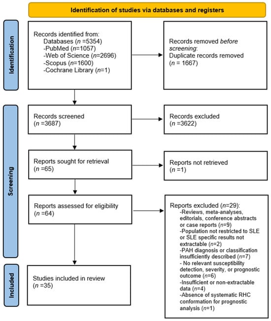
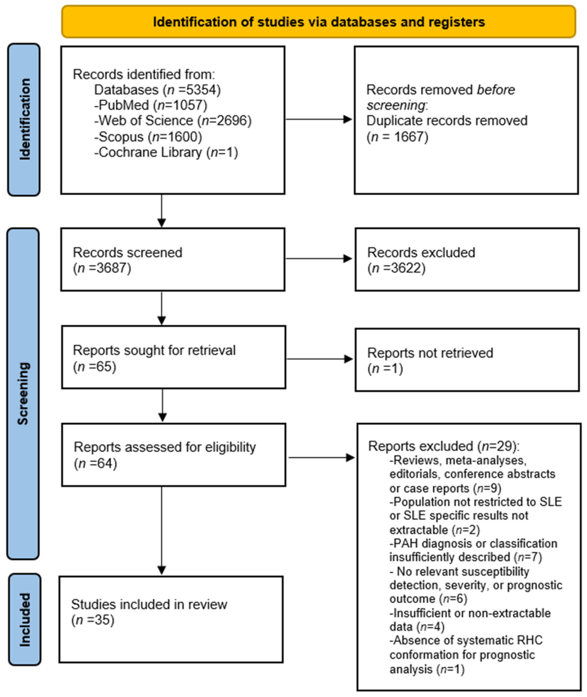

Disclaimer/Publisher’s Note: The statements, opinions and data contained in all publications are solely those of the individual author(s) and contributor(s) and not of MDPI and/or the editor(s). MDPI and/or the editor(s) disclaim responsibility for any injury to people or property resulting from any ideas, methods, instructions or products referred to in the content. |
© 2026 by the authors. Licensee MDPI, Basel, Switzerland. This article is an open access article distributed under the terms and conditions of the Creative Commons Attribution (CC BY) license.