


Abstract
Background: GPR146 is a G protein-coupled receptor involved in lipid metabolism and atherosclerosis. However, whether hepatic GPR146 exerts systemic metabolic effects that remotely modulate macrophage function remains unexplored. Methods: Hepatic GPR146 knockdown and negative control groups were established in high-fat diet-fed ApoE−/− mice. Untargeted metabolomics was performed on liver tissues, and peritoneal macrophages were isolated for Oil Red O, BODIPY 493/503, and Filipin staining, as well as RT-qPCR analysis of genes associated with lipid uptake, cholesterol efflux, and polarization markers. Results: Hepatic GPR146 knockdown significantly altered the hepatic metabolome, with arachidonic acid metabolism showing negative enrichment and the ABC transporters pathway showing positive enrichment. Macrophages from shGpr146 mice exhibited markedly reduced lipid accumulation upon ox-LDL stimulation, accompanied by decreased CD36 and SR-A1 mRNA expression, whereas ABCA1 and ABCG1 mRNA levels remained unchanged. Additionally, macrophages from shGpr146 mice showed reduced expression of the M1-associated markers iNOS and IL-1β and increased expression of the M2-associated marker Fizz1. Conclusions: Our findings reveal that hepatic GPR146 knockdown remodels the hepatic metabolome and may attenuate macrophage foam cell formation and pro-inflammatory polarization, suggesting a potential association between hepatic metabolic reprogramming and macrophage responses.
1. Introduction
Atherosclerosis and its related cardiovascular complications remain a leading cause of death worldwide [1]. Extensive studies have established the pivotal role of macrophages in the pathogenesis of atherosclerosis [2,3,4]. Upon exposure to oxidized low-density lipoprotein (ox-LDL) within the arterial microenvironment, macrophages actively internalize lipids via scavenger receptors, including CD36 and SR-A1, leading to the accumulation of cholesterol esters and neutral lipids, which ultimately drives their conversion into lipid-laden foam cells [5,6]. This pathological process is tightly orchestrated by an intricate network of metabolic and inflammatory signaling pathways [7]. Consequently, the identification of key molecular regulators that govern macrophage lipid metabolism has long been a central focus in cardiovascular research.
GPR146 is a G protein-coupled receptor that was long considered an orphan receptor and has recently emerged as an important regulator of lipid metabolism [8,9]. Human genetic studies have linked GPR146 variants, such as rs1997243, to plasma cholesterol levels and cardiometabolic risk [10]. Previous studies have demonstrated that GPR146 deficiency reduces circulating cholesterol levels and confers protection against atherosclerosis through modulation of the ERK1/2–SREBP2 axis [11]. Although cholesin has been identified as an endogenous ligand for GPR146 that inhibits hepatic PKA-ERK1/2 signaling, the role of cholesin-GPR146 signaling in macrophages remains unknown. Intriguingly, a very recent study reported that adipose tissue-specific, rather than liver-specific, GPR146 deficiency is responsible for the reduction in hepatic lipid accumulation [9], suggesting that the metabolic effects of GPR146 deficiency may involve interorgan crosstalk rather than being restricted to direct tissue-specific actions. These findings raise a compelling question: does hepatic GPR146 deficiency exert systemic metabolic effects that remotely modulate macrophage function, independent of its local actions?
As the liver serves as a central hub governing systemic metabolite production and secretion, alterations in hepatic metabolism are transmitted to the circulating metabolic milieu, where they can profoundly influence the functional status of peripheral immune cells. Nevertheless, whether hepatic GPR146 deficiency induces hepatic metabolic reprogramming that is associated with altered macrophage lipid metabolism and inflammatory phenotypes remains unclear. Here, by integrating hepatic untargeted metabolomics with functional and molecular analyses of peritoneal macrophages, we aimed to characterize hepatic metabolic alterations induced by GPR146 knockdown and determine whether these alterations are associated with changes in macrophage lipid metabolism and inflammatory phenotypes.
2. Materials and Methods
2.1. Animals
Male ApoE−/− mice (C57BL/6J background, 8 weeks old) were purchased from Charles River Laboratory Animal Technology Co., Ltd. (Beijing, China; certificate No.: SYXK(Jing)2024-0071). All mice were housed in a specific-pathogen-free facility under controlled temperature and humidity, with a 12 h light/dark cycle and free access to water and chow. The mice were fed a high-fat diet (HFD; D12108C, SPF Biotechnology Co., Ltd., Beijing, China) for 12 weeks to induce atherosclerotic lesion formation.
Following the 12-week HFD feeding, the mice were randomly assigned to two groups and received a single tail-vein injection of adeno-associated virus serotype 8 (AAV8) encoding either a short hairpin RNA targeting GPR146 (AAV-shGpr146) or a non-targeting control (AAV-shNC) under the control of a hepatocyte-specific thyroxine-binding globulin (TBG) promoter at a dose of 5 × 10¹² viral genomes per mouse (Beijing Likely Biotechnology, Beijing, China). All animal procedures were approved by the Animal Protection and Ethics Committee of Beijing Tsinghua Changgung Hospital (Approval No.: ZYZC202606033S) and were performed in accordance with the institutional guidelines for animal care and use.
2.2. Untargeted Metabolomics Analysis
Liver tissue samples were harvested from shNC and shGpr146 mice (n = 6 per group), immediately snap-frozen in liquid nitrogen, and stored at −80 °C until further processing. Untargeted metabolomics profiling was performed by Lianchuan Biotechnology Co., Ltd. (Hangzhou, China; Project No.: LC-P20260609150). Briefly, metabolites were extracted from each liver sample using a standardized methanol–acetonitrile–water extraction protocol, and the resulting extracts were analyzed using an ultra-high-performance liquid chromatography–tandem mass spectrometry (UHPLC–MS/MS) system. Raw data were processed for peak detection, alignment, and compound identification using the in-house metabolite database of Lianchuan Biotechnology (Hangzhou, China). All identified metabolites were annotated and quantified based on their retention times and mass spectral features.
2.3. Isolation and Culture of Primary Mouse Peritoneal Macrophages
Peritoneal macrophages were harvested from euthanized mice by peritoneal lavage. Briefly, the abdominal skin was disinfected with 75% ethanol, and blunt dissection was performed to expose the intact peritoneum. Prechilled sterile PBS (5 mL) was injected into the peritoneal cavity, followed by gentle abdominal massage. The lavage fluid was aspirated and collected, and this procedure was repeated three times. The pooled lavage fluids were centrifuged at 1500 rpm for 10 min at 4 °C. The cell pellet was resuspended in RPMI 1640 complete medium, and cell viability was assessed by Trypan blue exclusion. Macrophages were then seeded into culture plates for subsequent experiments.
2.4. Oil Red O Staining
For Oil Red O staining, cultured macrophages were fixed in Oil Red O fixative for 20 min, rinsed with distilled water, and briefly immersed in 60% isopropanol for 20 s. Cells were then stained with freshly prepared Oil Red O working solution (solution A: solution B = 3:2, v/v; filtered after 10 min of standing) for 20 min at room temperature. After sequential washing with 60% isopropanol and distilled water, nuclei were counterstained with Mayer’s hematoxylin for 1 min, followed by bluing in Oil Red O buffer for 1 min. Stained cells were visualized under a light microscope.
2.5. BODIPY 493/503 Staining
For BODIPY 493/503 staining, macrophages were washed with PBS, fixed in 4% paraformaldehyde for 10 min at room temperature, and permeabilized with 0.2% Triton X-100 for 5 min. After two washes with PBS (5 min each), cells were incubated with BODIPY 493/503 staining solution in the dark for 15 min at room temperature, followed by two additional PBS washes. Fluorescence images were captured under a microscope with 488 nm excitation, and green fluorescence was recorded as an indicator of neutral lipid content.
2.6. Filipin Staining
For Filipin staining, macrophages were washed with PBS, fixed with Fixative Solution for 15 min at room temperature, and washed twice with PBS. Cells were then incubated with Cholesterol Staining Solution in the dark for 60 min at room temperature. Following two additional PBS washes, stained cells were visualized under a fluorescence microscope with 340–380 nm excitation, and blue fluorescence was recorded as an indicator of free cholesterol content.
2.7. Quantitative Real-Time PCR (RT-qPCR)
Total RNA was extracted from peritoneal macrophages using TRIzol reagent and reverse-transcribed into cDNA using a PrimeScript™ RT Kit (TaKaRa, Kusatsu, Japan). Quantitative PCR was performed with TB Green® Premix Ex Taq™ II (TaKaRa) on a LightCycler® 480 system. The thermal cycling protocol consisted of an initial denaturation at 95 °C for 30 s, followed by 40 cycles of 95 °C for 5 s and 60 °C for 30 s. Target gene expression was normalized to β-actin and quantified using the 2−ΔΔCt method. Primer sequences are provided in Table 1 and supplementary Table S1.
Table 1.
List of oligonucleotide primer pairs used in qRT-PCR.
2.8. Western Blotting
GPR146 protein expression in liver tissues was assessed by Western blotting. Briefly, liver lysates were separated by SDS-PAGE and immunoblotted with anti-GPR146 and anti-tubulin antibodies. Detailed experimental procedures and uncropped Western blots are provided in Supplementary Material.
3. Statistical Analysis
Data are presented as mean ± SEM. Normality was tested using the Shapiro–Wilk test. Two-group comparisons were analyzed by two-tailed unpaired Student’s t-test (or Welch’s t-test when variances were unequal) for normally distributed data, or by Mann–Whitney U test for non-normally distributed data. Multi-group comparisons were performed using one-way ANOVA with Bonferroni’s post hoc test (normal distribution) or Kruskal–Wallis test with Dunn’s post hoc test (non-normal distribution). Two-way ANOVA with Bonferroni’s post hoc test was used for analyses involving two factors. Correlations were evaluated by Pearson’s or Spearman’s rank correlation test as appropriate. A p-value < 0.05 was considered statistically significant. All analyses and graphing were performed using GraphPad Prism 10.0 (GraphPad Software Inc., San Diego, CA, USA) and SPSS 21.0 (IBM, Armonk, NY, USA).
4. Results
4.1. Hepatic Metabolomic Profiling of shNC and shGpr146 ApoE−/− Mice
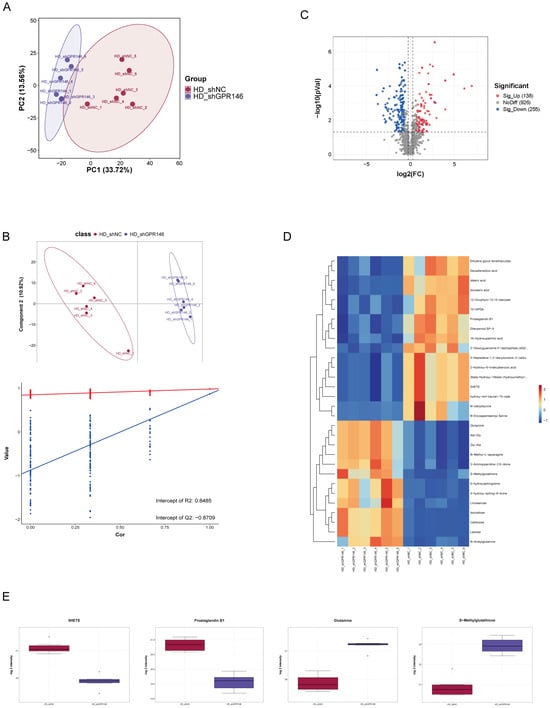
To characterize hepatic metabolic alterations following GPR146 knockdown, untargeted metabolomics profiling was performed on liver tissues harvested from shNC and shGpr146 ApoE−/− mice. Unsupervised principal component analysis (PCA) initially revealed a clear separation trend between the two experimental groups, suggesting a substantial metabolic shift induced by GPR146 knockdown (Figure 1A). To further discriminate the group-specific metabolic signatures, we constructed a supervised partial least-squares discriminant analysis (PLS-DA) model, which demonstrated distinct clustering of the metabolic profiles between shNC and shGpr146 mice (Figure 1B). The robustness of the PLS-DA model was validated by a permutation test (n = 200 permutations); the observed negative intercept of the Q2 regression line confirmed the absence of overfitting (inset of Figure 1B). Using a threshold of p < 0.05 and fold change > 1.5, a total of 393 differential metabolites were identified via volcano plot analysis, among which 138 metabolites were significantly upregulated and 255 were downregulated in the shGpr146 group compared to the shNC group (Figure 1C). Hierarchical clustering analysis of the top 30 differential metabolites generated a heatmap that clearly illustrated the distinct abundance patterns between the two cohorts (Figure 1D). Notably, given their well-established roles in inflammatory signaling and immunometabolism, we further scrutinized the relative abundances of selected metabolites, including 5-HETE, Prostaglandin B1, Glutamine, and S-Methylglutathione. Box-and-whisker plots revealed that the pro-inflammatory lipid mediators 5-HETE and Prostaglandin B1 were markedly downregulated in the livers of shGpr146 mice, whereas metabolites linked to redox homeostasis and amino acid metabolism, such as Glutamine and S-Methylglutathione, exhibited significantly elevated levels in the knockdown group (Figure 1E).
Figure 1.
Hepatic metabolomic profiling of shNC and shGpr146 ApoE−/− mice. (A) Principal component analysis (PCA) score plot demonstrating the overall separation trend between the two groups. (B) PLS-DA score plot illustrating the distinct metabolic clustering between the shNC and shGpr146 groups; the inset shows the permutation test (n = 200) confirming the model’s robustness with a negative Q2 intercept. (C) Volcano plot of differential metabolites in the liver. Red and blue dots represent significantly upregulated and downregulated metabolites, respectively, in the shGpr146 group compared to the shNC group (fold change > 1.5, p < 0.05). (D) Hierarchical clustering heatmap of the top 30 differential metabolites between the two groups. Red and blue indicate high and low relative metabolite abundances, respectively. (E) Box-and-whisker plots showing the relative abundances of selected differential metabolites (5-HETE, Prostaglandin B1, Glutamine, and S-Methylglutathione) in the liver tissues of each group. Abbreviations: PCA, principal component analysis; PLS-DA, partial least-squares discriminant analysis; Q2, cross-validated predictive ability; 5-HETE, 5-hydroxyeicosatetraenoic acid; PGB1, prostaglandin B1.
4.2. Functional Annotation of Differential Metabolites by KEGG and MSEA
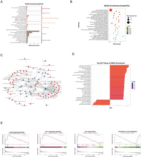
To gain mechanistic insights into the metabolic pathways affected by hepatic GPR146 deficiency, we performed KEGG pathway enrichment analysis on the identified differential metabolites. The results revealed that these metabolites were predominantly enriched in pathways including Metabolic pathways, Arachidonic acid metabolism, ABC transporters, and Efferocytosis (Figure 2A,B). Given the well-established roles of these pathways in atherosclerosis, we further constructed a metabolite–pathway interaction network to visualize their interconnections (Figure 2C). Notably, Metabolic pathways occupied the central hub of the network, with Efferocytosis and ABC transporters serving as bridging nodes connecting key metabolites such as Glucose and Glutamine.
Figure 2.
Functional annotation of differential metabolites by KEGG and MSEA. (A) KEGG pathway hierarchical classification bar plot showing the distribution of differential metabolites across primary and secondary pathway categories. (B) Bubble plot of KEGG enrichment analysis, where the bubble size represents the number of metabolites and the color gradient indicates the p-value. (C) Metabolite–pathway interaction network diagram visualizing the connections between key differential metabolites (nodes) and their associated KEGG pathways (edges). (D) Bar plot of KEGG enrichment scores displaying the top 30 pathways ranked by p-value from the MSEA. (E) Running enrichment score (ES) plots from MSEA illustrating the cumulative enrichment distribution of the top-ranked pathways across the ranked metabolite list. Abbreviations: KEGG, Kyoto Encyclopedia of Genes and Genomes; MSEA, metabolite set enrichment analysis; ES, enrichment score; ABC, ATP-binding cassette.
We next performed Metabolite Set Enrichment Analysis (MSEA) to evaluate the directional changes in these pathways. Among the top 30 pathways ranked by p-value, the ABC transporters pathway showed significant positive enrichment in the livers of shGpr146 mice compared to shNC controls (Figure 2D,E). Given that hepatic ABC transporters play a critical role in mediating cholesterol efflux into bile acids and HDL, this positive enrichment is consistent with our observed improvement in circulating lipid profiles and may indirectly contribute to the maintenance of systemic cholesterol homeostasis.
4.3. Hepatic GPR146 Knockdown Is Associated with Reduced ox-LDL-Induced Lipid Accumulation in Peritoneal Macrophages
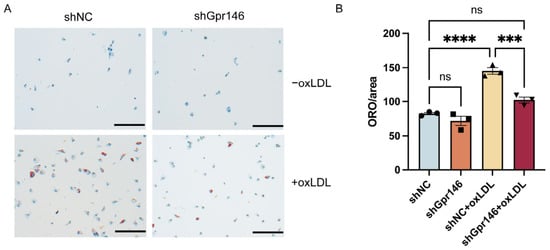
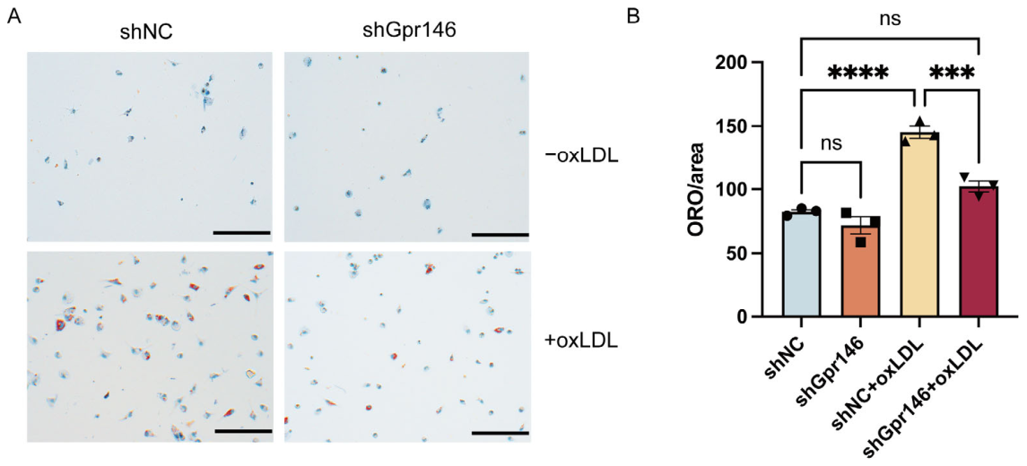
To further evaluate the role of GPR146 in cellular lipid accumulation, peritoneal macrophages were isolated from shNC and shGpr146 mice and cultured under basal conditions or stimulated with 50 μg/mL ox-LDL for 24 h to induce foam cell formation in vitro. Oil Red O staining revealed markedly reduced intracellular lipid accumulation in macrophages derived from shGpr146 mice compared with those from shNC controls (Figure 3A). Quantification of Oil Red O-positive area confirmed this reduction (Figure 3B). These data indicate that macrophages isolated from mice with hepatic GPR146 knockdown exhibit reduced ox-LDL-induced lipid accumulation.
Figure 3.
Oil Red O staining and quantification in peritoneal macrophages. (A) Representative Oil Red O staining of peritoneal macrophages from shNC and shGpr146 mice after ox-LDL stimulation (n = 3 per group). (B) Quantification of Oil Red O-positive area. Data are presented as mean ± SEM (n = 3 per group). *** p < 0.001, **** p < 0.0001, ns, not significant. Scale bar, 50 μm. Abbreviations: ORO, Oil Red O staining; oxLDL, oxidized low-density lipoprotein.
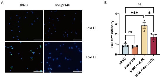
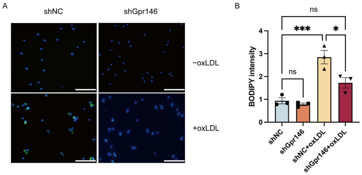
BODIPY 493/503 staining, which specifically labels neutral lipids, was performed on peritoneal macrophages following ox-LDL stimulation. Compared with the shNC group, macrophages from shGpr146 mice exhibited markedly reduced intracellular lipid accumulation (Figure 4A). Quantification of BODIPY 493/503 fluorescence intensity confirmed this reduction (Figure 4B).
Figure 4.
BODIPY staining and quantification in peritoneal macrophages. (A) Representative BODIPY staining of peritoneal macrophages from shNC and shGpr146 mice after ox-LDL stimulation (n = 3 per group). (B) Quantification of BODIPY intensity. Data are presented as mean ± SEM (n = 3 per group).* p < 0.05, *** p < 0.001, ns, not significant. Scale bar, 50 μm. Abbreviations: oxLDL, oxidized low-density lipoprotein; BODIPY, boron-dipyrromethene.
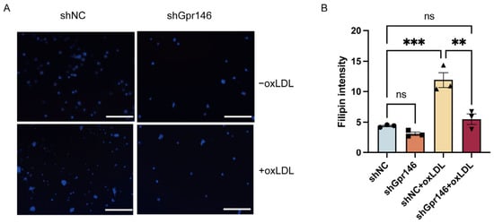
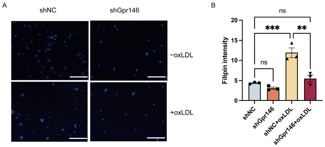
To further characterize the nature of lipid accumulation, we performed Filipin staining, which specifically labels unesterified (free) cholesterol, on peritoneal macrophages following ox-LDL stimulation. Consistent with the Oil Red O and BODIPY 493/503 results, macrophages from shGpr146 mice exhibited reduced Filipin fluorescence intensity compared to those from shNC controls (Figure 5A). Quantification of Filipin fluorescence intensity confirmed this reduction (Figure 5B).
Figure 5.
Filipin staining and quantification in peritoneal macrophages. (A) Representative Filipin staining of peritoneal macrophages from shNC and shGpr146 mice after ox-LDL stimulation (n = 3 per group). (B) Quantification of Filipin intensity. Data are presented as mean ± SEM (n = 3 per group). ** p < 0.01, *** p < 0.001, ns, not significant. Scale bar, 50 μm. Abbreviations: oxLDL, oxidized low-density lipoprotein.
4.4. Hepatic GPR146 Knockdown Is Associated with Altered Macrophage Lipid Metabolism- and Polarization-Related Gene Expression
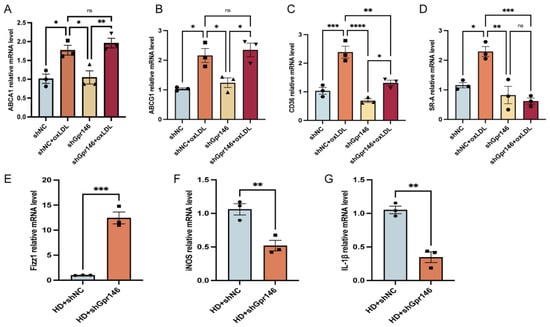
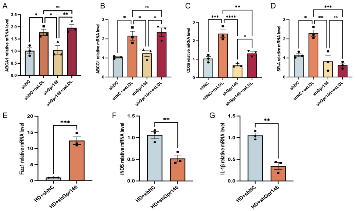
We next examined the mRNA expression levels of lipid uptake-related genes (CD36 and SR-A1), cholesterol efflux-related genes (ABCA1 and ABCG1), M1 polarization markers (iNOS and IL-1β), and the M2 polarization marker (Fizz1) in peritoneal macrophages by RT-qPCR. Compared to the shNC group, macrophages from shGpr146 mice exhibited significantly reduced mRNA levels of the lipid uptake-related genes CD36 and SR-A1 (Figure 6C,D), whereas no significant differences were observed in the cholesterol efflux-related genes ABCA1 and ABCG1 (Figure 6A,B). Regarding polarization markers, the M2 marker Fizz1 was significantly upregulated in the shGpr146 group (Figure 6E), while the M1 markers iNOS and IL-1β were markedly downregulated (Figure 6F,G).
Figure 6.
Hepatic GPR146 knockdown is associated with altered macrophage lipid metabolism- and polarization-related gene expression. Peritoneal macrophages were isolated from shNC and shGpr146 mice, and mRNA expression levels were analyzed by RT-qPCR. (A) ABCA1, (B) ABCG1, (C) CD36, (D) SR-A1, (E) Fizz1, (F) iNOS, and (G) IL-1β expression in peritoneal macrophages from each group. Data are presented as mean ± SEM (n = 3 per group). * p < 0.05, ** p < 0.01, *** p < 0.001, **** p < 0.0001, ns, not significant. Abbreviations: RT-qPCR, reverse transcription quantitative PCR; ABCA1, ATP-binding cassette subfamily A member 1; ABCG1, ATP-binding cassette subfamily G member 1; CD36, cluster of differentiation 36; SR-A1, scavenger receptor class A member 1; Fizz1, found in inflammatory zone 1; iNOS, inducible nitric oxide synthase; IL-1β, interleukin-1 beta.
5. Discussion
In this study, we demonstrate that hepatic GPR146 knockdown alters the hepatic metabolome and attenuates macrophage foam cell formation and pro-inflammatory polarization in ApoE−/− mice fed a high-fat diet. GPR146 has been established as a key regulator of lipid metabolism [12,13]. Previous studies have shown that GPR146 promotes hepatic SREBP2 activity through ERK1/2 activation, thereby regulating VLDL secretion and circulating LDL-cholesterol and triglyceride levels [11]. Beyond its metabolic functions, GPR146 has also been implicated in inflammatory signaling. GPR146 expression is upregulated in advanced human atherosclerotic plaques, and its silencing in THP-1 monocytes attenuates oxLDL-induced pro-inflammatory gene expression [8]. More recently, Shi et al. reported that adipose-specific, rather than hepatic, GPR146 deficiency reduces hepatic lipid accumulation by limiting free fatty acid influx, revealing an unexpected role in adipose–liver crosstalk [9]. By integrating untargeted hepatic metabolomics with functional and molecular analyses of peritoneal macrophages, we found that hepatic GPR146 knockdown was associated with negative enrichment of arachidonic acid metabolism and positive enrichment of the ABC transporters pathway, and that these hepatic metabolic alterations were accompanied by reduced macrophage lipid accumulation, diminished foam cell formation, and changes in inflammatory polarization markers. Collectively, our findings suggest a potential link between hepatic GPR146-dependent metabolic alterations and macrophage responses, providing a rationale for further investigation of liver–macrophage metabolic crosstalk in atherosclerosis.
Our untargeted metabolomics analysis revealed that hepatic GPR146 knockdown broadly reprogrammed the liver metabolic landscape, with the most prominent changes occurring in arachidonic acid metabolism, ABC transporter pathways, and glutathione metabolism. Arachidonic acid metabolism emerged as one of the most significantly downregulated pathways in GPR146-deficient livers. Arachidonic acid is a polyunsaturated fatty acid that serves as a precursor for a wide array of bioactive lipid mediators, including prostaglandins, leukotrienes, and hydroxyeicosatetraenoic acids (HETEs), which are centrally involved in the initiation and propagation of inflammatory responses [14,15]. Among the arachidonic acid-derived metabolites that were significantly reduced in our dataset were 5-HETE and prostaglandin B1. 5-HETE is a 5-lipoxygenase-derived arachidonic acid metabolite that participates in inflammatory lipid signaling and also serves as a precursor for the potent chemoattractant 5-oxo-ETE [16,17]. Prostaglandin B1, another member of the prostaglandin family derived from arachidonic acid, has been shown to modulate inflammatory processes and vascular tone, and its elevation has been associated with various cardiovascular pathologies [18,19,20]. The coordinated downregulation of these pro-inflammatory lipid mediators in GPR146-deficient livers suggests that GPR146 knockdown suppresses the hepatic production of inflammatory eicosanoids, which may contribute to an overall reduction in systemic inflammatory tone, a notion that aligns with our observation of reduced M1 and increased M2 polarization markers in peritoneal macrophages.
Our MSEA further revealed a significant positive enrichment of the ABC transporters pathway in GPR146-deficient livers. Hepatic ATP-binding cassette transporters play a critical role in maintaining systemic cholesterol homeostasis by mediating the biliary excretion of cholesterol and bile acids [21,22]. Members of the ABC transporter family, including ABCG5, ABCG8, and ABCB4, are responsible for the secretion of cholesterol, bile acids, and phospholipids into the bile, thereby facilitating the elimination of excess cholesterol from the body [23,24]. Our findings suggest that hepatic GPR146 knockdown is associated with alterations in metabolites linked to ABC transporter pathways. However, whether these metabolic changes reflect altered hepatic transporter expression or activity and contribute to cholesterol excretion remains to be determined.
Having identified hepatic metabolic alterations following GPR146 knockdown, we next examined macrophage phenotypes in the same experimental model. Peritoneal macrophages isolated from mice with hepatic GPR146 knockdown exhibited markedly reduced lipid accumulation upon ox-LDL stimulation, as evidenced by Oil Red O, BODIPY 493/503, and Filipin staining. This reduction in intracellular lipid accumulation was accompanied by significantly decreased mRNA expression of the scavenger receptors CD36 and SR-A1, whereas the expression of cholesterol efflux transporters ABCA1 and ABCG1 remained unchanged. These findings suggest that reduced expression of lipid uptake-associated receptors may contribute to decreased intracellular lipid accumulation. However, direct measurements of lipid uptake and cholesterol efflux are required to clarify the underlying mechanisms. Beyond lipid metabolism, macrophages from shGpr146 mice also exhibited a less pro-inflammatory expression profile, as evidenced by reduced iNOS and IL-1β expression and increased Fizz1 expression. These changes in inflammatory marker expression in peritoneal macrophages were accompanied by reduced levels of selected hepatic lipid metabolites, suggesting a potential association between hepatic metabolic alterations and macrophage inflammatory responses. The concept that liver-derived metabolites can remotely modulate macrophage function is supported by existing evidence. TMAO, generated by hepatic FMO3, upregulates macrophage CD36 and SR-A1 and impairs reverse cholesterol transport [25]. Furthermore, GPR146 deficiency upregulates hepatic CYP7A1 and alters hepatic SR-B1 protein levels, both of which would be expected to change the composition of circulating metabolites [26,27].
Several limitations of this study should be acknowledged. First, while our integrated analysis suggested an association between hepatic metabolomic alterations and macrophage phenotypes, plasma metabolites were not measured. This limits our ability to identify potential circulating mediators that may contribute to the associations between hepatic metabolic reprogramming and macrophage functional changes. Second, GPR146 expression was not directly assessed or manipulated in peritoneal macrophages. Although hepatic GPR146 knockdown was confirmed at the protein level (Supplementary Figure S1), tissue-specific transgene expression was not assessed, and potential off-target transduction cannot be completely excluded. In addition, endogenous GPR146 expression levels in the liver were not directly compared with those in other tissues. Previous studies have demonstrated hepatocyte-restricted expression of AAV8-TBG-driven transgenes, including the absence of detectable transgene expression in hepatic F4/80+ macrophages [28,29]. However, these findings do not establish the absence of AAV8-mediated transduction in extrahepatic macrophages, such as peritoneal macrophages. Therefore, potential macrophage-intrinsic effects of GPR146 silencing cannot be ruled out. If such off-target effects occurred, the observed macrophage phenotypes could not be exclusively attributed to hepatic GPR146 knockdown, and the proposed liver-macrophage crosstalk should be regarded as provisional. Further studies involving macrophage-specific GPR146 manipulation and tissue-specific transduction analysis will be necessary to distinguish these possibilities. Third, macrophage polarization was assessed by mRNA expression of iNOS, IL-1β, and Fizz1, which are well-established M1 and M2 markers. In addition, CD36 and SR-A1 were evaluated at the mRNA level. Validation at the protein level was not performed for these markers. Fifth, causal relationships between specific hepatic metabolites and macrophage functional changes await validation through metabolite supplementation or depletion experiments in vitro. Despite these limitations, our findings provide a basis for further investigating the potential relationship between hepatic GPR146-associated metabolic alterations and macrophage lipid and inflammatory responses. Future studies aimed at identifying the circulating metabolites that bridge hepatic metabolic reprogramming to macrophage responses will be essential to fully elucidate this pathway.
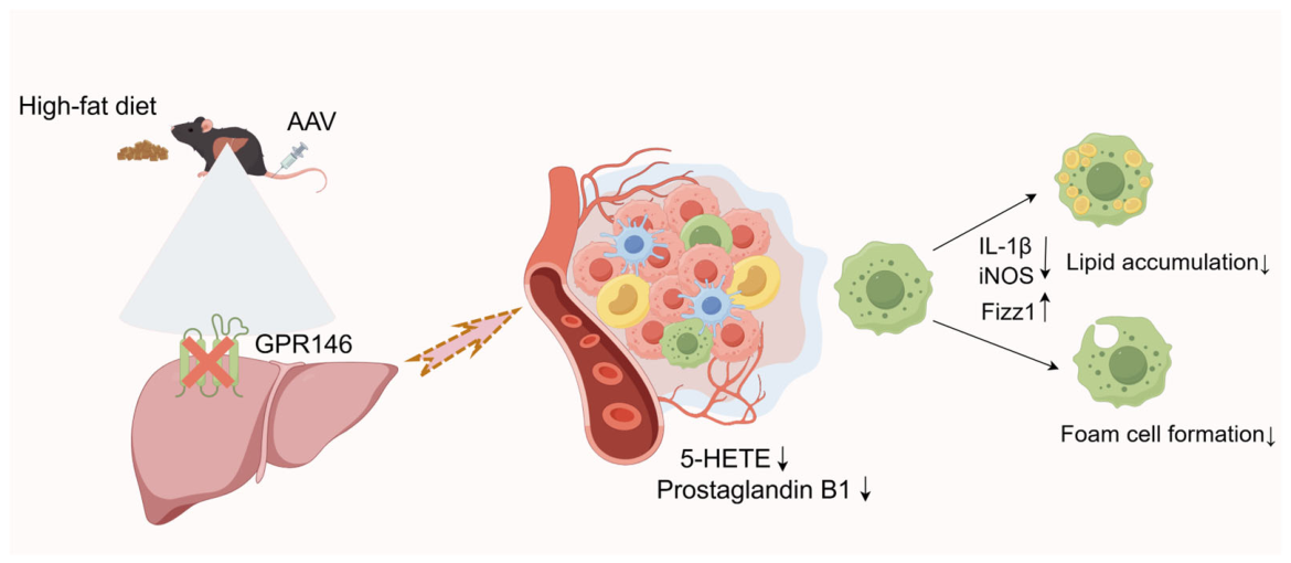
Based on our findings, we propose a working model of hepatic GPR146-mediated liver–macrophage metabolic crosstalk (Figure 7).
Figure 7.
A working model of hepatic GPR146-mediated liver-macrophage metabolic crosstalk. Hepatic GPR146 knockdown is associated with reduced hepatic levels of selected lipid metabolites, including 5-HETE and PGB1. We propose that these metabolic changes may influence circulating metabolic signals and contribute to a shift in the expression pattern of M1/M2-associated markers, as evidenced by reduced iNOS and IL-1β and increased Fizz1 expression. Dashed arrows indicate proposed but not directly verified connections. Figure created using FigDraw. Abbreviations: 5-HETE, 5-hydroxyeicosatetraenoic acid; PGB1, prostaglandin B1; iNOS, inducible nitric oxide synthase; IL-1β, interleukin-1 beta; Fizz1, found in inflammatory zone 1; M1, classically activated macrophage; M2, alternatively activated macrophage; AAV, adeno-associated virus.
6. Conclusions
In conclusion, hepatic GPR146 knockdown remodels the hepatic metabolome and is associated with reduced macrophage foam cell formation and a less pro-inflammatory gene expression profile. By integrating hepatic metabolomics with functional analyses of peritoneal macrophages, our findings suggest a potential link between hepatic metabolic alterations and macrophage responses mediated by hepatic GPR146. These findings may extend the current understanding of the atheroprotective effects of GPR 146 deficiency beyond its established cholesterol-lowering effects, suggesting a role for systemic metabolic regulation of immune cell function. These findings support hepatic GPR146 as a potential therapeutic target and warrant further investigation of its systemic metabolic and immunomodulatory effects.
Supplementary Materials
The following supporting information can be downloaded at: https://www.mdpi.com/article/10.3390/jcdd13110609/s1, Supplementary S1: Supplementary Method, Figure S1, and Table S1: The oligonucleotide primers used in qRT-PCR; Supplementary S2: The ARRIVE guidelines 2.0: author checklist.
Author Contributions
Q.L.: Writing—original draft, Methodology, Software. Y.W.: Writing—original draft, Software. Y.G.: Formal analysis, Validation. P.Z.: Writing—review & editing. T.L.: Writing—review & editing. All authors have read and agreed to the published version of the manuscript.
Funding
The authors declared that financial support was received for this work and its publication. This study was supported by the Beijing Natural Science Foundation (L242046) and the Beijing Hospitals Authority Clinical Medicine Development of special funding support (ZLRK202518).
Institutional Review Board Statement
All animal experiments were approved by the Animal Protection and Ethics Committee of Beijing Tsinghua Changgung Hospital (Approval No. ZYZC202606033S, approval date: 10 August 2025) and were conducted in accordance with the institutional guidelines for animal care and use.
Informed Consent Statement
Not applicable.
Data Availability Statement
The raw data supporting the conclusions of this article will be made available by the authors on request.
Acknowledgments
We sincerely thank Yiguo Wang of Tsinghua University for his valuable guidance and support during this study.
Conflicts of Interest
The authors declare that they have no known competing financial interests or personal relationships that could have appeared to influence the work reported in this paper.
References
- Salazar, J.; Morillo, V.; Suárez, M.K.; Castro, A.; Ramírez, P.; Rojas, M.; Añez, R.; D’MArco, L.; Chacín-González, M.; Bermúdez, V. Role of Gut Microbiome in Atherosclerosis: Molecular and Therapeutic Aspects. Curr. Cardiol. Rev. 2023, 19, e020223213408. [Google Scholar] [CrossRef] [Scilit] [PubMed]
- Hansson, G.K.; Hermansson, A. The immune system in atherosclerosis. Nat. Immunol. 2011, 12, 204–212. [Google Scholar] [CrossRef] [Scilit] [PubMed]
- Ley, K. Role of the adaptive immune system in atherosclerosis. Biochem. Soc. Trans. 2020, 48, 2273–2281. [Google Scholar] [CrossRef] [Scilit] [PubMed]
- Eshghjoo, S.; Jayaraman, A.; Sun, Y.; Alaniz, R.C. Microbiota-Mediated Immune Regulation in Atherosclerosis. Molecules 2021, 26, 179. [Google Scholar] [CrossRef] [Scilit] [PubMed]
- Chistiakov, D.A.; Melnichenko, A.A.; Myasoedova, V.A.; Grechko, A.V.; Orekhov, A.N. Mechanisms of foam cell formation in atherosclerosis. J. Mol. Med. 2017, 95, 1153–1165. [Google Scholar] [CrossRef] [Scilit] [PubMed]
- Chistiakov, D.A.; Bobryshev, Y.V.; Orekhov, A.N. Macrophage-mediated cholesterol handling in atherosclerosis. J. Cell. Mol. Med. 2016, 20, 17–28. [Google Scholar] [CrossRef] [Scilit] [PubMed]
- Wang, J.; Lei, L.; Wang, S.; Sun, Y. Asiaticoside alleviates atherosclerosis progression by suppressing RhoF–NF-κB/MAPK signaling and inflammation in macrophages. Phytomedicine 2025, 146, 157137. [Google Scholar] [CrossRef] [Scilit] [PubMed]
- Liu, Q.; Geng, Y.; Wang, Y.; Lv, T.; Zhang, P. Associations of Cholesin and GPR146 with atherosclerotic plaque stability: Integrated bioinformatics and functional analyses. Front. Cardiovasc. Med. 2026, 13, 1808833. [Google Scholar] [CrossRef] [Scilit] [PubMed]
- Shi, Y.; Cheng, K.Y.; Thi, T.T.; Wang, Y.; Yang, Y.; Cao, X.; Chhay, V.; Shen, Y.; He, Y.; Zhao, T.; et al. GPR146 in adipose tissue drives adipose-liver crosstalk and promotes hepatic steatosis in mice. Nat. Commun. 2026, 17, 3389. [Google Scholar] [CrossRef] [Scilit] [PubMed]
- Rimbert, A.; Yeung, M.W.; Dalila, N.; Thio, C.H.; Yu, H.; Loaiza, N.; Oldoni, F.; van der Graaf, A.; Wang, S.; Said, M.A.; et al. Variants in the GPR146 Gene Are Associated With a Favorable Cardiometabolic Risk Profile. Arterioscler. Thromb. Vasc. Biol. 2022, 42, 1262–1271. [Google Scholar] [CrossRef] [Scilit] [PubMed]
- Hu, X.; Chen, F.; Jia, L.; Long, A.; Peng, Y.; Li, X.; Huang, J.; Wei, X.; Fang, X.; Gao, Z.; et al. A gut-derived hormone regulates cholesterol metabolism. Cell 2024, 187, 1685–1700.e18. [Google Scholar] [CrossRef] [Scilit] [PubMed]
- Wilkins, B.P.; Finch, A.M.; Wang, Y.; Smith, N.J. Orphan GPR146: An alternative therapeutic pathway to achieve cholesterol homeostasis? Trends Endocrinol. Metab. 2022, 33, 481–492. [Google Scholar] [CrossRef] [Scilit] [PubMed]
- Yu, H.; Rimbert, A.; Palmer, A.E.; Toyohara, T.; Xia, Y.; Xia, F.; Ferreira, L.M.; Chen, Z.; Chen, T.; Loaiza, N.; et al. GPR146 Deficiency Protects against Hypercholesterolemia and Atherosclerosis. Cell 2019, 179, 1276–1288.e14. [Google Scholar] [CrossRef] [Scilit] [PubMed]
- Lubrano, V.; Ndreu, R.; Balzan, S. Classes of Lipid Mediators and Their Effects on Vascular Inflammation in Atherosclerosis. Int. J. Mol. Sci. 2023, 24, 1637. [Google Scholar] [CrossRef] [Scilit] [PubMed]
- Samuelsson, B. Role of basic science in the development of new medicines: Examples from the eicosanoid field. J. Biol. Chem. 2012, 287, 10070–10080. [Google Scholar] [CrossRef] [Scilit] [PubMed]
- Powell, W.S.; Rokach, J. The eosinophil chemoattractant 5-oxo-ETE and the OXE receptor. Prog. Lipid Res. 2013, 52, 651–665. [Google Scholar] [CrossRef] [Scilit] [PubMed]
- Erlemann, K.-R.; Rokach, J.; Powell, W.S. Oxidative stress stimulates the synthesis of the eosinophil chemoattractant 5-oxo-6,8,11,14-eicosatetraenoic acid by inflammatory cells. J. Biol. Chem. 2004, 279, 40376–40384. [Google Scholar] [CrossRef] [Scilit] [PubMed]
- Ricciotti, E.; FitzGerald, G.A. Prostaglandins and inflammation. Arterioscler. Thromb. Vasc. Biol. 2011, 31, 986–1000. [Google Scholar] [CrossRef] [Scilit] [PubMed]
- Wang, M.; Song, W.; Cheng, Y.; FitzGerald, G.A. Microsomal prostaglandin E synthase-1 inhibition in cardiovascular inflammatory disease. J. Intern. Med. 2008, 263, 500–505. [Google Scholar] [CrossRef] [Scilit] [PubMed]
- Beccacece, L.; Abondio, P.; Bini, C.; Pelotti, S.; Luiselli, D. The Link between Prostanoids and Cardiovascular Diseases. Int. J. Mol. Sci. 2023, 24, 4193. [Google Scholar] [CrossRef] [Scilit] [PubMed]
- VandeBerg, J.; Chan, J. Hepatobiliary transport in health and disease. Clin. Lipidol. 2012, 7, 189–202. [Google Scholar] [CrossRef] [Scilit] [PubMed]
- Cuperus, F.J.C.; Claudel, T.; Gautherot, J.; Halilbasic, E.; Trauner, M. The Role of canalicular ABC transporters in cholestasis. Drug Metab. Dispos. 2014, 42, 546–560. [Google Scholar] [CrossRef] [Scilit] [PubMed]
- Wang, H.H.; Liu, M.; Portincasa, P.; Wang, D.Q.-H. Recent Advances in the Critical Role of the Sterol Efflux Transporters ABCG5/G8 in Health and Disease. Adv. Exp. Med. Biol. 2020, 1276, 105–136. [Google Scholar] [CrossRef] [Scilit] [PubMed]
- Capitani, M.C.; Capiglioni, A.M.; Marinelli, R.A.; Marrone, J. Biliary elimination of cholesterol can be modulated by hepatocyte mitochondrial Aquaporin-8 in mice. Sci. Rep. 2026, 16, 7579. [Google Scholar] [CrossRef] [Scilit] [PubMed]
- Zhen, J.; Zhou, Z.; He, M.; Han, H.-X.; Lv, E.-H.; Wen, P.-B.; Liu, X.; Wang, Y.-T.; Cai, X.-C.; Tian, J.-Q.; et al. The gut microbial metabolite trimethylamine N-oxide and cardiovascular diseases. Front. Endocrinol. 2023, 14, 1085041. [Google Scholar] [CrossRef] [Scilit] [PubMed]
- Liu, X.; Han, J.; Li, X.; Zhang, Z.; Li, J.; Yao, Y. GPR146 regulates CYP7A1 transcription in cells and in vivo of mice. Biochem. Biophys. Res. Commun. 2025, 772, 152045. [Google Scholar] [CrossRef] [Scilit] [PubMed]
- Zhang, B.; Loaiza, N.; Rimbert, A.; Oldoni, F.; Blauw, L.L.; Rensen, P.C.N.; Martinez, L.O.; Robert, J.; von Eckardstein, A.; Wolters, J.C.; et al. Loss of GPR146 decreases plasma levels of HDL cholesterol via post-translational up-regulation of SR-B1 protein levels. Cardiovasc. Res. 2025, 121, 2860–2872. [Google Scholar] [CrossRef] [Scilit] [PubMed]
- Greenstein, A.W.; Majumdar, N.; Yang, P.; Subbaiah, P.V.; Kineman, R.D.; Cordoba-Chacon, J. Hepatocyte-specific, PPARγ-regulated mechanisms to promote steatosis in adult mice. J. Endocrinol. 2017, 232, 107–121. [Google Scholar] [CrossRef] [Scilit] [PubMed]
- Kiourtis, C.; Wilczynska, A.; Nixon, C.; Clark, W.; May, S.; Bird, T.G. Specificity and off-target effects of AAV8-TBG viral vectors for the manipulation of hepatocellular gene expression in mice. Biol. Open 2021, 10, bio058678. [Google Scholar] [CrossRef] [Scilit] [PubMed]

Disclaimer/Publisher’s Note: The statements, opinions and data contained in all publications are solely those of the individual author(s) and contributor(s) and not of MDPI and/or the editor(s). MDPI and/or the editor(s) disclaim responsibility for any injury to people or property resulting from any ideas, methods, instructions or products referred to in the content. |
© 2026 by the authors. Licensee MDPI, Basel, Switzerland. This article is an open access article distributed under the terms and conditions of the Creative Commons Attribution (CC BY) license.