


Abstract
Attributing exertional symptoms in patients with aortic stenosis (AS) and coronary artery disease is challenging. We performed a secondary analysis of a single-center prospective registry of 190 patients with severe high-gradient AS and a single intermediate left anterior descending lesion assessed with fractional flow reserve (FFR) and instantaneous wave-free ratio (iFR). The primary outcome was angina severity (Canadian Cardiovascular Society [CCS] class III–IV vs. II); New York Heart Association (NYHA) class III–IV vs. II functional limitation was secondary and exploratory. Among 185 complete cases, older age (odds ratio [OR] 3.00 per decade, 95% confidence interval [CI] 1.84–4.87; p < 0.001) and smaller measured aortic valve area (AVA; OR 1.39 per 0.1 cm2 decrease, 95% CI 1.08–1.79; p = 0.009) were independently associated with severe angina, whereas lesion morphology, FFR, and iFR were not. No variable was independently associated with NYHA class III–IV limitation, although this analysis was underpowered. Because AVA is flow dependent and transvalvular flow and coronary microvascular function were not directly measured, the AVA association does not establish fixed valvular obstruction as the mechanism of angina. The lack of association with FFR/iFR pertains to symptom severity and does not invalidate their use for lesion-level hemodynamic assessment.
1. Introduction
Severe aortic stenosis (AS) and coronary artery disease (CAD) frequently coexist in older patients with a high burden of cardiovascular comorbidity [1,2,3]. Both conditions can cause exertional angina and dyspnea, making it difficult to determine whether symptoms arise predominantly from the valvular lesion, an individual coronary stenosis, or both [4,5,6]. This uncertainty is particularly relevant in patients with angiographically intermediate coronary lesions, in whom revascularization decisions may be influenced by whether a lesion is considered a clinically relevant contributor to symptoms [4,7,8,9,10].
Invasive coronary physiology is routinely used to assess the hemodynamic significance of intermediate coronary stenoses [4,7,11]. However, pressure-derived indices require careful interpretation in severe AS [4,7,8,9]. AS is associated with left ventricular hypertrophy, elevated filling pressures, altered coronary flow patterns, and microvascular dysfunction, each of which may modify the relationship among epicardial stenosis severity, myocardial ischemia, and pressure-derived indices [4,12,13,14]. Hyperemic flow may increase after aortic valve replacement (AVR), potentially lowering fractional flow reserve (FFR), whereas non-hyperemic pressure ratios such as the instantaneous wave-free ratio (iFR) appear less affected at the group level; nevertheless, substantial individual variability and FFR/iFR discordance have been reported [9,14,15,16].
More fundamentally, the physiological threshold associated with myocardial ischemia may differ from the threshold at which an individual patient experiences angina [17]. In the recent ORBITA-FIRE study, physiological thresholds at angina onset varied substantially among patients and with cardiac workload and were generally lower than conventional ischemia-based thresholds used to guide revascularization [18]. Although these findings cannot be extrapolated directly to severe AS, they reinforce the concept that epicardial pressure-derived measurements are not direct surrogates of symptom burden. This distinction may be especially important in severe AS, in which symptoms can reflect the combined effects of valvular obstruction, CAD, ventricular remodeling, microvascular dysfunction, and non-cardiac comorbidities [4,12,13,14]. Although the interplay among AS, CAD, and invasive coronary physiology has been studied extensively, less is known about the relative associations of patient characteristics, valvular measurements, quantitative lesion morphology, and epicardial pressure-derived indices with symptom severity in the clinically ambiguous subgroup with an intermediate coronary lesion. Analyses that combine multiple coronary territories may also be difficult to interpret because subtended myocardial mass, lesion location, and concomitant CAD burden vary among vessels [19]. Restricting the primary analysis to a single left anterior descending (LAD) artery lesion therefore provides a more homogeneous setting for examining symptom attribution. Importantly, the present study addresses a within-cohort question in patients with intermediate coronary disease rather than comparing symptom burden across the spectrum of CAD severity. A comparison among patients with no CAD, intermediate CAD, and unequivocally severe CAD would address a different, between-cohort question that requires prospectively defined comparator populations.
Accordingly, we performed a secondary analysis of a prospective registry of patients with severe high-gradient AS and a single intermediate LAD lesion assessed with both FFR and iFR. We examined whether greater angina severity (primary outcome) and more severe functional limitation (secondary outcome) were associated with patient characteristics, valvular measurements, LAD lesion morphology, or epicardial pressure-derived indices.
2. Materials and Methods
2.1. Study Design and Population
This was a secondary, patient-level analysis of a prospective, single-center registry conducted at a tertiary academic center in Krakow, Poland, between January 2018 and January 2020 [20,21,22]. The registry enrolled consecutive patients with severe AS who underwent routine coronary angiography as part of pre-intervention assessment for transcatheter (TAVR) or surgical (SAVR) aortic valve replacement. Eligible patients had at least one angiographically intermediate coronary stenosis, defined visually as 40–90% diameter stenosis. Because eligibility for the parent registry required an intermediate lesion selected for invasive physiological assessment, patients without CAD were not enrolled as controls, and a separate cohort with unequivocally severe obstructive CAD was not prospectively assembled. Severe AS was defined echocardiographically as an aortic valve area (AVA) < 1.0 cm2 and a mean transaortic pressure gradient > 40 mmHg. Lesions were excluded from the parent registry when low frame-rate acquisition, inadequate image quality, or vessel overlap precluded reliable quantitative coronary angiography (QCA), or when ostial or tandem anatomy precluded reliable invasive physiological assessment.
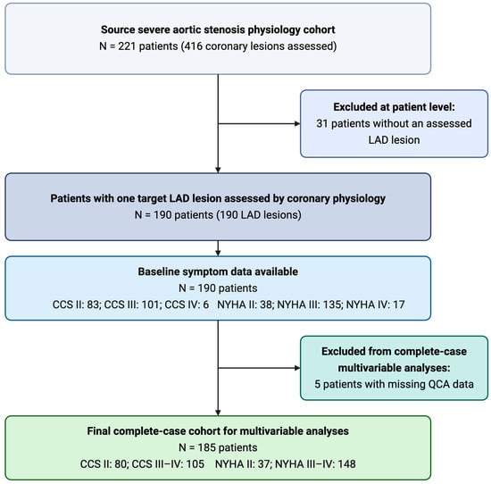
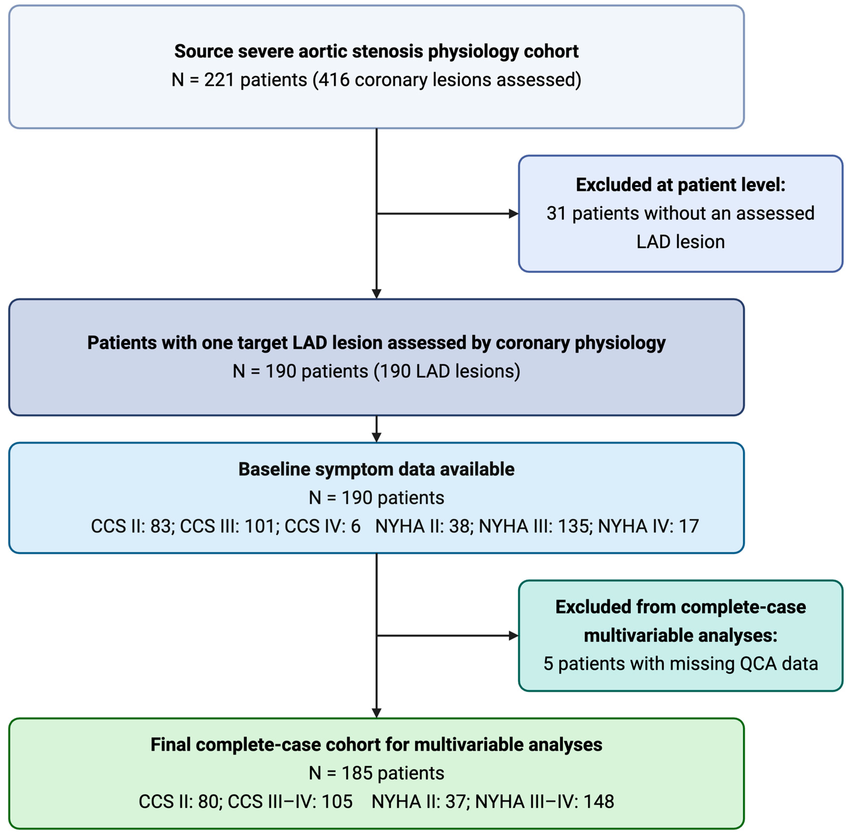
For the present analysis, the study population was restricted to patients with an LAD lesion assessed with both FFR and iFR. The LAD was selected to create a clinically homogeneous cohort, avoid mixing coronary territories with different subtended myocardial mass, and ensure one target lesion per patient. No patient contributed more than one LAD lesion. Patients with missing QCA measurements of lesion length or diameter stenosis were excluded only from complete-case multivariable analyses (Figure 1).
Figure 1.
Flowchart of patient selection and derivation of the complete-case cohort. Abbreviations: CCS, Canadian Cardiovascular Society; LAD, left anterior descending artery; NYHA, New York Heart Association; QCA, quantitative coronary angiography.
The study was approved by the Bioethics Committee of the Jagiellonian University (No. 1072.6120.1.2019) and was conducted in accordance with the Declaration of Helsinki. All participants provided written informed consent.
2.2. Clinical and Echocardiographic Assessment
Baseline demographic and clinical characteristics were recorded at the time of invasive coronary assessment. These included age, sex, body mass index (BMI), cardiovascular risk factors, relevant comorbidities, prior myocardial infarction, previous percutaneous coronary intervention (PCI), renal function, atrial fibrillation (AF), chronic obstructive pulmonary disease (COPD), and peripheral artery disease. Canadian Cardiovascular Society (CCS) angina class and New York Heart Association (NYHA) functional class were recorded at baseline by the treating physicians as part of routine clinical assessment. CCS class was used to grade exertional angina or chest discomfort, whereas NYHA class captured dyspnea and overall functional limitation. The parent registry did not include protocol-mandated central symptom adjudication or blinded symptom assessment, and the timing of symptom classification relative to the invasive physiological measurements was not documented in a way that permits patient-level verification. Therefore, we cannot exclude the possibility that knowledge of coronary anatomy or physiology influenced symptom classification. Transthoracic echocardiography was performed as part of routine preprocedural assessment by experienced echocardiographers. Left ventricular ejection fraction (LVEF) was assessed using standard echocardiographic methods. Peak and mean transaortic pressure gradients were derived from continuous-wave Doppler recordings using the highest reliably obtained transvalvular velocity. AVA was calculated with the continuity equation from left ventricular outflow tract (LVOT) diameter and Doppler-derived velocity-time integrals in the LVOT and across the aortic valve. For sensitivity analyses, AVA was indexed to body surface area (BSA), calculated with the Mosteller formula.
2.3. Coronary Angiography and Invasive Physiological Assessment
Coronary angiography was performed using standard radial or femoral access techniques. Invasive physiological assessment was performed with a commercially available pressure guidewire and dedicated console (Philips Volcano, San Diego, CA, USA). Pressure-wire equalization was performed before each measurement, and drift was checked after withdrawal of the pressure sensor to the guiding-catheter tip; recordings with drift >0.02 were repeated. iFR was measured at rest, and the mean of three recordings was used for analysis. FFR was measured during steady-state maximal hyperemia induced by intravenous adenosine at 140 µg/kg/min and calculated as the ratio of mean distal coronary pressure to mean aortic pressure. QCA was performed offline using CAAS 5.7 QCA software (Pie Medical, Maastricht, The Netherlands) by two experienced analysts blinded to invasive physiological results. Assessed variables included diameter stenosis, lesion length, reference vessel diameter, and minimal lumen diameter. Systematic FFR or iFR pullback assessment, including pullback pressure gradient (PPG) analysis, was not performed. Direct measures of coronary microvascular function, including coronary flow reserve (CFR) and the index of microcirculatory resistance (IMR), were unavailable. Additional non-LAD lesions were assessed when clinically indicated. For descriptive analyses, additional CAD was defined at the patient level as at least one non-LAD lesion undergoing invasive physiological assessment. A patient was considered to have physiologically significant additional non-LAD disease when at least one such lesion had FFR ≤ 0.80 or iFR ≤ 0.89. These measurements were used only to describe the burden of additional epicardial disease and were not included in the primary LAD-specific analyses.
2.4. Study Outcomes and Definitions
The primary outcome was greater angina severity, defined as CCS class III–IV versus CCS class II angina within the symptomatic study cohort. The secondary outcome was severe functional limitation, defined as NYHA class III–IV versus NYHA class II; because of the small NYHA class II reference group, this secondary analysis was interpreted as exploratory. The principal variables of interest were age, sex, LVEF, measured AVA, LAD lesion morphology (diameter stenosis and lesion length), and epicardial pressure-derived indices (FFR and iFR). FFR and iFR were analyzed as continuous measures and according to conventional dichotomous thresholds for hemodynamic significance: FFR ≤ 0.80 and iFR ≤ 0.89. For integrated symptom analyses, patients were categorized into four angina–functional limitation phenotypes according to CCS and NYHA class: CCS II/NYHA II, CCS II/NYHA III–IV, CCS III–IV/NYHA II, and CCS III–IV/NYHA III–IV.
2.5. Statistical Analysis
The primary analyses were performed at the patient level. Categorical variables are presented as counts and percentages (%), and continuous variables as medians with interquartile ranges (IQRs). Baseline characteristics were compared between CCS and NYHA groups using the χ2 test or Fisher exact test for categorical variables and the Mann–Whitney U test for continuous variables. Comparisons across the four combined angina–functional limitation phenotypes were performed using the Kruskal–Wallis test. Analyses of additional non-LAD lesions were conducted at the patient level. Among patients with at least one additional non-LAD lesion assessed, the prevalence of at least one lesion with FFR ≤ 0.80 or iFR ≤ 0.89 was compared using Fisher exact tests. Multivariable logistic-regression models were used to identify factors associated with CCS class III–IV angina and, in a secondary exploratory analysis, NYHA class III–IV functional limitation. Covariates were selected a priori on the basis of clinical relevance and included age, sex, LVEF, measured AVA, diameter stenosis, and lesion length. FFR and iFR were evaluated in separate models because they are closely related pressure-derived indices. For clinical interpretation, age was expressed per 10-year increase; LVEF per 10-percentage-point increase; measured AVA per 0.1 cm2 decrease; diameter stenosis per 10% increase; lesion length per 10 mm increase; and FFR or iFR per 0.10 decrease. Results are reported as odds ratios (ORs) with 95% confidence intervals (CIs). The incremental value of clinical, valvular, angiographic, and physiological variables for prediction of CCS class III–IV angina was evaluated using nested logistic-regression models. The clinical model included age, sex, and LVEF. Measured AVA was then added, followed by lesion morphology (diameter stenosis and lesion length). FFR or iFR was subsequently added in separate alternative models. Incremental model performance was assessed using likelihood-ratio tests, Akaike information criterion (AIC) values, and the C-statistic. Internal validation was performed by bootstrap optimism correction using 1000 resamples. The robustness of the primary findings was examined in several additional analyses. First, three sensitivity analyses were performed: measured AVA was replaced by mean transaortic gradient; the models were additionally adjusted for AF, COPD, and estimated glomerular filtration rate (eGFR); and AVA indexed to BSA was substituted for absolute AVA. Second, to examine potential confounding by transvalvular hemodynamics and ventricular systolic function, mean or peak transaortic gradient was added to the primary models with measured AVA retained, and the primary models were repeated in the subgroup with preserved LVEF (≥50%). Third, the linearity-in-the-log-odds assumption for age and AVA was assessed using the Box–Tidwell approach; because this suggested possible non-linearity for AVA, a further sensitivity analysis added a quadratic term for centered AVA to determine whether the associations of FFR and iFR with severe angina were robust to this alternative specification. Effect modification was assessed by adding separate interaction terms (AVA × FFR, AVA × iFR, AVA × mean transaortic gradient, and AVA × LVEF) to the fully adjusted models. Continuous variables were centered at their cohort median values before interaction terms were constructed. An analogous analysis used peak rather than mean transaortic gradient. Exploratory discrimination analyses of age, AVA, FFR, and iFR for each outcome were performed at the patient level using univariable receiver-operating-characteristic (ROC) curves, and areas under the curve (AUCs) were reported with 95% CIs. Finally, the associations of FFR and iFR with each outcome were assessed in the parent physiology cohort, including all assessed LAD and non-LAD lesions. Lesion-level ROC analyses were performed with 95% CIs estimated by patient-clustered bootstrap resampling, and univariable generalized estimating equation (GEE) models with an independence working correlation structure and patient-clustered robust standard errors were used to account for multiple lesions per patient. Missing data were not imputed. Descriptive analyses used available data, whereas multivariable analyses were based on complete cases. The study size was determined by the number of eligible patients in the prospective registry; no a priori sample-size calculation was performed. To characterize the statistical sensitivity of the secondary NYHA analysis, post hoc power was estimated for the principal continuous predictors using the observed Wald effect sizes from the complete-case logistic-regression models and a two-sided α of 0.05. These estimates were considered descriptive and were used only to indicate the limited ability of the secondary analysis to detect associations of the observed magnitude. All tests were two-sided, and a p-value < 0.05 was considered statistically significant. All analyses other than the primary multivariable models were considered secondary or exploratory, and their p-values were interpreted descriptively without adjustment for multiple comparisons. Analyses were performed using Python (version 3.11; Python Software Foundation, Wilmington, DE, USA) with the pandas (version 2.2.3), NumPy (version 2.1.3), SciPy (version 1.14.1), statsmodels (version 0.14.4), scikit-learn (version 1.6.1), and Matplotlib (version 3.10.0) libraries.
3. Results
3.1. Study Cohort and Symptom Burden
Among 221 patients with severe AS who underwent physiological assessment of 416 coronary lesions, 190 had a single assessed LAD lesion and were included in the present patient-level analysis (Figure 1). No patient contributed more than one LAD lesion. Baseline CCS and NYHA functional-class data were available for all 190 patients; five patients had missing QCA measurements of lesion length or diameter stenosis and were excluded from complete-case multivariable analyses.
CCS class II angina was present in 83 patients (43.7%), whereas 101 (53.2%) and 6 (3.2%) patients had CCS class III and IV angina, respectively. Thus, 107 patients (56.3%) were classified as having CCS class III–IV angina. NYHA class II functional limitation was observed in 38 patients (20.0%), whereas 135 (71.1%) and 17 (8.9%) patients had NYHA class III and IV functional limitation, respectively. No patient had CCS class I or NYHA class I symptoms; therefore, the study compares symptom severity within an already symptomatic cohort rather than the presence versus absence of angina.
When CCS and NYHA classes were considered jointly, 20 patients (10.5%) had CCS class II and NYHA class II symptoms, 63 (33.2%) had CCS class II and NYHA class III–IV functional limitation, 18 (9.5%) had CCS class III–IV angina and NYHA class II functional limitation, and 89 (46.8%) had both CCS class III–IV angina and NYHA class III–IV functional limitation.
3.2. Characteristics According to Angina Severity
Baseline clinical, valvular, angiographic, and physiological characteristics according to CCS class are presented in Table 1. Compared with patients with CCS class II angina, those with CCS class III–IV angina were older (median, 85 [IQR, 81–89] versus 75 [69–83] years; p < 0.001) and had a smaller measured AVA (0.50 [0.40–0.55] versus 0.60 [0.50–0.80] cm2; p < 0.001). Notably, despite the smaller measured AVA, the peak transaortic gradient was lower in the CCS class III–IV group (89 [73–102] versus 96 [87–108] mmHg; p = 0.024), whereas the mean gradient did not differ significantly (49 [43–54] versus 50 [45–58] mmHg; p = 0.16). The CCS class III–IV group also had a numerically higher proportion of women and a lower LVEF; both differences were of borderline statistical significance (both p = 0.05). In contrast, lesion-level characteristics were similar between CCS groups. Median FFR was 0.86 (0.80–0.89) in the CCS class III–IV group and 0.87 (0.79–0.89) in the CCS class II group (p = 0.81), whereas median iFR was 0.91 in both groups (p = 0.54). The prevalence of FFR ≤ 0.80 and iFR ≤ 0.89 did not differ between groups, and diameter stenosis and lesion length were also similar. At least one additional non-LAD lesion was physiologically assessed in 135 patients (71.1%), with no difference between CCS groups. Among patients with at least one additional non-LAD lesion assessed, the prevalence of at least one FFR-positive or iFR-positive lesion was likewise similar between CCS groups (Table 1).
Table 1.
Baseline clinical, valvular, angiographic, and physiological characteristics according to angina severity in patients with severe aortic stenosis and a single left anterior descending artery lesion.
3.3. Factors Associated with CCS Class III–IV Angina
In complete-case multivariable analyses including 185 patients, older age and smaller measured AVA were independently associated with CCS class III–IV angina (Table 2). In the FFR model, the OR for CCS class III–IV angina was 3.00 per 10-year increase in age (95% CI: 1.84–4.87; p < 0.001) and 1.39 per 0.1 cm2 decrease in measured AVA (95% CI: 1.08–1.79; p = 0.009). Similar estimates were observed in the corresponding iFR model. No independent association was observed between angina severity and either FFR or iFR. Diameter stenosis and lesion length were likewise not independently associated with severe angina in either model.
Table 2.
Multivariable logistic-regression analyses of factors associated with CCS class III–IV angina in patients with severe aortic stenosis and a single LAD lesion.
Incremental model-performance analyses are shown in Table 3. Adding measured AVA to the clinical model comprising age, sex, and LVEF significantly improved model fit (incremental likelihood-ratio χ2 = 6.67; p = 0.010), reduced the AIC by 4.67 points, and increased the optimism-corrected C-statistic from 0.761 to 0.787. Subsequent addition of lesion morphology, FFR, or iFR did not improve model fit or discrimination.
Table 3.
Incremental model performance for CCS class III–IV angina.
3.4. Functional Limitation
Baseline characteristics according to NYHA functional class are presented in Table 4. Patients with NYHA class III–IV functional limitation were older than those with NYHA class II symptoms (84 [76–89] versus 76 [70–85] years; p = 0.003) and had a smaller measured AVA (0.50 [0.49–0.70] versus 0.60 [0.50–0.80] cm2; p = 0.008). FFR, iFR, lesion length, diameter stenosis, and the prevalence of additional physiologically significant non-LAD lesions did not differ significantly between NYHA groups. In multivariable analyses, no included variable was independently associated with NYHA class III–IV functional limitation (Table 5), although this secondary exploratory analysis was underpowered to detect modest associations. Among the 185 complete cases, 148 had NYHA class III–IV functional limitation and only 37 had NYHA class II symptoms. Estimated post hoc power at the observed effect sizes was approximately 38% for age, 24–25% for measured AVA (across the FFR and iFR models), 16% for FFR, and 11% for iFR. Accordingly, the non-significant estimates in the NYHA analysis should not be interpreted as evidence that no association exists.
Table 4.
Baseline clinical, valvular, angiographic, and physiological characteristics according to functional limitation in patients with severe aortic stenosis and a single left anterior descending artery lesion.
Table 5.
Multivariable logistic-regression analyses of factors associated with NYHA class III–IV functional limitation in patients with severe aortic stenosis and a single LAD lesion.
3.5. Integrated Symptom Phenotype Analyses
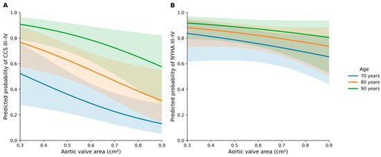
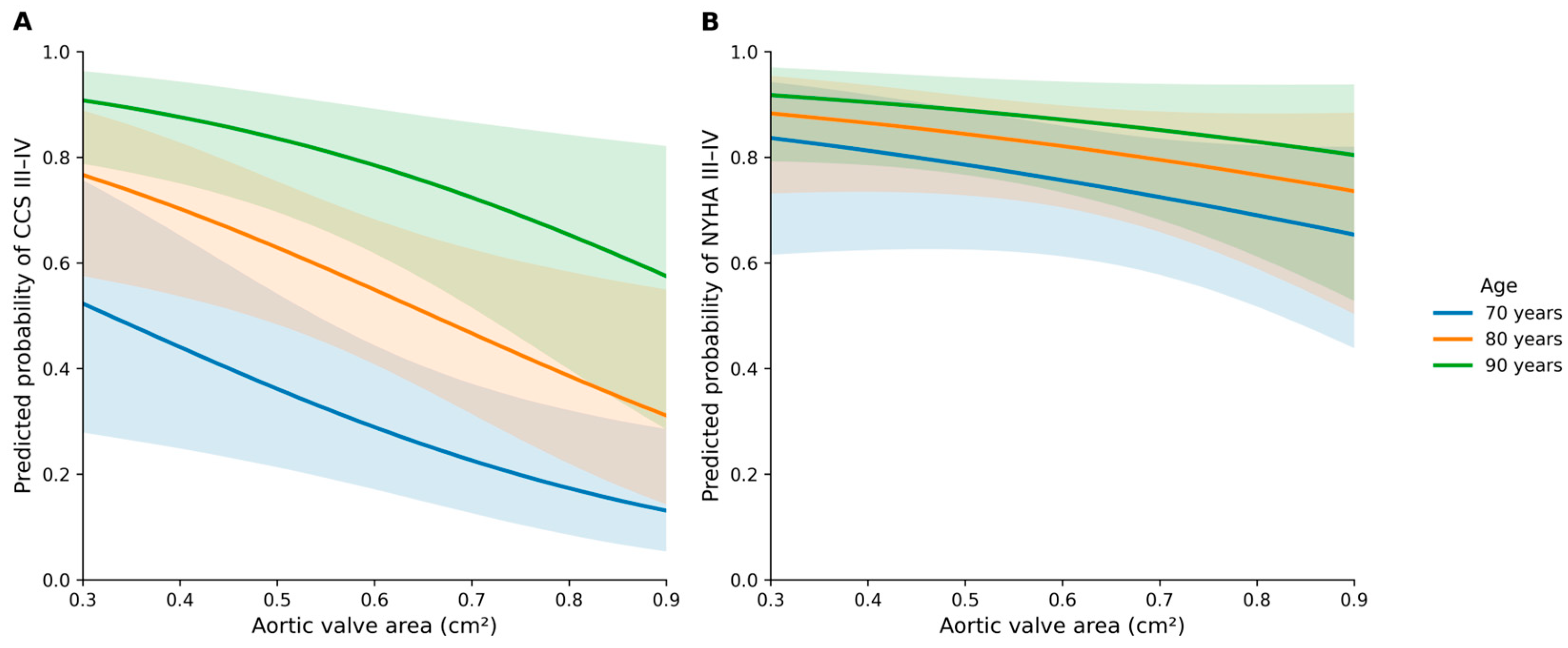
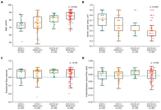
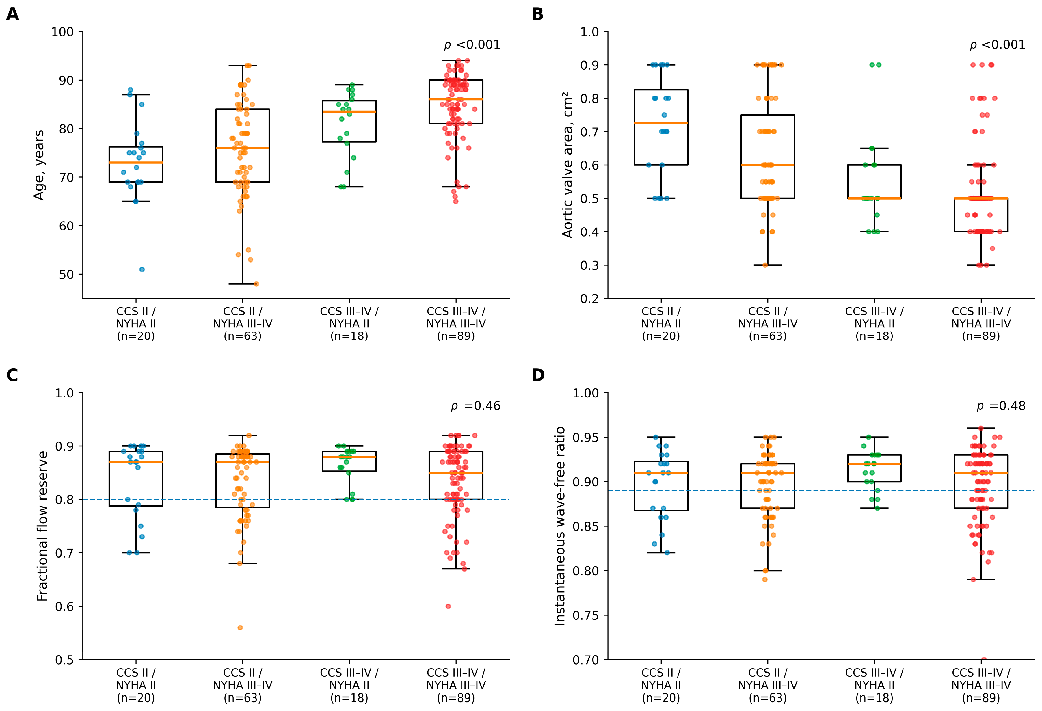
Figure 2 illustrates the model-predicted probability of CCS class III–IV angina and NYHA class III–IV functional limitation across the range of measured AVA for representative ages. The association between smaller measured AVA and severe angina was more pronounced than that observed for functional limitation. Confidence intervals widened toward the extremes of the AVA distribution, particularly for age-specific curves; therefore, apparent separation between curves at these extremes should not be overinterpreted. Across the four combined angina–functional limitation phenotypes, age and measured AVA differed significantly (both global p < 0.001), whereas FFR and iFR distributions were similar (p = 0.46 and p = 0.48, respectively; Figure 3).
Figure 2.
Predicted probabilities of severe angina and functional limitation according to measured aortic valve area, derived from the FFR-based models. (A) Predicted probability of Canadian Cardiovascular Society (CCS) class III–IV angina and (B) predicted probability of New York Heart Association (NYHA) class III–IV functional limitation according to measured aortic valve area (AVA). Curves were derived from separate multivariable logistic-regression models including age, sex, left ventricular ejection fraction, AVA, diameter stenosis, lesion length, and fractional flow reserve (FFR). Curves are shown for representative ages of 70, 80, and 90 years; sex was fixed at the male reference category, and continuous covariates other than age and AVA were fixed at cohort median values. Shaded areas indicate 95% confidence intervals, which widen toward the extremes of the AVA distribution; apparent separation between age-specific curves in these regions should therefore be interpreted cautiously.
Figure 3.
Distribution of age, measured aortic valve area, and epicardial pressure-derived indices across angina–functional limitation phenotypes: (A) age; (B) measured aortic valve area; (C) fractional flow reserve (FFR); (D) instantaneous wave-free ratio (iFR). Box plots indicate medians and interquartile ranges; points represent individual patients. Dashed lines indicate conventional thresholds for hemodynamic significance (FFR ≤ 0.80 and iFR ≤ 0.89). p values were derived from global Kruskal–Wallis tests across the four phenotype groups.
3.6. Sensitivity and Exploratory Analyses
Replacing measured AVA with mean transaortic gradient did not identify an association between mean gradient and CCS class III–IV angina in either physiological model (both p = 0.27). In an expanded model additionally adjusted for AF, COPD, and eGFR (n = 177), smaller measured AVA remained associated with CCS class III–IV angina (OR 1.44 per 0.1 cm2 decrease; p = 0.008), whereas FFR and iFR remained non-significant. When AVA indexed to BSA was substituted for absolute AVA, lower indexed AVA remained associated with CCS class III–IV angina (OR 1.68 per 0.1 cm2/m2 decrease; 95% CI: 1.11–2.52; p = 0.013). In contrast, indexed AVA was not independently associated with NYHA class III–IV functional limitation (OR 1.13 per 0.1 cm2/m2 decrease; 95% CI: 0.74–1.72; p = 0.56). When mean transaortic gradient was added to the primary models with measured AVA retained, smaller measured AVA remained associated with CCS class III–IV angina (OR 1.38 per 0.1 cm2 decrease; 95% CI: 1.08–1.78; p = 0.011), whereas mean gradient was not (p = 0.35); FFR and iFR again showed no association. Results were similar after adjustment for peak gradient (measured AVA: OR 1.38; 95% CI: 1.07–1.77; p = 0.013; peak gradient: p = 0.11). In an exploratory analysis restricted to patients with preserved LVEF (≥50%; n = 84), smaller measured AVA remained associated with severe angina in both the FFR model (OR 1.65; 95% CI: 1.08–2.52; p = 0.021) and the iFR model (OR 1.64; 95% CI: 1.08–2.51; p = 0.021), whereas neither FFR (p = 0.86) nor iFR (p = 0.79) was associated with angina.
The association between measured AVA and severe angina did not vary significantly according to LVEF (AVA × LVEF interaction: p = 0.57 in the FFR model and p = 0.56 in the iFR model; Supplementary Table S1). These analyses support the robustness of the association with measured AVA but cannot exclude a contribution from low transvalvular flow, which was not directly assessed. Similarly, no statistically significant interaction was observed between AVA and FFR (p = 0.21) or between AVA and iFR (p = 0.39) for severe angina. The Box–Tidwell assessment showed no evidence of departure from linearity for age (p = 0.96 in both models) but suggested possible non-linearity for AVA (p = 0.04 in both models). After a quadratic AVA term was added to the primary models, FFR (p = 0.74) and iFR (p = 0.93) were still not associated with CCS class III–IV angina. No significant AVA × mean-gradient interaction was observed for severe angina (p = 0.21) or functional limitation (Supplementary Table S1); the AVA × peak-gradient interaction was likewise non-significant (p = 0.37 for severe angina). Because the cohort was not powered for interaction testing, these non-significant interaction tests should not be interpreted as evidence of equivalence across subgroups.
Exploratory ROC analyses (Supplementary Table S2) showed moderate discrimination of age and measured AVA for severe angina, whereas AUCs for FFR and iFR were close to 0.50. For the secondary NYHA outcome, all ROC estimates should be interpreted cautiously because the analysis had limited statistical power. Extending the physiological analysis to all assessed coronary lesions did not materially alter the findings. In GEE models accounting for clustering of lesions within patients, FFR was not associated with CCS class III–IV angina (OR 1.03 per 0.10 decrease; 95% CI: 0.74–1.43; p = 0.85) or NYHA class III–IV functional limitation (OR 1.48; 95% CI: 0.91–2.41; p = 0.11). Similarly, iFR was not associated with severe angina (OR 0.92; 95% CI: 0.51–1.64; p = 0.77) or functional limitation (OR 1.85; 95% CI: 0.88–3.91; p = 0.11). Including non-LAD lesions therefore did not reveal an association between symptom severity and epicardial pressure-derived indices that might have been obscured by restricting the primary analysis to a single LAD lesion (Supplementary Table S3).
4. Discussion
In this secondary analysis of patients with severe high-gradient AS and a single intermediate LAD lesion, greater angina severity within an already symptomatic cohort was independently associated with older age and smaller measured AVA, whereas no independent association was observed with LAD lesion morphology or epicardial pressure-derived indices. In particular, neither FFR nor iFR was independently associated with CCS class III–IV angina after adjustment for clinical variables, measured AVA, and quantitative angiographic characteristics. Consistent with these findings, adding measured AVA to a clinical model improved model fit and discrimination, whereas subsequently adding lesion morphology, FFR, or iFR provided no incremental discriminatory information. For the secondary NYHA outcome, no included variable reached statistical significance; however, this exploratory analysis was underpowered to detect modest associations and should not be interpreted as evidence that such associations are absent.
These findings illustrate the difficulty of attributing symptom severity to a single coronary lesion in patients with severe AS [3,4]. Angina is a hallmark symptom of severe AS and can occur without obstructive epicardial CAD. Potential mechanisms include increased left ventricular afterload and myocardial oxygen demand, left ventricular hypertrophy, impaired subendocardial perfusion, elevated filling pressures, and coronary microvascular dysfunction [1,2,4,8]. An intermediate LAD lesion further complicates clinical interpretation, but its anatomical severity and pressure-derived hemodynamic significance may not be the dominant determinants of symptom burden [1]. The strong independent association between age and severe angina also warrants cautious interpretation. Age may act as a marker for unmeasured or incompletely measured factors such as frailty, diffuse atherosclerotic burden, microvascular dysfunction, reduced coronary reserve, anemia, or other comorbidities. Conversely, lower habitual activity in older patients could reduce exertional exposure and lead to under-recognition, rather than over-reporting, of angina; this would, if anything, bias the observed association toward the null. Because frailty, hemoglobin concentration, detailed activity level, and direct microvascular measurements were unavailable, age should be viewed as a patient-level marker associated with symptom severity rather than as a demonstrated causal mechanism.
The association between smaller measured AVA and severe angina requires particularly careful physiological interpretation [23]. All participants met criteria for severe high-gradient AS, confirming hemodynamically important valvular obstruction throughout the cohort [10]. However, continuity-equation AVA is flow dependent [10,24], and patients with CCS class III–IV angina had a smaller measured AVA despite a lower peak transaortic gradient and a similar mean gradient, as well as a lower LVEF. This pattern raises the possibility that differences in transvalvular flow and ventricular function contributed both to the measured AVA and to symptom burden [24]. Stroke volume index and the dimensionless index could not be derived because the individual LVOT diameter and Doppler velocity-time-integral measurements required for reliable retrospective calculation were not retained in the analytic dataset. Accordingly, a low-flow state cannot be formally excluded, including among patients with preserved LVEF [10,24]. Additional analyses support the robustness of the statistical association but do not establish its mechanism. Smaller measured AVA remained associated with severe angina after adjustment for either mean or peak transaortic gradient and in an exploratory subgroup with LVEF ≥ 50%, and no significant AVA × LVEF or AVA × transaortic-gradient interaction was identified. Nevertheless, LVEF and transaortic gradients are not direct measures of forward flow, and preserved LVEF does not exclude a low-flow state [10,24]. The observed association should therefore be interpreted as an association with measured AVA—which may reflect both fixed valvular obstruction and prevailing flow conditions—rather than as evidence that greater fixed obstruction caused angina [10,24]. This flow dependence is the central caveat in translating the finding into clinical practice.
The clinical implications depend on separating two distinct questions: whether an intermediate coronary lesion is physiologically significant and whether that lesion explains the severity of a patient’s symptoms. FFR and iFR remain important tools for addressing the first question and can appropriately inform revascularization decisions [1,3,7,8,9,10]. The present study addresses the second question. The absence of an observed association between angina severity and FFR/iFR should therefore not be interpreted as evidence that these indices are clinically unhelpful or that a physiologically significant lesion should not be revascularized. Rather, an FFR or iFR value above or below a conventional threshold should not, by itself, be used to infer whether the lesion is responsible for the patient’s angina. This distinction is consistent with ORBITA-FIRE, in which physiological thresholds associated with angina onset varied among individuals and with cardiac workload [18], although those findings cannot be extrapolated directly to severe AS. In severe AS, symptoms may arise from multiple coexisting mechanisms, including valvular obstruction, ventricular remodeling, altered coronary hemodynamics, and microvascular dysfunction [12,20]. Microvascular vasodilator capacity can influence the hyperemic response underlying FFR, and neither FFR nor iFR directly quantifies microvascular ischemia [25,26,27]. Because CFR, IMR, and other direct measures of microvascular function were unavailable, the present study cannot distinguish symptoms arising predominantly from an epicardial stenosis from those related to microvascular dysfunction or global myocardial oxygen supply–demand imbalance. Accordingly, our finding is best described as no observed association between angina severity and epicardial pressure-derived indices, rather than as evidence against a coronary ischemic mechanism. The 2025 ESC/EACTS Guidelines similarly support integrating coronary anatomy, symptoms, the planned valve intervention strategy, and patient-level characteristics while recognizing the complexity of coronary physiology in severe AS [10].
The exploratory NYHA analysis requires separate consideration [5,10]. NYHA class incorporates exertional dyspnea, fatigue, and reduced functional capacity and is therefore broader and less lesion-specific than angina severity [28]. Patients with NYHA class III–IV symptoms were older and had smaller measured AVA in univariable analyses, but these associations did not reach statistical significance after multivariable adjustment. Because only 37 of 185 complete cases were in NYHA class II and post hoc power was low for the principal predictors, the non-significant multivariable and ROC results cannot reliably distinguish between a true absence of association and limited statistical sensitivity. These findings should therefore be regarded as exploratory and hypothesis-generating. Symptom classification itself is also inherently uncertain: CCS and NYHA classes reflect clinically interpreted, patient-reported exertional symptoms rather than direct mechanistic measures of valvular or coronary disease. In older patients with severe AS, habitual activity, frailty, pulmonary disease, ventricular dysfunction, anemia, and other comorbidities may influence symptom class independently of lesion-level physiology, potentially attenuating observed associations.
The descriptive assessment of additional non-LAD disease provides further context. The frequency of physiologically positive non-LAD lesions did not differ significantly between CCS or NYHA groups. These analyses should be interpreted cautiously because additional lesions were assessed only when clinically indicated; nevertheless, they provide no evidence that more severe symptoms were simply explained by a greater burden of physiologically significant epicardial disease outside the target LAD lesion.
The specific contribution of the present analysis lies in the simultaneous comparison of valvular measurements, quantitative lesion morphology, and both hyperemic and resting epicardial pressure-derived indices within a homogeneous intermediate-LAD cohort, rather than in demonstrating the broader concept that CAD and symptoms may be dissociated in severe AS. This distinction is important when considering the absence of comparator groups with no CAD or unequivocally severe CAD. Such groups would be required to determine whether symptom burden differs across categories of coronary disease severity, but they are not needed to examine within-cohort associations among valvular, anatomical, and physiological variables in a predefined intermediate-lesion population. A valid three-group comparison would require prospectively assembled populations undergoing comparable clinical, angiographic, and physiological characterization; constructing such groups retrospectively from unrelated cohorts could introduce substantial selection and measurement bias.
Several features strengthen the clinical interpretability of this analysis. Restriction to a single LAD lesion avoided mixing coronary territories with different subtended myocardial mass and limited confounding from multiple target lesions per patient [17,19]. The availability of both FFR and iFR allowed hyperemic and resting pressure-derived indices to be assessed in parallel, and the main findings were consistent across multivariable and sensitivity analyses. Clinically, these data support a comprehensive rather than lesion-centered approach to symptom interpretation: lesion-level hemodynamic significance should be assessed with appropriate physiological tools, whereas attribution of symptom severity should additionally consider measured AVA and its flow dependence, ventricular and flow status, patient characteristics, coronary anatomy, and potential microvascular mechanisms.
This study has several limitations. First, it was a secondary analysis of a prospective, single-center registry with a relatively modest sample size. In particular, only 38 patients had NYHA class II symptoms (37 among complete cases), which limited statistical power for the secondary functional-limitation outcome; these analyses are therefore exploratory and cannot exclude modest associations. The observational, cross-sectional design precludes causal inference, and residual confounding remains possible despite multivariable adjustment. Second, symptom classification was based on CCS and NYHA categories assigned in routine clinical practice rather than by a standardized, centrally adjudicated protocol. The parent registry did not mandate blinded symptom assessment, and the timing of symptom classification relative to completion of invasive physiological measurements cannot be verified at the patient level; knowledge of coronary anatomy or physiology may therefore have influenced classification. Moreover, all participants were symptomatic (CCS II–IV), and the study therefore examined determinants of greater angina severity rather than the presence versus absence of angina. This restricted symptom range may also have reduced the ability to detect associations with epicardial pressure-derived indices. Antianginal and other background therapies, as well as hemoglobin concentration, were not prespecified study variables and were not systematically captured for all participants. Their absence is an important source of residual confounding because antianginal treatment may reduce reported symptoms independently of lesion anatomy or physiology, whereas anemia may contribute to dyspnea and myocardial oxygen supply–demand imbalance. The analytic dataset recorded prior myocardial infarction and prior PCI at the patient level but did not reliably identify whether a prior infarction involved the LAD territory or whether the study lesion overlapped with, or was adjacent to, a previously treated LAD segment; therefore, effects of prior infarction or intervention on symptoms and invasive physiology cannot be excluded. Third, and most importantly for interpretation, transvalvular flow was not directly assessed. Stroke volume index and the dimensionless index were unavailable because the source measurements required for reliable retrospective calculation were not retained in the analytic dataset. Consequently, the smaller measured AVA observed in more symptomatic patients may partly reflect lower transvalvular flow and impaired ventricular function rather than greater fixed valvular obstruction. The association with measured AVA should therefore not be interpreted as evidence that greater fixed obstruction caused severe angina. Fourth, the analysis was restricted to patients with severe high-gradient AS and one physiologically assessed LAD lesion. This approach provided a homogeneous patient-level cohort but limits generalizability to other coronary territories, multivessel CAD, left main disease, non-obstructive CAD, or low-flow, low-gradient AS. The cohort comprised older patients undergoing assessment for TAVR or SAVR and may not represent younger patients or those managed conservatively. All included lesions were angiographically intermediate and selected for invasive physiological assessment, which may also have introduced selection bias. The parent registry was not designed as a three-group study spanning no CAD, intermediate CAD, and severe obstructive CAD; accordingly, the present findings address within-cohort associations in a physiology-selected intermediate-lesion population and cannot determine whether symptom burden differs across these CAD categories. Fifth, direct assessment of coronary microvascular function, including CFR and IMR, was not performed [25,26,27]. This is an important mechanistic limitation because microvascular dysfunction is common in severe AS, may contribute to myocardial ischemia and angina independently of epicardial stenosis severity [12], and may influence the hyperemic response underlying FFR. Systematic pressure-wire pullback and PPG measurements were likewise unavailable. Finally, analyses of non-LAD CAD were descriptive because additional lesions were assessed only when clinically indicated. Interaction analyses and several sensitivity analyses were exploratory and may have been underpowered to detect modest effects; non-significant interaction tests should not be interpreted as evidence of equivalence across subgroups. Although model performance was internally validated using bootstrap optimism correction, no external validation was performed. The findings should therefore be regarded as hypothesis-generating and require confirmation in larger multicenter cohorts with systematic assessment of transvalvular flow and both epicardial and microvascular coronary physiology.
5. Conclusions
Within this already symptomatic cohort of patients with severe high-gradient AS and a single intermediate LAD lesion, greater angina severity was independently associated with older age and smaller measured AVA, whereas no independent association was observed with LAD lesion morphology or epicardial pressure-derived indices, including FFR and iFR. Because continuity-equation AVA is flow dependent and direct measures of transvalvular flow were unavailable, the association with measured AVA does not establish that greater fixed valvular obstruction caused angina. Likewise, the absence of an association between angina severity and FFR/iFR should not be interpreted as evidence against a coronary ischemic mechanism or as invalidating these indices for assessment of lesion-level hemodynamic significance. Rather, symptom attribution in severe AS should integrate valvular measurements interpreted in the context of flow status, ventricular function, patient characteristics, coronary anatomy, epicardial physiological assessment, and potential microvascular mechanisms. The secondary NYHA analysis was exploratory and underpowered, and its non-significant findings should be interpreted accordingly. These findings are specific to the intermediate-lesion population studied and do not compare symptom burden across the full spectrum of CAD severity. Larger prospective studies incorporating stroke volume index, CFR, and IMR are needed to clarify the mechanisms underlying symptom severity in this population.
Supplementary Materials
The following supporting information can be downloaded at https://www.mdpi.com/article/10.3390/jcdd13110553/s1. Table S1: Exploratory interaction analyses of measured AVA with transaortic gradient and LVEF for symptom severity; Table S2: Exploratory univariable receiver-operating-characteristic analysis of age, measured AVA, FFR, and iFR for severe angina and functional limitation in the 190-patient LAD cohort; Table S3: Exploratory lesion-level analysis of FFR and iFR for severe angina and functional limitation across the parent physiology cohort.
Author Contributions
Conceptualization, A.D. and P.K.; methodology, A.D. and P.K.; formal analysis, A.D.; investigation, B.Z., T.R., Ł.N., P.S., J.L. and P.K.; data curation, B.Z., T.R., Ł.N. and P.S.; writing—original draft preparation, A.D.; writing—review and editing, B.Z., T.R., Ł.N., P.S., J.L. and P.K.; funding acquisition, P.K.; supervision, J.L. and P.K. All authors have read and agreed to the published version of the manuscript.
Funding
This research was funded by the National Science Center, grant number 2018/02/X/NZ5/02648 (to P.K.).
Institutional Review Board Statement
The study was conducted in accordance with the Declaration of Helsinki and approved by the Bioethics Committee of the Jagiellonian University on 31 January 2018 (protocol code 1072.6120.1.2019).
Informed Consent Statement
Informed consent was obtained from all subjects involved in the study.
Data Availability Statement
The data presented in this study are available from the corresponding author on reasonable request.
Acknowledgments
The graphical abstract was created with BioRender (Dziewierz, A. 2026. The authors declare that generative artificial intelligence tools (Claude Opus 4.8) were used during the preparation and revision of this manuscript for language editing and stylistic improvement. Following their use, the authors reviewed and edited the content as needed and take full responsibility for the content of the published article.
Conflicts of Interest
The authors declare no conflicts of interest. The funders had no role in the design of the study; in the collection, analyses, or interpretation of data; in the writing of the manuscript; or in the decision to publish the results.
Abbreviations
The following abbreviations are used in this manuscript:
| AF | Atrial fibrillation |
| AIC | Akaike information criterion |
| AS | Aortic stenosis |
| AUC | Area under the curve |
| AVA | Aortic valve area |
| AVR | Aortic valve replacement |
| BMI | Body mass index |
| BSA | Body surface area |
| CAD | Coronary artery disease |
| CCS | Canadian Cardiovascular Society |
| CFR | Coronary flow reserve |
| CI | Confidence interval |
| COPD | Chronic obstructive pulmonary disease |
| eGFR | Estimated glomerular filtration rate |
| FFR | Fractional flow reserve |
| GEE | Generalized estimating equation |
| iFR | Instantaneous wave-free ratio |
| IMR | Index of microcirculatory resistance |
| IQR | Interquartile range |
| LAD | Left anterior descending artery |
| LR | Likelihood ratio |
| LVEF | Left ventricular ejection fraction |
| LVOT | Left ventricular outflow tract |
| NYHA | New York Heart Association |
| OR | Odds ratio |
| PCI | Percutaneous coronary intervention |
| PPG | Pullback pressure gradient |
| QCA | Quantitative coronary angiography |
| ROC | Receiver-operating-characteristic |
| SAVR | Surgical aortic valve replacement |
| TAVR | Transcatheter aortic valve replacement |
References
- Faroux, L.; Villecourt, A.; Metz, D. The management of coronary artery disease in TAVR patients. J. Clin. Med. 2023, 12, 7126. [Google Scholar] [CrossRef] [Scilit] [PubMed]
- Stähli, B.E.; Linke, A.; Westermann, D.; Van Mieghem, N.M.; Leistner, D.M.; Massberg, S.; Alber, H.; Mügge, A.; Musumeci, G.; Kesterke, R.; et al. A randomized comparison of the treatment sequence of percutaneous coronary intervention and transcatheter aortic valve implantation: Rationale and design of the TAVI PCI trial. Am. Heart J. 2024, 277, 104–113. [Google Scholar] [CrossRef] [Scilit] [PubMed]
- Mafrica, D.; Dziewierz, A.; Dobrev, D.; Pelliccia, F. Transcatheter aortic valve implantation: Latest evidence, gaps in knowledge, and future directions. Adv. Interv. Cardiol. 2025, 21, 487–495. [Google Scholar] [CrossRef] [Scilit] [PubMed]
- Weferling, M.; Kim, W.K. Invasive functional assessment of coronary artery disease in patients with severe aortic stenosis in the TAVI era. J. Clin. Med. 2023, 12, 5414. [Google Scholar] [CrossRef] [Scilit] [PubMed]
- Garbacz, J.; Priadka, A.; Lemek, T.; Roczniak, J.; Rajzer, M.; Bartuś, S.; Surdacki, A.; Chyrchel, M. Factors influencing dyspnea occurrence in patients with moderate and severe aortic stenosis. Kardiol. Pol. 2025, 83, 485–488. [Google Scholar] [CrossRef] [Scilit] [PubMed]
- Kupatt, C.; Lettmann, N. Aortic stenosis combined with coronary artery disease in intermediate risk: Which way to go? JACC Cardiovasc. Interv. 2024, 17, 2486–2487. [Google Scholar] [CrossRef] [Scilit] [PubMed]
- Dowling, C.; Michail, M.; Zhang, J.M.; Comella, A.; Thakur, U.; Gooley, R.; McCormick, L.; Brown, A.J.; Wong, D.T.L. Diagnostic performance of quantitative flow ratio, non-hyperaemic pressure indices and fractional flow reserve for the assessment of coronary lesions in severe aortic stenosis. Cardiovasc. Diagn. Ther. 2022, 12, 314–324. [Google Scholar] [CrossRef] [Scilit] [PubMed]
- Di Gioia, G.; Pellicano, M.; Toth, G.G.; Casselman, F.; Adjedj, J.; Van Praet, F.; Ferrara, A.; Stockman, B.; Degrieck, I.; Bartunek, J.; et al. Fractional flow reserve-guided revascularization in patients with aortic stenosis. Am. J. Cardiol. 2016, 117, 1511–1515. [Google Scholar] [CrossRef] [Scilit] [PubMed]
- Sabbah, M.; Engstrøm, T.; Lønborg, J. Invasive pressure indices in aortic stenosis: The key role of resting flow after valve replacement. Front. Cardiovasc. Med. 2023, 10, 1179346. [Google Scholar] [CrossRef] [Scilit] [PubMed]
- Praz, F.; Borger, M.A.; Lanz, J.; Marin-Cuartas, M.; Abreu, A.; Adamo, M.; Ajmone Marsan, N.; Barili, F.; Bonaros, N.; Cosyns, B.; et al. 2025 ESC/EACTS Guidelines for the management of valvular heart disease. Eur. Heart J. 2025, 46, 4635–4736. [Google Scholar] [CrossRef] [Scilit] [PubMed]
- Siudak, Z.; Huczek, Z.; Grygier, M.; Tomaniak, M.; Kałużna-Oleksy, M.; Kleczyński, P.; Milewski, K.; Opolski, M.P.; Smolka, G.; Sabiniewicz, R.; et al. Interventional cardiology in Poland in 2024. Annual summary report of the Association of Cardiovascular Interventions of the Polish Cardiac Society and Jagiellonian University Medical College. Adv. Interv. Cardiol. 2025, 21, 469–472. [Google Scholar] [CrossRef] [Scilit] [PubMed]
- Michail, M.; Davies, J.E.; Cameron, J.D.; Parker, K.H.; Brown, A.J. Pathophysiological coronary and microcirculatory flow alterations in aortic stenosis. Nat. Rev. Cardiol. 2018, 15, 420–431. [Google Scholar] [CrossRef] [Scilit] [PubMed]
- Ahmad, Y.; Vendrik, J.; Eftekhari, A.; Howard, J.P.; Cook, C.; Rajkumar, C.; Malik, I.; Mikhail, G.; Ruparelia, N.; Hadjiloizou, N.; et al. Determining the predominant lesion in patients with severe aortic stenosis and coronary stenoses: A multicenter study using intracoronary pressure and flow. Circ. Cardiovasc. Interv. 2019, 12, e008263. [Google Scholar] [CrossRef] [Scilit] [PubMed]
- Ahmad, Y.; Götberg, M.; Cook, C.; Howard, J.P.; Malik, I.; Mikhail, G.; Frame, A.; Petraco, R.; Rajkumar, C.; Demir, O.; et al. Coronary hemodynamics in patients with severe aortic stenosis and coronary artery disease undergoing transcatheter aortic valve replacement: Implications for clinical indices of coronary stenosis severity. JACC Cardiovasc. Interv. 2018, 11, 2019–2031. [Google Scholar] [CrossRef] [Scilit] [PubMed]
- Scarsini, R.; Pesarini, G.; Zivelonghi, C.; Piccoli, A.; Ferrero, V.; Lunardi, M.; Gottin, L.; Zanetti, C.; Faggian, G.; Ribichini, F. Physiologic evaluation of coronary lesions using instantaneous wave-free ratio in patients with severe aortic stenosis undergoing transcatheter aortic valve implantation. EuroIntervention 2018, 13, 1512–1519. [Google Scholar] [CrossRef] [Scilit] [PubMed]
- Minten, L.; Bennett, J.; Otsuki, H.; Takahashi, K.; Fearon, W.F.; Dubois, C. Differential effect of aortic valve replacement for severe aortic stenosis on hyperemic and resting epicardial coronary pressure indices. J. Am. Heart Assoc. 2024, 13, e034401. [Google Scholar] [CrossRef] [Scilit] [PubMed]
- Fogelson, B.; Tahir, H.; Livesay, J.; Baljepally, R. Pathophysiological factors contributing to fractional flow reserve and instantaneous wave-free ratio discordance. Rev. Cardiovasc. Med. 2022, 23, 70. [Google Scholar] [CrossRef] [Scilit] [PubMed]
- Ahmed-Jushuf, F.; Foley, M.J.; Chotai, S.; Rajkumar, C.A.; Wang, D.; Simader, F.A.; Macierzanka, K.; Chiew, K.; Misra, S.; Williams, R.; et al. Determining the physiological threshold for angina (ORBITA-FIRE): A double-blind, randomized, placebo-controlled study. Circulation 2026, 153, 1795–1812. [Google Scholar] [CrossRef] [Scilit] [PubMed]
- Leone, A.M.; De Caterina, A.R.; Basile, E.; Gardi, A.; Laezza, D.; Mazzari, M.A.; Mongiardo, R.; Kharbanda, R.; Cuculi, F.; Porto, I.; et al. Influence of the amount of myocardium subtended by a stenosis on fractional flow reserve. Circ. Cardiovasc. Interv. 2013, 6, 29–36. [Google Scholar] [CrossRef] [Scilit] [PubMed]
- Dziewierz, A.; Zdzierak, B.; Rzeszutko, Ł.; Niewiara, Ł.; Legutko, J.; Kleczyński, P. Angiographic phenotypes predict FFR/iFR discordance in severe aortic stenosis: A cluster analysis. Cardiovasc. Revasc. Med. 2026, 89, 52–58. [Google Scholar] [CrossRef] [Scilit] [PubMed]
- Dziewierz, A.; Rzeszutko, Ł.; Niewiara, Ł.; Legutko, J.; Kleczyński, P. The role of aortic valve area in the quantitative flow ratio-fractional flow reserve discrepancy in patients with coronary artery disease and severe aortic stenosis. Adv. Interv. Cardiol. 2025, 21, 80–87. [Google Scholar] [CrossRef] [Scilit] [PubMed]
- Dziewierz, A.; Rzeszutko, Ł.; Dudek, D.; Legutko, J.; Kleczyński, P. Impact of chronic kidney disease on fractional flow reserve accuracy in severe aortic stenosis. Pol. Arch. Intern. Med. 2024, 134, 16649. [Google Scholar] [CrossRef] [Scilit] [PubMed]
- Keller, L.S.; Nuche, J.; Avvedimento, M.; Real, C.; Farjat-Pasos, J.; Paradis, J.M.; DeLarochellière, R.; Poulin, A.; Kalavrouziotis, D.; Dumont, E.; et al. Angina in patients with severe aortic stenosis undergoing transcatheter aortic valve replacement. Rev. Esp. Cardiol. (Engl. Ed.) 2023, 76, 991–1002. [Google Scholar] [CrossRef] [Scilit] [PubMed]
- La Canna, G.; Habjan, S.; Scarfò, I. Understanding the transvalvular gradient in aortic stenosis: A multifaceted perspective. J. Clin. Med. 2025, 14, 7916. [Google Scholar] [CrossRef] [Scilit] [PubMed]
- McConkey, H.Z.R.; Marber, M.; Chiribiri, A.; Pibarot, P.; Redwood, S.R.; Prendergast, B.D. Coronary microcirculation in aortic stenosis. Circ. Cardiovasc. Interv. 2019, 12, e007547. [Google Scholar] [CrossRef] [Scilit] [PubMed]
- Martínez Pereyra, V.; Seitz, A.; Mahrholdt, H.; Bekeredjian, R.; Sechtem, U.; Ong, P. Coronary microvascular dysfunction in patients with mild-to-moderate aortic stenosis—Insights from intracoronary acetylcholine testing. Int. J. Cardiol. Heart Vasc. 2020, 31, 100658. [Google Scholar] [CrossRef] [Scilit] [PubMed]
- Aleksandric, S.; Banovic, M.; Beleslin, B. Challenges in diagnosis and functional assessment of coronary artery disease in patients with severe aortic stenosis. Front. Cardiovasc. Med. 2022, 9, 849032. [Google Scholar] [CrossRef] [Scilit] [PubMed]
- Raphael, C.; Briscoe, C.; Davies, J.; Whinnett, Z.I.; Manisty, C.; Sutton, R.; Mayet, J.; Francis, D.P. Limitations of the New York Heart Association functional classification system and self-reported walking distances in chronic heart failure. Heart 2007, 93, 476–482. [Google Scholar] [CrossRef] [Scilit] [PubMed]

Disclaimer/Publisher’s Note: The statements, opinions and data contained in all publications are solely those of the individual author(s) and contributor(s) and not of MDPI and/or the editor(s). MDPI and/or the editor(s) disclaim responsibility for any injury to people or property resulting from any ideas, methods, instructions or products referred to in the content. |
© 2026 by the authors. Licensee MDPI, Basel, Switzerland. This article is an open access article distributed under the terms and conditions of the Creative Commons Attribution (CC BY) license.