


Simple Summary
In a farrow-to-finish farm, two sows developed red-to-purple cutaneous lesions localised on the udder, flanks, and hind limbs. The aim of this study was to investigate the origin of these lesions. The lesions appeared a few days after the sows were vaccinated. Clinical examination and laboratory findings were not consistent with bacterial or viral infectious etiology. Immunofluorescence was performed to detect antibodies and complement deposits in order to investigate a hypersensitivity reaction. No antibody or complement deposits were detected. Nevertheless, a vaccine-induced adverse reaction associated with hypersensitivity remains the most plausible explanation, as immune complexes are transient and may no longer be detectable at the time of sampling.
Abstract
This case report describes the investigation of two cases of cutaneous vasculitis in sows following vaccination. The herd was porcine reproductive and respiratory syndrome virus (PRRSV)-stable, with routine PRRSV-1 and porcine circovirus type 2 (PCV2) sows’ vaccination. Two sows developed red-to-purple macules on the udder, flanks, and hind limbs, occurring two and three days after vaccination, respectively. Diagnostic investigations included biochemistry, complete blood count, PCR, immunohistochemistry (IHC), serology, histopathology, Gram staining, and immunofluorescence (IF) to assess the involvement of type III hypersensitivity. Clinical examination and herd evaluation revealed no significant abnormalities. Both sows showed biochemistry results within the reference intervals. One sow had lymphocytosis. All PCR PCV2, PCV3 and PRRSV-1/2 on serum and IHC PCV2 conducted on skin biopsies were negative, although both animals were seropositive for PCV2. Histopathology revealed necrotizing leukocytoclastic vasculitis, and Gram staining detected no bacteria. IF failed to identify immune complex or complement deposition. However, type III hypersensitivity could not be excluded, as immune complexes are transient and biopsies were collected about 48 h after lesion onset. Overall, the findings support a vaccine-related adverse reaction. Vaccine adverse effects should therefore be considered in the differential diagnosis of cutaneous vasculitis in swine.
1. Introduction
Hypersensitivity reactions are defined by exacerbated immune responses classified into four main types. Type III hypersensitivity involves immune complex formation and complement activation, leading to vascular injuries, especially vasculitis. Lesions can appear immediately or several days after the antigen exposure. In human medicine the antigen triggering the type III hypersensitivity may be of infectious origin, or non-infectious origin like drugs or vaccines [1]. Several vaccines has been suspected of causing vasculitis (Varicella-Zoster virus, influenza and COVID-19 vaccines) [2,3]. In pig production, vasculitis lesions are regularly triggered by infectious causes: porcine circovirus type 2 (PCV2) and 3 (PCV3), Betaarterivirus suid 1 and 2, also known as porcine reproductive and respiratory syndrome virus type 1 (PRRSV-1) and type 2 (PRRSV-2), Actinobacillus sp., Erysipelothrix rhusiopathiae, Escherichia coli, Glaesserella parasuis, Leptospira interrogans, Salmonella sp., Streptococcus suis and Treponema pedis [4,5,6,7]. Vasculitides of non-infectious origin are less documented and no vaccine used in pig production mentions any cases of vasculitis in their summary of product characteristics.
This clinical case aims to investigate two cases of leukocytoclastic vasculitis following vaccination of sows occurring in the absence of detectable infectious triggers. The vaccines suspected of causing vasculitis in this case were Rhiniseng® (Laboratorios Hipra S.A., Spain), Suigen Rota Coli® (Virbac, France) and Respiporc Flu 3® (CEVA santé Animale, France). All three vaccines were inactivated vaccines. However, adjuvants and excipients differ from one another (except sodium chloride).
2. Case Presentation
2.1. Anamnesis and Clinical Examination
The case report concerns two sows of a 600-sow farrow-to-finish farm located in Brittany, France. The herd follows batch-production practices with farrowings scheduled every 2 weeks with a weaning age of approximately 21 days. Biosecurity measures are very well implemented according to French regulation. Regarding PRRSV-1, the herd is supposed to be positive stable with vaccination without evidence of sow-to-piglet transmission and no viral circulation on the finishing stage. The vaccination protocol is presented in Table 1.
Table 1.
Gilts and sows vaccination protocol.
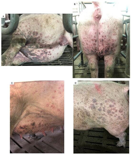
Seven weeks before farrowing, the first sow (Sow 1) received the rhinitis vaccine. Six hours later, she developed fever (40 °C) and anorexia for 2 days. Two days after the vaccination, multifocal, well-circumscribed, slightly raised and non-suppurative, red-to-purple macules occurred. They were not pruritic and ranged in diameter from 1 to 2 cm. They were localised to the udder, flanks and hind limbs, where they tended to coalesce (Figure 1). A clinical examination was carried out two days after macules appeared. The multiparous sow (parity 5) was in good body condition with a normal breathing pattern and normothermia. The sow demonstrated no reluctance to move and a good appetite.
Figure 1.
Red-to-purple macules localised on the hind limbs, udder and flanks of sow 1 (photos (A,B)) and sow 2 (photos (C,D)).
Five months later, the second sow (Sow 2) received swine influenza vaccine, colibacillosis and rotavirus vaccines on the same week. Three days after swine influenza vaccination and five days after colibacillosis and rotavirus vaccination, sow 2 developed similar skin lesions (Figure 1). No hyperthermia was noticed by the farmer. The clinical examination was carried out two days after macules appeared. Sow 2 was a parity 8 and the clinical examination’s result was similar to sow 1.
At the herd level, the fertility rate was normal (92%) as was prolificacy (16.8 piglets born alive per litter). Stillbirth and mummification rates were stable (respectively 6.1% and 2.4%). No abortion was noticed.
2.2. Samples Collection
For both sows, all samples were taken two days after the skin lesions occurred, after the clinical examination.
Blood samples were taken from the external jugular vein with an 18G x 1 1/2” needle in dry, heparin and EDTA tubes.
Biochemistry assessments were conducted on the heparin tubes. Urea (UR3825, Randox®, Ireland), creatinine (CR3814, Randox®, Ireland), total protein (TP3869, Randox®, Ireland), albumin (AB3800, Randox®, Ireland), calcium (CA3871, Randox®, Ireland) and phosphorus (PH3872, Randox®, Ireland) were assessed, using colorimetric tests with a RX IMOLA device (Randox®, Ireland). Serum amyloid A was measured using ELISA assay (Serum Amyloid A TP802, Tridelta®, Ireland) and read with a SUNRISE device (Tecan®, Switzerland).
The complete blood count was performed on EDTA blood samples using Vet abc Plus+ system (Scil®, Germany), based on impedance and spectrophotometric measurements. Blood smears were stained using the RAL 555 kit.
Extraction methods, automated extraction systems, PCR reagents and PCR instruments used for qPCR PCV2, PCV3 and RT-PCR PRRSV-1/PRRSV-2 are presented in Table 2.
Table 2.
Extraction methods, automated extraction system, PCR reagents and PCR instruments used for qPCR PCV2, PCV3 and RT-PCR PRRSV-1/PRRSV-2.
PCV2 serologies were performed with the ELISA Ingezim Circo IgG (11.PCV.K.1/2, Ingenasa®, Spain) test.
Urine samples were collected from sow 1 during a spontaneous voiding in a sterile sample container with boric acid. No urine was collected from sow 2 due to the practical impossibility to do it. A urinary analysis was performed for sow 1 by macroscopic and microscopic examination using a Kova® slide (Glasstic slide 10, Kova®, USA) and a urinary stick (Multistix 8 SG, Siemens®, USA).
Punch biopsies were done on the edge of 2 macules for both sows. Biopsies were kept in a 4% formaldehyde solution respecting a 1 to 10 volume ratio at room temperature.
For histopathological examination, formalin-fixed samples were embedded in paraffin wax, cut into 3 µm sections and stained with haematoxylin, eosin and saffron. Two to three cut levels per sample were evaluated and examined using a Leica DM 2500 microscope (Leica Microsystems®, Germany). Pictures were taken using an XCD U100CR digital camera (Sony®, Japan) and registered using the Archimed imaging software (version 12.3.11).
For immunohistochemistry (IHC), 4 μm thick sections of all tissues were cut and placed on silane-coated slides (SuperFrost Plus, Thermofisher®, USA). IHC was performed with a monoclonal rabbit anti-PCV2 antibody (in-house antibody produced by Anses Ploufragan, France) at 1/200 concentration.
Immunofluorescences (IF) were conducted to detect IgG, IgM, IgA, C3. Antibody references used are presented in Table 3. Antibodies were used at different concentrations (1/100, 1/50 and 1/20). Slides were read with a fluorescent microscope using a filter adapted to fluorochrome (Zeiss®, France). Analysis was performed with ZEN software (version ZEN 2). Slides incubated with PBS solution instead of antibodies served as negative controls.
All samples were directly sent to an external laboratory (LABOCEA, Ploufragan, France) and kept in positive cold (2° to 8 °C) during transportation. Biochemistry was assessed in LABEO (Normandy, France), urinary analysis, qPCR PCV2, RT-PCR PRRSV1/PRRSV-2, histolopathological examination, IHC and PCV2 serology were assessed by LABOCEA (Ploufragan, France). qPCR PCV3 was conducted by Anses Ploufragan-Plouzané-Niort laboratory (Ploufragan, France). Direct immunofluorescence (DIF) and Gram staining were conducted by VetAgro Sup laboratory (Marcy l’Etoile, France).
Table 3.
Antibody references used for IF.
3. Results
3.1. Biochemistry and Haematological Findings
For both sows, biochemistry values were within the reference ranges, particularly the blood urea and creatinine concentrations (Table 4).
Table 4.
Biochemistry findings.
Haematological findings are presented in Table 5. A slight increase in MCHC was observed for both sows but was considered of doubtful clinical significance, as it was isolated and not associated with other haematological abnormalities. In the absence of leukocytosis, the slight variations in the proportions of lymphocytes, monocytes, neutrophils and basophils in sow 1 and of monocytes and basophils in sow 2, compared with the reference intervals, are unlikely to be clinically significant.
Table 5.
Haematological findings.
3.2. Urinary Analysis
The urinary examination for sow 1 is presented in Table 6. The urine specific gravity (1.010) was within the reference interval (1.010–1.050) [12]. Crystals were observed but not red blood cells or leukocytes. The urinary pH was within the reference interval (5.5–7.5) [4].
Table 6.
Sow 1 urinary examination by macroscopic and microscopic examination and urinary stick.
3.3. PCV2-, PCV3- and PRRS-Related Investigations
All serum samples tested negative for PCV2 and PCV3 DNA and PRRSV-1/PRRSV-2 RNA. PCV2 was not detected by IHC (Table 7).
Table 7.
IHC PCV2 and qPCR PCV2, PCV3 and RT-PCR PRRSV results for both sows.
3.4. Histological, Gram Staining and Immunofluorescence Examination
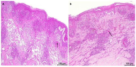
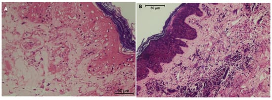
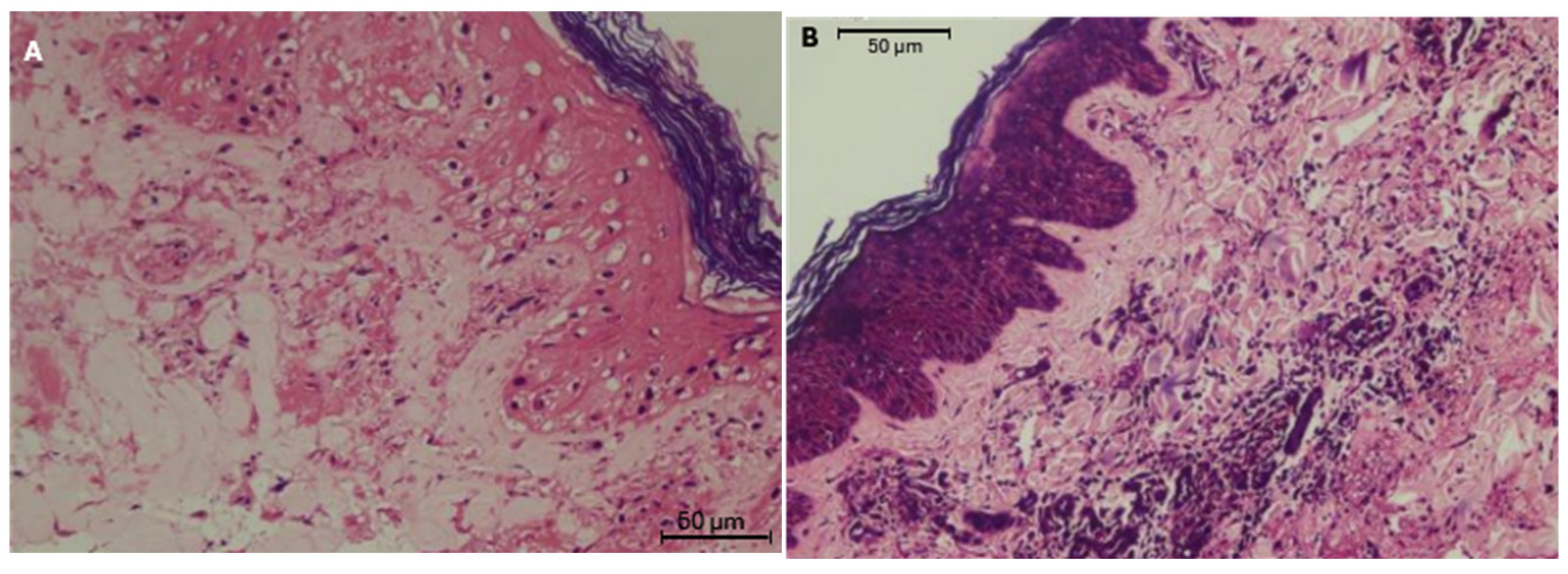
Histological examination of the punch biopsies revealed mild epithelial hyperplasia and mild to moderate infiltration of the perivascular and vascular walls by hypersegmented neutrophils in both sows. Some eosinophils were also present in the perivascular and vascular walls of sow 2. Mild necrotising vasculitis and fibrinoid degeneration, characterised by perivascular fibrinous exudates and cell debris, were also observed (Figure 2). The upper dermis and mid-dermal blood vessels were affected.
Figure 2.
Histopathological skin lesion HE staining showing necrotizing vasculitis (arrows) for sow 1 (A) and sow 2 (B). Scale bar: 100 µm.
Skin biopsy Gram staining was negative for both sows (Figure 3). The observation of several slides of each biopsy at a magnification of ×1000 did not reveal the presence of bacteria.
Figure 3.
Gram staining on skin biopsies for sow 1 (A) and sow 2 (B). Magnification of the image: (A) ×400 and (B) ×200. No bacteria observed. Scale bar: 50 µm.
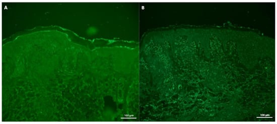
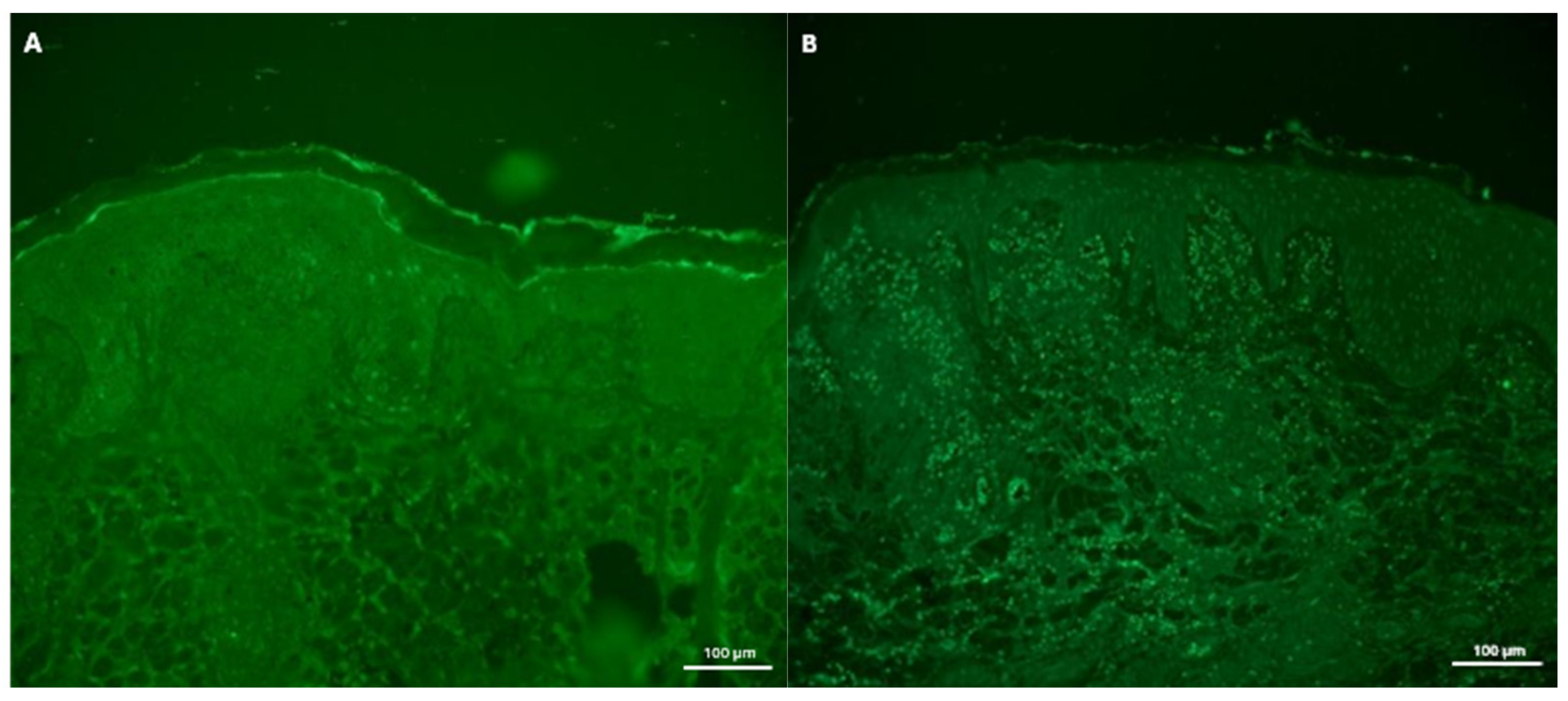
Direct immunofluorescence did not detect IgG, IgM, IgA or C3 deposition (Figure 4).
Figure 4.
Skin biopsies direct immunofluorescence using rabbit polyclonal anti-C3 pig and Goat Anti-rabbit IgG, FITC for sow 1 (A) and sow 2 (B). No immunofluorescence signal detected. Scale bar: 100 µm.
Both sows received amoxicillin (Amoxipro injectable, Ceva Santé Animale, Libourne, France) 15 mg/kg intramusculary and ketoprofen (Rifen 100 mg/mL, Vetviva Richter GMBH, Wels, Austria) the day skin lesions appeared and 48 h after. Papules and macules regressed within one week after their appearance for sow 1. Two days after treatment, scabs replaced some lesions for sow 2.
The sows farrowed 6 weeks after vaccination. Parturition and prolificacy were normal for both.
4. Discussion
Post-vaccination reaction is commonly described in the vaccine’s summary of product characteristics (section adverse events) used in pig production. In our case report, a few days after vaccination, two sows developed a purpuric macular eruption. The clinicopathological findings were consistent with dermal leukocytoclastic vasculitis. Infectious causes such as PCV2, PCV3, PRRSV-1/PRRSV-2 and bacteria were considered unlikely based on laboratory results, absence of systemic signs and normal renal function.
Similar cases have been reported in humans. Cutaneous reactions were delayed and occurred several days after vaccination [3,13]. Even though cutaneous reactions occurring at a distance from the injection are rare, vasculitis have been described after Varicella-Zoster virus, influenza and COVID-19 vaccination [2,13]. In human medicine, no risk factors have been identified in patients who developed post-vaccination vasculitis [14]. Missoum et al. [3] described one case with leukocytoclasic vasculitis lesions developing after SARS-CoV-2 inactivated vaccination. The vaccine used in this human case was an aluminum hydroxide-adjuvanted vaccine. Leukocytoclastic vasculitis was observed on histopathological examination of skin lesions and plasma protein C level increased 9 days after vaccination. The patient suffered from a renal failure characterised by hyperuremia and hypercreatininaemia. In our case, the two sows presented leukocytoclastic vasculitis too and a normal SAA level without renal failure at sampling time (four days and five days after the last vaccination for sow 1 and sow 2, respectively). Similarities between humans’ lesions after SARS-CoV-2 vaccination and sows’ lesions after vaccination may suggest a similar cause. The three vaccines suspected to be involved in the sows’ vasculitis were all inactivated vaccines. However, their compositions differ. Among them, only Rhiniseng® is adjuvanted with aluminum hydroxide as with the human case mentioned above. Differences in vaccine composition did not allow the identification of any specific component that could have contributed to the development of vasculitis in sows.
Infection causes were thoroughly investigated. A bacteria-induced vasculitis was considered less likely as sow 2 exhibited no additional clinical signs suggestive of systemic infection (no fever, no anorexia, clinically healthy). Several bacterial pathogens have been associated with vasculitis in pigs, including Actinobacillus sp., Erysipelothrix rhusiopathiae, Escherichia coli, Glaesserella parasuis, Leptospira interrogans, Salmonella sp., Streptococcus suis and Treponema pedis [5,6,7]. In our case, bacterial culture on skin biopsies could not be carried out, but Gram staining showed no bacteria on the slides although this staining might not highlight all the aforementioned bacteria. Main viral pathogens causing vasculitis in pigs are circoviruses and PRRSV-1/PRRSV-2 [4]. Vasculitis associated with PCV2 or PCV3 has been well described in porcine dermatitis and nephropathy syndrome (PDNS) [15,16,17]. Affected pigs are usually depressed, prostrate and anorexic. Mortality approaches 100% in animals over three months of age due to renal failure caused by glomerulonephritis [4,18]. Sipos et al. [19] also reported elevated urea and creatinine values in PDNS-affected pigs compared to controls. In our cases, biochemistry and urine analysis did not reveal kidney lesions. Although isolated skin lesions may occur in rare instances, PDNS-affected herds generally display additional circovirus-associated diseases [16,20]. No such conditions were observed in the herd before or after the present cases. In addition, qPCR for PCV2, PCV3 and RT-PCR PRRSV-1/PRRSV-2 as well as PCV2 IHC were all negative, which did not support a viral etiology. While PCV2 DNA may be inconsistently detected in skin lesions, it is typically present in serum from PDNS-affected pigs [21] which was not the case here.
DIF was performed to investigate a potential hypersensitivity reaction following vaccination. No IgG, IgM, IgA or C3 deposition was detected in the skin lesions. In the SARS-CoV-2 post-vaccination vasculitis case described by Missoum et al. [3], skin immunofluorescence was negative too.
This case has potential limitations. Bacteriology on skin biopsies could have been conducted to explore bacterial origin. During our initial investigations, we found this cause unlikely, which is why all skin biopsies sampled were fixed in formaldehyde solution. Subsequently, when investigating potential bacterial causes, we decided to carry out Gram staining. However, in the absence of systemic reactions such as hyperthermia, an infectious origin is unlikely.
Concerning direct immunofluorescence, no signal was detected. The delay between the lesion onset and sample collection could have influenced the results. Indeed, Thibault et al. [22] conducted DIF in suspected PDNS cases. C3 and IgM signals were the two most detected signals. However, these signals were no longer detected a few days after the onset of clinical lesions, supporting that antigen–antibody complex could be transient. In human medicine, skin biopsy had to be sampled rapidly (within 48 h of lesion onset) because leukocytoclastic vasculitis tends to evolute in non-specific lymphocytic perivascular lesions after 48 h [23,24]. In the present cases, biopsies were sampled approximately 48 h after skin lesions appeared, which may explain the absence of detectable immune deposits. Moreover, little is known about the sensitivity and specificity of IF in pigs. A lack of sensitivity could also explain the absence of detectable signals.
5. Conclusions
Vasculitis has not been previously described as an adverse reaction to any vaccines used in this herd. Nevertheless, in the absence of an identifiable infectious pathogen after differential diagnosis was done, a vaccine-associated hypersensitivity reaction remains the most plausible explanation for the vasculitis observed in these sows. Therefore, vaccine-induced adverse effects should be included in the differential diagnosis of porcine dermal vasculitis.
Author Contributions
Conceptualization, J.-F.D.-C. and A.L.; methodology, J.-F.D.-C. and A.L.; resources, N.A.-B., S.L., M.M. and D.P.; writing—original draft preparation, J.-F.D.-C.; writing—review and editing, A.L., G.B., V.N., J.F., M.B., N.A.-B., S.L., M.M. and D.P. All authors have read and agreed to the published version of the manuscript.
Funding
This research received no external funding.
Institutional Review Board Statement
The need for ethical review and approval was waived for this study. Sows included in this survey were commercial sows kept on farms in accordance with French and European regulations and standards for pig breeding and rearing (European directive 2010/63/EU). The biopsies were taken according to the procedure described in Diseases of Swine (Torrison J, Ranald Cameron R. Integumentary system. In: Zimmerman JJ, Karriker LA, Ramirez A, Schwartz KJ, Stevenson GW, Zhang J, editors. Diseases of swine. 11th ed. Ames: Wiley; 2019. pp. 292–312) using a biopsy punch of 3 mm diameter and lidocaïne as local anaesthetic. Consent was obtained from herd owners.
Informed Consent Statement
Informed consent was obtained from the animals’ owners.
Data Availability Statement
The original contributions presented in this study are included in the article. Further inquiries can be directed to the corresponding author.
Acknowledgments
The authors would like to thank the herd owners for their hospitality and cooperation.
Conflicts of Interest
The authors declare no conflicts of interest.
References
- Torres, J.S.S.; Annamaraju, P. Type III Hypersensitivity Reaction; StatPearls Publishing: Treasure Island, FL, USA, 2025. [Google Scholar]
- Nikkels, A.F.; Nikkels-Tassoudji, N.; Piérard, G.E. Cutaneous Adverse Reactions Following Anti-Infective Vaccinations. Am. J. Clin. Dermatol. 2005, 6, 79–87. [Google Scholar] [CrossRef] [Scilit] [PubMed]
- Missoum, S.; Lahmar, M.; Khellaf, G. Vascularite Leucocytoclasique et Néphrite Interstitielle Aiguë Après Vaccin à SARS-CoV-2 Inactivé. Nephrol. Ther. 2022, 18, 287–290. [Google Scholar] [CrossRef] [Scilit] [PubMed]
- Drolet, R. Urinary System. In Diseases of Swine, Zimmerman, J., Karriker, L., Ramirez, A., Schwartz, K., Stevenson, G., Zhang, J., Eds.; 11th ed.; John Wiley & Sons: Hoboken, NJ, USA, 2019; pp. 408–424. [Google Scholar]
- Robinson, N.A.; Loynachan, A.T. Cardiovascular and Hematopoietic Systems. In Diseases of Swine, Zimmerman, J., Karriker, L., Ramirez, A., Schwartz, K., Stevenson, G., Zhang, J., Eds.; 11th ed.; John Wiley & Sons: Hoboken, NJ, USA, 2019; pp. 223–233. [Google Scholar]
- Broes, A.; Taylor, D.J.; Martineau, G.P. Miscellaneous Bacterial Infections. In Diseases of Swine, Zimmerman, J., Karriker, L., Ramirez, A., Schwartz, K., Stevenson, G., Zhang, J., Eds.; 11th ed.; John Wiley & Sons: Hoboken, NJ, USA, 2019; pp. 981–1001. [Google Scholar]
- Torrison, J.; Cameron, R. Integumentary System: Skin, Hoof and Claws. In Diseases of Swine, Zimmerman, J., Karriker, L., Ramirez, A., Schwartz, K., Stevenson, G., Zhang, J., Eds.; 11th ed.; John Wiley & Sons: Hoboken, NJ, USA, 2019; pp. 292–312. [Google Scholar]
- Franzo, G.; Legnardi, M.; Centelleghe, C.; Tucciarone, C.M.; Cecchinato, M.; Cortey, M.; Segalés, J.; Drigo, M. Development and Validation of Direct PCR and Quantitative PCR Assays for the Rapid, Sensitive, and Economical Detection of Porcine Circovirus 3. J. Vet. Diagn. Investig. 2018, 30, 538–544. [Google Scholar] [CrossRef] [Scilit] [PubMed]
- Verheyen, A.J.; Maes, D.G.; Mateusen, B.; Deprez, P.; Janssens, G.P.; de Lange, L.; Counotte, G. Serum Biochemical Reference Values for Gestating and Lactating Sows. Vet. J. 2007, 174, 92–98. [Google Scholar] [CrossRef] [Scilit] [PubMed]
- Boulbria, G.; Costa, C.T.; Normand, V.; Bachy, V.; Rochel, D.; Brissonnier, M.; Berton, P.; Bouchet, F.; Lebret, A. Haematological Reference Intervals of Sows at End Gestation in Ten French Herds, the Impact of Parity on Haematological Parameters and the Consequences on Reproductive Performance. Porc. Health Manag. 2021, 7, 47. [Google Scholar] [CrossRef] [Scilit] [PubMed]
- Bhattarai, S.; Framstad, T.; Nielsen, J.P. Hematologic Reference Intervals of Danish Sows at Mid-Gestation. Acta Vet. Scand. 2019, 61, 16. [Google Scholar] [CrossRef] [Scilit] [PubMed]
- Reece, W.O. Functional Anatomy and Physiology of Domestic Animals, 4th ed.; Wiley-Blackwell: Ames, IA, USA, 2009. [Google Scholar]
- Gambichler, T.; Boms, S.; Susok, L.; Dickel, H.; Finis, C.; Abu Rached, N.; Barras, M.; Stücker, M.; Kasakovski, D. Cutaneous Findings Following COVID-19 Vaccination: Review of World Literature and Own Experience. J. Eur. Acad. Dermatol. Venereol. 2022, 36, 172–180. [Google Scholar] [CrossRef] [Scilit] [PubMed]
- Bonetto, C.; Trotta, F.; Felicetti, P.; Alarcón, G.S.; Santuccio, C.; Bachtiar, N.S.; Pernus, Y.B.; Chandler, R.; Girolomoni, G.; Hadden, R.D.M.; et al. Vasculitis as an Adverse Event Following Immunization—Systematic Literature Review. Vaccine 2016, 34, 6641–6651. [Google Scholar] [CrossRef] [Scilit] [PubMed]
- Cobos, À.; Domingo, M.; Pérez, M.; Huerta, E.; Llorens, A.; Segalés, J.; Sibila, M. Retrospective Investigation of Porcine Circoviruses in Cases of Porcine Dermatitis and Nephropathy Syndrome. Vet. Res. 2024, 55, 146. [Google Scholar] [CrossRef] [Scilit] [PubMed]
- Palinski, R.; Piñeyro, P.; Shang, P.; Yuan, F.; Guo, R.; Fang, Y.; Byers, E.; Hause, B.M. A Novel Porcine Circovirus Distantly Related to Known Circoviruses Is Associated with Porcine Dermatitis and Nephropathy Syndrome and Reproductive Failure. J. Virol. 2016, 91, e01879-16. [Google Scholar] [CrossRef] [Scilit] [PubMed]
- Segalés, J.; Sibila, M. Revisiting Porcine Circovirus Disease Diagnostic Criteria in the Current Porcine Circovirus 2 Epidemiological Context. Vet. Sci. 2022, 9, 110. [Google Scholar] [CrossRef] [Scilit] [PubMed]
- Rosell, C.; Segalés, J.; Ramos-Vara, J.A.; Folch, J.M.; Rodríguez-Arrioja, G.M.; Duran, C.O.; Balasch, M.; Plana-Durán, J.; Domingo, M. Identification of Porcine Circovirus in Tissues of Pigs with Porcine Dermatitis and Nephropathy Syndrome. Vet. Rec. 2000, 146, 40–43. [Google Scholar] [CrossRef] [Scilit] [PubMed]
- Sipos, W.; Duvigneau, J.C.; Pietschmann, P.; Schilcher, F.; Hofbauer, G.; Hartl, R.T.; Schmoll, F. Porcine Dermatitis and Nephropathy Syndrome (PDNS) Is Associated with a Systemic Cytokine Expression Profile Indicative of Proinflammation and a Th1 Bias. Vet. Immunol. Immunopathol. 2005, 107, 303–313. [Google Scholar] [CrossRef] [Scilit]
- Wellenberg, G.; Stockhofe-Zurwieden, N.; De Jong, M.; Boersma, W.; Elbers, A. Excessive Porcine Circovirus Type 2 Antibody Titres May Trigger the Development of Porcine Dermatitis and Nephropathy Syndrome: A Case-Control Study. Vet. Microbiol. 2004, 99, 203–214. [Google Scholar] [CrossRef] [Scilit] [PubMed]
- Olvera, A.; Sibila, M.; Calsamiglia, M.; Segalés, J.; Domingo, M. Comparison of Porcine Circovirus Type 2 Load in Serum Quantified by a Real Time PCR in Postweaning Multisystemic Wasting Syndrome and Porcine Dermatitis and Nephropathy Syndrome Naturally Affected Pigs. J. Virol. Methods 2004, 117, 75–80. [Google Scholar] [CrossRef] [Scilit] [PubMed]
- Thibault, S.; Drolet, R.; Germain, M.C.; D’Allaire, S.; Larochelle, R.; Magar, R. Cutaneous and Systemic Necrotizing Vasculitis in Swine. Vet. Pathol. 1998, 35, 108–116. [Google Scholar] [CrossRef] [Scilit] [PubMed]
- Balme, B.; Grossin, M. Apport de l’immunofluorescence Cutanée Directe Au Diagnostic Dermatologique. Ann. Dermatol. Venereol. 2009, 136, 175–181. [Google Scholar] [CrossRef] [Scilit] [PubMed]
- Jorizzo, J.L. Classification of Vasculitis. J. Investig. Dermatol. 1993, 100, 106–110. [Google Scholar] [CrossRef] [Scilit][Green Version]

Disclaimer/Publisher’s Note: The statements, opinions and data contained in all publications are solely those of the individual author(s) and contributor(s) and not of MDPI and/or the editor(s). MDPI and/or the editor(s) disclaim responsibility for any injury to people or property resulting from any ideas, methods, instructions or products referred to in the content. |
© 2026 by the authors. Licensee MDPI, Basel, Switzerland. This article is an open access article distributed under the terms and conditions of the Creative Commons Attribution (CC BY) license.