


Simple Summary
Primary hepatic masses are uncommon in dogs and can be difficult to diagnose before surgery because clinical signs and imaging findings are often nonspecific. This retrospective case series describes ten dogs with primary hepatic masses that underwent surgical treatment at a referral veterinary hospital. Information regarding clinical presentation, diagnostic evaluation, surgical findings, histopathological diagnosis, and follow-up was reviewed. A variety of benign and malignant hepatic masses were identified, and discrepancies between some preoperative diagnostic findings and final histopathological diagnoses were observed. Because of the small number of dogs and the retrospective nature of the study, the findings should be interpreted as descriptive observations. Nevertheless, this report contributes additional clinical information regarding the presentation and surgical management of canine primary hepatic masses and highlights areas for future investigation.
Abstract
Primary hepatic masses in dogs represent a heterogeneous group of lesions with variable biological behavior and challenging preoperative characterization. The objective of this retrospective study was to describe the clinical presentation, diagnostic findings, surgical management, and outcome of dogs with primary hepatic lesions treated surgically. Ten dogs with resectable hepatic lesions and no evidence of extrahepatic metastasis were included. Clinical records, imaging findings, histopathological diagnoses, treatment, and follow-up data were reviewed. Histopathological diagnoses included hepatocellular carcinoma (n = 3), nodular hyperplasia (n = 2), lobular hyperplasia (n = 1), hepatocellular adenoma (n = 1), undifferentiated sarcoma (n = 1), osteosarcoma (n = 1), and one case without a definitive histological diagnosis. Tumor size ranged from 3.3 to 18 cm. Both benign and malignant lesions were represented across this size range. Cytological findings were discordant with the final histopathological diagnosis in three dogs, whereas one sample was non-diagnostic due to blood contamination. Abdominal ultrasound identified solitary lesions in all evaluated dogs, although surgery revealed previously undetected multifocal disease in two cases. Six of the ten lesions involved the right medial hepatic lobe, although the small sample size precludes conclusions regarding lesion distribution. All dogs underwent surgical resection. Two perioperative deaths occurred secondary to postoperative renal failure. Among the surviving dogs, clinical status during follow-up was generally reported as improved compared with preoperative presentation, and survival times ranged from 4 to more than 730 days, including dogs with malignant neoplasms. Four dogs remained alive and disease-free at the end of the follow-up period (>730 days). This retrospective case series illustrates the clinical and pathological heterogeneity of primary hepatic masses in dogs. Discrepancies between preoperative assessment and intraoperative or histopathological findings were observed in some cases. Given the small sample size, heterogeneous diagnoses and non-standardized diagnostic workup, these observations should be interpreted cautiously and considered hypothesis-generating.
1. Introduction
Primary hepatic tumors are uncommon in dogs, accounting for approximately 0.6% to 1.3% of all canine neoplasms [1,2,3,4,5,6]. According to their gross morphological presentation, these lesions are traditionally classified into three distinct categories: massive, nodular, or diffuse tumors [5]. Massive tumors are typically characterized by a solitary lesion confined to a single liver lobe, whereas nodular lesions present as multifocal lesions affecting several lobes, and diffuse lesions involve the liver parenchyma extensively [2,4]. This morphological classification is not merely descriptive but clinically fundamental, as tumor distribution strongly influences surgical resectability, metastatic behavior, and the overall long-term prognosis.
Canine primary hepatic masses comprise a heterogeneous group of lesions with distinct cellular origins and biological behaviors [7]. Tumors arising from hepatocytes are classified as hepatocellular adenomas or hepatocellular carcinomas (HCC), whereas those originating from the biliary epithelium include biliary adenomas and cholangiocarcinomas. Additional primary hepatic masses, though less frequent, include sarcomas of mesenchymal origin and neuroendocrine carcinomas [8]. Among malignant primary liver tumors, HCC is considered the most frequently diagnosed entity in dogs, often presenting as the massive morphological type [8,9].
The biological behavior of hepatic tumors is highly variable and depends on both the histological subtype and the morphological pattern. Massive and well-differentiated lesions are generally associated with lower metastatic rates and more favorable outcomes compared with diffuse or highly aggressive neoplasms [2,8]. However, reported metastatic rates range widely from 37% to nearly 100%, reflecting the marked heterogeneity of these neoplasms [6]. In addition to the risk of metastatic spread, spontaneous rupture and secondary intra-abdominal hemorrhage represent critical causes of morbidity and mortality in dogs with hepatic neoplasia, particularly in cases of large hepatocellular carcinomas [10].
Surgical resection remains the treatment of choice for resectable primary hepatic masses and has been associated with significantly prolonged survival times compared with non-surgical management [2,11]. Nevertheless, the prognosis is influenced by multiple factors, including neoplasm morphology, histopathological diagnosis, the completeness of surgical margins, and the presence of metastatic disease [4]. Consequently, accurate preoperative characterization and staging of liver lesions play a major role in clinical decision-making and surgical planning.
Several studies have attempted to identify reliable predictors of malignancy in canine liver masses. Clinical and ultrasonographic features, such as thrombocytosis, a lesion size greater than 4.1 cm, and a heterogeneous echotexture, have been associated with malignant behavior [12]. However, imaging findings alone frequently lack the necessary specificity, and distinguishing benign from malignant lesions before surgery remains a formidable challenge for the clinician [13,14]. While ultrasonography remains the primary screening tool for hepatic masses in Veterinary Medicine, its limitations in accurately predicting malignancy and defining exact anatomical extent are well-documented [12,13].
In this context, the implementation of multi-phase contrast-enhanced computed tomography (CT) has emerged as a superior modality for characterizing complex vascular patterns and detecting small satellite nodules that may be overlooked during routine ultrasound examinations [14,15]. As noted by Fukushima et al. (2012) [15], the use of arterial, portal, and delayed phases allows for a more precise characterization of neoplasm vascularity, which is essential for differentiating between hepatocellular carcinoma, typically showing hyperattenuation in the arterial phase, and other malignant lesions like cholangiocarcinoma or metastatic disease [16]. The discrepancy between preoperative imaging and the actual surgical reality is a critical factor in surgical planning; the involvement of major vascular structures, such as the caudal vena cava or the portal vein, or the unexpected presence of multifocal disease, can significantly alter the surgical approach and the overall prognosis [3]. Understanding these discrepancies is essential for improving preoperative staging, optimizing surgical success, and ensuring transparent client communication regarding potential intraoperative complications and the likelihood of a curative resection.
Furthermore, the role of preoperative sampling, such as fine-needle aspiration or needle-core biopsy, in the management of canine hepatic masses remains a subject of ongoing clinical debate [16,17,18]. While some authors advocate for ultrasound-guided biopsies to guide treatment decisions, others emphasize the inherent risks of hemorrhage and the high probability of obtaining non-diagnostic samples due to the marked intratumoral heterogeneity typical of large, voluminous masses [3,18]. In many clinical settings, the decision to proceed directly to surgical resection is based on the presence of a resectable mass and the known diagnostic limitations of minimally invasive techniques, as a definitive diagnosis usually requires a comprehensive histopathological examination [3,18].
Despite the increasing use of advanced imaging and cytological techniques, discrepancies between preoperative assessment and final histopathological diagnosis continue to complicate the management of canine hepatic masses. Moreover, information regarding the clinical behavior and surgical outcome of uncommon hepatic lesions, such as primary sarcomas, remains limited in the current literature.
This study aimed to describe the clinical presentation, imaging findings, surgical management, histopathological diagnoses and outcomes of dogs undergoing surgery for primary hepatic masses in a referral clinical setting.
2. Materials and Methods
This retrospective study evaluated dogs diagnosed with primary hepatic neoplasms and managed surgically at the Veterinary Oncology Service of GICOREC-IUSA, Universidad de Las Palmas de Gran Canaria (ULPGC, Gran Canaria, Las Palmas de Gran Canaria, Spain), between January 2013 and December 2020. All procedures were performed according to Good Clinical Practice guidelines for animal clinical studies and approved by the Bioethics Committee of ULPGC (OEBA-ULPGC 33/2020R1). Informed owner consent was obtained in all cases.
Dogs were eligible for inclusion if they had one or more hepatic masses considered amenable to surgical excision based on preoperative abdominal ultrasonography and/or computed tomography (CT), showed no evidence of extrahepatic metastatic disease on thoracic radiographs, abdominal ultrasonography, and/or whole-body CT, and had available follow-up information. Dogs with diffuse hepatic involvement not considered suitable for surgery or with distant metastasis detected at diagnosis were excluded.
Eligibility for surgery was determined retrospectively from the available imaging studies and the surgeon’s clinical judgment. In general, candidates were required to have a disease apparently confined to the liver, without diffuse parenchymal involvement or major vascular invasion that would preclude excision, and with a reasonable expectation that complete gross removal could be achieved while preserving sufficient functional hepatic tissue. As this was a retrospective study spanning several years, these considerations were not prospectively standardized and surgical candidacy was ultimately assessed on an individual basis.
A total of 10 dogs met the inclusion criteria. Fine-needle aspiration (FNA) cytology was performed in four cases at the discretion of the attending clinician as part of the routine diagnostic evaluation. Because surgical management was primarily guided by imaging findings and assessment of lesion resectability, cytological confirmation was not required either for study inclusion or for surgical decision-making.
Medical records were reviewed retrospectively. Data extracted included signalment, presenting clinical signs, hematological and biochemical findings, imaging characteristics, surgical and histopathological findings, affected hepatic lobe, perioperative complications, adjuvant treatment, disease-free interval (DFI), and overall survival time (ST).
Abdominal ultrasonography was the primary imaging modality used for preoperative evaluation. Computed tomography (CT) was performed in two dogs when additional anatomical detail was considered necessary to assess lesion extent and facilitate surgical planning. Consequently, preoperative imaging protocols were not standardized across all patients, and surgical decision-making was based on the diagnostic information available in each case. Surgical management consisted of liver lobectomy or partial hepatectomy according to lesion location, extent of involvement, and intraoperative findings.
Histopathological examination was performed on all surgically excised specimens, except in one dog that died during the perioperative period and for which a definitive diagnosis could not be established. Follow-up information was obtained from medical records and, when available, through direct communication with owners or referring veterinarians. Survival time (ST) was defined as the interval between surgery and death or last follow-up, whereas disease-free interval (DFI) was defined as the time from surgery to documented recurrence or disease progression.
3. Results
3.1. Case Population
Ten dogs with surgically managed hepatic masses were included. The mean age at presentation was 10.4 years (range 7–13 years). Seven dogs were male, and three were female, including one spayed female. Most dogs were purebred, representing eight different breeds, while one dog was mixed-breed. Detailed patient information, histopathological diagnosis, disease-free interval (DFI), survival time (ST), and adjuvant treatment are summarized in Table 1.
Table 1.
Patient data, affected lobe, histopathological type, disease-free period, survival time, and treatment received in 10 dogs with liver tumors.
3.2. Clinical Presentation
Clinical signs were non-specific and mainly included weight loss (8/10), hyporexia or anorexia (8/10), diarrhea (5/10), vomiting (1/10), and abdominal distension (1/10). Two dogs were diagnosed incidentally during imaging studies performed for unrelated reasons.
3.3. Clinicopathological Findings
Clinicopathological abnormalities identified in the study population are summarized in Table 2. Serum biochemical alterations most commonly included increased ALT and ALP activities, whereas hematological abnormalities mainly consisted of mild anemia, thrombocytosis, neutrophilia and monocytosis.
Table 2.
Clinicopathological abnormalities identified in dogs with primary hepatic neoplasms.
3.4. Cytological Evaluation
Preoperative fine-needle aspiration cytology was performed in four dogs. Cytological findings were discordant with the final histopathological diagnosis in three cases, while one sample was considered non-diagnostic because of marked blood contamination.
3.5. Diagnostic Imaging
An abdominal ultrasound was performed in eight dogs, and a solitary hepatic mass was identified in all cases. Surgical exploration subsequently revealed additional lesions in two of these dogs, including one dog with multifocal nodules and another with two large masses affecting different hepatic lobes. Computed tomography (CT) was performed in two dogs and identified solitary hepatic lesions that were confirmed intraoperatively, with no additional lesions detected at surgery. Therefore, the discrepancy between preoperative imaging and surgical findings occurred only in dogs evaluated by ultrasonography.
Thoracic radiographs were performed in all patients, and none had evidence of metastatic disease.
3.6. Surgical Findings
The mean maximal tumor diameter was 9.33 cm (range 3.3–18 cm) (Table 3). Single lesions were confirmed intraoperatively in eight dogs, whereas multifocal disease was identified in two. Most lesions involved the right medial lobe (6/10), followed by the left lateral lobe (2/10) and right lateral lobe (1/10). One dog presented lesions affecting both the quadrate and right lateral lobes.
Table 3.
Histological type, tumor size and dog weight.
A ventral midline celiotomy was performed in all dogs. Following complete abdominal exploration, the liver was systematically evaluated to confirm lesion location, assess the extent of hepatic involvement, and identify any additional lesions not detected during preoperative imaging. In two dogs, additional hepatic lesions were identified intraoperatively despite preoperative imaging findings suggestive of solitary disease.
Complete liver lobectomy was performed in all surgically resectable lesions. Hepatic resection was achieved using standard hilar dissection techniques, with individual isolation and ligation of the vascular and biliary structures supplying the affected lobe whenever anatomically feasible. The extent of resection was determined intraoperatively according to lesion location, involvement of adjacent hepatic tissue, and the possibility of achieving complete gross excision while preserving adequate functional liver parenchyma.
Particular attention was required in dogs with right-sided lesions because of their proximity to major vascular structures, including the portal vein and caudal vena cava. In these cases, careful hilar dissection and vascular control were performed before parenchymal transection. An endoscopic stapling device was required in one patient because of the lesion location and vascular anatomy.
In the dog with quadrate lobe involvement, surgical resection additionally included the right medial lobe, gallbladder, and caudate lobe to achieve complete gross removal of the affected tissue. No major intraoperative complications requiring abandonment of the procedure were recorded in the available medical records. However, because of the retrospective nature of the study, detailed information regarding operative time, estimated blood loss, transfusion requirements, and intraoperative hemodynamic variables was not consistently available.
3.7. Histological Findings
Histopathological examination revealed three hepatocellular carcinomas, two nodular hyperplasias, one lobular hyperplasia, one hepatocellular adenoma, one undifferentiated sarcoma, and one osteosarcoma. One dog did not have a definitive histopathological diagnosis. The case finally classified as osteosarcoma had initially been diagnosed as undifferentiated sarcoma, but postmortem immunohistochemical evaluation performed for a separate study supported the diagnosis of osteosarcoma.
Gross complete excision was considered to have been achieved intraoperatively in all surgically resected lesions. However, standardized histopathological assessment of surgical margins was not available for all cases; therefore, histological completeness of excision could not be consistently confirmed. Histopathological examination was used primarily to establish the definitive diagnosis of each lesion.
3.8. Treatment and Outcome
All dogs underwent surgical treatment. Adjuvant therapy was administered in one dog diagnosed with hepatic osteosarcoma. This dog received three cycles of doxorubicin (30 mg/m2) and vincristine (0.5 mg/m2), followed by masitinib (9 mg/kg once daily) and metronomic cyclophosphamide (15 mg/m2) for two months. Recurrence involving the liver and major omentum, consistent with abdominal sarcomatosis, was detected 210 days after surgery. Overall survival time in this dog was 399 days.
Two perioperative deaths occurred secondary to postoperative acute renal failure. Necropsy was performed in one case and revealed moderate-to-severe chronic interstitial nephritis, suggesting that pre-existing renal disease may have been exacerbated by perioperative hypotension.
Overall survival ranged from 4 to >730 days. Four dogs remained alive and disease-free at the end of the study period (>730 days), including two dogs with nodular hyperplasia and two with hepatocellular carcinoma. Dog 4, diagnosed with lobular hyperplasia, survived 365 days and remained disease-free until death from unrelated causes. Dog 10, diagnosed with hepatocellular adenoma, survived 150 days and remained disease-free until death from unrelated causes. In contrast, dogs diagnosed with undifferentiated sarcoma and osteosarcoma showed shorter survival times and disease progression.
A comparison between the findings of the present study and those reported in previous studies is presented in Table 4.
Table 4.
Exploratory observations in the present case series are compared with previous reports.
4. Discussion
This retrospective study describes the clinical presentation, diagnostic findings, surgical management, and outcome of dogs with surgically treated hepatic masses. Despite the heterogeneity of the histopathological diagnoses included in this series, clinical improvement was observed in the dogs that survived the perioperative period, and survival times varied considerably among cases. Given the small sample size, the diversity of diagnoses, and the retrospective nature of the study, these findings should be interpreted as descriptive observations rather than evidence of treatment efficacy.
The signalment of the dogs included in this study was generally consistent with previous reports describing hepatic neoplasms as diseases affecting predominantly older dogs without a clear breed predisposition [2,5]. Similarly, clinical signs were mostly non-specific and included weight loss, hyporexia, diarrhea, and abdominal distension. Interestingly, clinical presentation was similar among dogs regardless of histopathological diagnosis, suggesting that benign and malignant hepatic masses may be clinically indistinguishable at presentation.
Clinicopathological abnormalities were variable and generally non-specific. Increased ALT and ALP activities represented the most frequent biochemical alterations, in agreement with previous studies [2,5]. However, two dogs with clinically significant hepatic lesions showed no biochemical abnormalities, indicating that normal liver enzyme activities do not exclude the presence of surgically relevant hepatic disease. Hematological alterations such as anemia, thrombocytosis, neutrophilia, and monocytosis were also identified, although no consistent association with specific histopathological diagnoses could be established because of the low number of cases.
Fine-needle aspiration cytology was performed in only four dogs, and therefore, no formal assessment of diagnostic performance was possible. In three dogs, cytological findings differed from the final histopathological diagnosis, whereas one sample was considered non-diagnostic because of blood contamination. These observations should be interpreted cautiously, given the very limited number of cytological samples evaluated. Sampling error may occur because areas of necrosis, hemorrhage, inflammation, hyperplasia, and neoplasia can coexist within the same lesion, particularly in large hepatic masses such as those included in the present series (mean diameter 9.33 cm). Consequently, cytological samples may not always be representative of the predominant histopathological process. In addition, distinguishing nodular hyperplasia, hepatocellular adenoma, and well-differentiated hepatocellular carcinoma on cytological examination can be challenging. In the surgically treated cases included in this study, definitive classification was ultimately established through histopathological examination.
Complementary to these diagnostic challenges, the search for a reliable serum biomarker for hepatic neoplasia in veterinary medicine continues to be a persistent challenge. Alpha-fetoprotein (AFP) has been investigated as a potential diagnostic tool, similar to its widespread use in human oncology for the screening of hepatocellular carcinoma. However, as discussed by Lowseth et al. (1991) [19], while elevated serum AFP levels are frequently associated with canine hepatocellular carcinoma, they lack the necessary specificity as they can also be present in dogs with non-neoplastic regenerative hepatic diseases, nodular hyperplasia, or other types of carcinomas. This lack of absolute specificity significantly limits the utility of AFP as a standalone diagnostic tool in clinical practice. In the surgically treated cases included in this study, definitive classification was ultimately established through histopathological examination. These observations should be interpreted within the context of the study design and do not permit conclusions regarding the diagnostic role of surgical exploration in dogs with hepatic lesions [19].
Moving from laboratory to imaging assessment, abdominal ultrasound successfully identified hepatic masses in all examined dogs; however, disease extent was underestimated in two cases in which additional lesions were detected only during surgery. Previous studies have shown that ultrasonography is useful for identifying liver lesions and determining their morphology, although its specificity for differentiating benign from malignant lesions remains limited [13,14]. Similarly, ultrasonographic appearance did not allow reliable discrimination between benign and malignant masses in our study. The presence of occult multifocal disease identified intraoperatively reinforces the importance of careful surgical exploration even in cases apparently presenting as solitary lesions on imaging studies.
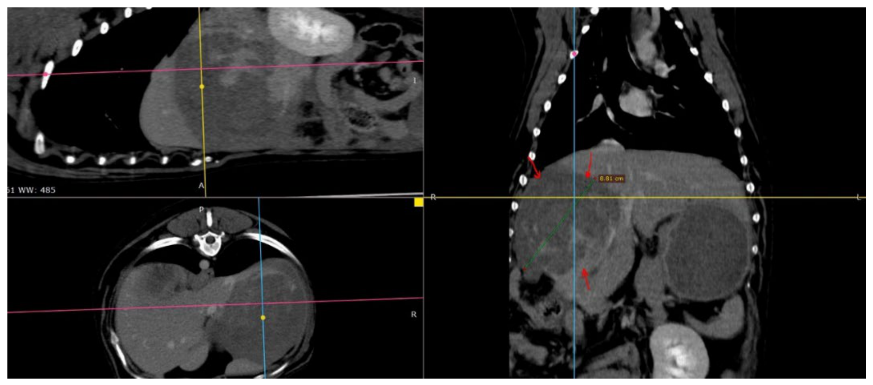
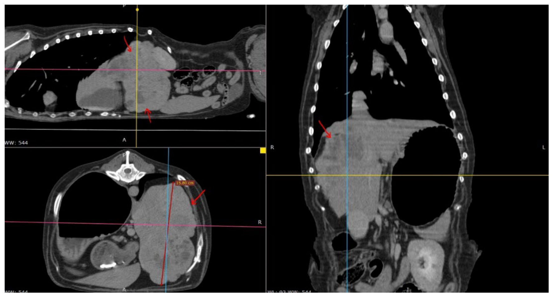
Computed tomography provided additional anatomical information regarding lesion size, lobe involvement and relationship with adjacent structures [20]. In Patient 4 (Figure 1), CT demonstrated a well-circumscribed solitary lesion subsequently diagnosed as lobular hyperplasia. In contrast, Patient 8 (Figure 2) showed a markedly larger mass occupying most of the right cranial abdomen and was later confirmed as hepatocellular carcinoma. Despite the clear difference in size and local mass effect, imaging findings alone were insufficient to reliably distinguish benign from malignant disease.
Figure 1.
Contrast-enhanced computed tomography (CT) images of Patient 4, diagnosed with lobular hyperplasia. Multiplanar reconstructions are shown, including transverse (left panels) and dorsal/coronal (right panels) views. Red arrows delineate a well-defined solitary hepatic mass located in the right medial liver lobe. The lesion measured approximately 9 cm in maximal diameter and showed no evidence of invasion of adjacent structures or detectable metastatic disease on CT examination.
Figure 2.
Contrast-enhanced computed tomography (CT) images of Patient 8, diagnosed with hepatocellular carcinoma. Multiplanar reconstructions are shown, including transverse (left panels) and dorsal/coronal (right panels) views. Red arrows delineate a large hepatic mass arising from the right medial liver lobe. The lesion measured approximately 16 cm in maximal diameter and occupied a substantial portion of the cranial abdomen, causing displacement of adjacent abdominal organs. No distant metastatic lesions were identified on the preoperative CT study.
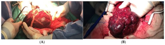
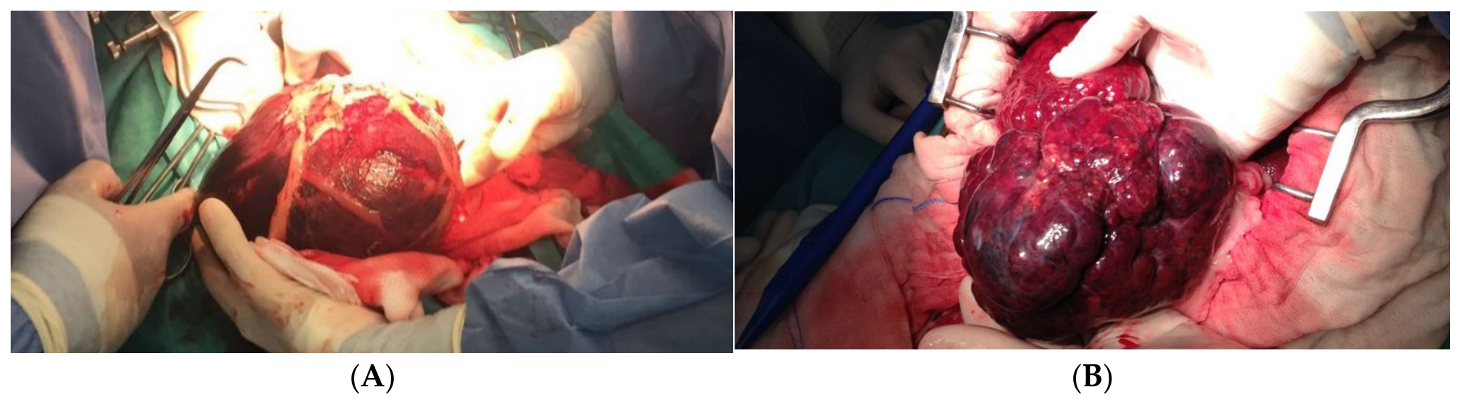
Representative macroscopic appearances of malignant and benign hepatic lesions are shown in Figure 3, illustrating that gross appearance alone may not reliably distinguish lesion type.
Figure 3.
(A) Patient 8 with a malignant neoplasm, hepatocellular carcinoma. (B) Patient 2 with benign neoplasms, nodular hyperplasia and hepatoma.
Tumor size varied substantially among both benign and malignant lesions in this series. Both benign and malignant lesions were observed across a wide range of lesion sizes, with large masses identified in both categories and some malignant lesions being comparatively small. However, given the limited number of cases and the heterogeneous nature of the diagnoses included, no conclusions regarding the relationship between lesion size, malignancy, prognosis, or biological behavior can be drawn from the present series. Although previous studies have investigated associations between prognosis and factors such as tumor morphology, histological type, and stage [11], the present study was not designed to evaluate these relationships.
Most lesions in this series involved the right medial hepatic lobe [21]. Although this distribution differs from previous reports describing a predominance of left-sided hepatic tumors [2,5], the small sample size precludes any conclusions regarding anatomical predilection. Consequently, this finding should be regarded as exploratory and interpreted with caution. The discrepancy may reflect referral bias, random variation, or true differences in lesion distribution among study populations. Regardless of the underlying cause, the present findings emphasize that clinically significant hepatic masses may arise in any liver lobe and should not be considered uncommon in the right hepatic divisions.
From a surgical standpoint, right-sided lesions are often more challenging to resect than those affecting the left liver lobes. The left lateral and medial lobes possess relatively long and accessible vascular pedicles, facilitating lobectomy, whereas the right-sided lobes have a broader attachment and lie in close proximity to major vascular structures, including the portal vein and caudal vena cava. Consequently, resection of masses involving the right medial or quadrate lobes requires meticulous hilar dissection and vascular control. The short hepatic veins draining directly into the vena cava may increase the risk of intraoperative hemorrhage, while the proximity of the gallbladder occasionally necessitates concurrent cholecystectomy to facilitate exposure or achieve complete excision. Several lesions involving the right medial lobe were surgically managed in the present series. However, given the small number of cases and the absence of a comparison group, no conclusions regarding comparative surgical risk among hepatic lobes can be drawn.
Two perioperative deaths occurred secondary to postoperative acute renal failure (ARF), representing a perioperative mortality rate of 20% in this cohort. While these complications were not directly attributable to the hepatic lesions, they highlight the systemic vulnerability of geriatric patients undergoing major hepatectomy. Although a necropsy in one case revealed pre-existing chronic interstitial nephritis, it is likely that the physiological response to surgical trauma acted as a triggering factor. During the surgery of large hepatic masses, transient hypotensive episodes can occur due to blood loss or compression of the caudal vena cava during neoplasm manipulation. In patients with limited renal reserve, even brief periods of renal hypoperfusion may contribute to postoperative renal injury. Furthermore, systemic inflammatory responses associated with major surgery may adversely affect renal microcirculation. However, because of the retrospective nature of this study, detailed information regarding intraoperative blood pressure, blood loss, surgical duration, anesthetic variables, perioperative fluid therapy, or sepsis was not consistently available. Consequently, the precise contribution of these factors to postoperative renal failure could not be determined. Future studies should evaluate these variables prospectively to better characterize perioperative risk factors in dogs undergoing hepatic surgery.
Survival times varied widely among the dogs included in this series. Differences in survival were observed among individual cases with different histopathological diagnoses, including hepatocellular carcinoma, benign hyperplastic lesions, undifferentiated sarcoma, and osteosarcoma. However, because of the small sample size, heterogeneous diagnoses, and absence of a comparison group, these observations should be interpreted solely as descriptive findings. No conclusions regarding prognosis, treatment efficacy, biological behavior, or the impact of surgery on survival can be drawn from the present data. A particularly noteworthy finding in this series was the diagnosis of a primary hepatic osteosarcoma (PHO) in Patient 1, an exceedingly rare entity in veterinary medicine with only a limited number of cases reported in the literature. Unlike its skeletal counterpart, primary extra-skeletal osteosarcoma of the liver is thought to arise from pluripotent mesenchymal cells undergoing malignant osteoblastic differentiation within the hepatic parenchyma. The diagnosis was established through histopathological evaluation and subsequently supported by immunohistochemical findings obtained during postmortem examination. Patient 1 survived 399 days following surgery and adjuvant treatment. Given the rarity of this tumor and the descriptive nature of the present study, no conclusions can be drawn regarding prognosis or treatment efficacy from this single case. Nevertheless, the case contributes additional clinical information on a rarely reported hepatic tumor type in dogs.
5. Limitations
This study has several important limitations. First, the number of cases included was small, limiting the ability to identify meaningful associations or draw robust conclusions regarding prognosis, lesion distribution, diagnostic accuracy, or treatment outcome. Second, the retrospective and single-institution design may have introduced referral and selection biases.
The cohort was also highly heterogeneous, including both benign and malignant lesions with distinct biological behaviors. Consequently, interpretation of survival times and disease-free intervals across the entire population should be undertaken with caution.
Diagnostic evaluation was not standardized. Abdominal ultrasonography was performed in most dogs, whereas computed tomography was only available in two cases. Similarly, fine-needle aspiration cytology was performed in only four dogs. Therefore, the study was not designed to formally assess the diagnostic performance of imaging modalities, cytology, or other diagnostic approaches. In addition, the small sample size and heterogeneous nature of the study population do not permit evaluation of lesion size as a predictor of biological behavior, anatomical predilection, comparative surgical risk among hepatic lobes, recurrence risk, or prognostic differences among histopathological diagnoses. Standardized histopathological reporting was not available for all cases, and variables such as surgical margin status, vascular or lymphatic invasion, tumor grading, and immunohistochemical characterization were not consistently recorded. Consequently, the completeness of excision, recurrence risk, and the prognostic significance of these histopathological features could not be systematically assessed.
Additional limitations include the lack of standardized follow-up protocols, incomplete clinicopathological data in some patients, the absence of formal statistical analysis, and the lack of a non-surgical comparison group. These factors preclude causal inferences regarding the impact of surgery on survival or clinical outcome. Accordingly, postoperative clinical improvement and survival times reported in this study should be interpreted solely as descriptive observations and not as evidence of therapeutic benefit.
Accordingly, the findings reported here should be considered descriptive and hypothesis-generating. Larger prospective studies using standardized diagnostic and follow-up protocols are needed to further clarify the diagnosis, biological behavior, and management of canine hepatic masses and lesions.
6. Conclusions
This retrospective case series describes the clinical presentation, diagnostic findings, surgical management and outcomes of ten dogs with surgically treated primary hepatic masses. Considerable heterogeneity was observed in histopathological diagnoses, imaging findings and clinical outcomes.
Discrepancies between preoperative assessment and intraoperative or histopathological findings occurred in several cases, highlighting the challenges associated with the diagnosis and surgical management of canine hepatic lesions.
Given the small sample size and methodological limitations, the results should be interpreted cautiously. Nevertheless, the study provides clinically relevant descriptive information and may serve as a basis for future investigations involving larger and more standardized populations.
Author Contributions
C.G.P.-S.: Resources, Project administration, Investigation, Supervision, Writing—original draft preparation, Writing—review and editing. S.C.-R.: Project administration, Investigation, Supervision, Writing—review and editing. E.R.G.-B.: Conceptualization, Resources, Methodology, Project administration, Funding acquisition. Investigation. B.C.: Writing—review and editing. F.R.-E.: Resources, Methodology, Project administration, Investigation, Writing—review and editing. All authors have read and agreed to the published version of the manuscript.
Funding
This article has been funded by the G. PASCH project; CANCERSICO Siftung, a charitable foundation under Liechtenstein law, holder of the tax identification number 0002217375, and it is registered at Neugasse 15, 9490 Vaduz, Liechtenstein, and it was partially supported by grant CGC/2025/12551 from the Cabildo de Gran Canaria (Las Palmas, Spain).
Institutional Review Board Statement
The study was conducted according to the guidelines of the Declaration of Helsinki and approved by the Institutional Review Board (or Ethics Committee) of ULPGC (protocol code OEBA-ULPGC 33/2020R1, approval date: 23 October 2020).
Informed Consent Statement
Informed consent was obtained from all subjects involved in the study.
Data Availability Statement
The data presented in this study are available from the corresponding author upon reasonable request. The data are not publicly available due to privacy and ethical restrictions.
Acknowledgments
The authors would like to thank CANCERSICO Siftung for supporting this article.
Conflicts of Interest
The authors declare no conflicts of interest.
References
- Guilford, W.G.; Strombeck, D.R.; Williams, D.A. (Eds.) Hepatic neoplasms. In Strombeck’s Small Animal Gastroenterology, 3rd ed.; Saunders Co. Ltd.: Saunders, NB, USA, 1996; pp. 847–859. [Google Scholar]
- Liptak, J.M.; Dernell, W.S.; Monnet, E.; Powers, B.E.; Bachand, A.M.; Kenney, J.G.; Withrow, S.J. Massive hepatocellular carcinoma in dogs: 48 cases (1992–2002). J. Am. Vet. Med. Assoc. 2004, 225, 1225–1230. [Google Scholar] [CrossRef] [Scilit] [PubMed]
- Liptak, J.M.; Dernell, W.S.; Withrow, S.J. Liver tumors in cats and dogs. Compend. Contin. Educ. Pract. Vet. 2004, 26, 50–56. [Google Scholar]
- Liptak, J.M. Hepatobiliary tumors. In Withrow & MacEwen’s Small Animal Clinical Oncology; Withrow, S.J., Vail, D.M., Eds.; Elsevier: Amsterdam, The Netherlands, 2007; pp. 483–490. [Google Scholar]
- Patnaik, A.K.; Hurvitz, A.I.; Lieberman, P.H. Canine hepatic neoplasms: A clinicopathologic study. Vet. Pathol. 1980, 17, 553–564. [Google Scholar] [CrossRef] [Scilit] [PubMed]
- Monnet, E.; Kudnig, S. Alimentary tract, liver, and pancreas. In Textbook of Small Animal Surgery; Slatter, D.H., Ed.; WB Saunders: Saunders, NB, USA, 1993; pp. 2378–2382. [Google Scholar]
- Terai, K.; Ishigaki, K.; Kagawa, Y.; Okada, K.; Yoshida, O.; Sakurai, N.; Heishima, T.; Asano, K. Clinical, diagnostic, and pathologic features and surgical outcomes of combined hepatocellular-cholangiocarcinoma in dogs: 14 cases (2009–2021). J. Am. Vet. Med. Assoc. 2022, 260, 1668–1674. [Google Scholar] [CrossRef] [Scilit] [PubMed]
- Balkman, C. Hepatobiliary neoplasia in dogs and cats. Vet. Clin. N. Am. Small Anim. Pract. 2009, 39, 617–625. [Google Scholar] [CrossRef] [Scilit] [PubMed]
- Kew, M.C. Hepatocellular carcinoma: Epidemiology and risk factors. J. Hepatocell. Carcinoma. 2014, 1, 115–125. [Google Scholar] [CrossRef] [Scilit] [PubMed]
- Cullen, J.M.; Stalker, M.J. Liver and Biliary System. In Jubb, Kennedy & Palmer’s Pathology of Domestic Animals, 6th ed.; Maxie, M.G., Ed.; Elsevier: Amsterdam, The Netherlands, 2016; Volume 2, pp. 258–352. [Google Scholar]
- Kinsey, J.R.; Gilson, S.D.; Hauptman, J.; Mehler, S.J.; May, L.R. Factors associated with long-term survival in dogs undergoing liver lobectomy as treatment for liver tumors. Can. Vet. J. 2015, 56, 598–604. [Google Scholar] [PubMed]
- Leela-Arporn, R.; Ohta, H.; Tamura, M.; Nagata, N.; Sasaoka, K.; Dermlim, A.; Nisa, K.; Morishita, K.; Sasaki, N.; Nakamura, K.; et al. Predictive factors of malignancy in dogs with focal liver lesions using clinical data and ultrasonographic features. J. Vet. Med. Sci. 2019, 81, 723–729. [Google Scholar] [CrossRef] [Scilit] [PubMed]
- Vörös, K.; Vrabély, T.; Papp, L.; Horváth, L.; Karsai, F. Correlation of ultrasonographic and pathomorphological findings in canine hepatic diseases. J. Small Anim. Pract. 1991, 32, 627–634. [Google Scholar] [CrossRef] [Scilit]
- Wormser, C.; Reetz, J.A.; Giuffrida, M.A. Diagnostic accuracy of ultrasound to predict the location of solitary hepatic masses in dogs. Vet. Surg. 2016, 45, 208–213. [Google Scholar] [CrossRef] [Scilit] [PubMed]
- Fukushima, K.; Kanemoto, H.; Ohno, K.; Takahashi, M.; Nakashima, K.; Fujino, Y.; Uchida, K.; Fujiwara, R.; Nishimura, R.; Tsujimoto, H. CT characteristics of primary hepatic tumors in dogs. Vet. Radiol. Ultrasound. 2012, 53, 252–257. [Google Scholar] [CrossRef] [Scilit] [PubMed]
- Rothuizen, J.; Twedt, D.C. Liver biopsy techniques. Vet. Clin. N. Am. Small Anim. Pract. 2009, 39, 469–480. [Google Scholar] [CrossRef] [Scilit] [PubMed]
- Hammer, A.S.; Sikkema, D.A. Hepatic neoplasia in the dog and cat. Vet. Clin. N. Am. Small Anim. Pract. 1995, 25, 419–435. [Google Scholar] [CrossRef] [Scilit] [PubMed]
- Roth, L. Comparison of liver cytology and biopsy diagnoses in dogs and cats: 56 cases. Vet. Clin. Pathol. 2001, 30, 35–38. [Google Scholar] [CrossRef] [Scilit] [PubMed]
- Lowseth, L.A.; Gerlach, R.F.; Gillett, N.A.; Muggenburg, B.A. Detection of serum alpha-fetoprotein in dogs with hepatic tumors. J. Am. Vet. Med. Assoc. 1991, 199, 1738–1741. [Google Scholar]
- Kurokawa, S.; Tanaka, T.; Yamazaki, H.; Noguchi, S.; Wada, Y.; Nishida, H.; Akiyoshi, H. Comparing the CT and MRI findings for canine primary hepatocellular lesions. Vet. Rec. 2022, 190, e1083. [Google Scholar] [CrossRef] [Scilit] [PubMed]
- Foster, H.D.; Matz, B.M.; Grimes, J.A.; Mankin, K.M.T.; Vinayak, A.; Liptak, J.M.; Linden, D.S.; Kennedy, E.J.; Korchek, K.A.; Kim, W.S. Outcomes and prognostic variables associated with right divisional hepatic lobectomies in 70 dogs. Vet. Surg. 2026, 55, 187–196. [Google Scholar] [CrossRef] [Scilit] [PubMed]

Disclaimer/Publisher’s Note: The statements, opinions and data contained in all publications are solely those of the individual author(s) and contributor(s) and not of MDPI and/or the editor(s). MDPI and/or the editor(s) disclaim responsibility for any injury to people or property resulting from any ideas, methods, instructions or products referred to in the content. |
© 2026 by the authors. Licensee MDPI, Basel, Switzerland. This article is an open access article distributed under the terms and conditions of the Creative Commons Attribution (CC BY) license.