


Simple Summary
Rabies threatens both animal and human health, highlighting the need for research tools that help scientists study the virus. Antibodies can serve as tools for detecting viral proteins, but their usefulness depends on how they recognize their targets. This study focused on the rabies virus matrix protein, which helps the virus form and release new particles. We produced six antibodies that recognized a laboratory-produced version of this protein. Although all six recognized the same small region, they differed in their dependence on individual protein building blocks for binding. This finding shows that antibodies recognizing the same region do not necessarily recognize it in the same way. The antibodies provide tools for further studies of matrix protein recognition and may support future work on protein detection. Their ability to recognize the protein in infected samples still needs to be established before diagnostic applications can be assessed.
Abstract
Rabies is a fatal zoonotic disease caused by rabies virus (RABV), with mortality reaching 100% after disease onset, posing a serious threat to public health. The RABV matrix (M) protein plays a critical role in virion assembly and budding. However, its immunogenicity and antigenic epitope structure have not yet been systematically elucidated. Identifying the amino acid residues involved in antibody recognition of the RABV M protein can improve understanding of its antigenicity. This study generated six monoclonal antibodies (mAbs) against recombinant RABV M protein and compared their recognition of a previously reported antigenic region. Western blotting and indirect immunofluorescence demonstrated that all six mAbs recognized recombinant M protein and M protein expressed in HEK293T cells. Sequential truncation analysis mapped the antibody-binding region to residues 21–35 (21PVSAPPYDDDLWLPP35), encompassing the previously reported 25PPYDDD30 epitope. Alanine scanning of residues 24–31 showed that D30 substitution reduced binding by all six mAbs, whereas L31 substitution reduced binding by the other five mAbs but not 14F8. Sequence analysis revealed that D30 and L31 were highly conserved among the analyzed strains despite variation within the epitope region. Structural mapping onto an AlphaFold 3 model predicted that this region was surface-exposed and adopted a coil conformation. This study identified shared and antibody-dependent contributions of individual residues to antibody recognition, extending the characterization of this antigenic region. The antibodies and binding data provide a foundation for further investigation of RABV M protein antigenicity. This study refines the antigenic epitope map of the RABV M protein, providing valuable tools for future studies of its structure and function.
1. Introduction
Rabies is an acute zoonosis characterized by progressive involvement of the central nervous system. Its causative agent, RABV, typically gains entry into the human body through damaged skin or mucosal surfaces. Clinical manifestations include agitation, hydrophobia, aerophobia, and progressive paralysis. Once clinical signs develop, the case fatality rate approaches 100% [1,2]. Although rabies is vaccine-preventable and modern inactivated cell-culture vaccines are safe and highly effective, conventional regimens generally require multiple doses [3]. Protection is assessed primarily through virus-neutralizing antibody responses; compared with replicating vaccine platforms, inactivated vaccines tend to induce more limited CD8+ T-cell and type-1 cellular immune responses [4]. Although effective vaccines are available, rabies persists in over 150 countries and territories, posing a major challenge to public health worldwide [5]. A study estimated that dog-mediated rabies causes approximately 59,000 human deaths annually [6].
RABV, a member of the species Lyssavirus rabies, belongs to the genus Lyssavirus in the family Rhabdoviridae [7]. Its genome is a linear, unsegmented, negative-sense single-stranded RNA molecule of approximately 11.9 kb [8]. The genome contains five major genes arranged in the order 3′-N-P-M-G-L-5′, encoding the nucleoprotein (N), phosphoprotein (P), matrix protein (M), glycoprotein (G), and large protein (L), respectively [5]. In commonly studied RABV strains, the matrix protein comprises 202 amino acids and has a predicted molecular mass of approximately 23 kDa, making it the smallest of the five major viral structural proteins [9]. The M protein coats and condenses the viral ribonucleoprotein complex and couples it to the viral envelope through interactions with the membrane-associated glycoprotein G [10]. M is also a major determinant of virion morphogenesis during budding, including formation of the characteristic bullet-shaped particle [11]. M-deficient recombinant RABV produces dramatically fewer cell-free infectious particles and forms predominantly elongated, rod-like particles rather than typical bullet-shaped virions [12]. Late-domain motifs in M, including the PPEY/PPxY and YL motifs, promote virion release by recruiting or interacting with host factors such as WW-domain proteins, NEDD4 and the ESCRT-I component TSG101 [13].
Beyond its roles in virion assembly and budding, the RABV M protein has several regulatory functions. Finke et al. demonstrated that M shifts viral RNA synthesis away from mRNA transcription and toward genome replication [14]. Recombinant-virus studies further indicate that M and G act jointly to modulate viral replication, pathogenicity and cell-to-cell spread, although these findings do not by themselves establish that direct M–G binding causes the altered pathogenic phenotype [15]. In cell-culture models, lyssavirus M proteins can promote TRAIL-dependent apoptosis through caspase-8 activation [16]. RABV M can also partially localize to mitochondria and activate the intrinsic apoptotic pathway by disrupting mitochondrial membrane potential and promoting the release of cytochrome c and apoptosis-inducing factors [17].
RABV strains can be classified into three clades based on the M gene [18]. However, the relationship between genetic variation and antigenic differences among these clades requires further clarification. Monoclonal antibodies (mAbs) with virus-neutralizing activity and high antigen specificity have applications in diagnosis and therapy. Antibodies targeting viral proteins also provide valuable tools for characterizing antigenic differences among strains [19]. Previous research showed that the M protein-specific mAb 3-9-16 recognizes an N-terminal linear epitope. Two M protein isoforms, designated Mα and Mβ, have been distinguished according to the accessibility of this epitope [20]. Furthermore, substitution of the amino acid at position 95 has been reported to influence RABV virulence [21]. Whether this substitution also affects antigenic properties remains unclear.
In 2019, Liu et al. mapped a linear epitope in the N-terminal region of the RABV M protein to residues 25–30 (25PPYDDD30) using five M-specific monoclonal antibodies. Sequence variation within this region was associated with strain-dependent antibody reactivity, and a D28G substitution abolished recognition by all five antibodies [22]. This study generated six monoclonal antibodies against recombinant RABV M protein and characterized their reactivity and the regions recognized by each antibody. The contribution of each amino acid to antibody binding was evaluated through alanine-scanning mutagenesis of the N-terminal epitope (24APPYDDDL31). Complementary sequence conservation analysis and structural mapping aided the interpretation of residue-specific differences. Collectively, these analyses extended previous characterization of this antigenic region by distinguishing recognition features shared across the antibody panel from residue dependencies specific to individual antibodies.
2. Materials and Methods
2.1. Cells, Plasmids, Bacterial Strains, and Experimental Animals
SP2/0 myeloma cells and human embryonic kidney 293T (HEK293T) cells were maintained at the Longhu Laboratory (hereafter referred to as our laboratory). The pET-28a(+), pET-32a(+), pEGFP-C1, and pGEX-6P-1 vectors, together with Rosetta (DE3) and Escherichia coli DH5α strains, were maintained in our laboratory. Female BALB/c mice aged 6–8 weeks were purchased from the Laboratory Animal Center of Zhengzhou University. The M protein sequence of the CVS-11 strain (GenBank:GQ918139.1) served as the template for synthesis by Tsingke Biotechnology Co., Ltd. (Beijing, China).
2.2. Expression of Recombinant RABV M Protein
The M gene fragment containing BamHI and XhoI restriction sites was amplified from the synthesized M gene sequence. The amplified fragment was cloned into the prokaryotic expression vector pET-28a(+) using T4 DNA ligase (2011A; Takara, Beijing, China). The sequence-verified recombinant plasmid was transformed into Rosetta (DE3) cells for M protein expression. Protein expression was induced in logarithmic-phase cultures by adding isopropyl β-D-1-thiogalactopyranoside (IPTG) to a final concentration of 1 mmol/L, followed by incubation at 16 °C for 12 h. Bacterial cells were disrupted by sonication using an ultrasonic cell disruptor (Sonic). Whole-cell samples and the supernatant and pellet fractions were prepared separately. Expression of the His-tagged recombinant protein was assessed by sodium dodecyl sulfate–polyacrylamide gel electrophoresis (SDS-PAGE). The M protein was expressed at high levels as inclusion bodies and subsequently subjected to denaturation and refolding. The purity of the resulting M protein was assessed by SDS-PAGE, and its identity was further examined by Western blotting. The concentration of the M protein was quantified with a bicinchoninic acid (BCA) assay kit (PC0020; Solarbio, Beijing, China).
2.3. Generation of RABV M Protein-Specific Monoclonal Antibodies
Purified recombinant M protein was emulsified with an equal volume of Freund’s complete adjuvant (F5881; Sigma, St. Louis, MO, USA). Two 6-week-old female BALB/c mice were immunized subcutaneously with 50 μg of protein per mouse. Two subsequent immunizations were administered at 2-week intervals using M protein emulsified with Freund’s incomplete adjuvant (F5506; Sigma, USA). Blood was obtained from the tail tips one week after the third immunization, and serum antibody titers were measured by indirect enzyme-linked immunosorbent assay (ELISA) using immunogen-coated plates. Two weeks after the third immunization, the mouse exhibiting the highest antibody titer was given an intraperitoneal booster of 50 μg of M protein without adjuvant.
Three days after the booster, the mouse was euthanized, and splenocytes were isolated. The splenocytes were fused with SP2/0 myeloma cells using polyethylene glycol 1500 (PEG-1500; Roche, Penzberg, Germany). The fused cells were cultured in hypoxanthine–aminopterin–thymidine (HAT) selective medium (H0262; Sigma) to select hybridomas. After 10 days of culture, antibody titers in the culture supernatants were determined by indirect ELISA. Hybridomas underwent at least three rounds of subcloning by limiting dilution. All clones obtained from the final round tested positive by indirect immunofluorescence assay (IFA). The resulting hybridoma clones were expanded and stored.
For monoclonal antibody production in ascites, healthy 6-week-old female BALB/c mice received an intraperitoneal injection of Freund’s incomplete adjuvant. After 5–7 days, monoclonal hybridoma cells in the logarithmic growth phase were injected intraperitoneally. Ascites fluid was collected 7 days after cell injection and centrifuged at 10,000× g for 20 min at 4 °C. The middle layer was carefully collected, aliquoted, and stored at −80 °C. Monoclonal antibodies were isolated by Protein A affinity chromatography using a commercial column (36401ES25; Yeasen, Shanghai, China) and eluted with glycine buffer at pH 3.0 (Solarbio, China). To ensure immediate neutralization, the eluate was collected into tubes containing 33 μL of Tris-HCl buffer (pH 8.5). Antibody isotypes were identified with a mouse monoclonal antibody isotyping kit (PK20002; Proteintech, Wuhan, China), while titers and binding affinities were assessed by indirect ELISA.
2.4. Determination of Monoclonal Antibody Affinity
Monoclonal antibody affinity was determined by indirect ELISA. Recombinant M protein was diluted in carbonate buffer (CBS) to 1 μg/mL and 2 μg/mL. The diluted antigen was added to 96-well plates at 100 μL per well and incubated at 37 °C for 2 h. The plates were washed three times with phosphate-buffered saline supplemented with Tween 20 (PBST) and then incubated with 5% skim milk for 2 h at 37 °C to block nonspecific binding. Purified monoclonal antibodies were prepared at 1 mg/mL and subjected to serial dilution, beginning with a 1:1000 dilution. The plates were sequentially incubated with the diluted monoclonal antibodies as primary antibodies and HRP-conjugated goat anti-mouse IgG as the secondary antibody. Following washing, color development was initiated by adding 3,3′,5,5′-tetramethylbenzidine (TMB) substrate and allowing the plates to incubate in the dark for 5 min. Stop solution was then added to terminate the reaction, and the optical density at 450 nm (OD450) was recorded with a microplate reader.
Double-reciprocal plots were generated by plotting 1/OD450 against 1/[Ab], where [Ab] denotes the molar concentration of the monoclonal antibody, and fitted by linear regression. For each antigen coating concentration, the antibody concentration required to achieve half-maximal binding was derived from the corresponding regression equation. The affinity constant (Kaff) was subsequently calculated using the following formula:
In this equation, n is the ratio between the two antigen coating concentrations, while [Ab′]t and [Ab]t denote the molar antibody concentrations required to achieve half-maximal binding at the corresponding coating concentrations.
2.5. Identification of Antigenic Epitopes in the RABV M Protein
To identify the RABV M protein epitopes recognized by the six monoclonal antibodies, four truncated M gene fragments were individually cloned into pET-32a(+) to generate recombinant prokaryotic expression plasmids. The corresponding truncated M proteins were expressed following induction, and protein samples were prepared. The epitopes were initially mapped based on the reactivity of the six monoclonal antibodies with each truncated protein.
The coding sequences corresponding to the initially mapped epitope regions were further truncated into five fragments. These fragments were individually cloned into pEGFP-C1 to generate recombinant eukaryotic expression plasmids. HEK293T cells were transfected with the recombinant plasmids using jetPRIME® reagent (Polyplus, Strasbourg, France), enabling expression of the respective truncated proteins. Cell samples were collected at 48 h post-transfection for Western blotting analysis. The epitope regions were further delineated based on the reactivity of the six monoclonal antibodies with each truncated protein.
2.6. Western Blotting Analysis
Following the addition of 5× SDS loading buffer, protein samples were denatured by heating in a boiling water bath for 10 min. The gel plates were mounted in the electrophoresis apparatus, and 1× SDS-PAGE running buffer was added. Samples were loaded at 10–20 μL per well. Electrophoresis was initially performed at 80 V, and the voltage was increased to 120 V when bromophenol blue entered the resolving gel. Following electrophoresis, proteins were electrotransferred onto polyvinylidene difluoride (PVDF) membranes.
Nonspecific binding was blocked by incubating the membranes in PBST containing 5% skim milk for 2 h at room temperature. Following three PBST washes, the membranes were probed overnight at room temperature with supernatants from positive hybridoma cultures as the primary antibody source. The membranes were subsequently washed with PBST and incubated for 1 h at room temperature with HRP-conjugated rabbit anti-mouse IgG (A21040; Abbkine, Wuhan, China). Following four washes with PBST, enhanced chemiluminescence (ECL) reagent (CW0049PM; Cwbio, Taizhou, China) was applied evenly to the membrane surface, and chemiluminescent signals were captured.
2.7. Indirect Immunofluorescence Assay
The recombinant expression plasmid was transfected into HEK293T cells. After 48 h of culture, the cells were washed twice with PBST, fixed with 4% paraformaldehyde for 20 min at room temperature, and then permeabilized with 0.3% Triton X-100 for 20 min. After three washes, cells were blocked with 5% skim milk at 37 °C for 1 h. Following five washes, cells were incubated with culture supernatants from the six positive hybridoma clones as primary antibodies at 37 °C for 1 h. After another five washes, cells were incubated with DyLight 594-conjugated goat anti-mouse IgG (1:200; A23410; Abbkine, China) at 37 °C for 45 min. Cells were washed five times and examined using an inverted fluorescence microscope (Ti2-U; Nikon, Tokyo, Japan).
2.8. Alanine Scanning
A panel of single-point mutant peptides was generated to identify residues critical for antibody recognition of the 24APPYDDDL31 epitope. Each amino acid was individually replaced with alanine, except for the native alanine, which was substituted with glycine (Table 1). The coding sequences of the mutant peptides were individually cloned into pGEX-6P-1, and protein samples were prepared following induced expression. Residues affecting antigen–antibody binding were identified based on the reactivity of the six monoclonal antibodies with each mutant peptide, with equal molar amounts of samples maintained throughout the identification process.
Table 1.
Alanine-scanning mutagenesis scheme for the epitope peptide 24APPYDDDL31.
2.9. Epitope Conservation Analysis and Structural Mapping
Conservation of the identified B-cell epitopes was evaluated by comparing M protein amino acid sequences from multiple RABV strains obtained from GenBank. The sequences were aligned with ClustalW (https://www.genome.jp/tools-bin/clustalw, accessed on 25 June 2026), and conservation patterns were visualized in Jalview. A three-dimensional model of the M protein was generated using AlphaFold 3, onto which the epitopes were mapped in PyMOL version 3.1.9 to examine their spatial distribution and predicted surface accessibility.
3. Results
3.1. Expression and Purification of Recombinant RABV M Protein
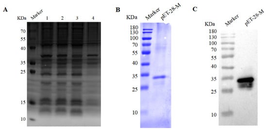
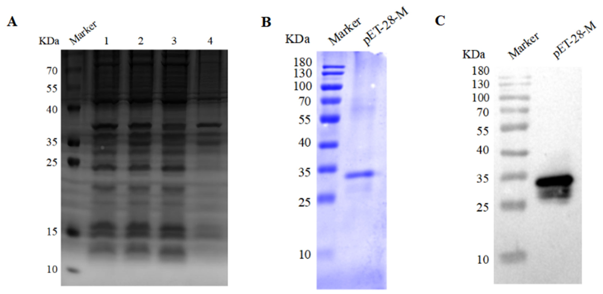
SDS-PAGE analysis of the supernatant and pellet fractions following sonication revealed that the recombinant M protein migrated at approximately 31 kDa and accumulated mainly in inclusion bodies (Figure 1A). The protein was subsequently subjected to urea-mediated denaturation and refolding. Western blotting showed that the purified recombinant M protein was recognized by a mouse anti-His antibody (Figure 1C).
Figure 1.
Production and characterization of recombinant RABV M protein. (A) SDS–PAGE analysis of recombinant M protein expressed using the pET-28a(+) vector. Lanes 1–4 represent the whole-cell lysate before induction, whole-cell lysate after induction, supernatant after sonication, and pellet after sonication, respectively. (B) SDS-PAGE analysis of refolded recombinant M protein. (C) Detection of purified recombinant M protein by Western blotting with an anti-His antibody.
3.2. Generation and Characterization of Monoclonal Antibodies Against the RABV M Protein
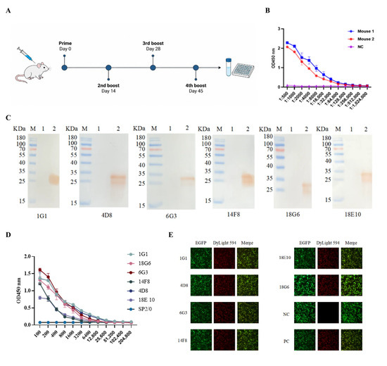
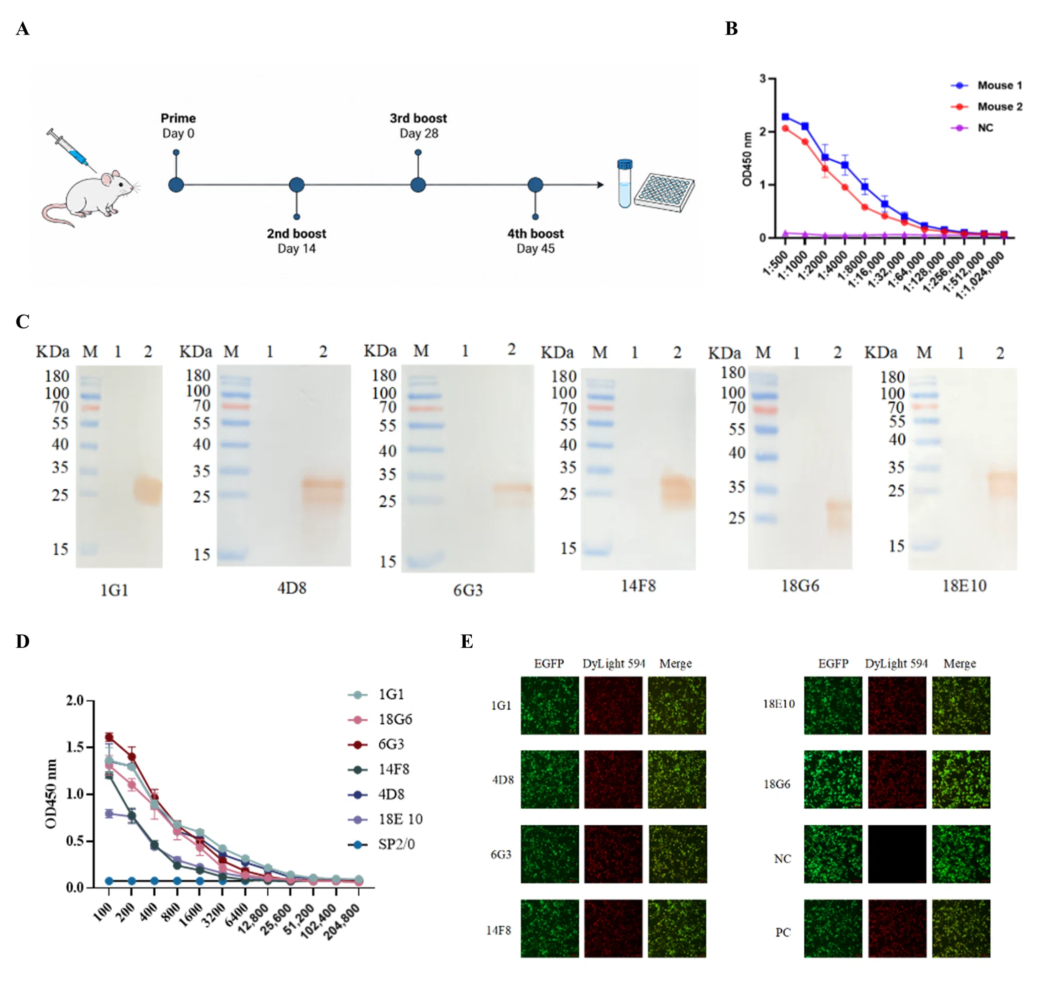
Following the immunization regimen outlined in Figure 2A, all mice developed serum antibody titers of 1:64,000 (Figure 2B), demonstrating a robust humoral response to the recombinant M protein.
Figure 2.
Generation and characterization of monoclonal antibodies against the RABV M protein. (A) Overview of the immunization regimen in BALB/c mice. (B) Indirect ELISA assessment of serum antibody titers. NC, negative control serum obtained from tail-tip blood of unimmunized mice. (C) Western blotting analysis of monoclonal antibody reactivity with prokaryotically expressed recombinant M protein. M, protein molecular weight marker (10–180 kDa); lane 1, pET-28a(+) empty-vector control; lane 2, pET-28-M recombinant protein. (D) Antibody titers in hybridoma culture supernatants determined by indirect ELISA. (E) Immunofluorescence analysis of monoclonal antibody recognition of recombinant M protein expressed from pEGFP-C1-M in HEK293T cells. Green fluorescence indicates EGFP expression, and red fluorescence indicates antibody staining. Culture supernatants from positive hybridomas were used as primary antibodies. NC, unimmunized mouse serum; PC, immunized mouse serum.
Six hybridoma cell lines secreting RABV M protein-specific monoclonal antibodies were obtained through cell fusion, limiting-dilution subcloning, and indirect ELISA screening. These cell lines were designated 1G1, 4D8, 6G3, 14F8, 18G6, and 18E10 (Figure 2D). Western blotting showed that all six monoclonal antibodies reacted with the prokaryotically expressed His-tagged recombinant M protein. The protein exhibited an apparent molecular weight of approximately 31 kDa (Figure 2C).
IFA revealed green EGFP fluorescence in pEGFP-C1-M-transfected cells. Specific red fluorescence was detected when culture supernatants from positive hybridomas were used as primary antibodies. No red fluorescence was observed with unimmunized mouse serum as the negative control, whereas immunized mouse serum produced red fluorescence as the positive control (Figure 2E). These results showed that monoclonal antibodies generated against prokaryotically expressed recombinant M protein recognized M protein expressed in HEK293T cells.
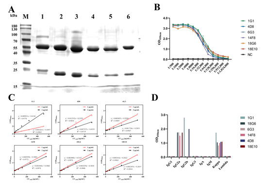
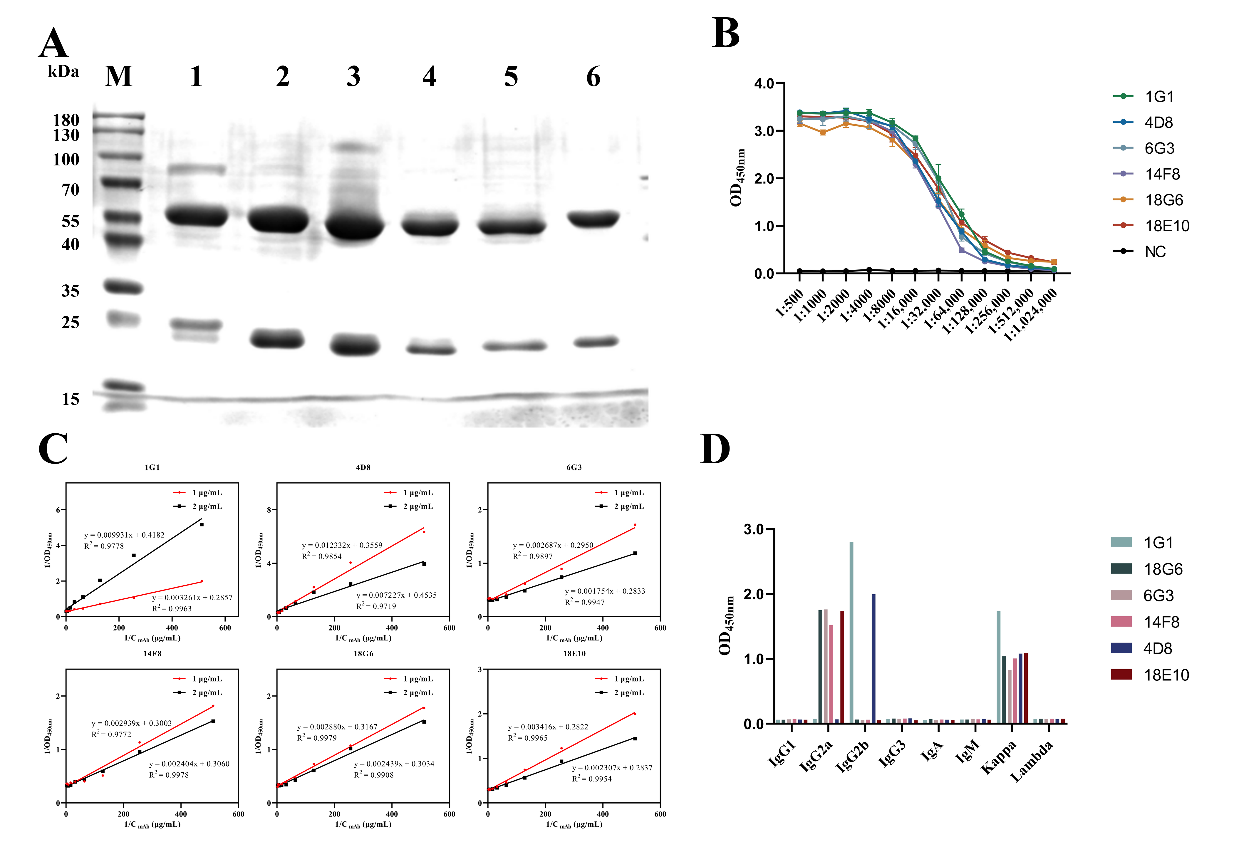
SDS-PAGE analysis of monoclonal antibodies purified from ascitic fluid revealed distinct bands at approximately 55 kDa for the heavy chains and 25 kDa for the light chains, consistent with a high degree of purity. (Figure 3A). Isotyping identified 1G1 and 4D8 as IgG2b and the remaining four monoclonal antibodies as IgG2a. All six antibodies possessed κ light chains (Figure 3D). After antibody concentrations were standardized to 1 mg/mL, indirect ELISA showed titers of 1:1,024,000 for 18E10 and 18G6 and 1:512,000 for the remaining four antibodies (Figure 3B). The affinity constants for 1G1, 4D8, and 6G3 were 2.02 × 10⁹, 1.921 × 10⁹, and 8.36 × 10¹⁰ L/mol, respectively. The corresponding values for 14F8, 18G6, and 18E10 were 1.143 × 10⁹, 6.145 × 10⁹, and 5.066 × 10⁹ L/mol, respectively (Figure 3C).
Figure 3.
Production and characterization of monoclonal antibodies. (A) Electrophoretic profiles of purified monoclonal antibodies resolved by SDS-PAGE. M, protein molecular weight marker (10–180 kDa); lanes 1–6, purified monoclonal antibodies 1G1, 4D8, 6G3, 14F8, 18G6, and 18E10, respectively. (B) Titers of the six monoclonal antibodies determined by indirect ELISA. (C) Affinities of the six monoclonal antibodies determined using an ELISA-based assay. (D) Isotyping of the six monoclonal antibodies.
3.3. Identification of M Protein B-Cell Epitopes Recognized by Monoclonal Antibodies
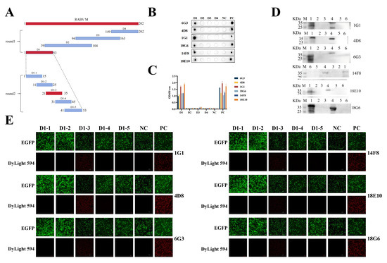
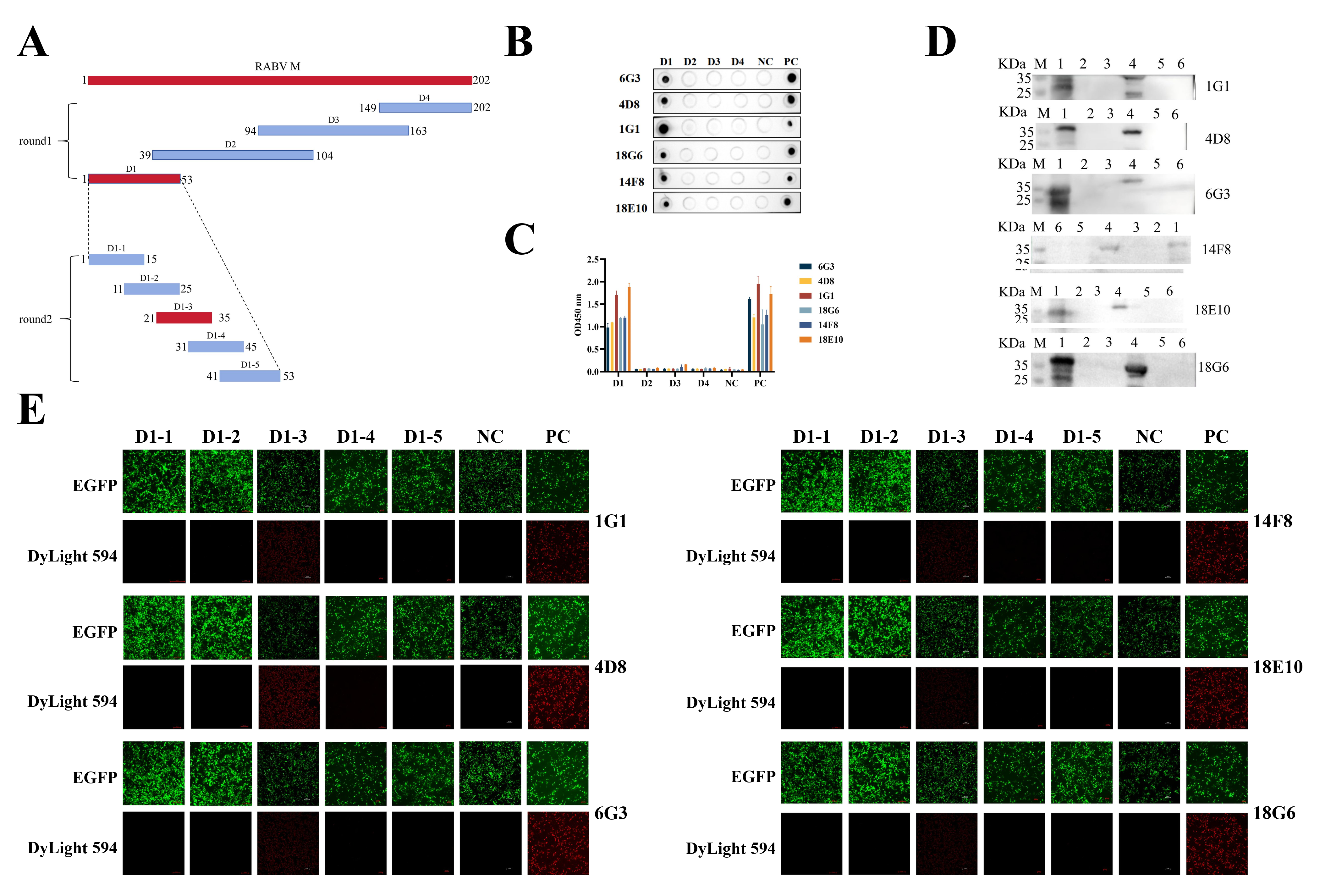
The M protein epitopes recognized by the monoclonal antibodies were mapped through two rounds of truncation analysis, as illustrated in Figure 4A. Dot blot and indirect ELISA analyses showed that the monoclonal antibodies specifically recognized the first-round fragment D1, encompassing amino acids 1–53 (Figure 4B,C). D1 was subsequently truncated further, and the corresponding coding sequences were cloned into pEGFP-C1 to refine epitope localization. Western blotting and Indirect Immunofluorescence Assay showed that the monoclonal antibodies recognized the second-round fragment D1-3, encompassing amino acids 21–35 (Figure 4D). These results localized the recognized epitopes to the D1-3 region.
Figure 4.
Mapping of the RABV M protein epitope region recognized by the mAbs. (A) Diagram of truncated M protein constructs employed in epitope mapping. Fragments recognized by the mAbs are highlighted in red. (B,C) Reactivity of the mAbs with the first-round truncated proteins D1–D4 assessed by Dot blot (B) and indirect ELISA (C). PC, full-length M protein expressed from pET-32a(+); NC, the empty pET-32a(+) vector expression control; D1, D2, D3, and D4, the corresponding truncated proteins expressed from pET-32a(+), respectively. (D) Western blotting analysis of mAb reactivity with the second-round truncated proteins. M, protein molecular weight marker (10–180 kDa); Lanes 1–6 correspond to the full-length M protein and truncated proteins D1-1, D1-2, D1-3, D1-4, and D1-5, respectively. (E) Indirect immunofluorescence assay (IFA) analysis of mAb reactivity with the second-round truncated proteins. PC, cells transfected with pEGFP-C1 encoding full-length M protein; NC, cells transfected with the empty pEGFP-C1 vector; D1-1, D1-2, D1-3, D1-4, and D1-5, cells transfected with pEGFP-C1 encoding the corresponding truncated proteins, respectively.
3.4. Identification of Key Amino Acid Residues Within the Epitope
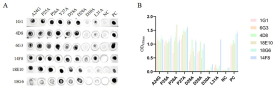
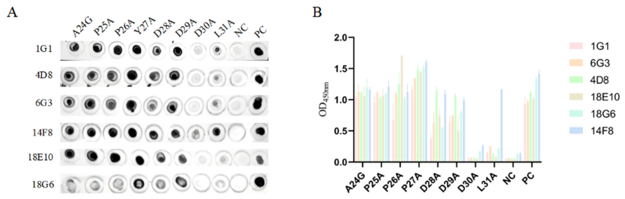
The contribution of individual residues in the 24APPYDDDL31 epitope to monoclonal antibody binding was assessed by alanine-scanning mutagenesis. Dot blot and indirect ELISA analyses showed that substitution of D30 significantly reduced peptide binding to all six monoclonal antibodies. Substitution of L31 also reduced binding to varying degrees for the other five antibodies, with 14F8 being the exception (Figure 5). These findings suggest that D30 and L31 may play key roles in antigen–antibody binding.
Figure 5.
Identification of critical residues by alanine scanning. (A) Identification of critical residues by dot blot. (B) Identification of critical residues by indirect ELISA.
3.5. Conservation Analysis and Structural Visualization of the Antigenic Epitope
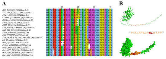
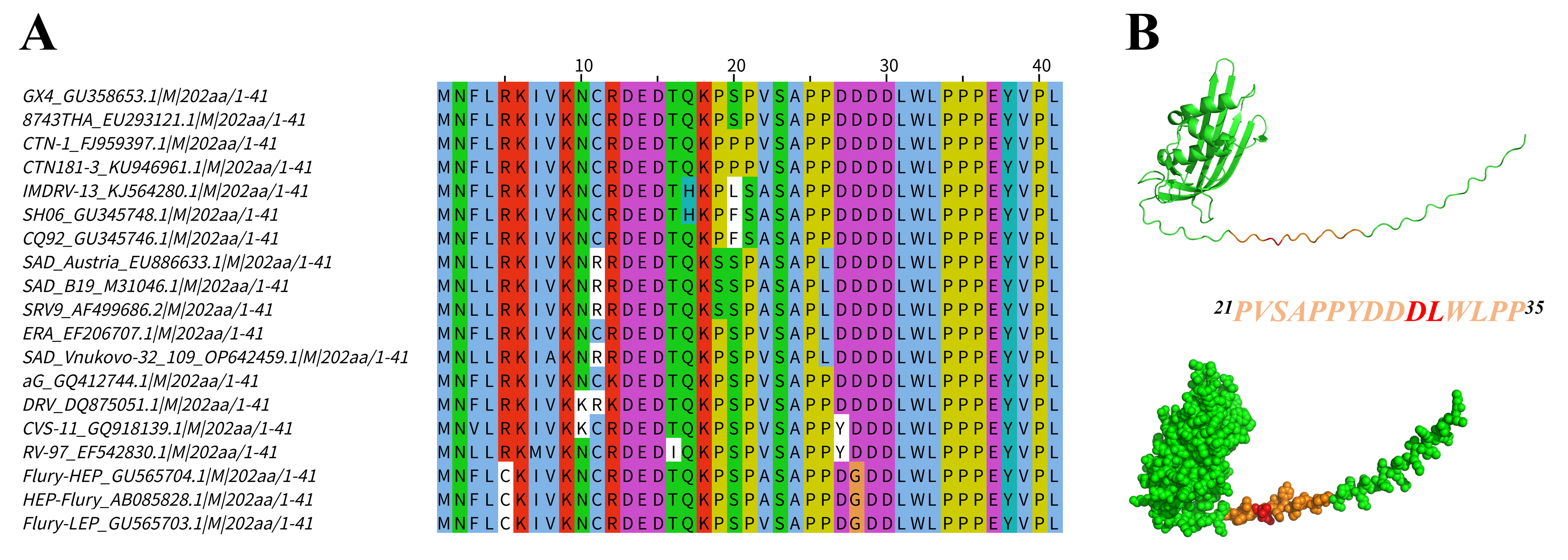
Conservation of the identified epitope was evaluated by aligning M protein sequences from laboratory-adapted RABV strains and representative street isolates. The epitope 21PVSAPPYDDDLWLPP35 exhibited low overall sequence conservation among the analyzed strains. However, the key residues D30 and L31 were highly conserved across all strains included in the analysis (Figure 6A).
Figure 6.
Sequence conservation analysis and structural visualization of the identified epitope. (A) Multiple sequence alignment of the epitope 21PVSAPPYDDDLWLPP35 performed using ClustalW, with the alignment results visualized. The red box indicates the epitope region recognized by the monoclonal antibodies. (B) Mapping of the epitope onto the M protein structure predicted by AlphaFold 3. The epitope region (amino acids 21–35) is shown in brown, and key residues are highlighted in red.
The identified linear epitope was mapped onto the M protein structure predicted by AlphaFold 3 using PyMOL. The model predicted that the epitope was surface-exposed and adopted a random-coil conformation (Figure 6B). This surface exposure may facilitate antibody recognition and binding. However, these predictions neither establish direct antibody–antigen contacts nor confirm the structural roles of D30 and L31. Further experimental validation is required to address these points.
4. Discussion
Rabies is a severe zoonotic infection associated with an exceptionally poor prognosis once clinical symptoms emerge, with the case fatality rate approaching 100% at this stage [23,24]. The global occurrence of rabies poses a significant threat to animal health and public health worldwide. In addition to its roles in viral particle assembly and release through budding, the RABV M protein inhibits nuclear transport, regulates host transcriptional activity, and promotes apoptosis. It also participates in host immune responses through interactions with the G protein [15].
In this study, the M gene sequence of the RABV CVS-11 strain was used to construct the prokaryotic expression vector pET-28-M. The resulting recombinant M protein was used to immunize healthy BALB/c mice. Cell fusion and limiting-dilution subcloning yielded six hybridoma cell lines that stably secreted M protein-specific monoclonal antibodies. However, the antibody variable regions were not sequenced; therefore, whether these antibodies represent genetically distinct clones could not be determined. Western blotting and IFA showed that all six antibodies recognized recombinant M protein, supporting their potential use as tools for its detection. However, their specificity for native M protein has not yet been verified. Evidence of their recognition capability therefore remains limited to recombinant protein systems.
Monoclonal antibodies are valuable tools for epitope mapping owing to their high specificity for antigen recognition. Studies of RABV monoclonal antibodies were reported as early as 1978 [25]. Subsequently, antibodies targeting the RABV P, N, and G proteins were generated and used for epitope identification. In 2005, two G protein-specific monoclonal antibodies were used to identify 226KLCGVL231 as the minimal binding region [26]. A study published in 2019 identified 25PPYDDD30 as the minimal region recognized by M protein-specific monoclonal antibodies [22]. The epitope region mapped in this study, 21PVSAPPYDDDLWLPP35, encompasses this previously reported sequence, indicating overlap between the antibody recognition regions identified in the two studies.
Sequence alignment revealed low overall conservation of the mapped epitope region among the analyzed RABV strains. However, D30 and L31 were highly conserved across all strains included in the analysis. Guided by the previously reported 25PPYDDD30 epitope and sequence conservation analysis, this study performed systematic alanine scanning of the 24APPYDDDL31 region. Dot Blot and indirect ELISA analyses showed that substitution of D30 significantly reduced antigen binding by all six monoclonal antibodies. Substitution of L31 affected binding to varying degrees for the other five antibodies, with 14F8 being the exception. These findings suggest that D30 contributes to binding by all six antibodies, whereas the contribution of L31 may vary among antibodies. Nevertheless, the molecular mechanisms underlying the involvement of these residues in antibody recognition remain to be elucidated.
This study produced recombinant RABV M protein using a prokaryotic expression system and generated six monoclonal antibodies that recognized the recombinant protein. Although the mapped linear epitope region overlapped with a previously reported epitope, alanine scanning further clarified the contributions of D30 and L31 to antibody binding. However, whether these residues directly contact the antibody remains to be established. These findings extend our understanding of RABV M protein antigen recognition and provide antibody tools and experimental evidence for further studies of its structure, function, and antigenicity.
5. Conclusions
This study mapped a linear B-cell epitope of the RABV M protein to the 21PVSAPPYDDDLWLPP35 region using the generated specific monoclonal antibodies. Alanine scanning further indicated that D30 and L31 can affect antigen–antibody binding. These findings provide experimental evidence and antibody tools for further investigations into the structure, function, and antigenicity of the RABV M protein.
Supplementary Materials
The following supporting information can be downloaded at: https://www.mdpi.com/article/10.3390/vetsci13111263/s1. Table S1. Rabies virus (RABV, Lyssavirus genotype 1) sequences used in this study (n = 19).
Author Contributions
Writing—original draft, Methodology, Investigation, Formal analysis, Data curation, Z.Z. and Y.C. (Yanhui Chen); Writing—review & editing, H.L.; Investigation, Formal analysis, Data curation, J.Z.: Investigation, Formal analysis, Data curation, Y.C. (Yumei Chen); Investigation, Formal analysis, Data curation, X.Z.; Investigation, Formal analysis, Data curation, C.L.; Formal analysis, Data curation, E.L.; Formal analysis, Data curation, S.W.; Writing—review & editing, Supervision, Funding acquisition, Conceptualization, A.W. All authors have read and agreed to the published version of the manuscript.
Funding
This research was funded by Major Science and Technology Projects in Henan Province (grant 241110310100) and the Major Research Program of Longhu Laboratory of Advanced Immunology (grant LHLab_ZD20230007).
Institutional Review Board Statement
This study was approved by the Animal Ethics Committee of the School of Life Sciences, Zhengzhou University, and was performed in accordance with animal ethics guidelines and approved protocols. The Animal Ethics Committee Approval Number was LHLab-HNZD-2023001 (23 January 2024).
Informed Consent Statement
Not applicable.
Data Availability Statement
The original contributions presented in this study are included in the article/supplementary material. Further inquiries can be directed to the corresponding author(s).
Acknowledgments
The authors would like to acknowledge the Major Science and Technology Projects in Henan Province and the Major Research Program of Longhu Laboratory of Advanced Immunology.
Conflicts of Interest
The authors declare no conflicts of interest.
References
- Wilde, H.; Hemachudha, T.; Tantawichien, T.; Khawplod, P. Rabies and other lyssavirus diseases. Lancet 2004, 363, 1906. [Google Scholar] [CrossRef] [Scilit] [PubMed]
- Singh, R.; Singh, K.P.; Cherian, S.; Saminathan, M.; Kapoor, S.; Manjunatha Reddy, G.B.; Panda, S.; Dhama, K. Rabies—Epidemiology, pathogenesis, public health concerns and advances in diagnosis and control: A comprehensive review. Vet. Q. 2017, 37, 212–251. [Google Scholar] [CrossRef] [Scilit] [PubMed]
- Ravish, H.S.; Khobragade, A.; Satapathy, D.; Gupta, M.; Kumar, S.; Bhomia, V.; Ramasubramanian, V.; Desai, M.; Agrawal, A.D. Safety and Immunogenicity of a novel three-dose recombinant nanoparticle rabies G protein vaccine administered as simulated post exposure immunization: A randomized, comparator controlled, multicenter, phase III clinical study. Hum. Vaccines Immunother. 2021, 17, 4239–4245. [Google Scholar] [CrossRef] [Scilit] [PubMed]
- Zhu, S.; Guo, C. Rabies Control and Treatment: From Prophylaxis to Strategies with Curative Potential. Viruses 2016, 8, 279. [Google Scholar] [CrossRef] [Scilit] [PubMed]
- Fooks, A.R.; Cliquet, F.; Finke, S.; Freuling, C.; Hemachudha, T.; Mani, R.S.; Müller, T.; Nadin-Davis, S.; Picard-Meyer, E.; Wilde, H.; et al. Rabies. Nat. Rev. Dis. Primers 2017, 3, 17091. [Google Scholar] [CrossRef] [Scilit] [PubMed]
- Hampson, K.; Coudeville, L.; Lembo, T.; Sambo, M.; Kieffer, A.; Attlan, M.; Barrat, J.; Blanton, J.D.; Briggs, D.J.; Cleaveland, S.; et al. Correction: Estimating the Global Burden of Endemic Canine Rabies. PLoS Neglected Trop. Dis. 2015, 9, e0003786. [Google Scholar] [CrossRef] [Scilit] [PubMed]
- Walker, P.J.; Bejerman, N.; Blasdell, K.R.; Debat, H.; Dietzgen, R.G.; Fooks, A.R.; Freitas-Astúa, J.; Garver, K.; Kondo, H.; Ramos-González, P.L.; et al. ICTV Virus Taxonomy Profile: Rhabdoviridae 2026. J. Gen. Virol. 2026, 107, 002255. [Google Scholar] [CrossRef] [Scilit] [PubMed]
- Tordo, N.; Poch, O.; Ermine, A.; Keith, G.; Rougeon, F. Completion of the rabies virus genome sequence determination: Highly conserved domains among the L (polymerase) proteins of unsegmented negative-strand RNA viruses. Virology 1988, 165, 565–576. [Google Scholar] [CrossRef] [Scilit] [PubMed]
- Wang, P.-H.; Xing, L. The roles of rabies virus structural proteins in immune evasion and implications for vaccine development. Can. J. Microbiol. 2024, 70, 461–469. [Google Scholar] [CrossRef] [Scilit] [PubMed]
- Cai, X.; Zhou, K.; Alvarez-Cabrera, A.L.; Si, Z.; Wang, H.; He, Y.; Li, C.; Zhou, Z.H. Structural heterogeneity of the rabies virus virion. Viruses 2024, 16, 1447. [Google Scholar] [CrossRef] [Scilit] [PubMed]
- Itakura, Y.; Tabata, K.; Saito, T.; Intaruck, K.; Kawaguchi, N.; Kishimoto, M.; Torii, S.; Kobayashi, S.; Ito, N.; Harada, M.; et al. Morphogenesis of bullet-shaped rabies virus particles regulated by TSG101. J. Virol. 2023, 97, e0043823. [Google Scholar] [CrossRef] [Scilit] [PubMed]
- Mebatsion, T.; Weiland, F.; Conzelmann, K.-K. Matrix protein of rabies virus is responsible for the assembly and budding of bullet-shaped particles and interacts with the transmembrane spike glycoprotein G. J. Virol. 1999, 73, 242–250. [Google Scholar] [CrossRef] [Scilit] [PubMed]
- Yuan, Y.; Fang, A.; Wang, Z.; Chen, H.; Fu, Z.F.; Zhou, M.; Zhao, L. The matrix protein of lyssavirus hijacks autophagosome for efficient egress by recruiting NEDD4 through its PPxY motif. Autophagy 2024, 20, 1723–1740. [Google Scholar] [CrossRef] [Scilit] [PubMed]
- Finke, S.; Mueller-Waldeck, R.; Conzelmann, K.-K. Rabies virus matrix protein regulates the balance of virus transcription and replication. J. Gen. Virol. 2003, 84, 1613–1621. [Google Scholar] [CrossRef] [Scilit] [PubMed]
- Pulmanausahakul, R.; Li, J.; Schnell, M.J.; Dietzschold, B. The glycoprotein and the matrix protein of rabies virus affect pathogenicity by regulating viral replication and facilitating cell-to-cell spread. J. Virol. 2008, 82, 2330–2338. [Google Scholar] [CrossRef] [Scilit] [PubMed]
- Kassis, R.; Larrous, F.; Estaquier, J.; Bourhy, H. Lyssavirus matrix protein induces apoptosis by a TRAIL-dependent mechanism involving caspase-8 activation. J. Virol. 2004, 78, 6543–6555. [Google Scholar] [CrossRef] [Scilit] [PubMed]
- Wang, S.; Zhou, H.; Zhang, X.; Cao, J.; Lei, T.; Chen, M.; Zhang, Y.; Xu, Y. Dynamic bidirectional regulation between Stk38 and rabies virus M protein coordinates apoptosis progression during neurotropic infection. Int. J. Biol. Macromol. 2025, 315, 144398. [Google Scholar] [CrossRef] [Scilit] [PubMed]
- Wu, H.; Wang, L.; Tao, X.; Li, H.; Rayner, S.; Liang, G.; Tang, Q. Genetic diversity and molecular evolution of the rabies virus matrix protein gene in China. Infect. Genet. Evol. 2013, 16, 248–253. [Google Scholar] [CrossRef] [Scilit] [PubMed]
- Bi, Z.; Xia, X.; Wang, Y.; Mei, Y. Development and characterization of neutralizing monoclonal antibodies against canine distemper virus hemagglutinin protein. Microbiol. Immunol. 2015, 59, 202–208. [Google Scholar] [CrossRef] [Scilit] [PubMed]
- Ameyama, S.; Toriumi, H.; Takahashi, T.; Shimura, Y.; Nakahara, T.; Honda, Y.; Mifune, K.; Uchiyama, T.; Kawai, A. Monoclonal antibody #3-9-16 recognizes one of the two isoforms of rabies virus matrix protein that exposes its N-terminus on the virion surface. Microbiol. Immunol. 2003, 47, 639–651. [Google Scholar] [CrossRef] [Scilit] [PubMed]
- Ito, N.; Mita, T.; Shimizu, K.; Ito, Y.; Masatani, T.; Nakagawa, K.; Yamaoka, S.; Abe, M.; Okadera, K.; Minamoto, N.; et al. Amino acid substitution at position 95 in rabies virus matrix protein affects viral pathogenicity. J. Vet. Med. Sci. 2011, 73, 1363–1366. [Google Scholar] [CrossRef] [Scilit] [PubMed]
- Liu, J.; Zhao, W.; He, W.; Wang, N.; Su, J.; Ji, S.; Chen, J.; Wang, D.; Zhou, J.; Su, S. Generation of Monoclonal Antibodies against Variable Epitopes of the M Protein of Rabies Virus. Viruses 2019, 11, 375. [Google Scholar] [CrossRef] [Scilit] [PubMed]
- Davis, B.M.; Rall, G.F.; Schnell, M.J. Everything You Always Wanted to Know About Rabies Virus (But Were Afraid to Ask). Annu. Rev. Virol. 2015, 2, 451–471. [Google Scholar] [CrossRef] [Scilit] [PubMed]
- Rupprecht, C.E.; Hanlon, C.A.; Hemachudha, T. Rabies re-examined. Lancet Infect. Dis. 2002, 2, 327–343. [Google Scholar] [CrossRef] [Scilit] [PubMed]
- Wiktor, T.J.; Koprowski, H. Monoclonal antibodies against rabies virus produced by somatic cell hybridization: Detection of antigenic variants. Proc. Natl. Acad. Sci. USA 1978, 75, 3938–3942. [Google Scholar] [CrossRef] [Scilit] [PubMed]
- Marissen, W.E.; Kramer, R.A.; Rice, A.; Weldon, W.C.; Niezgoda, M.; Faber, M.; Slootstra, J.W.; Meloen, R.H.; Clijsters-van der Horst, M.; Visser, T.J.; et al. Novel Rabies Virus-Neutralizing Epitope Recognized by Human Monoclonal Antibody: Fine Mapping and Escape Mutant Analysis. J. Virol. 2005, 79, 4672–4678. [Google Scholar] [CrossRef] [Scilit] [PubMed]

Disclaimer/Publisher’s Note: The statements, opinions and data contained in all publications are solely those of the individual author(s) and contributor(s) and not of MDPI and/or the editor(s). MDPI and/or the editor(s) disclaim responsibility for any injury to people or property resulting from any ideas, methods, instructions or products referred to in the content. |
© 2026 by the authors. Licensee MDPI, Basel, Switzerland. This article is an open access article distributed under the terms and conditions of the Creative Commons Attribution (CC BY) license.