


Simple Summary
Dogs with atopic dermatitis often develop recurrent bacterial and yeast infections of the skin and ears. Biofilms can make these infections harder to control, while bacterial quorum sensing coordinates some of the behaviors involved in infection. Propolis is a bee-collected resin containing compounds with reported antibacterial, antibiofilm, and quorum-sensing-related activity. Most findings come from laboratory strains or organisms isolated from humans. Studies using canine isolates have mainly measured growth inhibition. A controlled study of one propolis ear-drop formulation reported short-term improvement in dogs with otitis externa without assessing quorum sensing or biofilms. This review separates direct canine evidence from findings in other organisms and examines how propolis chemistry and assay design affect interpretation. It also considers computational studies, which can suggest targets but cannot demonstrate biological activity on their own. A proposed research framework links chemical characterization, canine microbiology, computational hypotheses, and topical safety assessment. Further evidence is needed before propolis can be recommended as an adjunct for canine skin or ear infections.
Abstract
Background/Objectives: Secondary bacterial and yeast infections contribute to antimicrobial use in dogs with atopic dermatitis. This review examines whether propolis modulates quorum sensing (QS) or biofilm-associated traits relevant to canine skin and ear infections. Methods: The narrative synthesis was informed by a structured PubMed search on 12 October 2026, sensitivity searches, and targeted retrieval. Results: Propolis preparations and constituents altered QS-associated phenotypes and biofilm endpoints, mostly in biosensors and non-canine models. Studies of canine isolates mainly measured growth inhibition. No study identified in this review directly evaluated propolis-mediated modulation of the accessory gene regulator (agr) system in canine Staphylococcus pseudintermedius. A controlled canine otitis trial reported short-term improvement with one formulation and did not assess QS or biofilms. Conclusions: Structure prediction for AgrA, AgrC, and autoinducing peptide–AgrC complexes could guide functional studies that assess toxin production and biofilm persistence together. Clinical evaluation as a QS-directed adjunct requires reproducible activity in canine isolates at locally tolerated exposures.
1. Introduction
Canine atopic dermatitis (cAD) is a common, genetically predisposed, inflammatory, and pruritic skin disease with characteristic clinical features [1,2]. Its diagnosis rests on compatible historical and clinical findings and the exclusion of other pruritic dermatoses [1,2]. Skin barrier impairment and a dysregulated immune response promote cutaneous dysbiosis and allergen penetration [3]. In a longitudinal 16S rRNA study of dogs with cAD, disease flare-ups were associated with reduced bacterial diversity and increased proportions of Staphylococcus—S. pseudintermedius in particular—and Corynebacterium species; antimicrobial therapy restored diversity in parallel with improvement in clinical severity, transepidermal water loss, and skin pH [4]. Superficial bacterial folliculitis, usually caused by S. pseudintermedius, develops on the background of altered barrier function and underlying disease, is frequently recurrent, and often requires repeated treatment; the emergence of methicillin-resistant S. pseudintermedius (MRSP) has made optimal antimicrobial stewardship in these patients a priority [5]; in Hungary, 19.3% of 243 clinical canine skin isolates of S. pseudintermedius were methicillin-resistant and 50.6% were multidrug-resistant [6]. Current guidelines recommend cytology before any antimicrobial use and topical therapy alone as the treatment of choice for surface and superficial pyoderma [7], while the management of cAD flares starts with the identification and elimination of flare factors, including infections [8]. Otitis externa is a frequent companion problem in which P. aeruginosa, although not a typical member of the canine ear microbiota, is commonly isolated from chronic cases and is notoriously difficult to treat [9,10].
A common denominator of these secondary infections is biofilm formation. Among 237 coagulase-positive staphylococci from companion-animal skin infections, biofilm production was detected in 51.0% of S. pseudintermedius, 94.6% of S. aureus, and 88.9% of S. coagulans isolates, with NaCl supplementation acting as an inducer or inhibitor depending on the species [11]. The intercellular adhesion (ica) operon encoding polysaccharide intercellular adhesin/poly-N-acetylglucosamine (PIA/PNAG) was detected in all S. pseudintermedius and S. aureus isolates, and an in silico search confirmed the ica genes in 106 of 107 complete S. pseudintermedius genomes and in all >800 S. aureus genomes, whereas none were found in the two available S. coagulans genomes [11]. In a multinational collection of 253 P. aeruginosa isolates from canine otitis externa, 82% were strong biofilm formers and 25% were resistant to fluoroquinolones [12], and published estimates of biofilm formation among otitis isolates range from 40% to 95% [9]. Malassezia pachydermatis also forms biofilms: 43 of 52 isolates (82.7%) from the ear canals of healthy dogs were biofilm producers. Among these 43 isolates, mature-biofilm MICs exceeded the study-defined epidemiological cut-off values for itraconazole in 43 isolates (100%), posaconazole in 41 (95.3%), and voriconazole in 36 (83.7%) [13]. These findings indicate markedly reduced in vitro susceptibility in the biofilm state, in line with the general mechanisms of biofilm-associated tolerance [14,15], and should not be interpreted as validated clinical resistance categories for canine infection.
The problem has a clear One Health dimension. Of 35 genome-sequenced canine otitis isolates of P. aeruginosa, two belonged to the high-risk sequence types (STs) ST111 and ST244, associated with antimicrobial-resistant nosocomial infections in humans [12]; S. pseudintermedius is implicated in a growing number of human infections, and one third of 160 clinical canine isolates from Texas were methicillin-resistant [16]. Antimicrobial resistance is therefore a shared challenge for human and veterinary medicine, and multidrug-resistant staphylococci, together with P. aeruginosa (not tested for susceptibility), have been recovered from the environment of small-animal hospitals [17], which motivates the search for natural, sustainable alternatives [18].
In many bacterial pathogens, biofilm development and virulence factor production are coordinated by quorum sensing (QS), a cell-density-dependent communication system based on diffusible autoinducers. Quorum-sensing inhibitors (QSIs) have been proposed as anti-pathogenic drugs that attenuate virulence without directly killing bacteria [19]; a synthetic furanone provided an early proof of concept by blocking QS-controlled gene expression in P. aeruginosa, increasing biofilm susceptibility to tobramycin and promoting bacterial clearance in a mouse pulmonary infection model [20]. At the same time, the hierarchical organization and plasticity of QS networks, the limited predictive power of in vitro assays, and the poor pharmacological properties of many natural QSIs restrict clinical translation [21,22]. QS also shapes collective bacterial responses to antibiotic exposure and thus the population dynamics of resistance, and the evolutionary risks of QS inhibition need to be considered in a One Health context [23]. Rosignoli et al. recently reviewed how bioinformatics—protein structure prediction, virtual screening, molecular dynamics, and machine learning—can accelerate the discovery of plant-derived QSIs against P. aeruginosa and uropathogenic Escherichia coli, and proposed an integrated workflow linking ethnobotanical selection, chemical profiling, and QS/biofilm assays [22].
Propolis is a resinous material collected by honeybees from plant exudates, and its chemical composition varies with botanical and geographical origin, which complicates biological comparisons and standardization [24]. Poplar-type propolis, typical of temperate Europe, is dominated by flavonoid aglycones such as chrysin, pinocembrin, galangin, and pinobanksin and by phenolic acids and their esters, including caffeic acid phenethyl ester (CAPE) [25]. Propolis is used in veterinary apitherapy [26], and our earlier Hungarian-language review summarized its clinical applications in canine dermatology and other canine disorders [27]. Three recent reviews have summarized propolis among natural products used in veterinary medicine [28], among integrative therapies for wound healing in small animals [29], and as an alternative treatment for companion-animal skin infections in the context of antimicrobial resistance [30]; the last of these reports in vitro activity against Staphylococcus spp., P. aeruginosa, Malassezia pachydermatis, and dermatophytes and notes the lack of clinical trials in canine pyoderma. This review asks whether reported effects of propolis on QS and biofilms can be reproduced in pathogens from canine skin and ears and what evidence would justify topical development. It complements prior reviews of veterinary applications [27,28,29,30] by distinguishing target-level evidence from phenotypic findings and computational predictions. It extends the discovery framework of Rosignoli et al. [22] by linking chemically characterized preparations and canine isolates to three decisions: whether the measured effect can be interpreted, whether it is reproducible on an endpoint relevant to the intended indication, and whether the active exposure is locally tolerated.
We searched PubMed via NCBI E-utilities on 12 October 2026 at 00:18 CEST without applying language, publication date, or publication type filters. The main query retrieved 154 records. Nineteen additional records from the sensitivity searches were selected for separate tabulation. One author (Z.T.T.) screened the titles and abstracts of the 154 main search records for relevance. A second author (Á.K.) checked each of the 154 screening decisions and reviewed the final source selection. Record-level agreement is documented in Table S5. Targeted searches for canine evidence and reference checking supplemented the database search. Supplementary File S1 reports the search strings, the available screening records, and the sources cited from outside the tabulated search sets. Figure S1 summarizes these retrieval routes. The targeted searches and reference checks were not logged as a complete set of retrieved records. No formal full-text eligibility assessment was conducted for the original search. The Protein Data Bank search was conducted separately to identify target structures.
The evidence tables include primary studies reporting a measured signaling, biofilm, virulence, growth, formulation, or local tolerability endpoint relevant to the target organisms or intended application. Studies of dogs, canine isolates, and canine cells were retained for their relevance to canine disease, with the model identified for each finding. Non-canine studies and biosensors were included when they informed a specified mechanism, assay, or endpoint and were interpreted as indirect evidence. Compound-level studies were included when the tested compound was documented in propolis; synthetic derivatives and plant-resin comparators are identified separately. Reports without a relevant measured endpoint were not used as primary evidence of activity. Reviews and guidance provided context, and computational studies informed target selection. The same criteria were used to determine which sources retrieved through targeted searches or reference checking contributed primary evidence to the revised synthesis.
A focused appraisal was conducted for four primary studies representing target-level biochemical evidence, growth-monitored phenotypic outcomes, antibiofilm testing in canine isolates, and canine keratinocyte viability. Each report was examined for the identity of the tested material, the organism or model, endpoint definitions, and controls relevant to interpretation. This appraisal did not constitute a formal risk-of-bias assessment.
2. Biofilm and Quorum Sensing in Canine Skin and Ear Infections
2.1. The Atopic Skin and the Ear Canal as Niches for Biofilm-Associated Infection
The atopic skin offers S. pseudintermedius a niche in which reduced microbial diversity and barrier impairment co-exist [4], and recurrent folliculitis exposes the resident staphylococcal population to repeated antimicrobial courses [5]. In the ear canal, P. aeruginosa colonization is generally considered a consequence of dysbiosis, and the combination of biofilm formation with intrinsic and acquired resistance—especially to clinically important antimicrobial classes—underlies the need for alternative treatments [9]. Pathogens recovered from canine otitis externa, including S. pseudintermedius, P. aeruginosa, Streptococcus canis, Proteus mirabilis, E. coli, and M. pachydermatis, were screened for biofilm production by Congo red agar and crystal violet microtiter assays (which correlated poorly; no S. canis isolate was a strong producer), and their minimum biofilm eradication concentrations (MBECs) differed markedly between otic cleansers and species; M. pachydermatis biofilms in particular were largely refractory to N-acetylcysteine [31], although the broader evidence for N-acetylcysteine as an adjunct in dogs and cats has recently been reviewed [32]. Mixed communities are also relevant: a dual-species S. pseudintermedius–M. pachydermatis biofilm model yielded higher cell counts than mono-species biofilms in vitro and produced a highly structured, matrix-embedded biofilm on porcine skin ex vivo [33].
2.2. Canine-Specific Evidence on Biofilm Formation and Virulence
Table 1 summarizes the canine-specific studies that characterize biofilm formation, QS-related genotypes, or QS signals in the target pathogens. For S. pseudintermedius, biofilm production is common but strongly dependent on growth conditions: all methicillin-resistant and methicillin-susceptible isolates in one study formed biofilm in brain–heart infusion broth with 4% NaCl, whereas methicillin-susceptible isolates produced more biofilm in standard and glucose-supplemented media [34]. In one collection from infected and colonized dogs, all 56 S. pseudintermedius isolates were strong biofilm producers; 28 exhibited a polysaccharide matrix and 28 a non-polysaccharide matrix, while the complete icaADBC operon was detected in only a subset of isolates [35]. Moreover, isolates from healthy dogs formed significantly more biofilm than otitis isolates in glucose-supplemented medium (p < 0.01), with no difference between MRSP and methicillin-susceptible S. pseudintermedius (MSSP), although otitis isolates were more resistant to the canine cathelicidin (K9CATH) [36]. These findings indicate that biofilm phenotype alone does not discriminate between commensal and pathogenic S. pseudintermedius, and that host-defense interactions should be considered alongside biomass endpoints.
For P. aeruginosa, canine otitis isolates combine frequent strong biofilm formation with genomic diversity and occasional high-risk sequence types [12]. Forty P. aeruginosa isolates from canine otitis externa in Hungary were genetically unrelated, yet their mature biofilms were degraded by several essential oils (2.5% oil solution, corresponding to a final in-well concentration of 0.25%) [37], illustrating that plant-derived mixtures can be evaluated directly against canine biofilms; in a larger Hungarian collection of 110 isolates, 99.1% and 90.0% exceeded the interpretive thresholds for enrofloxacin and marbofloxacin, respectively [38]. QS signaling itself has been documented in canine material: N-acylhomoserine lactones (AHLs) were quantified by liquid chromatography–tandem mass spectrometry (LC-MS/MS) in ear rinses of dogs with P. aeruginosa otitis, and a time-dependent decrease in AHL concentrations was observed in two dogs during standard treatment [39]. In contrast, an earlier study reported that P. aeruginosa isolates from chronic canine otitis externa more often displayed reduced elastase activity, with transcriptional profiles consistent with a QS deficiency [40].
Table 1.
Canine-specific evidence on biofilm formation, QS-related genotypes, and QS signals in the target pathogens.
2.3. Biofilm-Associated Tolerance and the Rationale for Anti-Virulence Approaches
Biofilm-embedded cells are protected by restricted antimicrobial penetration, physiological heterogeneity, persister formation, and matrix-related mechanisms [14,15]. In P. aeruginosa, QS controls several of these traits, and its pharmacological inhibition increased biofilm susceptibility to tobramycin [20]. Because anti-virulence agents do not primarily target growth, they are expected to exert weaker selective pressure than conventional antimicrobials, although this assumption, the redundancy of QS networks, and the scarcity of predictive models remain important caveats [21]. For canine patients, two features make the QS concept particularly relevant: QS signals are measurable in clinical ear material [39], and many treatments are topical, which allows locally high concentrations of agents with poor systemic bioavailability—a constraint highlighted for plant-derived QSIs in general [22].
3. Quorum-Sensing Systems of the Target Pathogens
Figure 1 depicts the canonical Las, Rhl, and PQS circuits of P. aeruginosa and the staphylococcal agr circuit, with S. aureus as the mechanistic reference. Table 2 summarizes signaling and the evidence relevant to the target pathogens. Gram-negative P. aeruginosa uses AHL- and quinolone-based circuits sensed by cytoplasmic receptors, staphylococci and streptococci use secreted peptide pheromones sensed by membrane-bound or cytoplasmic receptors, and for Malassezia no QS system has been characterized.
Figure 1.
Reference quorum-sensing circuits in Pseudomonas aeruginosa and Staphylococcus aureus. (A) In the canonical P. aeruginosa network, LasI produces 3-oxo-C12-HSL, which activates LasR. Dashed arrows indicate regulatory relationships from LasR to the Rhl and PQS circuits. RhlI produces C4-HSL, which activates RhlR, and HHQ and PQS activate PqsR (MvfR) [42,43]. Altered QS-associated phenotypes have been reported in canine otitis isolates [40]. IQS is discussed in Section 3.1. (B) In the S. aureus agr reference circuit, extracellular AIP activates AgrC, which phosphorylates AgrA. Activated AgrA induces RNAIII transcription [44]. AIP, autoinducing peptide; agr, accessory gene regulator; HHQ, 2-heptyl-4-hydroxyquinoline; HSL, homoserine lactone; IQS, integrated quorum-sensing signal; PQS, Pseudomonas quinolone signal; RNAIII, regulatory RNA III. Solid arrows indicate signal synthesis, receptor activation or downstream signaling steps (phosphorylation and stranscriptional activation).
3.1. Pseudomonas aeruginosa: Las, Rhl, PQS, and IQS
In the canonical P. aeruginosa QS network, LasI produces 3-oxo-C12-homoserine lactone, which activates LasR. Activated LasR regulates the Rhl and PQS circuits (Figure 1A) [42,43]. These circuits regulate elastase, rhamnolipids, phenazines including pyocyanin, motility, and biofilm maturation, and their signal molecules have additional functions in environmental adaptation [43]. IQS was proposed to connect the Las network with phosphate-stress responses and downstream pqs/rhl signaling [45]. However, its biosynthetic origin and status as an independent QS system remain disputed; it is therefore considered here as a proposed QS-related signal rather than an established fourth circuit [46]. Structurally, the LasR ligand-binding domain bound to its autoinducer forms a symmetrical dimer with an α–β–α fold, and the crystal structure (Protein Data Bank, PDB, 2UV0) is widely used as a template for structure-based design of LasR inhibitors [47].
Some canine otitis isolates displayed reduced elastase activity and transcriptional profiles consistent with altered QS [40], while AHLs were detectable in canine ear rinses [39]. QS-associated genes were present in genomes of canine otitis isolates, but these studies did not assess their expression or IQS signaling [48,49]. These observations justify comparing Las, Rhl, and PQS responses across canine isolates. They do not establish that inhibition of multiple circuits is required.
3.2. Staphylococci: The agr/AIP System and Its Paradoxical Role in Biofilm
In staphylococci, the accessory gene regulator (agr) locus encodes a QS system in which an autoinducing peptide (AIP) activates the AgrC histidine kinase and the response regulator AgrA; the main effector of global gene regulation is the regulatory RNA III (RNAIII) [44]. Allelic agr variants produce AIPs that strongly cross-inhibit heterologous receptors [44], and staphylococcal species communicate via agr: culture supernatants of 37 of 52 non-aureus staphylococcal isolates representing 17 species inhibited S. aureus agr, and the AIP of Staphylococcus schleiferi (as named in the original study; the canine-associated subspecies is now classified as Staphylococcus coagulans) abolished agr induction in all four S. aureus agr classes and suppressed key virulence factors in Galleria mellonella [50]; several canine S. pseudintermedius isolates were among the inhibitory supernatants.
In S. pseudintermedius, four agr groups (I–IV) have been described. In 160 canine clinical isolates, agr II was significantly less common in diseased dogs and less likely to be multidrug-resistant or to carry the toxin genes expA and sec-canine; the authors noted that agr interference had produced clinical improvement in S. aureus animal models and that the therapeutic potential of agr in S. pseudintermedius was much less explored [16]. The agr III group predominated among MRSP in a Portuguese collection [34] and was also the most prevalent agr type among the sequenced Brazilian isolates [35], whereas a Korean study found such agr heterogeneity that no genotype–phenotype correlation could be established for otitis isolates [36].
In S. aureus, repression of agr favored biofilm accumulation under the conditions tested, whereas reactivation in established biofilms caused protease-dependent detachment [51]. This raises a potential trade-off: suppressing agr-regulated toxin production may prolong biofilm persistence, and dispersal may release viable cells. No study identified in this review directly tested how propolis affects agr signaling or RNAIII responses in canine S. pseudintermedius. In canine isolates, changes in agr activity and toxin output should therefore be interpreted alongside the viable burden in established biofilms and, where dispersal occurs, the viability of released cells. Reduced toxin output alone would not justify progression if the viable biofilm burden increased.
3.3. Streptococcus: Peptide Pheromone Signaling
Streptococci use peptide pheromones that can be classified into four groups—cyclical peptides of the Agr type; peptides with Gly-Gly processing motifs; Rap, NprR, PlcR, and PrgX (RNPP)-family sensory systems; and the Rgg-like regulatory family—which control competence, conjugation, biofilm development, and virulence [52]. Streptococcus canis is a recognized canine otitis externa pathogen that forms biofilm in vitro [31], but no study identified for this review characterized its QS systems or their responses to propolis compounds. Most propolis research on streptococci concerns oral species such as S. mutans and S. sobrinus, in which the propolis constituents apigenin and tt-farnesol affected glucosyltransferase activity, biofilm viability, and caries development [53]; these data cannot be extrapolated directly to β-hemolytic skin streptococci. No canine S. canis study measuring LuxS/autoinducer-2 (AI-2) signaling was identified in this review.
3.4. Malassezia pachydermatis: An Unexplored Communication System
For M. pachydermatis, no QS system or signal molecule has been characterized in the literature identified for this review. The best-known fungal QS molecule is farnesol, which mediates the inoculum-size effect in Candida albicans by preventing the yeast-to-mycelium conversion [54]. Exogenous farnesol reduced biofilm formation of M. pachydermatis isolates from canine otitis externa by up to 71% [41], and tt-farnesol is a sesquiterpene alcohol commonly found in propolis [53,55]. A recent review of propolis against Candida biofilms noted that in silico models have identified candidate QSIs such as kaempferol [56]. Whether farnesol acts as a signal in Malassezia or simply as a membrane-active antibiofilm agent is unknown, and this represents a clear knowledge gap.
3.5. Quorum-Sensing Inhibition as an Anti-Virulence Strategy
QS can be disrupted at three main levels: degradation or sequestration of the autoinducer, inhibition of autoinducer biosynthesis, and antagonism of the receptor [22]. Selected flavonoids inhibited LasR- and RhlR-dependent reporter activity. Biochemical experiments, performed with LasR as the representative receptor, supported non-competitive antagonism and impaired LasR–DNA binding; the equivalent mechanism was not directly tested for RhlR [57]. Poplar-type propolis contains flavones, flavonols, and flavanones, including chrysin, galangin, and pinocembrin [25]. Target-level evidence for individual compounds does not establish that the whole extract acts through the same mechanism (Section 5).
Table 2.
Representative QS systems and targets of canine skin and ear pathogens, canine-specific evidence, and reported propolis-related activity (format adapted from Rosignoli et al. [22]).
4. Propolis: Chemistry, Geographic Types, and Standardization
4.1. Chemical Diversity and Geographic Types
The chemical variability of propolis is the central obstacle to reproducible biological research, and studies with chemically characterized samples, bioassay-guided fractionation, and comparisons of propolis of different origins have been advocated to link particular chemical types to particular activities [24]. Table 3 summarizes the propolis types represented in the anti-QS and antibiofilm literature. Poplar-type propolis is rich in flavonoid aglycones and phenolic acid esters [25]; a Portuguese 70% ethanolic extract contained pinobanksin, chrysin, acacetin, apigenin, pinocembrin, and kaempferol dimethyl ether [71], and untargeted metabolomics showed that standardized Populus nigra bud extract clusters with European propolis, confirming the botanical link [72]. Brazilian green propolis shares chemical indicators, including artepillin C, with its botanical source Baccharis dracunculifolia [73], and also contains p-coumaric and caffeoylquinic acids, kaempferol, and kaempferide [74]. Brazilian brown propolis contains prenylated phenylpropanoid acids such as artepillin C, drupanin, and baccharin together with flavonoids and chlorogenic acids [75], whereas Brazilian red propolis is characterized by benzophenones such as guttiferone E and oblongifolin B [76] and by flavonoids including vestitone and thevetiaflavone [77]. Chinese red propolis was rich in pinobanksin, pinobanksin-3-acetate, and chrysin [78], and the main candidate QSI of manuka propolis was isoprenyl caffeate [79].
Outside these well-known types, propolis from the United States could be grouped into samples rich in cinnamic acid derivatives, flavonoids, or triterpenes [80]; Moroccan Euphorbia propolis was dominated by monoterpene hydrocarbons, with α-pinene as the major volatile [81]; cynarin was the major compound of an Algerian sample [82]; propolis from the Brazilian Atlantic Forest contained 16 triterpenoids, phenylpropanoids, and flavonoids [83]; Mexican stingless-bee propolis was dominated by sesquiterpenes [70]; Brazilian stingless-bee geopropolis yielded the 4-phenylcoumarin cinnamoyloxy-mammeisin [84]; and prenylated flavanones known as propolins, characteristic of Okinawan propolis, have been isolated from fruits of Macaranga tanarius [85]. Hungarian propolis samples from six regions differed in total phenolic and flavonoid contents, and the sample with the highest contents—the only one tested for antibacterial activity—inhibited S. aureus at 50 µg/mL [86].
Table 3.
Propolis types represented in the anti-QS and antibiofilm literature relevant to canine skin and ear pathogens.
4.2. Extraction Solvents and Their Influence on Activity
The extraction medium shapes both chemistry and activity. For an Albanian propolis, the ethanolic extract inhibited P. aeruginosa growth and biofilm more effectively than propylene glycol or polyethylene glycol (PEG) 400 extracts [62]. In a comparison of ethanol, methanol, acetone, and water, ethanolic extracts had the highest phenolic, flavonoid, and tannin contents and produced the highest biofilm inhibition, while P. aeruginosa was among the most tolerant organisms (minimum inhibitory concentration, MIC, 3.13–25 mg/mL across species and solvents) [94]. Non-aqueous PEG 400 yielded extracts with total phenolic content and antioxidant activity comparable to ethanol, contained all ten high-performance liquid chromatography (HPLC) markers (caffeic, p-coumaric, trans-ferulic, and trans-cinnamic acids, kaempferol, apigenin, pinocembrin, chrysin, CAPE, and galangin; all markers except kaempferol higher in the ethanolic extract of propolis, EEP), eradicated S. aureus, E. coli, and A. baumannii biofilms (MBEC), and was proposed as an alternative excipient for pediatric and veterinary formulations [95]; this does not establish local safety in canine skin or ears. Natural deep eutectic solvents (NADES) produced poplar propolis extracts with chemical profiles similar to 70% ethanol and preserved or even enhanced antimicrobial activity, although some NADES were cytotoxic or genotoxic [96]. A deep eutectic propolis extract has already been formulated into hydrogels, oleogels, and bigels intended for canine bacterial skin disease, with the highest release of phenolic compounds from hydrogels [97]. For M. pachydermatis, ethanolic red propolis extracts gave lower MICs than supercritical extracts (typically 4 vs. 4–8 mg/mL). The Results and Table 2 of the source report MICs of 16 or >16 mg/mL for the brown ethanolic extract, whereas the abstract gives the unit as µg/mL [98].
4.3. Standardization
Two studies on Polish poplar propolis illustrate why total phenolic content alone is an inadequate standard. Nine ethanolic extracts from the same region differed only slightly in chemical composition and antioxidant capacity, yet only four (P1–P4) inhibited biofilm of certified S. aureus/S. epidermidis (not clinical methicillin-resistant S. aureus, MRSA, or methicillin-resistant coagulase-negative staphylococci, MRCNS) [87]. Conversion of ethanolic extracts into dry concentrates equalized antioxidant capacity and total phenolic and flavonoid contents but not the concentrations of nine individual HPLC-quantified components, and comparisons with pure galangin, pinocembrin, and p-coumaric acid indicated that the antibacterial effect resulted from the combined action of flavonoids and phenolic acids [68]. P. nigra bud extract, which can be produced without bees, has been proposed as a standardizable alternative to European propolis [72], although its antibiofilm data so far concern S. pyogenes. For QS and biofilm research, we therefore recommend that every propolis batch be characterized by (i) origin, collection season, and storage; (ii) extraction solvent, ratio, and dry-matter yield, with doses expressed as mg dry extract per mL; (iii) total phenolics and flavonoids; and (iv) LC-MS/MS or HPLC quantification of a defined marker panel (for poplar-type propolis at least chrysin, pinocembrin, galangin, pinobanksin, and CAPE) [24,68,95].
5. Anti-Quorum-Sensing and Antibiofilm Evidence for Propolis and Its Constituents
5.1. Assays Used and Their Interpretation
Most anti-QS studies of propolis use violacein-producing Chromobacterium biosensors. C. violaceum ATCC 12472 produces violacein under AHL control. The AHL-deficient CV026 biosensor, derived from ATCC 31532 and historically reported as C. violaceum [99], belongs to C. subtsugae [100]. Its response to externally supplied AHLs allows screening for changes in AHL-dependent pigment production. These assays are convenient screens but have two important limitations. First, inhibition of violacein can reflect growth inhibition unless growth is monitored in parallel at sub-inhibitory concentrations; the propolis concentration used by Bulman et al. (0.0005% v/v) was explicitly selected because it did not affect biosensor growth [58]. Second, reduced pigment does not necessarily indicate QS antagonism: the manuka propolis fraction PF5 down-regulated vioA, vioD, and vioE, and its candidate active compound, isoprenyl caffeate, was predicted by docking to compete for the flavin adenine dinucleotide-binding pockets of the violacein biosynthetic enzymes VioD and VioA [79]. Biosensor results should therefore be confirmed in the target pathogen by QS-controlled phenotypes (elastase, pyocyanin, rhamnolipids, swarming, pyoverdine; hemolysis and protease activity in staphylococci), reporter assays, and gene expression.
Biofilm studies mostly use the crystal violet microtiter assay for biomass [101]; for staphylococci, standardized testing conditions and the classification of strains into non-, weak, moderate, and strong producers have been recommended [102]. Metabolic dyes (2,3-bis(2-methoxy-4-nitro-5-sulfophenyl)-2H-tetrazolium-5-carboxanilide [XTT], 3-(4,5-dimethylthiazol-2-yl)-2,5-diphenyltetrazolium bromide [MTT], and resazurin), colony-forming unit (CFU) counts, the MBEC, confocal laser scanning microscopy (CLSM) with live/dead staining, and scanning electron microscopy (SEM) complement biomass data, and inhibition of biofilm formation must be distinguished from eradication of mature biofilm. Gene-expression data should follow the Minimum Information for Publication of Quantitative Real-Time PCR Experiments (MIQE) 2.0 guidelines [103].
Solvent controls and cell-free extract blanks are essential because the color, turbidity, and redox activity of propolis preparations may interfere with optical and metabolic readouts. Putative anti-QS effects should therefore be supported by orthogonal functional measurements and concurrent growth and viability assessment under the same exposure conditions. Table 4, Table 5, Table 6 and Table 7 classify individual findings by evidence type. Direct mechanistic evidence (M) requires experimental support for a defined molecular target or a target-dependent effect. Phenotypic or pathway-associated evidence (P) includes growth, biomass, virulence, reporter, and transcriptional endpoints without direct target confirmation. Computational predictions (C) nominate targets for experimental testing. Clinical outcomes (CL) and formulation or analytical outcomes (F) are identified separately. These categories describe the evidence for each finding. The origin of the isolate or model is reported separately.
5.2. Pseudomonas aeruginosa and AHL Biosensors
At a concentration that did not inhibit biosensor growth, commercial propolis tinctures altered AHL-responsive reporter outputs and reduced swarming and QS-associated phenotypes in P. aeruginosa PAO1 [58]. These findings support pathway-associated activity without identifying a direct molecular target. Changes in biosensor responses or QS-associated phenotypic endpoints were also reported for US propolis samples [80], manuka propolis [79], Moroccan propolis [81,92], and Algerian propolis [82,104] (Table 4). These observations do not establish direct QS target engagement.
Additional studies broaden the chemical and extract-level evidence. Tamfu et al. evaluated a Cameroonian propolis extract and isolated cycloartane-type triterpene acids using violacein, P. aeruginosa swarming, and biofilm assays [105]. Endpoints included MIC-based exposures, which limit separation of antimicrobial effects from specific QS interference. Kolaylı et al. compared four ethanolic propolis extracts in antimicrobial, anti-QS, and antibiofilm assays [106].
In P. aeruginosa itself, ethanolic Albanian propolis decreased extracellular DNA (eDNA) release and phenazine production in addition to inhibiting growth and biofilm [62]. Propolis and poplar bud resin extracts inhibited biofilm formation at sub-MIC, partially inhibited swimming motility (bud resins also increased swarming), and reduced lipopolysaccharide (LPS)-induced tumor necrosis factor-α (TNF-α) and interleukin-1β (IL-1β) production by human peripheral blood mononuclear cells (PBMC) [107]. A propolis ethanolic extract (Hadjab et al.) reduced swarming and pyoverdine production of multidrug-resistant (MDR) P. aeruginosa isolates in a dose-dependent manner [104]. Brazilian green propolis extracts were active against MDR clinical P. aeruginosa in planktonic cultures and biofilms, with a five-minute exposure reducing biofilm metabolic activity to an extent similar to chlorhexidine [88], and, in combination with Cinnamomum verum, reduced biofilm formation or established biofilm metabolic activity in selected strains; cytotoxicity was assessed in human keratinocytes [74]. At ½ MIC (6.25 mg/mL), propolis reduced biofilm formation by 67.2% in the clinical isolate C21 and by 63.1% in the environmental isolate E81. Pyocyanin production decreased by 54.2% and 63.9%, respectively, and twitching motility by 74.6% and 75.9%, respectively. QS gene expression was examined only for the green tea and olive leaf extracts [108].
Negative results are equally informative. Brown propolis extracts and fractions that killed about 93% of S. aureus in biofilm had no antibacterial or antibiofilm activity against P. aeruginosa [75]; the hexane phase of an Atlantic Forest propolis inhibited P. aeruginosa growth by 92%, but none of the isolated triterpenoids was active [83]; propolis-loaded chitosan/poly(ethylene oxide) (PEO) nanofibrous mats showed moderate antibiofilm activity against S. aureus, S. epidermidis, and E. faecalis but a weaker antibiofilm effect on P. aeruginosa (tissue culture plate [TCP] assay: medium residual biofilm at 5 mg/mL) [109]; and the prenylated flavanone propolin D inhibited biofilm formation by several pathogens but not by two P. aeruginosa strains [85]. P. aeruginosa thus appears less susceptible than staphylococci to many propolis preparations, which makes sub-MIC anti-virulence endpoints, rather than growth inhibition, particularly important.
Table 4.
Extract-level evidence for anti-QS and antibiofilm activity of propolis against AHL biosensors and Pseudomonas aeruginosa.
5.3. Staphylococci: S. aureus, MRSA, and S. pseudintermedius
Evidence for staphylococci is more abundant but concerns mainly S. aureus and MRSA (Table 5). Early work showed that sub-inhibitory concentrations of an ethanolic extract inhibited lipase activity in 18 clinical Staphylococcus strains and coagulase in 11 S. aureus strains, reduced adhesion and biofilm formation, and enhanced the activity of several antibiotics [110]. More recent studies have documented changes in adhesion- and biofilm-related gene expression in MRSA. A Chinese poplar (Shandong) ethanolic extract (MIC 128 µg/mL against MRSA ATCC 43300 and clinical CI2) inhibited biofilm formation, reduced preformed biofilm biomass and metabolic activity, decreased adhesion and extracellular polysaccharide and protein production, and down-regulated icaADBC, fnbAB, clfAB, and sarA [65]. Propolis nanoparticles (mean diameter 118 nm) acted synergistically with ampicillin (fractional inhibitory concentration index 0.375–0.5) and down-regulated mecR1, mecA, blaZ, and icaADBC [111]. An Australian extract disrupted the cell envelope, reduced penicillin-binding protein 2a (PBP2a) expression and β-lactamase activity, and inhibited biofilm formation (crystal violet [CV]/XTT biomass and metabolic activity; reduced adhesion) at ≥½ MIC [112]. Chinese red propolis down-regulated genes related to biofilm formation, autolysis, cell-wall synthesis, and virulence [78].
Hungarian propolis is directly represented: of six regional ethanolic extracts, the one with the highest phenolic and flavonoid contents (EEP1) inhibited planktonic S. aureus at 50 µg/mL, showed checkerboard synergy with vancomycin against MRSA and methicillin-susceptible S. aureus (MSSA) (β-lactam synergy only against MSSA ATCC 29213), inhibited biofilm formation, and degraded mature biofilms at 50–200 µg/mL through a marked decrease in metabolic activity [86]. The Portuguese ethanolic extract was bactericidal against all 137 staphylococcal isolates from ovine and caprine mastitis milk at ≤6.68 mg/mL. At half the isolate-specific MBC, the extract reduced biofilm formation in 34 of 45 isolates and reduced established biofilm biomass in 36 of 45. The reported mean reductions among responsive isolates were 71% and 88.5%, respectively [71]. Brazilian red propolis was bacteriostatic against MRSA (MIC 0.0156–0.125 mg/mL), whereas brown propolis was inactive; red propolis combined with imipenem was the most effective combination against biofilm [91]. Brown propolis (minimum bactericidal concentration (MBC) 781–1563 µg/mL; MIC not determinable) also interfered with biofilm production of 40 bovine mastitis staphylococci at ½ MBC [89], and geopropolis extracts inhibited S. aureus biofilm by more than 65% at the highest concentration tested [93].
For S. pseudintermedius, only one study with a biofilm endpoint was identified. Ethanolic extracts of Brazilian green propolis and its botanical source, B. dracunculifolia, reduced crystal violet biomass during biofilm formation and after treatment of preformed biofilms in the canine clinical isolate SD55 and the reference strain ED99 [73]. For both strains, the green propolis MIC was reported as 0.156 mg/mL in the abstract and Results and 0.0156 mg/mL in Table II. The reported MBCs were 0.312 mg/mL for SD55 and 1.25 mg/mL for ED99 [73]. The MIC discrepancy precludes a reliable conversion of MIC-relative biofilm exposures to absolute concentrations. In S. aureus, chrysin reduced RNAIII transcription together with α-hemolysin expression [63], whereas other studies assessed α-hemolysin production or sarA/ica-associated biofilm regulation [64,65,66]. However, no propolis or constituent study identified in this review directly evaluated agr signaling or RNAIII responses in canine S. pseudintermedius.
Table 5.
Extract-level antibiofilm and anti-virulence evidence for propolis against staphylococci and streptococci.
5.4. Streptococcus and Malassezia
Data on streptococci relevant to the skin are scarce. Polish poplar propolis dry concentrates inhibited biofilm formation by Streptococcus agalactiae (to about 60% of control for the most active concentrate), an effect attributed to the combined action of flavonoids and phenolic acids rather than to individual polyphenols [68]; Actichelated propolis at 1/4 and 1/8 MIC reduced adhesion of S. pyogenes to human buccal cells [67]; and a P. nigra bud extract inhibited S. pyogenes biofilm at sub-MIC levels, and an oral spray formulation containing it dispersed preformed biofilms [72]. A deep eutectic propolis gel intended for canine skin disease was active against S. agalactiae among other species [97]. Among the propolis studies identified in this review, only one canine S. canis isolate was tested, and its growth was not inhibited at concentrations up to 8 mg/mL [69]. Although biofilm formation has been demonstrated in canine otitis isolates of S. canis [31], no study identified here evaluated the effects of propolis on its biofilm formation or QS-related responses.
In vitro studies of propolis against M. pachydermatis have primarily evaluated growth inhibition or fungicidal activity. A propolis extract had a minimum fungicidal concentration (MFC) for 90% of isolates (MFC90) of 5.3 mg/mL against canine otitis isolates [113]; red and green Brazilian propolis inhibited isolates with reduced susceptibility to commercial antifungals [98]; and Mexican stingless-bee propolis from Scaptotrigona mexicana and Tetragonisca angustula (MIC 7.11 mg/mL, MFC 21.33 mg/mL for both extracts) damaged the cell wall and plasma membrane, as shown by propidium iodide penetration [70]. No study identified in this review directly tested a propolis preparation against M. pachydermatis biofilms or endogenous signaling, although propolis inhibits C. albicans biofilms and isolated constituents such as pinocembrin, galangin, and chrysin have helped to identify mechanisms in that yeast [56], and farnesol—a propolis constituent—reduces M. pachydermatis biofilm [41,55].
5.5. Phytochemical Classes of Propolis with Anti-QS and Antibiofilm Activity
Following the class-based organization of Rosignoli et al. [22], Table 6 summarizes the compound-level evidence.
5.5.1. Flavones and Flavonols: Chrysin, Galangin, Apigenin, and Quercetin
Evidence for chrysin includes both QS reporter responses and a purified-protein assay. Chrysin and galangin (3,5,7-trihydroxyflavone in the original paper) inhibited both the LasR and RhlR reporters, whereas pinocembrin inhibited LasR but not RhlR [57]. Chrysin impaired LasR–DNA binding in a purified-protein assay [57]. In a separate study, the synthetic chrysin phosphate ester E6 showed greater anti-virulence activity than the parent compound and impaired LasR–DNA binding [59]. Chrysin was among five commercial phytochemicals (isoliquiritin, epigallocatechin gallate (EGCG), eugenol, chrysin, luteolin; not a propolis extract) identified as candidate QS and biofilm inhibitors of metallo-β-lactamase-producing P. aeruginosa, whose interactions with LasI, LasR, and homology-modeled RhlI/RhlR were examined by docking [61], and it ranked among the top-scoring polyphenols against the LasR ligand-binding domain in a virtual screen [60]. Against S. aureus, sub-inhibitory chrysin reduced RNAIII transcription and α-hemolysin expression and protected mice in a pneumonia model [63]. In separate CV assays, chrysin and galangin reduced biofilm biomass, with approximately 46% and 33% residual biomass at 20 µg/mL, respectively [77]. In molecular dynamics (MD) simulations from that study, chrysin and thevetiaflavone formed more stable sortase A (SrtA) complexes than galangin or vestitone [77]. Chrysin-loaded chitosan nanoparticles enhanced sub-MIC antibiofilm activity without a bactericidal effect and reduced cell-surface hydrophobicity and exopolysaccharide production [114]. Galangin inhibited pyoverdine production and biofilm formation of P. aeruginosa, and the combination of galangin and ciprofloxacin reduced the P. aeruginosa burden in a medaka wound model [115]. Quercetin was identified as an active constituent of an Atlantic Forest propolis fraction that prevented S. aureus biofilm formation by about 96% at 0.5 mg/mL [83].
5.5.2. Flavanones and Flavanonols: Pinocembrin, Pinobanksin, and Propolins
Pinocembrin was a constituent of the US propolis group with QS-inhibitory activity in CV026 [80]. At sub-inhibitory concentrations, it reduced α-hemolysin production by S. aureus and protected mice from pneumonia [64], and in silico it was selected, together with vestitol, as a candidate inhibitor of the P. aeruginosa biofilm regulator SagS (docking score −7.19 kcal/mol) with stable behavior in MD simulations [116]. Pinobanksin is the dominant quantified constituent of Chinese red propolis, whose extract (not the isolated compound) down-regulated MRSA biofilm/virulence genes [78], and it is also present in Portuguese propolis with antibiofilm activity [71], but no isolated-compound QS data were found. The prenylated flavanone propolin D inhibited biofilm formation by S. aureus at 5–10 µg/mL and reduced fimbrial gene expression in E. coli, but did not inhibit P. aeruginosa biofilm [85].
5.5.3. Phenolic Acid Esters: CAPE and Isoprenyl Caffeate
CAPE inhibited the formation and consolidation of MRSA biofilms (MIC 256 µg/mL against ATCC 33591 and clinical isolates CI2/CI3), reduced adhesion and extracellular polysaccharide synthesis, and affected icaADBC, sarA, fnbAB, and clfAB expression [66]; a synthetic caffeamide derivative (36M), developed against oral pathogens, also reduced MRSA biofilm formation and the expression of mecA, mecR1, and mecI [117]. CAPE and quercetin concentrations decreased when ethanolic propolis extract was exposed to P. aeruginosa, suggesting uptake or transformation by the bacteria [62]. Isoprenyl caffeate, a constituent identified in manuka propolis, completely inhibited violacein at ≥50 µg/mL, but its predicted target was the violacein biosynthetic pathway rather than an AHL receptor [79].
5.5.4. Prenylated Cinnamic Acids: Artepillin C, Drupanin, and Baccharin
Prenylated phenylpropanoic acids correlated positively with antibiofilm activity against S. aureus in a metabolomics–chemometrics analysis of brown propolis [75], and artepillin C was a shared marker of green propolis and B. dracunculifolia extracts that reduced biofilm of a canine S. pseudintermedius isolate [73] and was present in green propolis extracts active against MDR P. aeruginosa biofilms [74,88]. Isolated artepillin C has not, to our knowledge, been tested in QS assays.
5.5.5. Other Classes: Benzophenones, Coumarins, Terpenoids, and Sesquiterpenes
Guttiferone E from Brazilian red propolis exerted potent anti-MRSA activity, acted synergistically with carbapenems and vancomycin, inhibited biofilm formation, and enhanced the bactericidal activity of suppressed macrophages, and docking suggested interaction with PBP2a [76]. The geopropolis coumarin cinnamoyloxy-mammeisin (MIC 11.3 µM) reduced adherence of S. aureus to human keratinocytes at 5.7 µM, disrupted biofilm formation, and showed no significant acute toxicity in G. mellonella [84]. Triterpenes were among the constituents of QS-inhibitory US propolis samples, although the activity was not attributed to them [80], whereas triterpenoids isolated from an Atlantic Forest propolis were inactive against P. aeruginosa [83]. Farnesol, discussed in Section 3.4, is the only propolis-associated sesquiterpene with antibiofilm data against M. pachydermatis [41] (tt-farnesol is also documented as a propolis constituent in oral-biofilm studies [53,55]).
Table 6.
Compound-level evidence for propolis constituents with anti-QS, anti-virulence, or antibiofilm activity against the target pathogens or QS biosensors.
5.6. Canine and Companion-Animal Evidence
Table 7 distinguishes studies of canine isolates, studies of small-animal isolates or formulations intended for canine disease, and clinical evidence in dogs. Most isolate studies assessed susceptibility, whereas Barbosa et al. also measured S. pseudintermedius biomass during biofilm formation and after treatment of preformed biofilms [73]. In two studies that tested clinical small-animal P. aeruginosa isolates, the host was not specified for each isolate [97,118]. P. aeruginosa was recovered from dogs in a controlled otitis trial, but the trial did not assess QS or biofilm outcomes [119]. Concentrations in Table 7 retain the reported material and endpoint because a common unit does not make different extracts or assays directly comparable.
Table 7.
Propolis evidence from canine and companion-animal isolates, candidate topical formulations, and a canine otitis trial.
6. Computational Prioritization of Propolis Constituents for Canine Pathogens
For extract-specific predictions, ligand selection should start with constituents analytically confirmed in the tested propolis batch. Compound databases can support ligand annotation [122]. Published structural and computational studies have examined LasR (PDB 2UV0) [47,60,61], sortase A (PDB 1T2P) [77], and SagS [116]. These predictions guide experimental testing (Figure 2); they do not establish target engagement or functional inhibition in canine isolates.
Figure 2.
Computational prioritization of propolis constituents for experimental assessment, informed by the framework of Rosignoli et al. [22]. Batch-confirmed compounds and justified target structures enter a documented preparation and docking workflow. Simulations and topical-relevance assessment refine priorities but do not demonstrate target engagement or functional inhibition. Experimental findings provide feedback to compound and target selection. Solid arrows indicate the computational sequence; the dashed arrow indicates evidence-driven refinement. Reporting requirements are listed in Table 8; links to experimental evidence are shown in Figure 3.
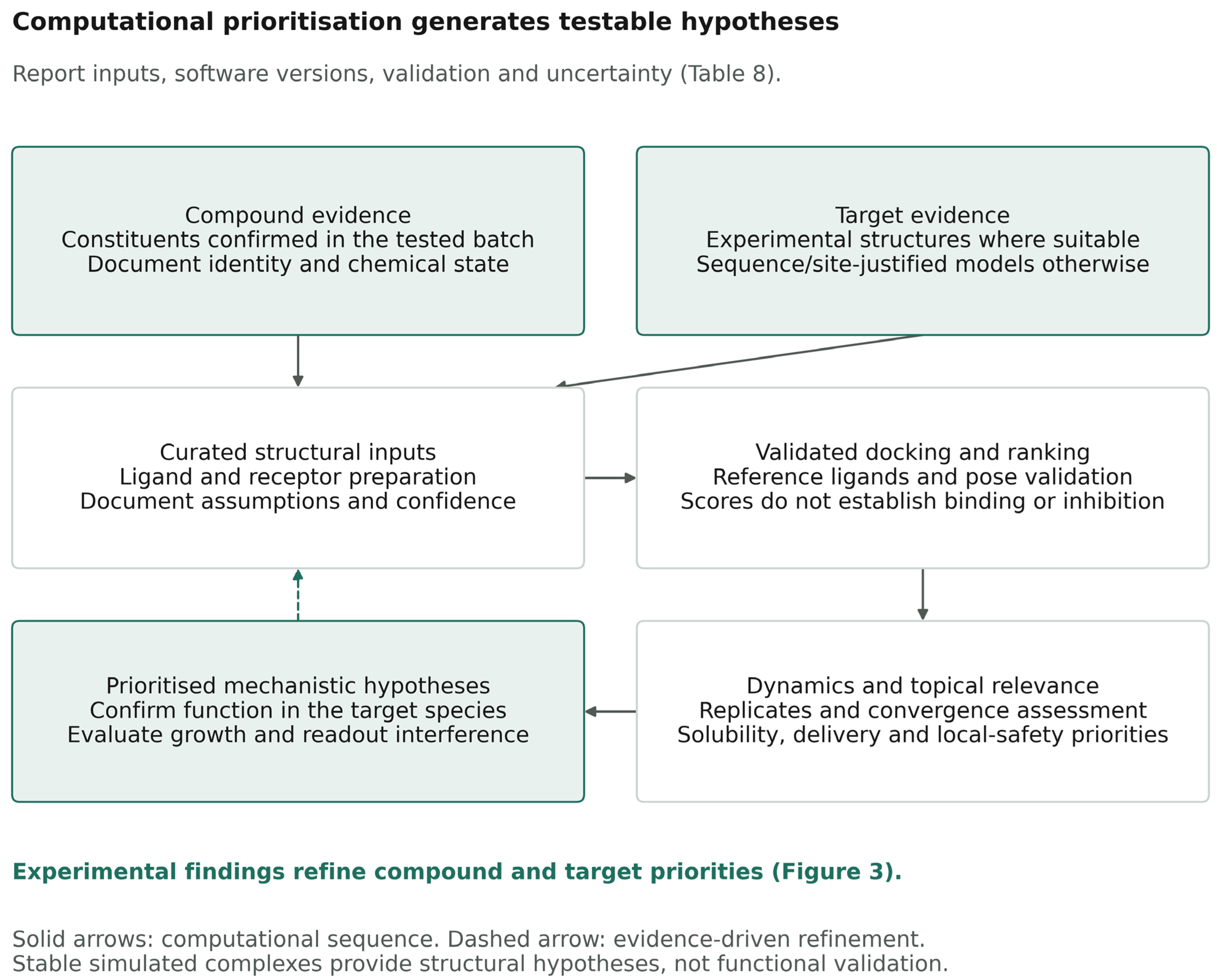
An organism-filtered search of the RCSB Protein Data Bank for Staphylococcus pseudintermedius on 25 October 2026 returned nine entries. None represented AgrA, AgrC, or an autoinducing peptide (AIP)–receptor complex. Homologous templates may still be suitable for specific targets. Template-based modeling and structure prediction [123,124,125] could support the development of sequence-specific AgrA, AgrC, and AIP–AgrC models. Model confidence and binding-site uncertainty should be reported, and predicted effects on agr signaling require functional testing.
Table S2 summarizes resources for molecular dynamics [126,127,128], physicochemical and pharmacokinetic prediction [129], docking [130,131], and QS screening workflows [132]. Input structures, parameters, and outputs should be documented [133,134,135]. Docking scores from different programs should not be interpreted as directly comparable binding energies. For topical applications, solubility, formulation stability, local exposure, and tolerability take precedence over oral drug-likeness criteria. Table 8 lists the minimum reporting requirements.
Figure 3.
Conceptual framework for evaluating propolis in canine skin and ear infections, informed by Rosignoli et al. [22]. Modules A and B provide chemically characterized material and a defined canine evidence base. Module C addresses antimicrobial context and assay interpretability; D and E separate QS/virulence evidence from biofilm endpoints; F provides transcriptional support; G generates computational hypotheses; and H addresses formulation and local safety before clinical evaluation. Solid arrows indicate evidence flow, and dashed arrows computational feedback. The decision checks require interpretable assays, reproducible activity on endpoints relevant to the intended indication in canine isolates, and activity at a locally tolerated exposure for the intended formulation. Computational agreement is not proof of biological activity. MIC, minimum inhibitory concentration; MIQE, Minimum Information for Publication of Quantitative Real-Time PCR Experiments; QS, quorum sensing.
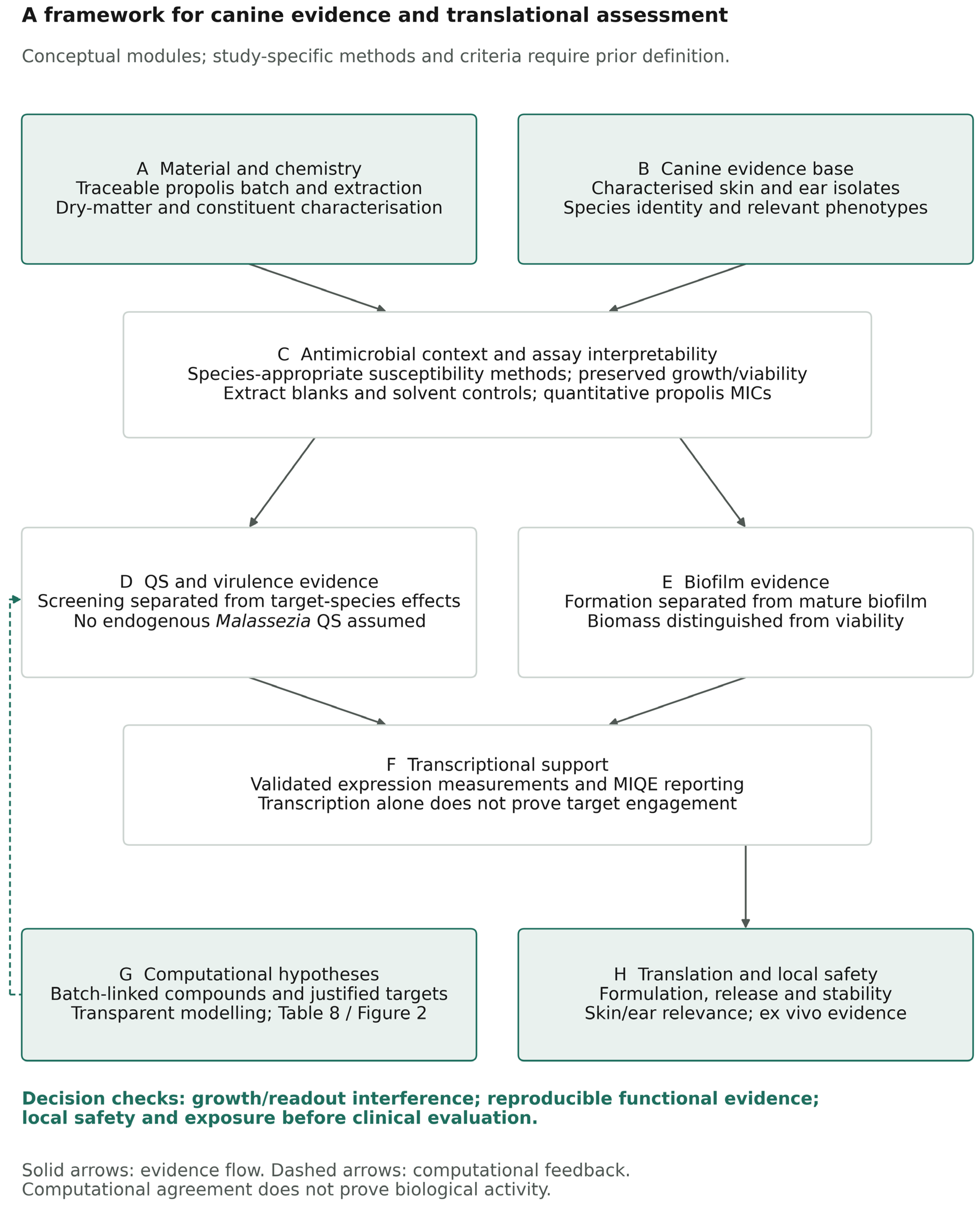
Table 8.
Proposed minimum reporting checklist for in silico studies of propolis constituents against QS and biofilm targets (adapted from the reproducibility recommendations summarized by Rosignoli et al. [22]).
7. Proposed Integrated Workflow for Canine Clinical Isolates
Figure 3 adapts the discovery workflow of Rosignoli et al. [22] to propolis and canine skin and ear pathogens. The sequence prioritizes experimental evidence in canine isolates and uses computational predictions to select targets for testing.
Priority 1: Establish assay validity and activity (Modules A–C). Use chemically characterized batches and canine isolates relevant to the intended indication. Prioritize S. pseudintermedius from skin and P. aeruginosa from ears. Include S. canis or M. pachydermatis where relevant to the indication. Establish species-appropriate susceptibility ranges. A growth-independent QS claim requires preserved growth and viability under the functional assay conditions and exclusion of vehicle and readout interference. An effect explained by these factors does not justify progression as a QS-directed candidate.
Priority 2: Demonstrate a reproducible effect on the intended endpoint (Modules D–G). Prespecify the primary endpoint and the minimum effect required relative to vehicle. Separate prevention of biofilm formation from activity against established biofilms and measure viable burden alongside biomass. For S. pseudintermedius, interpret toxin output alongside viable biofilm burden and viable dispersal. For P. aeruginosa, compare responses across canine isolates with characterized QS phenotypes [40]. A claim about a specific QS mechanism requires target-specific evidence; validated transcriptional endpoints can provide supporting data [103]. Computational predictions can guide these comparisons (Table 8). For a proposed adjunct, establish whether the combination improves the prespecified endpoint relative to standard treatment alone.
Priority 3: Confirm activity and local tolerability of the intended formulation (Module H). Determine whether the same formulation remains active at an achievable, locally tolerated exposure under conditions relevant to the intended site. Assess sensitization and, for otic application, ototoxicity as appropriate. Systemic interstitial-fluid measurements [136] do not establish exposure in the ear canal. Progression to an appropriately approved clinical study requires reproducible activity in relevant canine isolates and evidence of local tolerability at the active exposure.
8. Clinical Relevance, Translational Constraints, and Research Gaps
QS modulation is proposed as an adjunct to treatment for canine skin and ear infections [21,22]. Propolis preparations or constituents enhanced antimicrobial activity in selected non-canine studies [76,86,91,110,111,115]. These findings do not establish adjunctive benefit in dogs.
In a separate in vitro study that did not test propolis, the inhibitory concentration of the marbofloxacin component remained lower during serial passage of P. aeruginosa ATCC 27853 with Tris-ethylenediaminetetraacetic acid (Tris-EDTA), with or without geraniol, than during marbofloxacin-only exposure [137]. The comparison concerns concentrations within the tested regimens and does not establish a corresponding difference in the intrinsic susceptibility of the recovered isolates or efficacy in canine otitis.
Most studies of canine isolates measured growth inhibition or killing [69,70,73,98,113,120,121]. Barbosa et al. reported lower crystal violet biomass during biofilm formation and after treatment of preformed biofilms in one canine S. pseudintermedius isolate and one reference strain [73]. The study did not assess biofilm cell viability or agr activity. No study identified in this review demonstrated propolis-mediated QS modulation or antibiofilm activity in confirmed canine P. aeruginosa isolates. For S. canis, the evidence identified here is limited to growth testing of one canine isolate [69]. In vitro antifungal activity against M. pachydermatis does not establish an effect on its biofilms or signaling [70,98,113].
In a randomized, double-blind, vehicle-controlled trial of acute or subacute canine otitis externa, 27 of 38 dogs receiving a 2.5% propolis ear-drop formulation showed clinical and microbiological improvement or resolution after 14 days [119]. The mixed infections, unequal group sizes (38 and 10), short follow-up, and absence of an active comparator limit inference about pathogen-specific or adjunctive efficacy. The study did not measure QS or biofilm outcomes. No adverse effects were reported during follow-up; this observation does not establish an ototoxicity profile. The 11 nonresponders had mixed bacterial and fungal infections involving Proteus mirabilis (n = 6) or Pseudomonas aeruginosa (n = 5), indicating that the overall response rate should not be extrapolated to infections involving these pathogens [119].
Non-canine studies help prioritize targets. Biochemical assays support LasR-related effects for selected compounds [57,59], whereas changes in pigment production, adhesion, or biofilm-associated transcription alone do not establish target engagement [65,66,79,108]. Differences in extract composition and assay design limit comparisons between studies [68,87].
The only canine-cell-based cytotoxicity study identified in this review tested a Portuguese ethanolic propolis extract in canine progenitor epithelial keratinocytes over 72 h. Cell viability declined with increasing concentration over the tested range of 0.025–2.50 mg/mL. At 1.0 mg/mL, canine keratinocyte viability was approximately 20% of the untreated control (Figure 2 in [71]). This concentration is below some antimicrobial concentrations reported for other propolis preparations tested against canine isolates, including the MIC of 7.11 mg/mL reported for M. pachydermatis [70]. The studies used different preparations, assays, and exposure conditions, so these values cannot establish a therapeutic margin. Nor do they establish the concentration required for a growth-independent effect on QS. For topical development, activity against the intended pathogen and local tolerability should be assessed with the same formulation at matched exposures. For otic use, keratinocyte viability does not establish otic safety.
PubMed was the only database used for the structured bibliographic search. Sensitivity searches and targeted retrieval supplemented this search, but relevant studies indexed only in veterinary, agricultural, or multidisciplinary databases may have been missed. The synthesis therefore does not provide an exhaustive inventory of eligible studies.
9. Conclusions and Future Perspectives
Propolis preparations and individual constituents show antimicrobial and antibiofilm effects in vitro. A controlled canine otitis trial provides preliminary evidence for one formulation [119], but direct QS modulation in canine isolates and clinical benefit as a QS-directed adjunct remain unestablished. Structure prediction for AgrA, AgrC, and AIP–AgrC complexes of S. pseudintermedius can guide target selection for experimental testing [124,125]. Whether combined modulation of Las, Rhl, and PQS offers an advantage over modulation of a single circuit in canine P. aeruginosa otitis isolates remains to be tested [42,43]. The immediate priority is to establish whether a chemically characterized formulation produces a reproducible effect in relevant canine isolates at an exposure tolerated at the intended site.
Supplementary Materials
The following supporting information can be downloaded at https://www.mdpi.com/article/10.3390/vetsci13111260/s1, File S1: Search strings, search log, screening records, and other sources identified outside the structured search.
Author Contributions
Conceptualization, Z.T.T. and Á.K.; methodology, Z.T.T. and Á.K.; investigation, Z.T.T., Á.J. and Á.K.; writing—original draft preparation, Z.T.T.; writing—review and editing, N.K.T., H.F., M.N.-T., Á.J. and Á.K.; supervision, N.K.T. and Á.K.; project administration, Á.K.; funding acquisition, Á.J. and Á.K. All authors have read and agreed to the published version of the manuscript.
Funding
This research was funded by the Ministry of Science and Technology of Hungary from the National Research, Development and Innovation Fund, financed under the 2026-2.1.1-EKÖP funding scheme, grant number 2026-2.1.1-EKÖP-2026-00015.
Institutional Review Board Statement
Not applicable.
Informed Consent Statement
Not applicable.
Data Availability Statement
No new data were created or analyzed in this study. All data supporting the literature search, screening records, and source classification are provided in Supplementary File S1.
Conflicts of Interest
The authors declare no conflicts of interest. The funders had no role in the design of the study; in the collection, analyses, or interpretation of data; in the writing of the manuscript; or in the decision to publish the results.
Abbreviations
The following abbreviations are used in this manuscript:
| ADME | absorption, distribution, metabolism, and excretion |
| ADMET | absorption, distribution, metabolism, excretion, and toxicity |
| agr | accessory gene regulator |
| AHL | N-acylhomoserine lactone |
| AI-2 | autoinducer-2 |
| AIP | autoinducing peptide |
| AMR | antimicrobial resistance |
| ATCC | American Type Culture Collection |
| BHIB | brain–heart infusion broth |
| cAD | canine atopic dermatitis |
| CAMHB | cation-adjusted Mueller–Hinton broth |
| CAPE | caffeic acid phenethyl ester |
| CFU | colony-forming unit |
| CLSM | confocal laser scanning microscopy |
| CPEK | canine progenitor epithelial keratinocytes |
| CV | crystal violet |
| DES | deep eutectic solvent |
| ECOFF | epidemiological cut-off value |
| eDNA | extracellular DNA |
| EDTA | ethylenediaminetetraacetic acid |
| EEP | ethanolic extract of propolis |
| EGCG | epigallocatechin gallate |
| EMSA | electrophoretic mobility shift assay |
| EPS | extracellular polysaccharide |
| EtOH | ethanol |
| FICI | fractional inhibitory concentration index |
| GAE | gallic acid equivalents |
| GC-MS | gas chromatography–mass spectrometry |
| GG | glycine–glycine (processing motif) |
| HPLC | high-performance liquid chromatography |
| HPLC-DAD | high-performance liquid chromatography with diode-array detection |
| HSL | homoserine lactone |
| ica | intercellular adhesion (locus) |
| ID | identifier |
| IL-1β | interleukin-1β |
| IQS | integrated quorum-sensing signal |
| K9CATH | canine cathelicidin |
| LC-MS/MS | liquid chromatography–tandem mass spectrometry |
| logP | logarithm of the n-octanol/water partition coefficient |
| LPS | lipopolysaccharide |
| MBC | minimum bactericidal concentration |
| MBC90 | minimum bactericidal concentration for 90% of isolates |
| MBEC | minimum biofilm eradication concentration |
| MBL | metallo-β-lactamase |
| MD | molecular dynamics |
| MDR | multidrug-resistant |
| MFC | minimum fungicidal concentration |
| MFC90 | minimum fungicidal concentration for 90% of isolates |
| MIC | minimum inhibitory concentration |
| MIQE | Minimum Information for Publication of Quantitative Real-Time PCR Experiments |
| MLST | multilocus sequence typing |
| MRCNS | methicillin-resistant coagulase-negative staphylococci |
| MRSA | methicillin-resistant Staphylococcus aureus |
| MRSP | methicillin-resistant Staphylococcus pseudintermedius |
| MSSA | methicillin-susceptible Staphylococcus aureus |
| MSSP | methicillin-susceptible Staphylococcus pseudintermedius |
| MTT | 3-(4,5-dimethylthiazol-2-yl)-2,5-diphenyltetrazolium bromide |
| NAC | N-acetylcysteine |
| NADES | natural deep eutectic solvent |
| OD | optical density |
| PBMC | peripheral blood mononuclear cells |
| PBP2a | penicillin-binding protein 2a |
| PCR | polymerase chain reaction |
| PDB | Protein Data Bank |
| PEG | polyethylene glycol |
| PEO | poly(ethylene oxide) |
| PG | propylene glycol |
| PHMB | polyhexamethylene biguanide |
| PI | propidium iodide |
| PIA | polysaccharide intercellular adhesin |
| pLDDT | predicted local distance difference test |
| PNAG | poly-N-acetylglucosamine |
| PNPs | propolis nanoparticles |
| PQS | Pseudomonas quinolone signal |
| qPCR | quantitative polymerase chain reaction |
| QS | quorum sensing |
| QSI | quorum-sensing inhibitor |
| RMSD | root-mean-square deviation |
| RMSF | root-mean-square fluctuation |
| RNA-seq | RNA sequencing |
| RNAIII | regulatory RNA III of the agr locus |
| RNPP | Rap, NprR, PlcR, and PrgX (peptide-receptor family) |
| RT-qPCR | reverse-transcription quantitative polymerase chain reaction |
| SEM | scanning electron microscopy |
| SILE | size-independent ligand efficiency |
| SrtA | sortase A |
| ST | sequence type |
| TCP | tissue culture plate |
| TLC | thin-layer chromatography |
| TNF-α | tumor necrosis factor-α |
| WGS | whole-genome sequencing |
| XTT | 2,3-bis(2-methoxy-4-nitro-5-sulfophenyl)-2H-tetrazolium-5-carboxanilide |
References
- Hensel, P.; Santoro, D.; Favrot, C.; Hill, P.; Griffin, C. Canine atopic dermatitis: Detailed guidelines for diagnosis and allergen identification. BMC Vet. Res. 2015, 11, 196. [Google Scholar] [CrossRef] [Scilit] [PubMed]
- Favrot, C.; Steffan, J.; Seewald, W.; Picco, F. A prospective study on the clinical features of chronic canine atopic dermatitis and its diagnosis. Vet. Dermatol. 2010, 21, 23–31. [Google Scholar] [CrossRef] [Scilit] [PubMed]
- Marsella, R. Advances in our understanding of canine atopic dermatitis. Vet. Dermatol. 2021, 32, 547-e151. [Google Scholar] [CrossRef] [Scilit] [PubMed]
- Bradley, C.W.; Morris, D.O.; Rankin, S.C.; Cain, C.L.; Misic, A.M.; Houser, T.; Mauldin, E.A.; Grice, E.A. Longitudinal Evaluation of the Skin Microbiome and Association with Microenvironment and Treatment in Canine Atopic Dermatitis. J. Investig. Dermatol. 2016, 136, 1182–1190. [Google Scholar] [CrossRef] [Scilit] [PubMed]
- Hillier, A.; Lloyd, D.H.; Weese, J.S.; Blondeau, J.M.; Boothe, D.; Breitschwerdt, E.; Guardabassi, L.; Papich, M.G.; Rankin, S.; Turnidge, J.D.; et al. Guidelines for the diagnosis and antimicrobial therapy of canine superficial bacterial folliculitis (Antimicrobial Guidelines Working Group of the International Society for Companion Animal Infectious Diseases). Vet. Dermatol. 2014, 25, 163-e43. [Google Scholar] [CrossRef] [Scilit] [PubMed]
- Mag, P.; Fehér, E.; Kaszab, E.; Máté, D.; Tarpataki, N.; Jerzsele, Á. Comparative Phenotypic and PCR-Based Methicillin Resistance Characterization of Clinical Canine Staphylococcus pseudintermedius. Antibiotics 2026, 15, 473. [Google Scholar] [CrossRef] [Scilit] [PubMed]
- Loeffler, A.; Cain, C.L.; Ferrer, L.; Nishifuji, K.; Varjonen, K.; Papich, M.G.; Guardabassi, L.; Frosini, S.M.; Barker, E.N.; Weese, J.S. Antimicrobial use guidelines for canine pyoderma by the International Society for Companion Animal Infectious Diseases (ISCAID). Vet. Dermatol. 2025, 36, 234–282. [Google Scholar] [CrossRef] [Scilit] [PubMed]
- Olivry, T.; DeBoer, D.J.; Favrot, C.; Jackson, H.A.; Mueller, R.S.; Nuttall, T.; Prélaud, P. Treatment of canine atopic dermatitis: 2015 updated guidelines from the International Committee on Allergic Diseases of Animals (ICADA). BMC Vet. Res. 2015, 11, 210. [Google Scholar] [CrossRef] [Scilit] [PubMed]
- Secker, B.; Shaw, S.; Atterbury, R.J. Pseudomonas spp. in Canine Otitis Externa. Microorganisms 2023, 11, 2650. [Google Scholar] [CrossRef] [Scilit] [PubMed]
- Veres, M.A.; Tóth, Z.A.; Illés, E.; Mag, P.; Kaszab, E.; Fehér, E.; Jerzsele, Á.; Kerek, Á. Phenotypic Susceptibility, Resistance-Associated Genomic Determinants, and Mobile-Genetic-Element Context of Canine Otitis Externa-Associated Pseudomonas aeruginosa Collected in Hungary in 2010 and 2017. Vet. Sci. 2026, 13, 855. [Google Scholar] [CrossRef] [Scilit] [PubMed]
- Andrade, M.; Oliveira, K.; Morais, C.; Abrantes, P.; Pomba, C.; Rosato, A.E.; Couto, I.; Costa, S.S. Virulence Potential of Biofilm-Producing Staphylococcus pseudintermedius, Staphylococcus aureus and Staphylococcus coagulans Causing Skin Infections in Companion Animals. Antibiotics 2022, 11, 1339. [Google Scholar] [CrossRef] [Scilit] [PubMed]
- Secker, B.; Shaw, S.; Hobley, L.; Atterbury, R.J. Genomic and phenotypic characterisation of Pseudomonas aeruginosa isolates from canine otitis externa reveals high-risk sequence types identical to those found in human nosocomial infections. Front. Microbiol. 2025, 16, 1526843. [Google Scholar] [CrossRef] [Scilit] [PubMed]
- Čonková, E.; Proškovcová, M.; Váczi, P.; Malinovská, Z. In Vitro Biofilm Formation by Malassezia pachydermatis Isolates and Its Susceptibility to Azole Antifungals. J. Fungi 2022, 8, 1209. [Google Scholar] [CrossRef] [Scilit] [PubMed]
- Hall, C.W.; Mah, T.F. Molecular mechanisms of biofilm-based antibiotic resistance and tolerance in pathogenic bacteria. FEMS Microbiol. Rev. 2017, 41, 276–301. [Google Scholar] [CrossRef] [Scilit] [PubMed]
- Araújo, D.; Silva, A.R.; Fernandes, R.; Serra, P.; Barros, M.M.; Campos, A.M.; Oliveira, R.; Silva, S.; Almeida, C.; Castro, J. Emerging Approaches for Mitigating Biofilm-Formation-Associated Infections in Farm, Wild, and Companion Animals. Pathogens 2024, 13, 320. [Google Scholar] [CrossRef] [Scilit] [PubMed]
- Little, S.V.; Bryan, L.K.; Hillhouse, A.E.; Cohen, N.D.; Lawhon, S.D. Characterization of agr Groups of Staphylococcus pseudintermedius Isolates from Dogs in Texas. mSphere 2019, 4, e00033-19. [Google Scholar] [CrossRef] [Scilit] [PubMed]
- Kerek, Á.; Sterczer, Á.; Somogyi, Z.; Kovács, D.; Jerzsele, Á. Investigation of the environmental presence of multidrug-resistant bacteria at small animal hospitals in Hungary. Acta Vet. Hung. 2021, 68, 387–392. [Google Scholar] [CrossRef] [Scilit] [PubMed]
- Kerek, Á.; Yurt, A.; Szabó, Á.; Tuska-Szalay, B.; Jerzsele, Á. Antimicrobial resistance and natural alternatives: In vitro efficacy of Hungarian propolis against feline and bovine Tritrichomonas foetus. Front. Vet. Sci. 2025, 12, 1635358. [Google Scholar] [CrossRef] [Scilit] [PubMed]
- Rasmussen, T.B.; Givskov, M. Quorum-sensing inhibitors as anti-pathogenic drugs. Int. J. Med. Microbiol. 2006, 296, 149–161. [Google Scholar] [CrossRef] [Scilit] [PubMed]
- Hentzer, M.; Wu, H.; Andersen, J.B.; Riedel, K.; Rasmussen, T.B.; Bagge, N.; Kumar, N.; Schembri, M.A.; Song, Z.; Kristoffersen, P.; et al. Attenuation of Pseudomonas aeruginosa virulence by quorum sensing inhibitors. EMBO J. 2003, 22, 3803–3815. [Google Scholar] [CrossRef] [Scilit] [PubMed]
- Krzyżek, P. Challenges and Limitations of Anti-quorum Sensing Therapies. Front. Microbiol. 2019, 10, 2473. [Google Scholar] [CrossRef] [Scilit] [PubMed]
- Rosignoli, S.; Lustrino, E.; Shevchuk, O.; Rinaldo, S.; Rubini, E.; Paiardini, A.; Carev, I. Bioinformatics-Driven, Plant-Based Antibiotic Research Against Quorum Sensing and Biofilm Formation in Pseudomonas aeruginosa and Escherichia coli Multiresistant Microbes. Biomolecules 2026, 16, 197. [Google Scholar] [CrossRef] [Scilit] [PubMed]
- Kerek, Á.; Tornyos, G.Á.; Husz, L.H.; Jerzsele, Á. The role of quorum sensing in the population dynamics of antimicrobial resistance. Magy. Állatorv. Lapja 2026, 148, 415–426. (In Hungarian) [Google Scholar] [CrossRef] [Scilit]
- Bankova, V. Recent trends and important developments in propolis research. Evid. Based Complement. Altern. Med. 2005, 2, 29–32. [Google Scholar] [CrossRef] [Scilit] [PubMed]
- Ristivojević, P.; Trifković, J.; Andrić, F.; Milojković-Opsenica, D. Poplar-type propolis: Chemical composition, botanical origin and biological activity. Nat. Prod. Commun. 2015, 10, 1869–1876. [Google Scholar] [CrossRef] [Scilit]
- Stevanović, J.; Glavinić, U.; Ristanić, M.; Erjavec, V.; Denk, B.; Dolašević, S.; Stanimirović, Z. Bee-Inspired Healing: Apitherapy in Veterinary Medicine for Maintenance and Improvement Animal Health and Well-Being. Pharmaceuticals 2024, 17, 1050. [Google Scholar] [CrossRef] [Scilit] [PubMed]
- Tóth, Z.T.; Tarpataki, N.; Nemes-Terényi, M.; Jerzsele, Á.; Kerek, Á. The potential role of propolis in the complementary therapy of dermatological and other disorders of dogs: A literature review. Magy. Állatorv. Lapja 2026, 148, 605–619. [Google Scholar] [CrossRef] [Scilit]
- Đuričić, D.; Žura Žaja, I.; Kowalczyk, A.; Vlahović, K.; Valpotić, H.; Kovačić, M.; Pećin, M.; Samardžija, M. Evidence and Clinical Applications of Natural Products in Veterinary Medicine: A Systematic Review of Clinoptilolite, Ozone Therapy, Propolis, and Phytotherapy. Vet. Sci. 2026, 13, 483. [Google Scholar] [CrossRef] [Scilit] [PubMed]
- Santos, J.K.V.R.S.; Costa, E.D.S.; Fernandes, C.C.L.; Cortez, A.A.; Fonsêca, A.D.V.; Guedes, R.F.M.; Araújo, P.R.M. Integrative Therapies in Wound Healing in Small Animals: An Approach Beyond Traditional Medicine. Vet. Sci. 2026, 13, 418. [Google Scholar] [CrossRef] [Scilit] [PubMed]
- Adra, C.; Olsson, M.A.; Horn, R.; Tran, T.D. Propolis as an Alternative Treatment for Companion Animal Skin Infections: Addressing Antimicrobial Resistance. Front. Vet. Sci. 2026, 13, 1880076. [Google Scholar] [CrossRef] [Scilit] [PubMed]
- Savaliya, B.F.; Kim, S.; Veltman, T.; Trott, D.J. Comparison of the in vitro antibiofilm activities of otic cleansers against canine otitis externa pathogens. Vet. Dermatol. 2025, 36, 148–158. [Google Scholar] [CrossRef] [Scilit] [PubMed]
- Tóth, Z.T.; Tarpataki, N.K.; Nemes-Terényi, M.; Jerzsele, Á.; Kerek, Á. From antidote to multimodal adjunct: A systematic review of N-acetylcysteine in dogs and cats. Front. Vet. Sci. 2026, 13, 1959626. [Google Scholar] [CrossRef] [Scilit] [PubMed]
- Castelo-Branco, D.S.C.M.; Aguiar, L.; Guedes, G.M.M.; Pereira-Neto, W.A.; Cordeiro, R.A.; Brilhante, R.S.N.; Sidrim, J.J.C.; Rocha, M.F.G. Standardization of in vitro dual-species biofilms of Staphylococcus pseudintermedius and Malassezia pachydermatis: A strategy to establish an ex vivo biofilm model. J. Microbiol. Methods 2023, 208, 106721. [Google Scholar] [CrossRef] [Scilit] [PubMed]
- Couto, N.; Belas, A.; Oliveira, M.; Almeida, P.; Clemente, C.; Pomba, C. Comparative RNA-seq-Based Transcriptome Analysis of the Virulence Characteristics of Methicillin-Resistant and -Susceptible Staphylococcus pseudintermedius Strains Isolated from Small Animals. Antimicrob. Agents Chemother. 2016, 60, 962–967. [Google Scholar] [CrossRef] [Scilit] [PubMed]
- Teixeira, I.M.; de Moraes Assumpção, Y.; Paletta, A.C.C.; Antunes, M.; da Silva, I.T.; Jaeger, L.H.; Ferreira, R.F.; de Oliveira Ferreira, E.; de Araújo Penna, B. Investigation on biofilm composition and virulence traits of S. pseudintermedius isolated from infected and colonized dogs. Braz. J. Microbiol. 2024, 55, 2923–2936. [Google Scholar] [CrossRef] [Scilit] [PubMed]
- Lee, G.Y.; Yang, S.J. Comparative assessment of genotypic and phenotypic correlates of Staphylococcus pseudintermedius strains isolated from dogs with otitis externa and healthy dogs. Comp. Immunol. Microbiol. Infect. Dis. 2020, 70, 101376. [Google Scholar] [CrossRef] [Scilit] [PubMed]
- Seres-Steinbach, A.; Schneider-Patkó, B.; Jerzsele, Á.; Veres, A.M.; Sonnevend, Á.; Bányai, K.; Schneider, G. Characterization of Canine Otitis Externa Pseudomonas aeruginosa Isolates and Their Sensitivities to Different Essential Oils. Animals 2025, 15, 826. [Google Scholar] [CrossRef] [Scilit] [PubMed]
- Veres, M.A.; Tóth, Z.A.; Illés, E.; Mag, P.; Kaszab, E.; Fehér, E.; Kerek, Á.; Jerzsele, Á. Phenotypic antimicrobial susceptibility and genomic resistance determinants in canine otitis-associated Pseudomonas aeruginosa isolates. Front. Vet. Sci. 2026, 13, 1902036. [Google Scholar] [CrossRef] [Scilit] [PubMed]
- Kušar, D.; Šrimpf, K.; Isaković, P.; Kalšek, L.; Hosseini, J.; Zdovc, I.; Kotnik, T.; Vengušt, M.; Tavčar-Kalcher, G. Determination of N-acylhomoserine lactones of Pseudomonas aeruginosa in clinical samples from dogs with otitis externa. BMC Vet. Res. 2016, 12, 233. [Google Scholar] [CrossRef] [Scilit] [PubMed]
- Tron, E.A.; Wilke, H.L.; Petermann, S.R.; Rust, L. Pseudomonas aeruginosa from canine otitis externa exhibit a quorum sensing deficiency. Vet. Microbiol. 2004, 99, 121–129. [Google Scholar] [CrossRef] [Scilit] [PubMed]
- Olabode, I.R.; Sachivkina, N.; Karamyan, A.; Mannapova, R.; Kuznetsova, O.; Bobunova, A.; Zhabo, N.; Avdonina, M.; Gurina, R. In Vitro Activity of Farnesol against Malassezia pachydermatis Isolates from Otitis Externa Cases in Dogs. Animals 2023, 13, 1259. [Google Scholar] [CrossRef] [Scilit] [PubMed]
- Lee, J.; Zhang, L. The hierarchy quorum sensing network in Pseudomonas aeruginosa. Protein Cell 2015, 6, 26–41. [Google Scholar] [CrossRef] [Scilit] [PubMed]
- Williams, P.; Cámara, M. Quorum sensing and environmental adaptation in Pseudomonas aeruginosa: A tale of regulatory networks and multifunctional signal molecules. Curr. Opin. Microbiol. 2009, 12, 182–191. [Google Scholar] [CrossRef] [Scilit] [PubMed]
- Novick, R.P.; Geisinger, E. Quorum sensing in staphylococci. Annu. Rev. Genet. 2008, 42, 541–564. [Google Scholar] [CrossRef] [Scilit] [PubMed]
- Lee, J.; Wu, J.; Deng, Y.; Wang, J.; Wang, C.; Wang, J.; Chang, C.; Dong, Y.; Williams, P.; Zhang, L.H. A cell-cell communication signal integrates quorum sensing and stress response. Nat. Chem. Biol. 2013, 9, 339–343. [Google Scholar] [CrossRef] [Scilit] [PubMed]
- Raya, J.; Montagut, E.J.; Marco, M.P. Analysing the integrated quorum sensing (iqs) system and its potential role in Pseudomonas aeruginosa pathogenesis. Front. Cell. Infect. Microbiol. 2025, 15, 1575421. [Google Scholar] [CrossRef] [Scilit] [PubMed]
- Bottomley, M.J.; Muraglia, E.; Bazzo, R.; Carfì, A. Molecular insights into quorum sensing in the human pathogen Pseudomonas aeruginosa from the structure of the virulence regulator LasR bound to its autoinducer. J. Biol. Chem. 2007, 282, 13592–13600. [Google Scholar] [CrossRef] [Scilit] [PubMed]
- Veres, M.A.; Tóth, Z.A.; Illés, E.; Mag, P.; Kaszab, E.; Fehér, E.; Jerzsele, Á.; Kerek, Á. Virulence-Associated Genomic Architecture of Canine Otitis Externa-Derived Pseudomonas aeruginosa Isolates from Hungary. Vet. Sci. 2026, 13, 664. [Google Scholar] [CrossRef] [Scilit] [PubMed]
- Veres, M.A.; Moravcsik, B.; Mag, P.; Kaszab, E.; Fehér, E.; Jerzsele, Á.; Kerek, Á. A conserved virulence backbone and heterogeneous type III secretion effector profiles in historical canine otitis-associated Pseudomonas aeruginosa from Hungary. Front. Vet. Sci. 2026, 13, 1951261. [Google Scholar] [CrossRef] [Scilit] [PubMed]
- Canovas, J.; Baldry, M.; Bojer, M.S.; Andersen, P.S.; Gless, B.H.; Grzeskowiak, P.K.; Stegger, M.; Damborg, P.; Olsen, C.A.; Ingmer, H. Cross-Talk between Staphylococcus aureus and Other Staphylococcal Species via the agr Quorum Sensing System. Front. Microbiol. 2016, 7, 1733. [Google Scholar] [CrossRef] [Scilit] [PubMed]
- Boles, B.R.; Horswill, A.R. Agr-mediated dispersal of Staphylococcus aureus biofilms. PLoS Pathog. 2008, 4, e1000052. [Google Scholar] [CrossRef] [Scilit] [PubMed]
- Cook, L.C.; Federle, M.J. Peptide pheromone signaling in Streptococcus and Enterococcus. FEMS Microbiol. Rev. 2014, 38, 473–492. [Google Scholar] [CrossRef] [Scilit] [PubMed]
- Koo, H.; Pearson, S.K.; Scott-Anne, K.; Abranches, J.; Cury, J.A.; Rosalen, P.L.; Park, Y.K.; Marquis, R.E.; Bowen, W.H. Effects of apigenin and tt-farnesol on glucosyltransferase activity, biofilm viability and caries development in rats. Oral Microbiol. Immunol. 2002, 17, 337–343. [Google Scholar] [CrossRef] [Scilit] [PubMed]
- Hornby, J.M.; Jensen, E.C.; Lisec, A.D.; Tasto, J.J.; Jahnke, B.; Shoemaker, R.; Dussault, P.; Nickerson, K.W. Quorum sensing in the dimorphic fungus Candida albicans is mediated by farnesol. Appl. Environ. Microbiol. 2001, 67, 2982–2992. [Google Scholar] [CrossRef] [Scilit] [PubMed]
- Jeon, J.G.; Pandit, S.; Xiao, J.; Gregoire, S.; Falsetta, M.L.; Klein, M.I.; Koo, H. Influences of trans-trans farnesol, a membrane-targeting sesquiterpenoid, on Streptococcus mutans physiology and survival within mixed-species oral biofilms. Int. J. Oral Sci. 2011, 3, 98–106. [Google Scholar] [CrossRef] [Scilit] [PubMed]
- Rivera-Yañez, N.; Hernández-Sánchez, K.M.; Hernández-Rosas, N.A.; Francisco-Cruz, L.; Nieto-Yañez, O.; Barrera-Ortega, C.C.; Pozo-Molina, G.; Méndez-Catalá, C.F.; Méndez-Cruz, A.R.; Ruiz-Hurtado, P.A.; et al. Understanding the Effect of Propolis and Its Derivatives Against Candida Biofilm: New Approaches in the Search for Alternative Therapies. J. Fungi 2026, 12, 301. [Google Scholar] [CrossRef] [Scilit] [PubMed]
- Paczkowski, J.E.; Mukherjee, S.; McCready, A.R.; Cong, J.P.; Aquino, C.J.; Kim, H.; Henke, B.R.; Smith, C.D.; Bassler, B.L. Flavonoids Suppress Pseudomonas aeruginosa Virulence through Allosteric Inhibition of Quorum-sensing Receptors. J. Biol. Chem. 2017, 292, 4064–4076. [Google Scholar] [CrossRef] [Scilit] [PubMed]
- Bulman, Z.; Le, P.; Hudson, A.O.; Savka, M.A. A novel property of propolis (bee glue): Anti-pathogenic activity by inhibition of N-acyl-homoserine lactone mediated signaling in bacteria. J. Ethnopharmacol. 2011, 138, 788–797. [Google Scholar] [CrossRef] [Scilit] [PubMed]
- Xie, Y.; Chen, J.; Wang, B.; Peng, A.Y.; Mao, Z.W.; Xia, W. Inhibition of Quorum-Sensing Regulator from Pseudomonas aeruginosa Using a Flavone Derivative. Molecules 2022, 27, 2439. [Google Scholar] [CrossRef] [Scilit] [PubMed]
- Lal, A.F.; Gupta, P.S.; Yadav, P.K. In silico study of Phenol Explorer database as potential inhibitors of quorum-sensing regulated pathogenicity in Pseudomonas aeruginosa. Biomed. Pharmacol. J. 2023, 16, 1577–1590. [Google Scholar] [CrossRef] [Scilit]
- Fakhar, M.; Ahmed, M.; Nasim Sabri, A. Computational and experimental strategies for combating MBL P. aeruginosa (MBLPA) biofilms using phytochemicals: Targeting the quorum sensing network. Saudi J. Biol. Sci. 2024, 31, 104001. [Google Scholar] [CrossRef] [Scilit] [PubMed]
- Meto, A.; Colombari, B.; Meto, A.; Boaretto, G.; Pinetti, D.; Marchetti, L.; Benvenuti, S.; Pellati, F.; Blasi, E. Propolis Affects Pseudomonas aeruginosa Growth, Biofilm Formation, eDNA Release and Phenazine Production: Potential Involvement of Polyphenols. Microorganisms 2020, 8, 243. [Google Scholar] [CrossRef] [Scilit] [PubMed]
- Wang, J.; Qiu, J.; Dong, J.; Li, H.; Luo, M.; Dai, X.; Zhang, Y.; Leng, B.; Niu, X.; Zhao, S.; et al. Chrysin protects mice from Staphylococcus aureus pneumonia. J. Appl. Microbiol. 2011, 111, 1551–1558. [Google Scholar] [CrossRef] [Scilit] [PubMed]
- Soromou, L.W.; Zhang, Y.; Cui, Y.; Wei, M.; Chen, N.; Yang, X.; Huo, M.; Baldé, A.; Guan, S.; Deng, X.; et al. Subinhibitory concentrations of pinocembrin exert anti-Staphylococcus aureus activity by reducing α-toxin expression. J. Appl. Microbiol. 2013, 115, 41–49. [Google Scholar] [CrossRef] [Scilit] [PubMed]
- Sang, H.; Feng, K.; Ju, Y.; Sun, Y.; Zhang, Y.; Xuan, H.; Wang, F. Propolis Exerts Antibiofilm Activity Against Methicillin-Resistant Staphylococcus aureus by Modulating Gene Expression to Suppress Adhesion. Microorganisms 2025, 13, 2810. [Google Scholar] [CrossRef] [Scilit] [PubMed]
- Feng, K.; Luan, H.; Sang, H.; Qiu, W.; Yang, R.; Cheng, J.; Feng, W.; Xu, W.; Song, P.; Wang, F. Caffeic Acid Phenethyl Ester Suppresses Adhesion to Mediate Its Antibiofilm Activity Against Methicillin-Resistant Staphylococcus aureus. Microorganisms 2026, 14, 1601. [Google Scholar] [CrossRef] [Scilit] [PubMed]
- Drago, L.; De Vecchi, E.; Nicola, L.; Gismondo, M.R. In vitro antimicrobial activity of a novel propolis formulation (Actichelated propolis). J. Appl. Microbiol. 2007, 103, 1914–1921. [Google Scholar] [CrossRef] [Scilit] [PubMed]
- Dżugan, M.; Miłek, M.; Ciszkowicz, E.; Łyskowski, A.; Tomczyk, M. Selected Polyphenols of Polish Poplar Propolis as a Key Component Shaping Its Antibacterial Properties—In Vitro and In Silico Approaches. Molecules 2025, 30, 2036. [Google Scholar] [CrossRef] [Scilit] [PubMed]
- Rodrigues, R.; Almeida, R.; Rodrigues, S.C.V.; Castro, J.; Oliveira, R.; Mendes, N.; Almeida, C.; Silva, S.; Araújo, D.; Almeida-Aguiar, C. In Vitro Investigation of the Antimicrobial Properties of Gerês Propolis in Bacteria Isolated from Companion Animals and Safety Profile Characterization Using the Galleria mellonella Model. Pathogens 2025, 14, 826. [Google Scholar] [CrossRef] [Scilit] [PubMed]
- Fuentes Esquivel, D.B.; Pérez, B.R.; Betancourt, N.T.; García Tovar, C.G.; Penieres Carrillo, J.G.; Galindo, F.H.; Flores, J.P.; Sánchez, T.A.C. In Vitro Antimycotic Activity and Structural Damage against Canine Malassezia pachydermatis Strains Caused by Mexican Stingless Bee Propolis. Vet. Sci. 2024, 11, 106. [Google Scholar] [CrossRef] [Scilit] [PubMed]
- Queiroga, M.C.; Laranjo, M.; Andrade, N.; Marques, M.; Costa, A.R.; Antunes, C.M. Antimicrobial, Antibiofilm and Toxicological Assessment of Propolis. Antibiotics 2023, 12, 347. [Google Scholar] [CrossRef] [Scilit] [PubMed]
- Mattoli, L.; Lugli, A.; Burico, M.; Fodaroni, G.; Decarli, D.; Gianni, M.; Maidecchi, A.; Antonini, G.; Tondi, S.; Gaetano, A.; et al. Populus nigra Bud Extract as a Standardized Alternative to Propolis: Evidence of Compositional Similarity-Functional Properties of an Oral Spray Containing Populus nigra Bud Extract. Molecules 2026, 31, 1836. [Google Scholar] [CrossRef] [Scilit] [PubMed]
- Barbosa, E.V.; Assumpção, Y.M.; Teixeira, I.M.; Pereira, R.F.A.; Ribeiro, V.P.; Bastos, J.K.; Cardoso, C.V.; Liberal, M.H.T.; Penna, B.A.; Rocha, L.M. In vitro comparison between antimicrobial and antibiofilm effects of Green Propolis and Baccharis dracunculifolia against Staphylococcus pseudintermedius isolate. An. Acad. Bras. Cienc. 2022, 94, e20211103. [Google Scholar] [CrossRef] [Scilit] [PubMed]
- Meccatti, V.M.; Martins, K.M.C.; Ramos, L.P.; Pereira, T.C.; de Menezes, R.T.; Marcucci, M.C.; Abu Hasna, A.; de Oliveira, L.D. Synergistic Antibiofilm Action of Cinnamomum verum and Brazilian Green Propolis Hydroethanolic Extracts against Multidrug-Resistant Strains of Acinetobacter baumannii and Pseudomonas aeruginosa and Their Biocompatibility on Human Keratinocytes. Molecules 2023, 28, 6904. [Google Scholar] [CrossRef] [Scilit] [PubMed]
- de Oliveira Dembogurski, D.S.; Silva Trentin, D.; Boaretto, A.G.; Rigo, G.V.; da Silva, R.C.; Tasca, T.; Macedo, A.J.; Carollo, C.A.; Silva, D.B. Brown propolis-metabolomic innovative approach to determine compounds capable of killing Staphylococcus aureus biofilm and Trichomonas vaginalis. Food Res. Int. 2018, 111, 661–673. [Google Scholar] [CrossRef] [Scilit] [PubMed]
- Ripari, N.; Lopes, E.C.; Francisco, Á.F.; de Almeida, J.F.S.; Honorio, M.D.S.; Júnior, A.F.; Fontes, M.R.M.; Tanimoto, M.H.; Calderon, L.A.; Bastos, J.K.; et al. Guttiferone E from Brazilian red propolis inhibited wound-isolated methicillin-resistant Staphylococcus aureus and enhanced the bactericidal action of suppressed macrophages. Phytomedicine 2025, 140, 156615. [Google Scholar] [CrossRef] [Scilit] [PubMed]
- Sarker, D.K.; Ratcliffe, E.; Burnett, F.; Uddin, S.J.; Shilpi, J.A.; Efthimiou, G.; Seidel, V. Flavonoid derivatives from red propolis: In silico predictions of their interactions with Staphylococcus aureus sortase A and studies on their antibiofilm potential. Z. Naturforsch. C J. Biosci. 2026, 81, 393–410. [Google Scholar] [CrossRef] [Scilit] [PubMed]
- Zhang, W.; Margarita, G.E.; Wu, D.; Yuan, W.; Yan, S.; Qi, S.; Xue, X.; Wang, K.; Wu, L. Antibacterial Activity of Chinese Red Propolis against Staphylococcus aureus and MRSA. Molecules 2022, 27, 1693. [Google Scholar] [CrossRef] [Scilit] [PubMed]
- Gemiarto, A.T.; Ninyio, N.N.; Lee, S.W.; Logis, J.; Fatima, A.; Chan, E.W.; Lim, C.S. Isoprenyl caffeate, a major compound in manuka propolis, is a quorum-sensing inhibitor in Chromobacterium violaceum. Antonie Van Leeuwenhoek 2015, 108, 491–504. [Google Scholar] [CrossRef] [Scilit] [PubMed]
- Savka, M.A.; Dailey, L.; Popova, M.; Mihaylova, R.; Merritt, B.; Masek, M.; Le, P.; Nor, S.R.; Ahmad, M.; Hudson, A.O.; et al. Chemical composition and disruption of quorum sensing signaling in geographically diverse United States propolis. Evid. Based Complement. Altern. Med. 2015, 2015, 472593. [Google Scholar] [CrossRef] [Scilit] [PubMed]
- Boutoub, O.; El-Guendouz, S.; Matos, I.; El Ghadraoui, L.; Costa, M.C.; Carlier, J.D.; Faleiro, M.L.; Figueiredo, A.C.; Estevinho, L.M.; Miguel, M.G. Chemical Characterization and Biological Properties Assessment of Euphorbia resinifera and Euphorbia officinarum Moroccan Propolis. Antibiotics 2024, 13, 230. [Google Scholar] [CrossRef] [Scilit] [PubMed]
- Hadjab, W.; Zellagui, A.; Mokrani, M.; Öztürk, M.; Ceylan, Ö.; Gherraf, N.; Bensouici, C. Pharmacological Potential Effects of Algerian Propolis Against Oxidative Stress, Multidrug-Resistant Pathogens Biofilm and Quorum-Sensing. Turk. J. Pharm. Sci. 2024, 21, 71–80. [Google Scholar] [CrossRef] [Scilit] [PubMed]
- da Silva Mirowski, P.; da Silva Coutinho de Araújo Bueno, G.; Elsner Rodrigues, V.; Fernandes Barros, T.; da Costa, A.G.; Yoshida, N.C.; da Rosa Guterres, Z.; Trentin, D.S.; Rodrigues Garcez, F. Chemical Composition and Evaluation of Antibacterial, Antibiofilm, and Mutagenic Potentials of a Propolis Sample from the Atlantic Forest of Midwest Brazil. Chem. Biodivers. 2023, 20, e202301238. [Google Scholar] [CrossRef] [Scilit] [PubMed]
- da Cunha, M.G.; de Cássia Orlandi Sardi, J.; Freires, I.A.; Franchin, M.; Rosalen, P.L. Antimicrobial, anti-adherence and antibiofilm activity against Staphylococcus aureus of a 4-phenyl coumarin derivative isolated from Brazilian geopropolis. Microb. Pathog. 2020, 139, 103855. [Google Scholar] [CrossRef] [Scilit] [PubMed]
- Lee, J.H.; Kim, Y.G.; Khadke, S.K.; Yamano, A.; Woo, J.T.; Lee, J. Antimicrobial and antibiofilm activities of prenylated flavanones from Macaranga tanarius. Phytomedicine 2019, 63, 153033. [Google Scholar] [CrossRef] [Scilit] [PubMed]
- Bouchelaghem, S.; Das, S.; Naorem, R.S.; Czuni, L.; Papp, G.; Kocsis, M. Evaluation of Total Phenolic and Flavonoid Contents, Antibacterial and Antibiofilm Activities of Hungarian Propolis Ethanolic Extract against Staphylococcus aureus. Molecules 2022, 27, 574. [Google Scholar] [CrossRef] [Scilit] [PubMed]
- Miłek, M.; Ciszkowicz, E.; Tomczyk, M.; Sidor, E.; Zaguła, G.; Lecka-Szlachta, K.; Pasternakiewicz, A.; Dżugan, M. The Study of Chemical Profile and Antioxidant Properties of Poplar-Type Polish Propolis Considering Local Flora Diversity in Relation to Antibacterial and Anticancer Activities in Human Breast Cancer Cells. Molecules 2022, 27, 725. [Google Scholar] [CrossRef] [Scilit] [PubMed]
- Santos, P.B.D.R.E.D.; Ávila, D.D.S.; Ramos, L.P.; Yu, A.R.; Santos, C.E.D.R.; Berretta, A.A.; Camargo, S.E.A.; Oliveira, J.R.; Oliveira, L.D. Effects of Brazilian green propolis extract on planktonic cells and biofilms of multidrug-resistant strains of Klebsiella pneumoniae and Pseudomonas aeruginosa. Biofouling 2020, 36, 834–845. [Google Scholar] [CrossRef] [Scilit] [PubMed]
- da Silva, V.F.; Rosa, D.S.; de Figueirêdo, P.I.; da Silva, T.M.S.; Peixoto, R.M.; da Costa, M.M. Antimicrobial activity of brown propolis ethanolic extract and its application in intramammary formulation for the treatment of bovine mastitis. Braz. J. Microbiol. 2026, 57, 30. [Google Scholar] [CrossRef] [Scilit] [PubMed]
- de Mélo Silva, I.S.; do Amorim Costa Gaspar, L.M.; Rocha, A.M.O.; da Costa, L.P.; Tada, D.B.; Franceschi, E.; Padilha, F.F. Encapsulation of Red Propolis in Polymer Nanoparticles for the Destruction of Pathogenic Biofilms. AAPS PharmSciTech 2020, 21, 49. [Google Scholar] [CrossRef] [Scilit] [PubMed]
- Ripari, N.; Pereira, A.F.M.; Júnior, A.F.; Rall, V.L.M.; Aldana-Mejía, J.A.; Bastos, J.K.; Sforcin, J.M. Brazilian red propolis in combination with β-lactams exerts an efficient antibacterial action over methicillin-resistant Staphylococcus aureus (MRSA) strains. J. Appl. Microbiol. 2023, 134, lxac080. [Google Scholar] [CrossRef] [Scilit] [PubMed]
- El-Guendouz, S.; Aazza, S.; Lyoussi, B.; Bankova, V.; Popova, M.; Neto, L.; Faleiro, M.L.; Miguel, M.D.G. Moroccan Propolis: A Natural Antioxidant, Antibacterial, and Antibiofilm against Staphylococcus aureus with No Induction of Resistance after Continuous Exposure. Evid. Based Complement. Altern. Med. 2018, 2018, 9759240. [Google Scholar] [CrossRef] [Scilit] [PubMed]
- Albuquerque Borges, M.G.D.S.; de Oliveira E Lima, L.; Veras, A.C.M.F.; Ferreira, R.C.; da Nóbrega Alves, D.; de Medeiros, I.A.; Magnani, M.; Sobral, M.V.; Dias de Castro, R.; Relison Tintino, S.; et al. Cytotoxicity and Biological Activities of Geopopolis Extract from the Stingless bee (Melipona scutellaris) in Isolates of Staphylococcus aureus. Chem. Biodivers. 2024, 21, e202301982. [Google Scholar] [CrossRef] [Scilit] [PubMed]
- Reyna-Fuentes, J.H.; González-Alanis, P.; Blanco-Martínez, Z.; Benavides-González, F.; Urbizu-González, A.L.; Vázquez-Sauceda, M.L.; Mireles-Villanueva, M.V. Antibacterial, Phytochemical, and Antioxidant Properties of Propolis Extracts Obtained Using Different Solvents Against Multidrug-Resistant Bacteria. Pathogens 2026, 15, 749. [Google Scholar] [CrossRef] [Scilit] [PubMed]
- Šuran, J.; Cepanec, I.; Mašek, T.; Starčević, K.; Tlak Gajger, I.; Vranješ, M.; Radić, B.; Radić, S.; Kosalec, I.; Vlainić, J. Nonaqueous Polyethylene Glycol as a Safer Alternative to Ethanolic Propolis Extracts with Comparable Antioxidant and Antimicrobial Activity. Antioxidants 2021, 10, 978. [Google Scholar] [CrossRef] [Scilit] [PubMed]
- Trusheva, B.; Petkov, H.; Chimshirova, R.; Popova, M.; Dimitrova, L.; Zaharieva, M.M.; Ilieva, Y.; Vasileva, B.; Tsvetkova, I.; Najdenski, H.; et al. Insight into the influence of natural deep eutectic solvents on the extraction of phenolic compounds from poplar type propolis: Composition and in vitro biological activity. Heliyon 2024, 10, e28621. [Google Scholar] [CrossRef] [Scilit] [PubMed]
- Svetikienė, D.; Jokubaite, M.; Zamokas, G.; Babickaite, L.; Šiugždiniene, R.; Ramanauskiene, K. Efficacy Study of Propolis Eutectic Extract in Gel Formulations for the Treatment of Bacterial Skin Diseases in Dogs. Animals 2025, 15, 1434. [Google Scholar] [CrossRef] [Scilit] [PubMed]
- Deegan, K.R.; Fonseca, M.S.; Oliveira, D.C.P.; Santos, L.M.; Fernandez, C.C.; Hanna, S.A.; Machado, B.A.S.; Umsza-Guez, M.A.; Meyer, R.; Portela, R.W. Susceptibility of Malassezia pachydermatis Clinical Isolates to Allopathic Antifungals and Brazilian Red, Green, and Brown Propolis Extracts. Front. Vet. Sci. 2019, 6, 460. [Google Scholar] [CrossRef] [Scilit] [PubMed]
- McClean, K.H.; Winson, M.K.; Fish, L.; Taylor, A.; Chhabra, S.R.; Camara, M.; Daykin, M.; Lamb, J.H.; Swift, S.; Bycroft, B.W.; et al. Quorum sensing and Chromobacterium violaceum: Exploitation of violacein production and inhibition for the detection of N-acylhomoserine lactones. Microbiology 1997, 143, 3703–3711. [Google Scholar] [CrossRef] [Scilit] [PubMed]
- Harrison, A.M.; Soby, S.D. Reclassification of Chromobacterium violaceum ATCC 31532 and its quorum biosensor mutant CV026 to Chromobacterium subtsugae. AMB Express 2020, 10, 202. [Google Scholar] [CrossRef] [Scilit] [PubMed]
- O’Toole, G.A. Microtiter dish biofilm formation assay. J. Vis. Exp. 2011, 47, e2437. [Google Scholar] [CrossRef] [Scilit] [PubMed]
- Stepanović, S.; Vuković, D.; Hola, V.; Di Bonaventura, G.; Djukić, S.; Cirković, I.; Ruzicka, F. Quantification of biofilm in microtiter plates: Overview of testing conditions and practical recommendations for assessment of biofilm production by staphylococci. APMIS 2007, 115, 891–899. [Google Scholar] [CrossRef] [Scilit] [PubMed]
- Bustin, S.A.; Ruijter, J.M.; van den Hoff, M.J.B.; Kubista, M.; Pfaffl, M.W.; Shipley, G.L.; Tran, N.; Rödiger, S.; Untergasser, A.; Mueller, R.; et al. MIQE 2.0: Revision of the Minimum Information for Publication of Quantitative Real-Time PCR Experiments Guidelines. Clin. Chem. 2025, 71, 634–651. [Google Scholar] [CrossRef] [Scilit] [PubMed]
- Hadjab, W.; Zellagui, A.; Mokrani, M.; Ceylan, Ö.; Öztürk, M.; Bensouici, C.; Banjer, H.J.; Sami, R.; Al-Mushhin, A.A.M.; Alharthi, S.; et al. Propolis Biomolecules Attenuates Virulence Factors of Multi-Drug Resistant Pseudomonas aeruginosa: In Vitro and In Silico Investigation. Sci. Adv. Mater. 2024, 16, 532–545. [Google Scholar] [CrossRef] [Scilit]
- Tamfu, A.N.; Ceylan, O.; Cârâc, G.; Talla, E.; Dinica, R.M. Antibiofilm and Anti-Quorum Sensing Potential of Cycloartane-Type Triterpene Acids from Cameroonian Grassland Propolis: Phenolic Profile and Antioxidant Activity of Crude Extract. Molecules 2022, 27, 4872. [Google Scholar] [CrossRef] [Scilit] [PubMed]
- Kolaylı, S.; Ureyen Esertas, U.Z.; Kara, Y. The Antimicrobial, Anti-Quorum Sensing, and Anti-Biofilm Activities of Ethanolic Propolis Extracts Used as Food Supplements. Biol. Bull. Russ. Acad. Sci. 2022, 49, S21–S30. [Google Scholar] [CrossRef] [Scilit]
- De Marco, S.; Piccioni, M.; Pagiotti, R.; Pietrella, D. Antibiofilm and Antioxidant Activity of Propolis and Bud Poplar Resins versus Pseudomonas aeruginosa. Evid. Based Complement. Altern. Med. 2017, 2017, 5163575. [Google Scholar] [CrossRef] [Scilit] [PubMed]
- El-Sayed, N.R.; Samir, R.; Abdel-Hafez, L.J.M.; Ramadan, M.A. Olive Leaf Extract Modulates Quorum Sensing Genes and Biofilm Formation in Multi-Drug Resistant Pseudomonas aeruginosa. Antibiotics 2020, 9, 526. [Google Scholar] [CrossRef] [Scilit] [PubMed]
- Ebrahimi, S.R.; Nikbakht, M.; Shams Abadi, M.S.; Gholipour, A.; Bagheri, M.; Validi, M. Investigation of biocompatibility and antibacterial properties of electrospun chitosan/polyethylene oxide-based scaffolds containing propolis extract against Enterococcus faecalis, Pseudomonas aeruginosa, Staphylococcus aureus and Staphylococcus epidermidis. Heliyon 2025, 11, e42228. [Google Scholar] [CrossRef] [Scilit] [PubMed]
- Scazzocchio, F.; D’Auria, F.D.; Alessandrini, D.; Pantanella, F. Multifactorial aspects of antimicrobial activity of propolis. Microbiol. Res. 2006, 161, 327–333. [Google Scholar] [CrossRef] [Scilit] [PubMed]
- Feng, K.; Sang, H.; Jin, H.; Song, P.; Xu, W.; Xuan, H.; Wang, F. Antimicrobial Activities of Propolis Nanoparticles in Combination with Ampicillin Sodium Against Methicillin-Resistant Staphylococcus aureus. Microorganisms 2025, 13, 1844. [Google Scholar] [CrossRef] [Scilit] [PubMed]
- Wang, F.; Liu, H.; Li, J.; Zhang, W.; Jiang, B.; Xuan, H. Australian propolis ethanol extract exerts antibacterial activity against methicillin-resistant Staphylococcus aureus by mechanisms of disrupting cell structure, reversing resistance, and resisting biofilm. Braz. J. Microbiol. 2021, 52, 1651–1664. [Google Scholar] [CrossRef] [Scilit] [PubMed]
- Cardoso, R.L.; Maboni, F.; Machado, G.; Alves, S.H.; de Vargas, A.C. Antimicrobial activity of propolis extract against Staphylococcus coagulase positive and Malassezia pachydermatis of canine otitis. Vet. Microbiol. 2010, 142, 432–434. [Google Scholar] [CrossRef] [Scilit] [PubMed]
- Siddhardha, B.; Pandey, U.; Kaviyarasu, K.; Pala, R.; Syed, A.; Bahkali, A.H.; Elgorban, A.M. Chrysin-Loaded Chitosan Nanoparticles Potentiates Antibiofilm Activity against Staphylococcus aureus. Pathogens 2020, 9, 115. [Google Scholar] [CrossRef] [Scilit] [PubMed]
- Liu, Y.S.; Zhang, C.; Khoo, B.L.; Hao, P.; Chua, S.L. Dual-species proteomics and targeted intervention of animal-pathogen interactions. J. Adv. Res. 2025, 73, 397–410. [Google Scholar] [CrossRef] [Scilit] [PubMed]
- Behera, S.K.; Panda, A.K.; Mishra, R.; Mahanty, A.; Bisht, S.S. Structure based virtual screening and molecular dynamics of natural antibiofilm compounds against SagS response regulator/sensor kinase in Pseudomonas aeruginosa. J. Biomol. Struct. Dyn. 2023, 41, 6011–6026. [Google Scholar] [CrossRef] [Scilit] [PubMed]
- Shih, Y.H.; Hsia, S.M.; Chiu, K.C.; Wang, T.H.; Chien, C.Y.; Li, P.J.; Kuo, Y.H.; Shieh, T.M. In Vitro Antimicrobial Potential of CAPE and Caffeamide Derivatives against Oral Microbes. Int. J. Mol. Sci. 2022, 23, 4099. [Google Scholar] [CrossRef] [Scilit] [PubMed]
- Svetikiene, D.; Zamokas, G.; Jokubaite, M.; Marksa, M.; Ivanauskas, L.; Babickaite, L.; Ramanauskiene, K. The Comparative Study of the Antioxidant and Antibacterial Effects of Propolis Extracts in Veterinary Medicine. Vet. Sci. 2024, 11, 375. [Google Scholar] [CrossRef] [Scilit] [PubMed]
- Lozina, L.A.; Peichoto, M.E.; Boehringer, S.I.; Koscinczuk, P.; Granero, G.E.; Acosta, O.C. Efficacy of Argentine Propolis Formulation for Topical Treatment of Canine Otitis Externa. Arq. Bras. Med. Vet. Zootec. 2010, 62, 1359–1366. [Google Scholar] [CrossRef] [Scilit]
- Dégi, J.; Herman, V.; Igna, V.; Dégi, D.M.; Hulea, A.; Muselin, F.; Cristina, R.T. Antibacterial Activity of Romanian Propolis against Staphylococcus aureus Isolated from Dogs with Superficial Pyoderma: In Vitro Test. Vet. Sci. 2022, 9, 299. [Google Scholar] [CrossRef] [Scilit] [PubMed]
- Meroni, G.; Cardin, E.; Rendina, C.; Herrera Millar, V.R.; Soares Filipe, J.F.; Martino, P.A. In Vitro Efficacy of Essential Oils from Melaleuca alternifolia and Rosmarinus officinalis, Manuka Honey-based Gel, and Propolis as Antibacterial Agents Against Canine Staphylococcus pseudintermedius Strains. Antibiotics 2020, 9, 344. [Google Scholar] [CrossRef] [Scilit] [PubMed]
- Sorokina, M.; Merseburger, P.; Rajan, K.; Yirik, M.A.; Steinbeck, C. COCONUT online: Collection of Open Natural Products database. J. Cheminform. 2021, 13, 2. [Google Scholar] [CrossRef] [Scilit] [PubMed]
- Waterhouse, A.; Bertoni, M.; Bienert, S.; Studer, G.; Tauriello, G.; Gumienny, R.; Heer, F.T.; de Beer, T.A.P.; Rempfer, C.; Bordoli, L.; et al. SWISS-MODEL: Homology modelling of protein structures and complexes. Nucleic Acids Res. 2018, 46, W296–W303. [Google Scholar] [CrossRef] [Scilit] [PubMed]
- Jumper, J.; Evans, R.; Pritzel, A.; Green, T.; Figurnov, M.; Ronneberger, O.; Tunyasuvunakool, K.; Bates, R.; Žídek, A.; Potapenko, A.; et al. Highly accurate protein structure prediction with AlphaFold. Nature 2021, 596, 583–589. [Google Scholar] [CrossRef] [Scilit] [PubMed]
- Abramson, J.; Adler, J.; Dunger, J.; Evans, R.; Green, T.; Pritzel, A.; Ronneberger, O.; Willmore, L.; Ballard, A.J.; Bambrick, J.; et al. Accurate structure prediction of biomolecular interactions with AlphaFold 3. Nature 2024, 630, 493–500. [Google Scholar] [CrossRef] [Scilit] [PubMed]
- Braun, E.; Gilmer, J.; Mayes, H.B.; Mobley, D.L.; Monroe, J.I.; Prasad, S.; Zuckerman, D.M. Best Practices for Foundations in Molecular Simulations [Article v1.0]. Living J. Comput. Mol. Sci. 2019, 1, 5957. [Google Scholar] [CrossRef] [Scilit] [PubMed]
- Abraham, M.J.; Murtola, T.; Schulz, R.; Páll, S.; Smith, J.C.; Hess, B.; Lindahl, E. GROMACS: High performance molecular simulations through multi-level parallelism from laptops to supercomputers. SoftwareX 2015, 1–2, 19–25. [Google Scholar] [CrossRef] [Scilit]
- Eastman, P.; Swails, J.; Chodera, J.D.; McGibbon, R.T.; Zhao, Y.; Beauchamp, K.A.; Wang, L.P.; Simmonett, A.C.; Harrigan, M.P.; Stern, C.D.; et al. OpenMM 7: Rapid development of high performance algorithms for molecular dynamics. PLoS Comput. Biol. 2017, 13, e1005659. [Google Scholar] [CrossRef] [Scilit] [PubMed]
- Daina, A.; Michielin, O.; Zoete, V. SwissADME: A free web tool to evaluate pharmacokinetics, drug-likeness and medicinal chemistry friendliness of small molecules. Sci. Rep. 2017, 7, 42717. [Google Scholar] [CrossRef] [Scilit] [PubMed]
- Morris, G.M.; Huey, R.; Lindstrom, W.; Sanner, M.F.; Belew, R.K.; Goodsell, D.S.; Olson, A.J. AutoDock4 and AutoDockTools4: Automated docking with selective receptor flexibility. J. Comput. Chem. 2009, 30, 2785–2791. [Google Scholar] [CrossRef] [Scilit] [PubMed]
- Eberhardt, J.; Santos-Martins, D.; Tillack, A.F.; Forli, S. AutoDock Vina 1.2.0: New Docking Methods, Expanded Force Field, and Python Bindings. J. Chem. Inf. Model. 2021, 61, 3891–3898. [Google Scholar] [CrossRef] [Scilit] [PubMed]
- Fernandes, S.; Sousa, M.; Martins, F.G.; Simões, M.; Sousa, S.F. Protocol for in silico characterization of natural-based molecules as quorum-sensing inhibitors. STAR Protoc. 2024, 5, 103367. [Google Scholar] [CrossRef] [Scilit] [PubMed]
- Sandve, G.K.; Nekrutenko, A.; Taylor, J.; Hovig, E. Ten simple rules for reproducible computational research. PLoS Comput. Biol. 2013, 9, e1003285. [Google Scholar] [CrossRef] [Scilit] [PubMed]
- Stodden, V.; McNutt, M.; Bailey, D.H.; Deelman, E.; Gil, Y.; Hanson, B.; Heroux, M.A.; Ioannidis, J.P.; Taufer, M. Enhancing reproducibility for computational methods. Science 2016, 354, 1240–1241. [Google Scholar] [CrossRef] [Scilit] [PubMed]
- Yuriev, E.; Agostino, M.; Ramsland, P.A. Challenges and advances in computational docking: 2009 in review. J. Mol. Recognit. 2011, 24, 149–164. [Google Scholar] [CrossRef] [Scilit] [PubMed]
- Mag, P.; Somogyi, Z.; Kertenics, A.; Kovács, M.; Szabó, P.; Jerzsele, Á. Exploratory Pharmacokinetic Characterization of Enrofloxacin and Ciprofloxacin in Plasma and Interstitial Fluid of Dogs: Nonlinear Mixed-Effects Modeling and PK/PD Target Attainment Analysis. Antibiotics 2026, 15, 716. [Google Scholar] [CrossRef] [Scilit] [PubMed]
- Veres, M.A.; Bodnár, B.; Takáts, L.; Kaszab, E.; Fehér, E.; Kerek, Á.; Jerzsele, Á. Combined Marbofloxacin-Geraniol-Tris-EDTA Exposure Delays Marbofloxacin Resistance Escalation in Pseudomonas aeruginosa While Early Genomic Changes Remain Limited. Antibiotics 2026, 15, 754. [Google Scholar] [CrossRef] [Scilit] [PubMed]
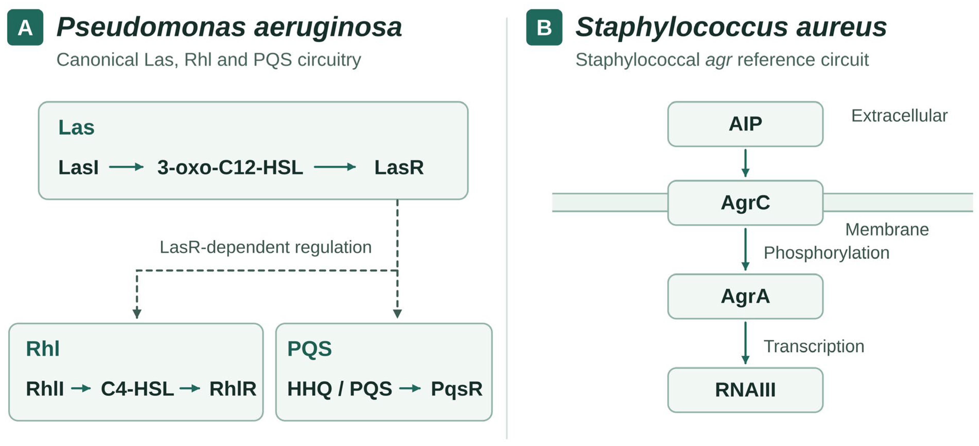
Disclaimer/Publisher’s Note: The statements, opinions and data contained in all publications are solely those of the individual author(s) and contributor(s) and not of MDPI and/or the editor(s). MDPI and/or the editor(s) disclaim responsibility for any injury to people or property resulting from any ideas, methods, instructions or products referred to in the content. |
© 2026 by the authors. Licensee MDPI, Basel, Switzerland. This article is an open access article distributed under the terms and conditions of the Creative Commons Attribution (CC BY) license.