


Simple Summary
Infectious bronchitis (IB) is a highly contagious respiratory viral disease of chickens, causing severe production losses in the poultry industry worldwide. The causative agent, infectious bronchitis virus (IBV), has already been classified into multiple genotypes and lineages, with new variants continually emerging. In this study, we isolated eight novel IBV strains in China during 2024–2026. Phylogenetic analysis of the full S1 gene showed that these strains clustered within the GVI genotype but were clearly distinct from the known GVI-1 lineage, together with two previously reported GVI strains. In addition, these ten GVI strains differed from the GVI-1 reference strain TC07-2 by 12.2–13.1% in nucleotide identity and 15.6–16.9% in amino acid identity across the full S1 gene. The whole-genome analysis of the representative strain HF240371 showed that two recombination events were detected with the complete S gene replaced. On the basis of the analysis of phylogeny, identity and recombination, we propose that these viruses represent the second lineage of GVI-genotype IBV, herein designated GVI-2. Antigenic assays confirmed that GVI-2 represents a serotype distinct from GVI-1. Our findings suggest that novel GVI-2 lineage IBVs have circulated locally in China, highlighting the need for continued molecular surveillance of IBV and for the development of updated vaccines against these emerging variants.
Abstract
As an important poultry pathogen, infectious bronchitis virus (IBV) has been designated as 1 to 10 genotypes comprising 44 lineages, with the continuous emergence of novel genotypes or lineages. In this study, eight novel IBV strains were isolated in China during 2024-2026. Phylogenetic and homology analysis of the whole S1 gene revealed that these novel strains with two previous reported GVI IBVs clustered into the GVI genotype, but were different from the GVI-1 lineage. The pairwise distances of nucleotide and amino acid sequences of the full S1 gene between the ten strains and TC07-2, the representative strain of the GVI-1 lineage, were 12.2–13.1% and 15.6–16.9%, respectively. Recombination analysis of the representative strain HF240371 showed that two recombination events were detected in the whole genome with replacement of the complete S gene. Here, in this study, these novel strains were designated as part of the GVI-2 lineage, representing a new lineage of GVI. Antigenic analysis indicated that these GVI-2 IBVs represent a unique serotype differing from GVI-1 IBVs. All these data not only highlight the emergence of the novel GVI-2 lineage in China, but also emphasize the significance of continued molecular epidemiology of IBV and development of new vaccines against these emerging novel GVI-2 strains.
1. Introduction
Infectious bronchitis virus (IBV), belonging to the gammacoronaviruses, was initially classified into 6 genotypes (GI to GVI), consisting of 32 different lineages based on the S1 gene [1]. Due to the high mutation and recombination rate [2], new genotypes or serotypes continuously emerge [3]. Currently, ten genotypes of IBV have been named, with 44 lineages [4,5,6,7,8,9,10,11,12]. Among these IBV lineages, GI-7, GI-19, GI-22, GI-28, GVI-1, GVII-1 and GVII-2 were first reported in China [9,10,13,14,15], while other genotypes or lineages are distributed worldwide or endemic in specific continents or countries. GVI-1 IBV was first isolated in Guangdong, China, in 2007 [16], and subsequently identified in Korea and Japan [17,18]. Currently, GVI-1 IBV shows an upward trend and tends to be a dominant lineage in some areas of China [19,20,21]. Notably, GVI-1 IBV was initially thought to be a genetically distinct lineage in Asia [1]. However, a recent phylogenetical analysis of IBVs isolated from commercial chickens in Botswana, Eswatini, Namibia and South Africa from 2010 to 2020 revealed that GVI-1 has been present in southern Africa since at least 2010 [22]. Although GVI-1 IBV has spread for a long time in Asia and southern Africa, only one lineage has been reported within the GVI genotype. In this study, eight novel IBVs were isolated from yellow-feather broiler chickens and laying hens with respiratory signs in distinct geographic regions of China (Anhui, Jiangsu, Shanxi and Shandong province) from 2024 to 2026, carrying an S1 gene with high identity with two previous reported strains [19]. Phylogenetic analysis indicated that these ten strains were assigned to monophyletic groups within the GVI genotype, but differed from the GVI-1 lineage. According to the reported designation criteria of “lineage”, a new lineage was arbitrarily assigned to monophyletic groups of at least three viruses sampled from at least two different outbreaks. New definitions of IBV lineages should be undertaken based on pairwise distance comparisons of 13% and 14% for nucleotide and amino acid sequences, respectively, and phylogenetic data together [1]. Based on the phylogeny and identity analysis of the whole S1 gene, these 10 strains identified from distinct provinces in China were designated as a new IBV lineage GVI-2, and the molecular and antigenic characteristics of the S1 gene of these GVI-2 IBVs were analyzed.
2. Materials and Methods
2.1. Samples Collection, Virus Isolation and Sequencing of S1 Gene and Complete Genome
2.1.1. Tissue Samples
The tissue samples, including trachea, lung, kidney and oral swabs, were collected from yellow-feather broiler chickens or laying hens with respiratory signs in the provinces of Anhui, Jiangsu, Shanxi and Shandong. The flocks were all previously immunized with IBV vaccines: spray-vaccinated with H120 (GI-1 lineage) and FNO-E55 (GI-13 lineage) at 1-day-old, followed by booster vaccinations of H120, FNO-E55 or QXL87 (GI-19 lineage) on day 13 or older by eye-drop inoculation.
2.1.2. Virus Isolation
To isolate the viruses, tissue samples were homogenized in phosphate-buffered saline (PBS) with 200 U/mL penicillin and 200 μg/mL streptomycin, followed by 3 freeze–thaw cycles to release virus. Then, the viral RNA of virus solution supernatant was extracted from the allantoic fluids using the VNP-32P automatic nucleic acid extractor (Vazyme, Nanjing, China). Then, the cDNA was synthesized using the HiScript II 1st Strand cDNA Synthesis Kit (Vazyme, Nanjing, China) and identified for IBV by quantitative reverse transcription polymerase chain reaction (RT-qPCR) of the 5b gene as previously described [20]. For the IBV-positive samples, the virus solution supernatant was inoculated into 9-day-old embryonated specific pathogen-free (SPF) chicken eggs, incubated at 37 °C for 72 h followed by 3 blind passages. Then, the IBV isolation was identified by subsequent RT-qPCR of the 5b gene.
2.1.3. RNA Extraction and RT-PCR of S1 Gene and Complete Genome of IBV Isolates
The viral RNA of IBV isolates was extracted and synthesized into cDNA as described in 2.1.2. The full S1 gene of the isolates was amplified as previously described [20]. To amplify the complete genome of IBV, 25 pairs of overlapping primers were designed, as listed in Table 1. The PCR procedures were as follows: 94 °C for 1 min, 35 cycles of 94 °C for 30 s, 53 °C for 30 s, and 72 °C for 1 min, and finally 72 °C for 10 min. The products of the S1 gene and the complete genome were sequenced by Sanger sequencing (Tsingke Biotech Co., Ltd, Shanghai, China). The full-length genomic sequence of the representative strain was assembled using the SeqMan program in DNAStar. To analyze the sequence identities between these novel strains and previously reported strains, the S1 gene sequences were compared against the NCBI (National Center for Biotechnology Information, Bethesda, MD, USA) database using the web-based BLASTN service, (https://blast.ncbi.nlm.nih.gov/Blast.cgi, accessed on 30 September 2026).
Table 1.
PCR amplification primers for the complete genome of IBV.
2.2. Recombination Analysis
The potential recombination events of the S1 gene and the complete genome were detected using the Recombination Detection Program 4.101 (RDP 4, version 4.101) by 7 methods (RDP, GENECONV, BOOTSCAN, MAXCHI, CHIMAERA, SISCAN and 3SEQ). The data sets of S1 and complete genome sequences contained the isolates and reference strains of 44 published lineages of IBV. The recombination events were considered to occur if at least 5 of the 7 methods yield a significance value lower than 10−20 (p value < 10−20) [23]. The recombination analysis was further verified by similarity plot analysis in SimPlot version 3.5.1.
2.3. Analysis of Phylogeny and Diversity of the Full S1 Gene
Phylogenetic analysis of novel IBV strains was performed according to the lineage-nomenclature scheme reported by Valastro [1]. The reference representative strains of 32 lineages ranging from GI-1 to GVI-1 were derived from Valastro’s study. Additional representative strains of newly reported genotypes or lineages in recent years were supplemented, including GI-28, GI-29, two GI-30 lineages, GI-31, GII-2, GVII-1, GVII-2, GVIII, GVIII-1, GIX-1 and GX-1 [4,5,6,7,8,9,10,11,12,24,25], retrieved from GenBank of the National Center for Biotechnology Information (NCBI). A Maximum likelihood tree of S1 nucleotide sequences of representative strains of 44 IBV lineages and ten novel IBV strains was constructed using MEGA 11.0. with 1 000 bootstrap replicates. The inter-genotype phylogenetic tree was constructed using only one reference strain of each lineage to avoid topological alteration caused by an excessive number of isolates. To reveal the phylogenetic relationships among GVI genotype IBV isolates, an intra-genotype phylogenetic tree was also constructed with novel IBVs and 53 GVI-1 lineage strains identified in our previous study. The best-fit nucleotide substitution model was selected by evaluating 24 candidate models in MEGA 11 according to the lowest Bayesian Information Criterion (BIC) score. A General Time Reversible model with Gamma-distributed rates among sites and a proportion of invariant sites (GTR+G+I) was identified as the optimal model. Pairwise identity analysis of novel GVI IBVs and GVI-1 linage strains were performed using Geneious prime (Version 2025), and presented as a heat map using TBtools-II version 2.542 [26].
2.4. Viral Cross-Neutralization Test and Antigenicity Analysis
2.4.1. Chickens, Chicken Embryos, Antisera, Vaccines and GVI Lineage Strains
Specific pathogen-free (SPF) chickens and SPF chicken embryos were purchased from Beijing Merial Vital Laboratory Animal Technology Co., Ltd. (Beijing, China). Vaccine strains H120 (GI-1 lineage), FNO-E55 (GI-13 lineage) and QXL87 (GI-19 lineage) were supplied by Sinopharm Yangzhou VAC Biological Engineering Co., Ltd. (Yangzhou, China), Ceva Animal Health Co., Ltd. (Hangzhou, China) and QYH Biotech Co., Ltd., respectively. The antisera of vaccines were prepared in our previous study. To investigate the molecular and antigenic characteristics of these novel strains within the GVI genotype, fifty-three GVI-1 lineage IBV strains identified between 2021 and 2024, including 24 strains isolated in our previous study [20], were included. All animal experiments were performed in accordance with the institutional animal care guidelines approved by the Animal Care Committee at Yangzhou University in China. The ethical permission code (no. 2025-SYXY-08, licensed on 21 April 2025) was provided by the Animal Care Committee of Yangzhou University.
2.4.2. Viral Cross-Neutralization Test
SPF chickens were used to prepare the antisera of GVI genotype IBV isolates, the procedures were as follows: 4-week-old SPF chickens were immunized with the live IBV isolates purified using the limited dilution method at a dose of 104 EID50 by the oculonasal route. After two weeks, every chicken received a booster immunization with the same inactivated IBV isolates at a dose of 105 EID50 by subcutaneous injection. Three weeks after booster immunization, the sera of immunized chickens were collected and checked for viral neutralization titers. For the beta (two-way) neutralization tests, antisera were first inactivated at 56 °C for 30 min, and then, the sera were diluted serially ten-fold or two-fold to mixed with the same amount of the purified IBV strains (100 EID50/0.1 mL). After incubation at 37 °C for 1 h, the mixture of virus and serum was inoculated into the allantoic cavity of 10-day-old SPF chicken embryos. The SPF chicken embryos were incubated at 37 °C for six days. For the weak lesion of embryo dwarfing by GVI genotype IBVs, virus proliferation in embryo allantoic fluid was checked by RT-qPCR as described in our previous study [20]. Finally, viral neutralization titers were calculated by the Reed–Muench method [27].
2.4.3. Antigenicity Analysis Between GVI Genotype IBV and Vaccines
To analyze the antigenic relationship among GVI-genotype IBVs, representative strains of GVI-1 lineage isolates were selected by the analysis of phylogeny and pairwise distance of the full S1 gene amino acid sequences. The isolate CK/CH/AH/HF240371 was selected as the representative strain of novel GVI-2 IBV to perform cross-neutralization tests with GVI-1 lineage isolate. The antigenic relatedness (R) between IBV isolates was calculated via double-direction VN titers according to the method of Archetti and Horsfall [28]. The criterion for serotype classification of IBV was performed according to Li et al. [29]: an R value ≥ 0.70 indicates antigenic identity, 0.70 > R ≥ 0.33 indicates antigenic relatedness (minor subtype difference), and 0.33 > R ≥ 0.11 indicates loose relatedness (major subtype difference), whereas R < 0.11 indicates no relatedness at all (serotype difference).
3. Results
3.1. Novel IBV Lineage GVI-2 Emerged in China
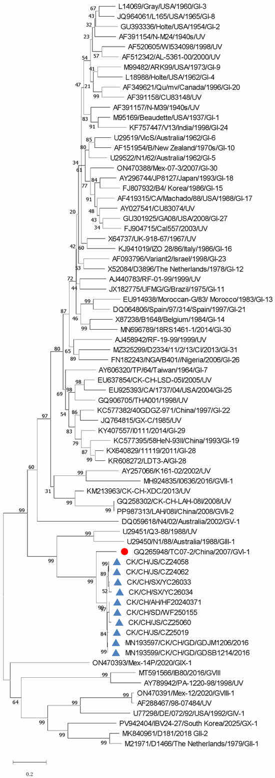
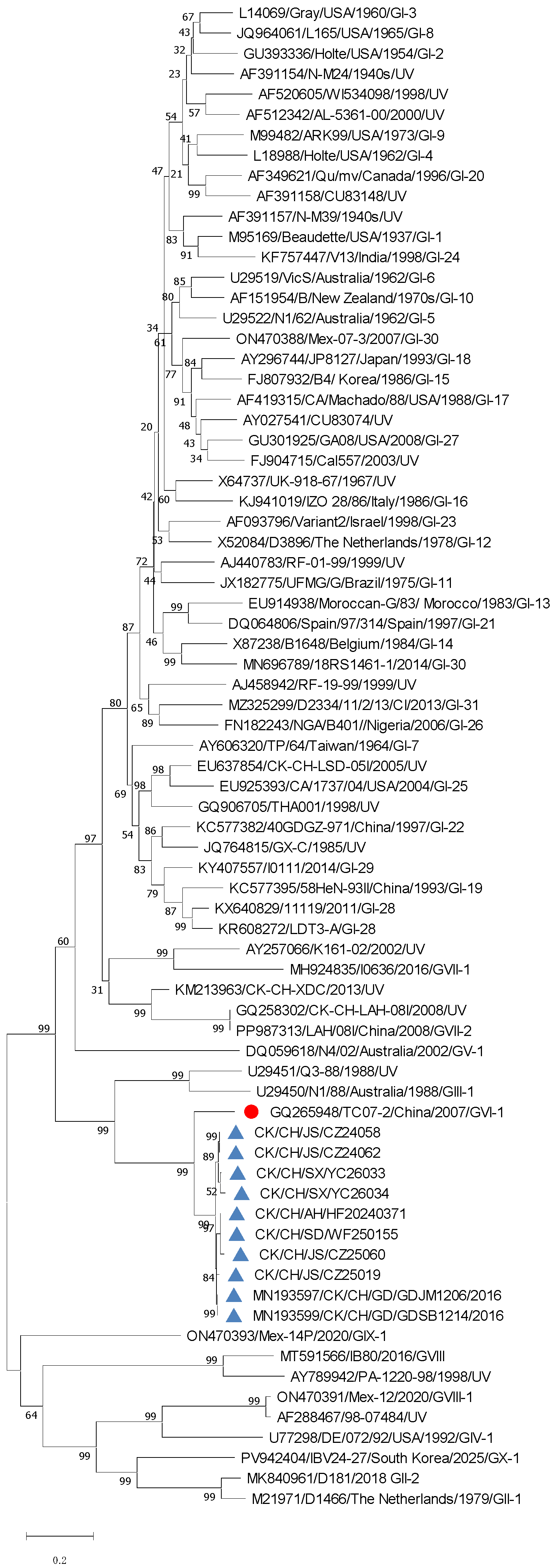
To identify the IBV related with the respiratory diseases in poultry farms with yellow-feather broiler chickens and laying hens, a total of 38 clinical samples including trachea, lung, kidney and oral swabs, were collected from the diseased chickens from Jan. 2024 to Jul. 2026. Eighteen IBV-positive samples were detected and inoculated into embryonated SPF chicken eggs for virus isolation. Finally, eight IBV strains were isolated according to a RT-qPCR assay of embryo allantoic fluid. The IBV strains were named CK/CH/JS/CZ24058(CZ24058), CK/CH/JS/CZ24062(CZ24062), CK/CH/AH/HF20240371(HF240371), CK/CH/JS/CZ25019(CZ25019), CK/CH/JS/CZ25060(CZ25060), CK/CH/SD/WF250155(WF250155), CK/CH/SX/YC26033(YC26033) and CK/CH/SX/YC26034(YC26034), respectively. For brevity, the abbreviations of the eight newly isolated IBV strains are shown in parentheses and the detailed information is listed in Table 2. Phylogenetic analysis of the complete S1 gene of these 8 IBV strains and representative strains of 44 reported IBV lineages showed that these 8 IBV strains have a close relationship with the representative GVI-1-lineage strain TC07-2 (Figure 1). Homology analysis of the full S1 gene indicated that these eight IBVs have 95.3–100% nucleotide identity and 93.6–100% amino acid identity, but only have 86.9–87.8% nucleotide identity (Table 3) and 83.1–84.4% amino acid identity (Table 4) with TC07-2, with the pairwise distances of 12.2–13.1% and 15.6–16.9% for nucleotide and amino acid sequences, respectively. Phylogeny and homology analysis showed that these eight IBVs were novel GVI genotype strains, having great variation from GVI-1 lineage IBVs. To verify this, BLAST analysis using NCBI and detailed phylogeny and homology analysis with more GVI-1 lineage IBV strains were conducted. The BLAST analysis of the full S1 gene of the representative strain HF240371 using NCBI showed that the S1 gene of HF240371 had 98.77% nucleotide similarity with two IBV strains: CK/CH/GD/GDJM1206 (accession no. MN193597) and CK/CH/GD/GDSB1214 (accession no. MN193599), isolated from Guangdong province in China in 2016, whereas the highest similarity with other uploaded IBV strains was only 87.48%.
Table 2.
The eight GVI-2 IBV strains isolated in this study.
Figure 1.
Phylogenetic analysis of ten novel GVI genotype IBVs. The phylogenetic tree was constructed using the full S1 gene of ten novel GVI genotype IBVs, the reference strains of the reported 44 IBV lineages of 10 genotypes, and unclassified Unique variant (UV) strains. Only one reference strain of each lineage was selected for tree construction. Filled red circle indicates the representative strain of the GVI-1 lineage, and blue triangles indicate the 8 strains isolated in our study and two previously reported strains identified in 2016.
Table 3.
The percent identity of S1 nucleotide sequence of 10 novel GVI type IBV isolates and TC07-2.
Table 4.
The percent identity of S1 amino acid sequence of 10 novel GVI type IBV isolates and TC07-2.
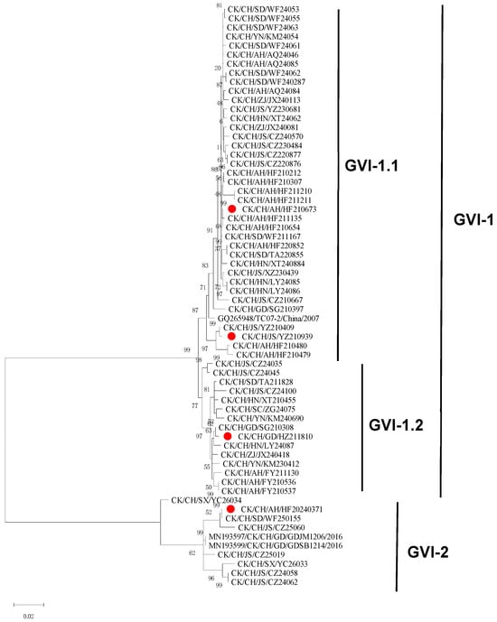
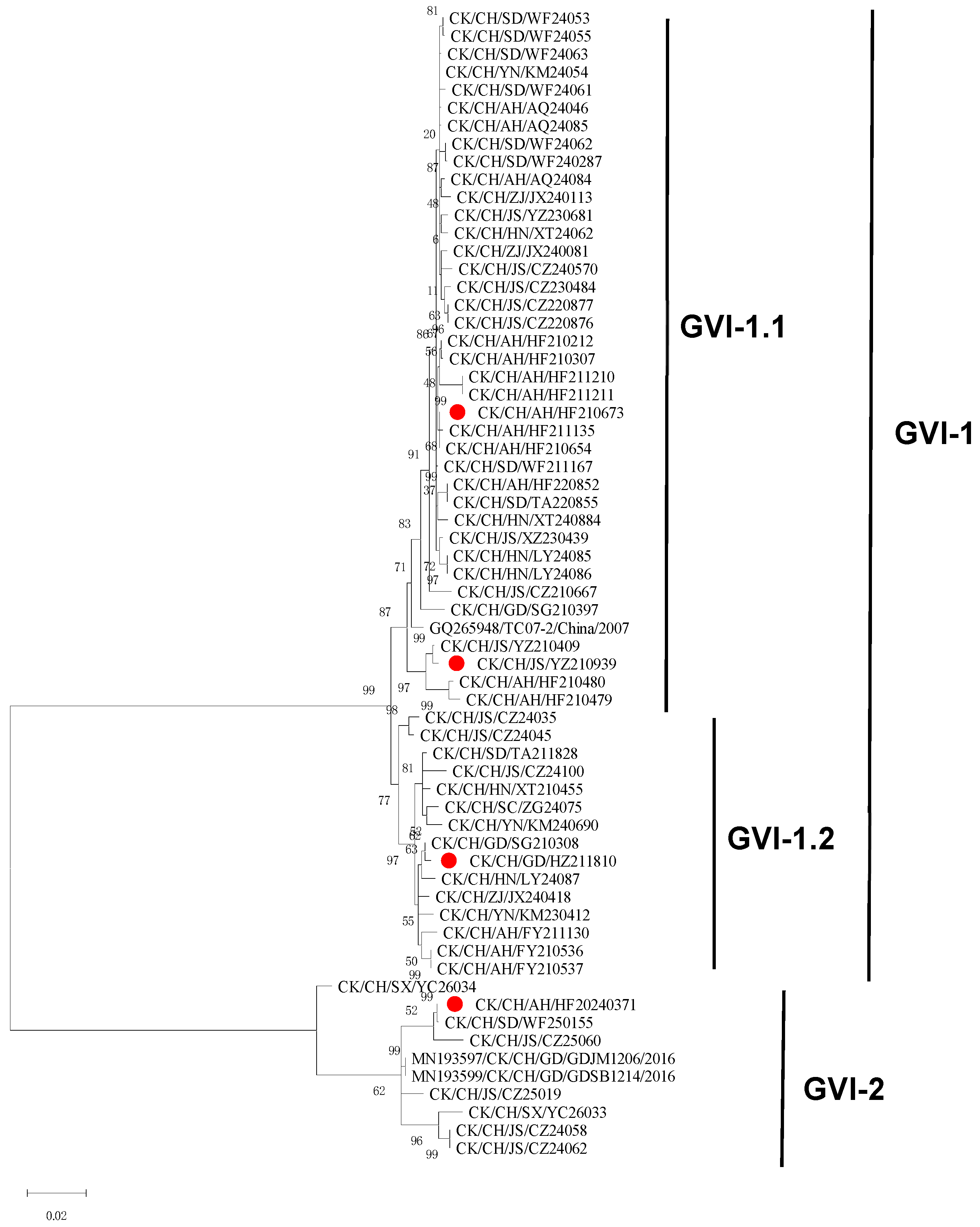
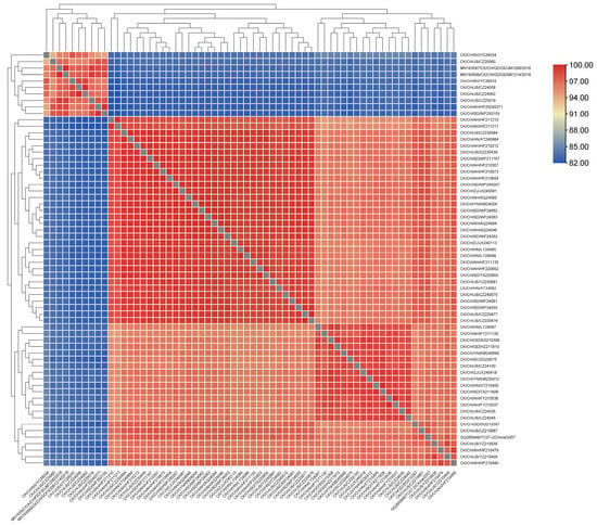
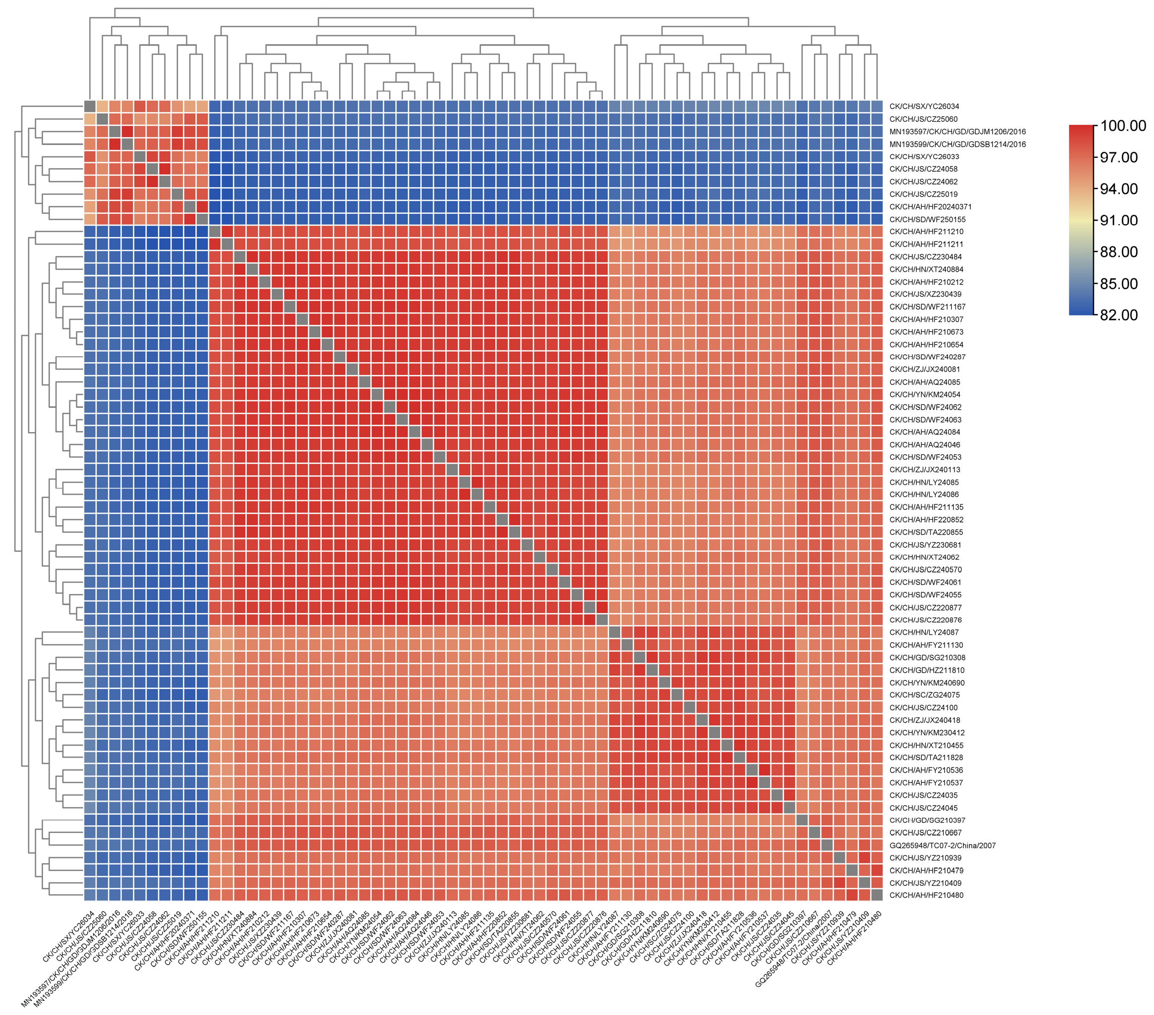
To confirm the variation in these novel GVI strains with GVI-1 lineage IBVs, a detailed phylogenetic analysis and homology analysis of these ten novel IBVs (including CK/CH/GD/GDJM1206 and CK/CH/GD/GDSB1214) and 53 GVI-1 lineage IBV isolates identified in our former study was performed. As shown in Figure 2, a distinct monophyletic branch was formed by these ten novel IBVs, differing from the branch formed by 53 GVI-1 lineage IBV isolates. Homology analysis of S1 gene amino acids (Figure 3) indicated that the ten novel GVI IBVs and 53 GVI-1 lineage IBV isolates formed two distinct monogroups with high intra-group similarity and low inter-group identity: the similarities between the ten novel GVI IBVs and GVI-1 lineage isolates were 93.6–100% and 94.7–100%, respectively, while similarities between the novel IBVs and GVI-1 isolates were 81.8–85.1%. In addition, these eight novel strains identified in our study broke out in different geographic regions far away from each other: HF240371 was isolated in Anhui, CZ24058, CZ24062, CZ25019 and CZ25060 were isolated in Jiangsu province, WF250155 was isolated in Shandong province and YC26033 and YC26034 were identified in Shanxi province in China. Hence, in consideration of the independent detection locations, clustering separately in the phylogenetic tree, and high pairwise distance from GVI-1 lineage IBVs, the novel IBV strains isolated in this study were designated as the new lineage GVI-2.
Figure 2.
Detailed intra-genotype phylogenetic tree of GVI built with 53 GVI-1 lineage IBV isolates identified in our previous study, and the ten novel GVI genotype strains. Filled red circles indicate IBV strains subsequently used for virus neutralization assays. Three strains from different subclades within the GVI-1 lineage and strain HF240371 were selected.
Figure 3.
S1 amino-acid identities among 53 GVI-1 lineage isolates and ten novel GVI genotype IBVs displayed with a color-coded matrix to represent different identity levels (red for high identity, blue for low identity).
3.2. Recombination of the Complete Genome of GVI-2
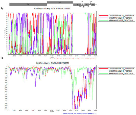
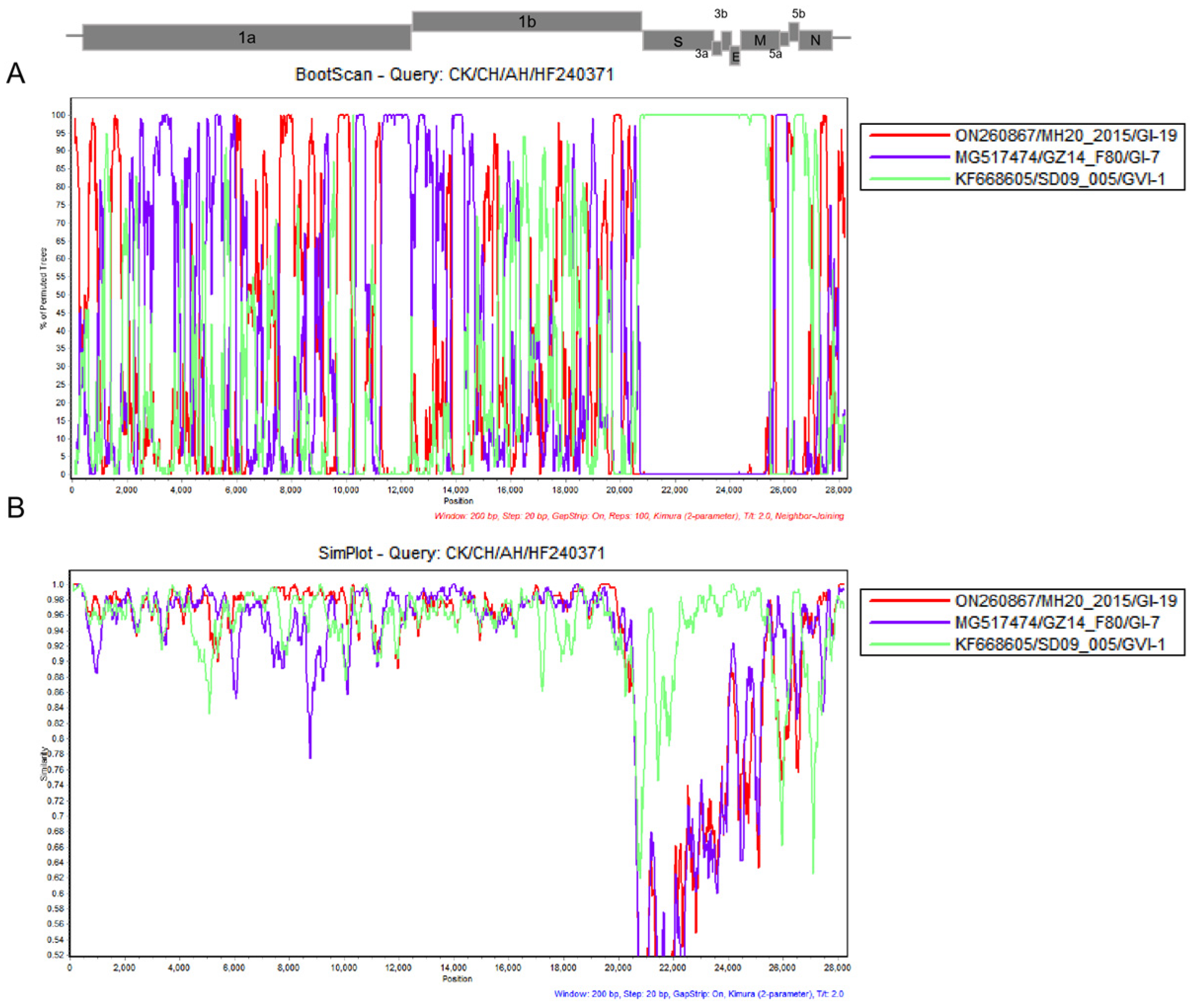
To investigate the origin and evolution of these novel GVI-2 viruses, the full-length genomic sequence of the strain HF240371 was sequenced to conduct genomic recombination analysis. The recombination analysis for the genome of HF240371 was first performed by RDP4. In RDP4, the criteria for identifying recombination events were as follows: a recombination event was considered valid when at least five out of the seven detection methods yielded a p-value < 10−20. As described in Table 5, two recombination events were detected across the whole genome of HF240371, with recombination breakpoints located at 5746/10253 nt and 20207/25008 nt. Notably, due to the limited reference parental sequence dataset, no high-confidence major parental strain was assigned for the recombination event (5746/10253 nt) in the 5′-proximal genomic region, with strain GZ14_F80 (GI-7 lineage) presented as a putative major parent and strain MH20 as a minor parent for this recombinant segment. For the recombination event at the 3′-proximal region (20207/25008 nt), the entire S gene and most of the M gene of HF240371 have relative high identity with GVI-1 lineage strain SD09_005, which was presented as a minor parent. The result of recombination analysis by RDP4 was further confirmed by Simplot (Figure 4). Our results demonstrated that the S1 gene of HF240371 resulted from genetic mutations, rather than recombination, further supporting the designation of the GVI-2 lineage and that recombination events contributed to the origin and evolution of HF240371.
Table 5.
Recombination analysis of the complete genome of CK/CH/AH/HF240371 by RDP4.
Figure 4.
Recombination analysis of strain CK/CH/AH/HF240371 using SimPlot. The complete genomic sequence of the CK/CH/AH/HF240371 strain was used as the query sequence in the SimPlot analysis. Analyses were performed with a window size of 200 bp and a step size of 20 bp. (A) BootScan plot. The y-axis shows the percentage of permuted trees, and the x-axis corresponds to nucleotide positions across the full-length IBV genome. A schematic of the IBV genomic organization is displayed at the top of the panel. (B) Sequence similarity plot. The y-axis indicates pairwise nucleotide sequence similarity, and the x-axis represents genome positions.
3.3. GVI-2 Lineage IBV Exhibited a Unique Serotype
To evaluate the antigenic variation in GVI genotype, IBV vaccine strains and the representative strains of GVI-1 and GVI-2 lineage isolates were selected to conduct the chicken embryo cross-neutralization assays. For the GVI-1 lineage, CK/CH/AH/HF210673 and CK/CH/GD/HZ211810 were selected as the representative strains of the two sub-branches GVI-1.1 and GVI-1.2 to prepare antisera. For sub-branch GVI-1.1, strains CK/CH/JS/YZ210939, CK/CH/AH/HF210480 and CK/CH/AH/HF210479 have relatively low S1 amino acid identities with CK/CH/AH/HF210673: 96.3–96.9% (Figure 3). To confirm the antigenic variation within sub-branch GVI-1.1, CK/CH/JS/YZ210939 was also used to prepare the antiserum for VN tests. For the novel GVI-2 lineage, CK/CH/AH/HF20240371 was selected as the representative strain to conduct the VN tests with GVI-1 lineage strains and vaccine strains. As shown in Table 6, the antisera of vaccine strains H120, FNO-E55 and QXL87 exhibited poor neutralizing activity against both GVI-1 and the newly identified GVI-2 IBV strains. This result indicated the weak cross-protection of current commercial vaccines against GVI-type IBV, which may contribute to the prevalence of GVI-type IBV. However, within the GVI-1 lineage, the antiserum of CK/CH/GD/HZ211810 also had low neutralizing activity against CK/CH/AH/HF210673 and CK/CH/JS/YZ210939, while the antiserum of CK/CH/JS/YZ210939 had a higher neutralizing activity against CK/CH/AH/HF210673 than CK/CH/GD/HZ211810. For GVI-2 lineage, the neutralizing titer of the antiserum of CK/CH/AH/HF20240371 was 3162.27, while the antiserum only had a neutralizing titer of 31.63 against strain CK/CH/AH/HF210673, with a decrease of about 99%. The antiserum of CK/CH/AH/HF210673 also had a weak neutralizing ability against CK/CH/AH/HF20240371, with a decrease of about 78%. These results show that the antigenicity of GVI genotype IBVs is diverse. To evaluate the serotypes of GVI genotype IBV, antigenic relatedness among different strains was calculated based on the criterion that strains with an antigenic relatedness R value < 0.11 belong to distinct serotypes. As shown in Table 7, GVI-2 lineage strain CK/CH/AH/HF20240371 only has a R value of 4.73% with GVI-1 lineage strain CK/CH/AH/HF210673, showing a unique serotype. For GVI-1 lineage strains, the R values of CK/CH/GD/HZ211810 with CK/CH/AH/HF210673 and CK/CH/JS/YZ210939 were 4.79% and 3.72%, respectively, while the R value of CK/CH/AH/HF210673 and CK/CH/JS/YZ210939 was 15.49%. The analysis of R values revealed that the GVI-1 isolates comprise two serotypes, with strain CK/CH/AH/HF210673 serving as the main serotype and the antigenicity of GVI-2 isolates was distinct from that of GVI-1 isolates, representing a novel serotype.
Table 6.
The VN tests of the representative IBV isolates of GVI and vaccine strains.
Table 7.
The antigenicity correlation R value of the strains of the representative GVI strains and vaccines.
4. Discussion
As a coherent and uniform nomenclature of IBV, the lineage classification method for IBV based on the S1 gene provides the rule to identify novel variants consistently. Since the 6 genotypes of IBV with 32 lineages were designated in 2016 [1], four novel genotypes with 12 lineages (GI-28, GI-29, two GI-30 lineages, GI-31, GII-2, GVII-1, GVII-2, GVIII, GVIII-1, GIX, GX-1) have been reported [4,5,6,7,8,9,10,11,12,24,25]. Most of these newly identified lineages were previously isolated in specific area(s) with subsequently increased prevalence in other regions, such as GI-28, GI-30, GVII-2, GVIII and GVIII-1. In this study, eight novel IBV strains were identified and classified as GVI genotype based on the full S1 gene. BLAST analysis showed that these novel strains only have up to 87.48% similarity in the S1 gene with the IBVs published, except for two strains CK/CH/GD/GDJM1206 and CK/CH/GD/GDSB1214 which had an identity of 98.77%. GDSB1214 and GDJM1206, initially classified as GVI genotype IBV, were identified in 2016 from Guangdong province of China, with S1 nucleotide identity of 100% [19]. The 8 novel IBV strains in our study were isolated from four provinces (Anhui, Jiangsu, Shandong and Shanxi), the main poultry farming provinces in eastern China during 2024 and 2026, which are far away from each other and from Guangdong province. In addition, detailed phylogenic analysis of S1 gene of these 10 strains and 53 GVI-1 lineage strains showed that 10 novel strains were assigned to a monophyletic group differing from the GVI-1 lineage. As the designation of “lineage” is assigned to monophyletic groups of at least three viruses sampled from at least two different outbreaks, the GVI-2 lineage was named in our study, increasing the number of IBV lineages to 45.
As the surveyed chicken flocks were all immunized with live vaccines H120 (GI-1 lineage), FNO-E55 (GI-13 lineage) and QXL87 (GI-19 lineage) or LDT3 (GI-28 lineage), the prevalence of GVI-2 may result from the poor protection from the current live IBV vaccines. To verify this, the antisera of vaccine strains H120, FNO-E55 and QXL87 prepared in our previous study were used to perform the viral neutralization test against HF240371, the representative strain of the GVI-2 lineage. The result indicated the poor neutralization ability of the above three vaccine strains against HF240371, which was consistent with the VN tests between with GVI-1 lineage IBV vaccine strains. Since GVI-1 lineage IBV has become one of the most prevalent lineages in recent epidemiological surveys in China and Africa [21,22], the GVI-2 lineage may also emerge as one of the dominant lineages. Notably, multiple serotypes have been identified within a single lineage, such as lineage GI-19 [20] and GI-22 [30,31,32]. For the GVI-1 lineage, antigenic variation was also found in a previous study [33]. In this study, VN tests were also performed to assess the antigenic relatedness among GVI IBV isolates and vaccines. To make a definitive judgment on the number of serotypes among GVI genotype, a relatively strict reference criterion was adopted [29]: strains with an antigenic relatedness coefficient R value < 0.11 were defined as distinct serotypes. The result of antigenic analysis indicated that GVI-1 lineage IBVs comprised 2 serotypes, differing from HF240371 (GVI-2 lineage). As the nucleotide and amino acid consistency of GVI-1 lineage isolates were higher than those of GVI-2, antigenic variation may also exist among GVI-2 lineage strains.
5. Conclusions
In summary, this is the first demonstration of the identification of novel IBV lineage GVI-2, which has circulated locally in China, and shows distinct antigenicity with the current IBV vaccines and GVI-1 lineage. Therefore, the continued study of the molecular epidemiology of GVI-2 and development of efficient control strategies against GVI-2 are badly needed.
Author Contributions
Conceptualization, X.M.; methodology, X.M.; validation, J.Z., X.C. and W.Z.; formal analysis, Z.W., S.W. and T.L.; investigation, X.M., X.C. and J.Z.; resources, J.Y., Q.S. and J.Z.; data curation, X.M. and Q.X.; writing—original draft preparation, X.M.; writing—review and editing, J.Y., Q.S., A.Q. and H.S.; visualization, J.Y.; supervision, J.Y. and Q.S.; project administration, J.Y. and Q.S.; funding acquisition, J.Y. and Q.S. All authors have read and agreed to the published version of the manuscript.
Funding
This study was funded by Poultry innovation team building projects of Shandong province modern agricultural industry technology system (SDAIT-13-011-05), Jiangsu Province Agricultural Science and Technology Independent Innovation Fund Project (CX(22)2047), the Basic Research Program of Jiangsu Province (BK20220579), the 111 Project (D18007), and the Priority Academic Program Development of Jiangsu Higher Education Institutions.
Institutional Review Board Statement
All animal experiments were performed in accordance with the institutional animal care guidelines approved by the Animal Care Committee at Yangzhou University in China. The ethical permission code (no. 2025-SYXY-08, licensed on 21 April 2025) was provided by the Animal Care Committee of Yangzhou University.
Informed Consent Statement
Written informed consent has been obtained from the animal owner to publish this paper.
Data Availability Statement
The original contributions presented in this study are included in the article. Further inquiries can be directed to the corresponding authors.
Conflicts of Interest
Jianjun Zhang, Wei Zhang and Shengnan Wang are from Sinopharm Yangzhou VAC Biological Engineering Co., Ltd., Yangzhou, China. The authors declare that the research was conducted in the absence of any commercial or financial relationships that could be construed as potential conflicts of interest.
Abbreviations
The following abbreviations are used in this manuscript:
| IB | Infectious Bronchitis |
| IBV | Infectious Bronchitis Virus |
| RT-PCR | Reverse Transcription Polymerase Chain Reaction |
| RT-qPCR | Reverse Transcription Quantitative Polymerase Chain Reaction |
| EID50 | Egg Infections Dose 50% |
References
- Valastro, V.; Holmes, E.C.; Britton, P.; Fusaro, A.; Jackwood, M.W.; Cattoli, G.; Monne, I. S1 gene-based phylogeny of infectious bronchitis virus: An attempt to harmonize virus classification. Infect. Genet. Evol. 2016, 39, 349–364. [Google Scholar] [CrossRef] [Scilit] [PubMed]
- Thor, S.W.; Hilt, D.A.; Kissinger, J.C.; Paterson, A.H.; Jackwood, M.W. Recombination in avian gamma-coronavirus infectious bronchitis virus. Viruses 2011, 3, 1777–1799. [Google Scholar] [CrossRef] [Scilit] [PubMed]
- Jackwood, M.W. Review of infectious bronchitis virus around the world. Avian Dis. 2012, 56, 634–641. [Google Scholar] [CrossRef] [Scilit] [PubMed]
- Petzoldt, D.; Vogel, N.; Bielenberg, W.; Haneke, J.; Bischoff, H.; Liman, M.; Rönchen, S.; Behr, K.P.; Menke, T. IB80—A Novel Infectious Bronchitis Virus Genotype (GVIII). Avian Dis. 2022, 66, 291–298. [Google Scholar] [CrossRef] [Scilit] [PubMed]
- Molenaar, R.J.; Dijkman, R.; de Wit, J.J. Characterization of infectious bronchitis virus D181, a new serotype (GII-2). Avian Pathol. 2020, 49, 243–250. [Google Scholar] [CrossRef] [Scilit] [PubMed]
- Mendoza-González, L.; Marandino, A.; Panzera, Y.; Tomás, G.; Williman, J.; Techera, C.; Gayosso-Vázquez, A.; Ramírez-Andoney, V.; Alonso-Morales, R.; Realpe-Quintero, M.; et al. Research Note: High genetic diversity of infectious bronchitis virus from Mexico. Poult. Sci. 2022, 101, 102076. [Google Scholar] [CrossRef] [Scilit] [PubMed]
- Marandino, A.; Mendoza-González, L.; Panzera, Y.; Tomás, G.; Williman, J.; Techera, C.; Gayosso-Vázquez, A.; Ramírez-Andoney, V.; Alonso-Morales, R.; Realpe-Quintero, M.; et al. Genome Variability of Infectious Bronchitis Virus in Mexico: High Lineage Diversity and Recurrent Recombination. Viruses 2023, 15, 1581. [Google Scholar] [CrossRef] [Scilit] [PubMed]
- Ma, T.; Xu, L.; Ren, M.; Shen, J.; Han, Z.; Sun, J.; Zhao, Y.; Liu, S. Novel genotype of infectious bronchitis virus isolated in China. Vet. Microbiol. 2019, 230, 178–186. [Google Scholar] [CrossRef] [Scilit] [PubMed]
- Jiang, L.; Zhao, W.; Han, Z.; Chen, Y.; Zhao, Y.; Sun, J.; Li, H.; Shao, Y.; Liu, L.; Liu, S. Genome characterization, antigenicity and pathogenicity of a novel infectious bronchitis virus type isolated from south China. Infect. Genet. Evol. 2017, 54, 437–446. [Google Scholar] [CrossRef] [Scilit] [PubMed]
- Chen, Y.; Jiang, L.; Zhao, W.; Liu, L.; Zhao, Y.; Shao, Y.; Li, H.; Han, Z.; Liu, S. Identification and molecular characterization of a novel serotype infectious bronchitis virus (GI-28) in China. Vet. Microbiol. 2017, 198, 108–115. [Google Scholar] [CrossRef] [Scilit] [PubMed]
- Brown Jordan, A.; Fusaro, A.; Blake, L.; Milani, A.; Zamperin, G.; Brown, G.; Carrington, C.V.F.; Monne, I.; Oura, C.A.L. Characterization of novel, pathogenic field strains of infectious bronchitis virus (IBV) in poultry in Trinidad and Tobago. Transbound. Emerg. Dis. 2020, 67, 2775–2788. [Google Scholar] [CrossRef] [Scilit] [PubMed]
- Bali, K.; Kaszab, E.; Marton, S.; Hamdiou, S.H.; Bentaleb, R.K.; Kiss, I.; Palya, V.; Bányai, K. Novel Lineage of Infectious Bronchitis Virus from Sub-Saharan Africa Identified by Random Amplification and Next-Generation Sequencing of Viral Genome. Life 2022, 12, 475. [Google Scholar] [CrossRef] [Scilit] [PubMed]
- Huang, Y.P.; Lee, H.C.; Cheng, M.C.; Wang, C.H. S1 and N gene analysis of avian infectious bronchitis viruses in Taiwan. Avian Dis. 2004, 48, 581–589. [Google Scholar] [CrossRef] [Scilit] [PubMed]
- Liu, S.W.; Zhang, Q.X.; Chen, J.D.; Han, Z.X.; Liu, X.; Feng, L.; Shao, Y.H.; Rong, J.G.; Kong, X.G.; Tong, G.Z. Genetic diversity of avian infectious bronchitis coronavirus strains isolated in China between 1995 and 2004. Arch. Virol. 2006, 151, 1133–1148. [Google Scholar] [CrossRef] [Scilit] [PubMed]
- Wang, Y.; Wang, Y.; Zhang, Z.; Fan, G.; Jiang, Y.; Liu, X.; Ding, J.; Wang, S. Isolation and Identification of Glandular Stomach Type IBV (QX IBV) in Chickens. Chin. J. Anim. Quar. 1998, 15, 1–13. [Google Scholar]
- Li, L.; Xue, C.; Chen, F.; Qin, J.; Xie, Q.; Bi, Y.; Cao, Y. Isolation and genetic analysis revealed no predominant new strains of avian infectious bronchitis virus circulating in South China during 2004–2008. Vet. Microbiol. 2010, 143, 145–154. [Google Scholar] [CrossRef] [Scilit] [PubMed]
- Mase, M.; Kawanishi, N.; Ootani, Y.; Murayama, K.; Karino, A.; Inoue, T.; Kawakami, J. A novel genotype of avian infectious bronchitis virus isolated in Japan in 2009. J. Vet. Med. Sci. 2010, 72, 1265–1268. [Google Scholar] [CrossRef] [Scilit] [PubMed]
- Lim, T.H.; Kim, M.S.; Jang, J.H.; Lee, D.H.; Park, J.K.; Youn, H.N.; Lee, J.B.; Park, S.Y.; Choi, I.S.; Song, C.S. Live attenuated nephropathogenic infectious bronchitis virus vaccine provides broad cross protection against new variant strains. Poult. Sci. 2012, 91, 89–94. [Google Scholar] [CrossRef] [Scilit] [PubMed]
- Chen, L.; Xiang, B.; Hong, Y.; Li, Q.; Du, H.; Lin, Q.; Liao, M.; Ren, T.; Xu, C. Phylogenetic analysis of infectious bronchitis virus circulating in southern China in 2016–2017 and evaluation of an attenuated strain as a vaccine candidate. Arch. Virol. 2021, 166, 73–81. [Google Scholar] [CrossRef] [Scilit] [PubMed]
- Meng, X.; Zhang, J.; Wan, Z.; Li, T.; Xie, Q.; Qin, A.; Shao, H.; Zhang, H.; Ye, J. Molecular epidemiology of infectious bronchitis virus in eastern and southern China during 2021–2023. Poult. Sci. 2024, 103, 103939. [Google Scholar] [CrossRef] [Scilit] [PubMed]
- Wang, Y.; Li, Y.; Yu, X.; Wang, Y.; Cai, C.; Li, W.; Liu, H.; Liang, L. Epidemiological surveillance of avian coronavirus in China in 2024. Poult. Sci. 2025, 104, 105251. [Google Scholar] [CrossRef] [Scilit] [PubMed]
- Strydom, C.; Abolnik, C. Seven infectious bronchitis virus genotypes including South American-origin G1-11 and Asian-origin GVI-1 circulated in southern African poultry from 2010 to 2020. Virus Res. 2025, 355, 199568. [Google Scholar] [CrossRef] [Scilit] [PubMed]
- Wu, Z.; Fang, H.; Xu, Z.; Lian, J.; Xie, Z.; Wang, Z.; Qin, J.; Huang, B.; Feng, K.; Zhang, X.; et al. Molecular Characterization Analysis of Prevalent Infectious Bronchitis Virus and Pathogenicity Assessment of Recombination Strain in China. Front. Vet. Sci. 2022, 9, 842179. [Google Scholar] [CrossRef] [Scilit] [PubMed]
- Wang, B.; Zhang, X.; Han, Z.; Li, H.; Sun, J.; Liu, S. A novel IBV Lineage within Genotype GVII (GVII-2) Representing a Unique Serotype Circulating in Chicken Flocks in China. Avian Dis. 2026, 70, 174–182. [Google Scholar] [CrossRef] [Scilit] [PubMed]
- Kim, S.-J.; Kim, H.-W.; Ahn, S.-M.; Son, S.-E.; Song, J.-H.; Kim, Y.-B.; Kwon, H.-J.; Choi, K.-S. Genetic, pathogenic, and antigenic characterization of GX-1, a novel infectious bronchitis virus genotype identified in South Korea. Poult. Sci. 2026, 105, 106217. [Google Scholar] [CrossRef] [Scilit] [PubMed]
- Feng, J.; Chen, C.; Wu, Y.; Fan, L.; Zhang, Z.; Chen, M.; Tang, H.; Yu, G.; Ren, J.; Luo, J.; et al. How to make big data accessible to plant biologists and beyond: Ten years of lessons from TBtools. Mol. Plant 2026, 19, 1378–1391. [Google Scholar] [CrossRef] [Scilit] [PubMed]
- Reed, L.J.; Muench, H. A simple method of estimating fifty percent endpoints. Am. J. Epidemiol. 1938, 27, 493–497. [Google Scholar] [CrossRef] [Scilit]
- Archetti, I.; Horsfall, F.L. Persistent antigenic variations of influenza A virus after incomplete neutralization in ovo with heterologous immune serum. Rend.—Ist. Super. Sanita 1951, 14, 909–936. [Google Scholar]
- Li, S.; Du, L.; Xia, J.; Du, J.; You, G.; Wen, Y.; Huang, X.; Zhao, Q.; Han, X.; Yan, Q.; et al. Antigenic and Pathogenic Characteristics of QX-Type Avian Infectious Bronchitis Virus Strains Isolated in Southwestern China. Viruses 2019, 11, 1154. [Google Scholar] [CrossRef] [Scilit] [PubMed]
- Li, M.; Wang, X.Y.; Wei, P.; Chen, Q.Y.; Wei, Z.J.; Mo, M.L. Serotype and genotype diversity of infectious bronchitis viruses isolated during 1985–2008 in Guangxi, China. Arch. Virol. 2012, 157, 467–474. [Google Scholar] [CrossRef] [Scilit] [PubMed]
- Wang, X.Y.; Li, M.; Wei, P.; Chen, Q.Y.; Wei, Z.J.; Mo, M.L.; Wei, T.C. Evaluation of antigenic relationship of Guangxi isolates of infectious bronchitis virus. Bing Du Xue Bao = Chin. J. Virol. 2012, 28, 621–627. [Google Scholar]
- Meng, X.; Li, N.; Xu, J.; Wei, Y.; Zhang, J.; Wan, Z.; Li, T.; Xie, Q.; Qin, A.; Shao, H.; et al. The molecular, antigenic, and pathogenic characterization of novel IBV of GI-22 lineage endemic in China. Virology 2026, 617, 110826. [Google Scholar] [CrossRef] [Scilit] [PubMed]
- Ren, M.; Sheng, J.; Ma, T.; Xu, L.; Han, Z.; Li, H.; Zhao, Y.; Sun, J.; Liu, S. Molecular and biological characteristics of the infectious bronchitis virus TC07-2/GVI-1 lineage isolated in China. Infect. Genet. Evol. 2019, 75, 103942. [Google Scholar] [CrossRef] [Scilit] [PubMed]

Disclaimer/Publisher’s Note: The statements, opinions and data contained in all publications are solely those of the individual author(s) and contributor(s) and not of MDPI and/or the editor(s). MDPI and/or the editor(s) disclaim responsibility for any injury to people or property resulting from any ideas, methods, instructions or products referred to in the content. |
© 2026 by the authors. Licensee MDPI, Basel, Switzerland. This article is an open access article distributed under the terms and conditions of the Creative Commons Attribution (CC BY) license.