


Simple Summary
Lipopolysaccharide is recognized as one of the commonly encountered stressors in production, exerting negative impacts on broiler performance. Chitooligosaccharides, the degraded derivatives of chitin and chitosan, possess potent anti-inflammatory, antioxidant, and gut microbiota-regulating properties. Therefore, this research aimed to explore the impacts of chitooligosaccharide on growth, immune response, antioxidant capacity, gut barrier integrity and microbiota in lipopolysaccharide-stimulated broilers. The results showed that dietary supplementation with chitooligosaccharide effectively alleviated lipopolysaccharide-induced acute BW loss and intestinal barrier dysfunction via suppressing inflammatory responses, alleviating oxidative stress, and regulating gut microbiota. These findings indicate a beneficial role of chitooligosaccharide in counteracting stress-evoked intestinal injury and acute BW loss in broilers.
Abstract
This research was performed to assess the impacts of chitooligosaccharide (COS) on growth, immune response, antioxidant status, intestinal barrier integrity and microbiota in lipopolysaccharide (LPS)-challenged broiler chickens. The 90-day-old chicks (Arbor Acres) were randomly divided into three treatments, with each treatment containing five replicates of six chicks. The treatments were the unchallenged group supplied a basal diet (CON), the LPS-challenged group supplied the same basal diet (LPS), and the LPS-challenged group supplied the basal diet supplemented with 300 mg/kg COS (LPS + COS). On d 21, birds in the LPS and LPS + COS groups were administered an intraperitoneal injection of LPS (5 mg/kg BW). The results showed that COS effectively alleviated (p < 0.05) LPS-induced BW loss. Dietary COS supplementation significantly suppressed (p < 0.05) LPS-induced serum interleukin-1β production and increased (p < 0.05) serum interleukin-10 concentration. COS also significantly attenuated (p < 0.05) the LPS-mediated reduction in serum and jejunal T-AOC activity and inhibited (p < 0.05) the elevation of jejunal MDA concentration. Relative to the LPS group, COS markedly decreased (p < 0.05) serum endotoxin concentration and elevated (p < 0.05) jejunal ZO-1 and occludin expression. In addition, COS supplementation decreased (p < 0.05) the abundance of Sellimonas relative to the LPS group, and enriched (p < 0.05) the abundance of Blautia compared to the CON group. Collectively, dietary COS supplementation could mitigate LPS-triggered inflammation, oxidative stress, intestinal barrier damage and microbiota dysbiosis, thus improving intestinal barrier function and attenuating acute BW loss in broilers. These findings suggest that COS is a potential feed additive for mitigating stress-induced intestinal injury and acute BW loss in broilers.
1. Introduction
Driven by the ever-growing demands for chicken meat, commercial broiler production has evolved into a modern intensive farming system, with the primary goal of maximizing productive efficiency [1]. Furthermore, advances in genetic selection and nutrition have collectively contributed to faster growth rates, lower feed conversion ratios and higher metabolic rates in broilers, which have also dramatically improved the productivity and efficiency of broiler production [2,3]. However, modern fast-growing strains have been shown to be highly vulnerable to stress, making them susceptible to diseases [4]. Unfortunately, in the intensive farming system, broilers are frequently confronted with a variety of stressors, which consequently results in immunological stress and impaired growth performance [5]. The intestine is extremely prone to immunological stress due to constant exposure to a myriad of antigens derived from feed, invading bacteria and viruses [6]. Immunological stress has been demonstrated to trigger intestinal inflammation, disrupt intestinal barrier function, induce intestinal microbial dysbiosis, ultimately leading to enteric diseases and compromised growth performance in broiler chickens [7,8,9].
For the past decades, antibiotics have been routinely employed in broiler production to mitigate immunological stress and control secondary bacterial infections with considerable success [10,11]. However, the rise and spread of antibiotic resistance has become a global public health concern, which resulted in regulatory restrictions on the use of antibiotics in broiler production [12]. Consequently, it is imperative to develop antibiotic-free solutions for alleviating immunological stress and maintaining intestinal health of broilers.
Chitooligosaccharide (COS) is the degradation product of chitosan or chitin prepared by chemical or enzymatic hydrolysis [13]. It is an oligomer of β-(1,4)-linked D-glucosamine with the degree of polymerization less than 20 [14]. Compared with chitosan and chitin, COS exhibits higher water solubility and lower viscosity, thereby rendering it readily absorbable by the intestinal tract [15]. In addition, COS has been demonstrated to exhibit diverse bioactive properties, such as anti-inflammation, anti-oxidation, anti-bacteria, anti-tumor, and anti-obesity [16,17,18,19]. Previous studies conducted on broilers have shown that COS can improve intestinal morphology, enhance immune function, antioxidant capacity and microbiota [20,21]. Furthermore, previous work has also demonstrated the protective effects of chitooligosaccharide against stress-induced intestinal injury [22,23,24]. Nevertheless, these findings were obtained from different animal models or in vitro systems. So far, the role of COS in relieving detrimental effects associated with immunological stress in broiler chickens remains unclear.
Lipopolysaccharide (LPS), an essential constituent of the outer membrane of Gram-negative bacteria, acts as a powerful inducer of the host’s inflammatory response [25]. A multitude of studies have confirmed that intraperitoneal injection of LPS has been widely adopted to establish an immunological stress model that can induce intestinal inflammation, impair intestinal barrier integrity and ultimately reduce growth performance in broilers [26,27]. Therefore, in the present study, an immunological stress model of broilers was established by intraperitoneal injection of LPS. The protective capacity of COS to mitigate LPS-provoked intestinal injury in broiler chickens was then investigated.
2. Materials and Methods
2.1. Animal Ethics
Animal procedures in the current study were examined and authorized by the Institutional Animal Care and Use Committee of Shandong Academy of Agricultural Sciences (Jinan, China; approval number: SAAS-2024-G24).
2.2. Broiler Husbandry
A total of 90 one-day-old male Arbor Acres broiler chicks were reared in stainless steel cages inside a climate-controlled room. Each cage was fitted with a feeding trough and nipple waterer to guarantee free access to feed and drinking water for broilers during the whole trial. The indoor temperature was kept at 33 °C in the initial 7 days, followed by a weekly decline of 3 °C until it stabilized at 27 °C. A 23 h light:1 h dark lighting regimen was applied across the entire experimental phase. All chicks received vaccinations with the inactivated Newcastle disease vaccine on day 7, as well as the inactivated infectious bursal disease vaccine on day 14.
2.3. Diets and Experimental Design
Following arrival, broilers were randomly assigned to 3 groups (5 replicates/group and 6 birds/replicate) in a completely randomized design. The 3 groups were as follows: the unchallenged group was offered the basal diet (CON), the LPS-challenged group was fed the basal diet (LPS), and the LPS-challenged group received the basal diet containing 300 mg/kg COS (LPS + COS). The COS with a deacetylation degree >90.00%, a degree of polymerization ranging from 2 to 6, and an average molecular weight <1000 Da was obtained from Dalian GlycoBio Company, Ltd. (Dalian, China). The COS was premixed with a small portion of the feed, and then the premix was blended gradually with the rest of the diet. The basal diet was prepared to fulfill the nutrient requirements of broilers set by the National Research Council (1994) [28] and supplied in mashed form (Table 1). On d 21, birds in the LPS and LPS + COS groups were intraperitoneally administered LPS from E. coli serotype O55:B5 (L-2880; Sigma-Aldrich, St. Louis, MO, USA) at 5 mg/kg body weight (BW) according to previous studies [29,30], whereas birds in the CON group were administered an equivalent volume of 0.9% physiological saline via the same route. After the LPS challenge, feed was removed while water was continuously available for birds.
Table 1.
Ingredients and nutrient levels of the basal diet (as fed basis, %).
2.4. Sample Collection
At 4 h post-challenge, 1 bird was randomly selected from each replicate cage (5 birds each treatment). Blood samples were collected via the wing vein into vacuum tubes without anticoagulant. Following centrifugation at 3000× g for 15 min under 4 °C conditions, serum was separated and preserved at −80 °C for later assays. Subsequently, the birds were euthanized by cervical dislocation, and the entire gastrointestinal tract was removed. Jejunal mucosal samples and cecal digesta were collected, rapidly frozen in liquid nitrogen and kept at −80 °C for subsequent assays.
2.5. Growth Performance and Body Weight Loss
Prior to the LPS injection, the BW of broilers in each cage was determined on days 1 and 21, and feed consumption per cage was documented. Average daily gain (ADG), average daily feed intake (ADFI), and feed conversion ratio (FCR) were then calculated for each cage. Four hours after injection, all broilers were reweighed, and BW loss was determined according to the formula presented below:
2.6. Determination of Inflammatory Cytokines
Diamine oxidase (DAO), D-lactate, tumor necrosis factor-α (TNF-α), interleukin-1β (IL-1β), IL-6, IL-10 and interferon-γ (IFN-γ) were measured by commercial kits (Nanjing Jiancheng Biotechnology Institute, Nanjing, China) following the guidelines provided by the manufacturer. Serum endotoxin concentration was determined by the Limulus amebocyte lysate assay (Nanjing Jiancheng Biotechnology Institute, Nanjing China). A bicinchoninic acid (BCA) protein quantification kit (Beyotime, Shanghai, China) was adopted to test the total protein concentration of jejunal mucosa. Values of inflammatory cytokines in the jejunum were presented as units per gram of protein.
2.7. Determination of Antioxidant Capacity
Measurements of antioxidant indicators in serum and jejunum were carried out using commercially available kits (Nanjing Jiancheng Biotechnology Institute, Nanjing, China) as guided by the manufacturer, covering superoxide dismutase (SOD), glutathione peroxidase (GSH-Px), catalase (CAT), malondialdehyde (MDA), and total antioxidant capacity (T-AOC). A bicinchoninic acid protein assay kit (Beyotime, Shanghai, China) was applied to assess the total protein concentration of jejunal mucosa. All results from jejunal tissues were normalized to total protein and presented as units per gram of protein.
2.8. Gene Expression Analysis
All assays were performed following protocols reported in our previous research [31]. Briefly, total RNA was isolated from jejunal mucosal samples with TRIZOL reagent (Invitrogen, Carlsbad, CA, USA). The RNA concentration was assessed by a NanoDrop 2000 spectrophotometer (Thermo Scientific, Wilmington, DE, USA), while RNA integrity was evaluated by ethidium bromide-stained agarose gel electrophoresis. Subsequently, reverse transcription was conducted with a PrimeScript® RT reagent kit (TaKaRa, Dalian, China).
RT-PCR was performed on a LightCycler® 480 II Real-time PCR Instrument (Roche, Basel, Switzerland). Primers were referenced in our previous work [29]. The total reaction volume of 10 μL contained 1 μL cDNA, 5 μL 2 × PerfectStartTM Green qPCR SuperMix, 0.2 μL of each primer and 3.6 μL nuclease-free water. The program was an initial pre-denaturation step at 94 °C for 30 s, 45 cycles of 94 °C for 5 s and 60 °C for 30 s. The specificity of each primer pair was assessed by melt curve analysis following RT-qPCR. The observation of a single melting peak for each assay indicated specific amplification of a single amplicon. Amplification efficiencies ranged from 95% to 105%, with the coefficient of determination (R2) exceeding 0.99 for all standard curves. Relative mRNA levels were normalized against GAPDH and quantified via the 2−ΔΔCt approach [32].
2.9. Cecal Microbiota Analysis
Bacterial genomic DNA was isolated from cecal content with a DNA kit (Omega Bio-tek, Norcross, GA, USA) following the manufacturer’s protocols. DNA concentration and integrity were determined following the same procedures described above for RNA. The V3–V4 region of the 16S rRNA gene was amplified with primers 338F and 806R.
The PCR amplification was carried out with the thermal profile described below: initial denaturation (95 °C, 3 min), 27 cycles of denaturation (95 °C, 30 s), annealing (55 °C, 30 s), and extension (72 °C, 45 s), with a final 10 min extension at 72 °C. Each amplification assay was performed in three technical replicates. Target PCR amplicons were separated on a 2% agarose gel, purified with the AxyPrep DNA Gel Extraction Kit (Axygen Biosciences, Union City, CA, USA), and quantified on a Quantus™ Fluorometer (Promega, Madison, WI, USA).
Equimolar mixtures of purified amplicons were prepared and subjected to paired-end sequencing on the Illumina MiSeq PE300 platform (Illumina, San Diego, CA, USA). Raw sequencing data were first demultiplexed, quality trimmed with fastp (v0.20.0), and then assembled using FLASH (v1.2.7) under the filtering thresholds. Sequences were clustered into operational taxonomic units (OTUs) at a 97% identity threshold via UPARSE v7.1, and chimeric sequences were detected and eliminated in this process. Taxonomic annotation for representative OTU sequences was implemented using RDP Classifier v2.2 against the Silva v138 16S rRNA reference database, with a confidence cutoff set at 0.7.
2.10. Statistical Analysis
Data analyses were conducted with SAS 9.2 software (SAS Inst. Inc., Cary, NC, USA). The Shapiro–Wilk test and Levene’s test were separately applied to test data normality and variance homogeneity. Principal coordinate analysis (PCoA) based on Bray–Curtis dissimilarity matrices was adopted to characterize beta diversity. The relative abundance of gut microbiota was compared using the Kruskal–Wallis test. The rest of the data were analyzed via one-way ANOVA followed by Tukey’s test. p < 0.05 was regarded as statistically significant.
3. Results
3.1. Growth Performance and BW Loss
As shown in Table 2, prior to the LPS challenge, dietary COS addition exerted no marked impacts on BW, ADG, ADFI or FCR of broiler chickens. Nevertheless, the LPS challenge remarkably elevated (p < 0.05) BW loss relative to the CON group. Supplementing COS in the diet effectively mitigated (p < 0.05) the BW loss triggered by LPS. Nevertheless, the BW loss remained markedly higher in the LPS + COS group relative to the CON group (p < 0.05).
Table 2.
Effects of chitooligosaccharide on growth performance and body weight loss of broilers before challenge.
3.2. Inflammatory Cytokines in the Serum and Jejunum
Effects of COS on inflammatory cytokines in the serum and jejunum of LPS-challenged broilers were presented in Table 3. The LPS administration led to significantly higher serum TNF-α and IL-1β levels (p < 0.05) in comparison with the CON group. Broilers in the LPS + COS group exhibited an intermediate serum TNF-α concentration, which did not differ significantly from that of either the CON or LPS group. Moreover, dietary COS supplementation markedly inhibited (p < 0.05) serum IL-1β production compared with the LPS group, whereas this cytokine concentration was still higher (p < 0.05) than that of the CON group. Furthermore, supplementation of COS in the basal diet resulted in an elevated (p < 0.05) serum IL-10 concentration compared to both the CON and LPS groups. Serum IL-6 and IFN-γ concentrations were not affected by the treatment.
Table 3.
Effects of chitooligosaccharide on inflammatory cytokines in the serum and jejunum of broilers challenged with LPS.
In the jejunum, the LPS challenge markedly increased (p < 0.05) TNF-α and IL-1β concentrations relative to the CON group. However, the LPS + COS group exhibited similar levels to both the CON group and the LPS group for these two cytokines. No significant differences were found for the concentrations of IL-6, IL-10 and IFN-γ.
3.3. Relative mRNA Expression Levels of Jejunal TLR4 and NF-κB p65
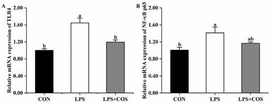
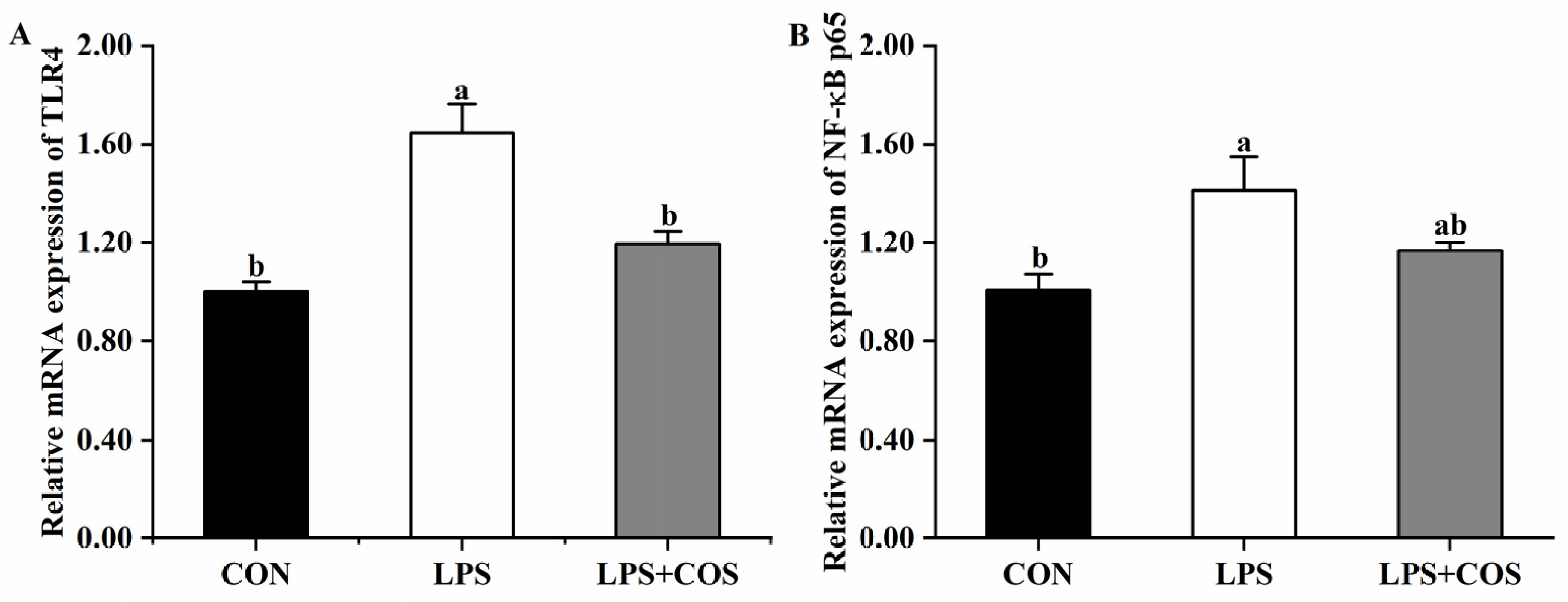
As illustrated in Figure 1, relative to the CON group, LPS significantly upregulated (p < 0.05) jejunal mRNA expression of TLR4 and NF-κB p65, whereas supplementation with COS exerted a marked suppressive effect on the LPS-mediated elevation of TLR4 mRNA expression (p < 0.05). Notably, the LPS + COS group exhibited an intermediate level of NF-κB p65 mRNA expression, which did not differ significantly from that detected in either the CON or LPS group.
Figure 1.
Effects of chitooligosaccharide on relative mRNA expression levels of TLR4 (A) and NF-κB p65 (B) in the jejunum of broilers challenged with LPS. CON, the unchallenged group, fed a basal diet; LPS, the LPS-challenged group, fed the same basal diet; LPS + COS, the LPS-challenged group, fed the basal diet supplemented with 300 mg/kg COS. Values are presented as mean ± SEM (n = 5). Bars without a common letter are significantly different (p < 0.05).
3.4. Antioxidant Capacity in the Serum and Jejunum
Table 4 shows the effects of COS on the antioxidant capacity of serum and jejunum in LPS-challenged broilers. Exposure to LPS markedly reduced (p < 0.05) serum SOD activity and T-AOC compared with the CON group. The LPS + COS group showed an intermediate SOD activity, with no significant difference from the CON group or the LPS group. Supplementation of COS exerted a marked ameliorative effect on the reduction in T-AOC triggered by LPS (p < 0.05); however, the T-AOC level was not restored to the baseline and remained markedly lower compared with the CON group (p < 0.05). Serum GSH-Px and CAT activities, together with MDA concentration, did not differ significantly across treatment groups.
Table 4.
Effects of chitooligosaccharide on antioxidant capacity in the serum and jejunum of broilers challenged with LPS.
In the jejunum, LPS challenge markedly reduced (p < 0.05) SOD activity relative to the CON group, whereas COS supplementation slightly improved SOD activity, with no significant difference from the other two groups. The MDA concentration exhibited remarkable differences across treatment groups, with the maximum level recorded in the LPS group, followed by the LPS + COS group, and the minimum level in the CON group (p < 0.05). Furthermore, LPS challenge reduced (p < 0.05) the T-AOC, while COS supplementation effectively abrogated the LPS-induced reduction in T-AOC (p < 0.05), restoring it to a level comparable to that of the CON group. No marked variations in GSH-Px and CAT activities were detected among treatment groups.
3.5. Intestinal Permeability
As presented in Table 5, a significant increase in serum DAO activity and endotoxin concentration (p < 0.05) was observed in LPS-challenged broilers compared with the CON group. Inclusion of COS remarkably abrogated (p < 0.05) LPS-triggered increase in endotoxin concentration. In contrast, COS addition only caused a marginal reduction in serum DAO activity, with no significant difference detected relative to the other two experimental groups. Furthermore, no notable difference in serum D-lactate level was detected across treatment groups.
Table 5.
Effects of chitooligosaccharide on intestinal permeability in broilers challenged with LPS.
3.6. Jejunal Tight Junction Protein Gene Expression
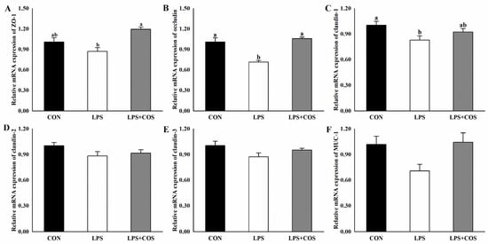
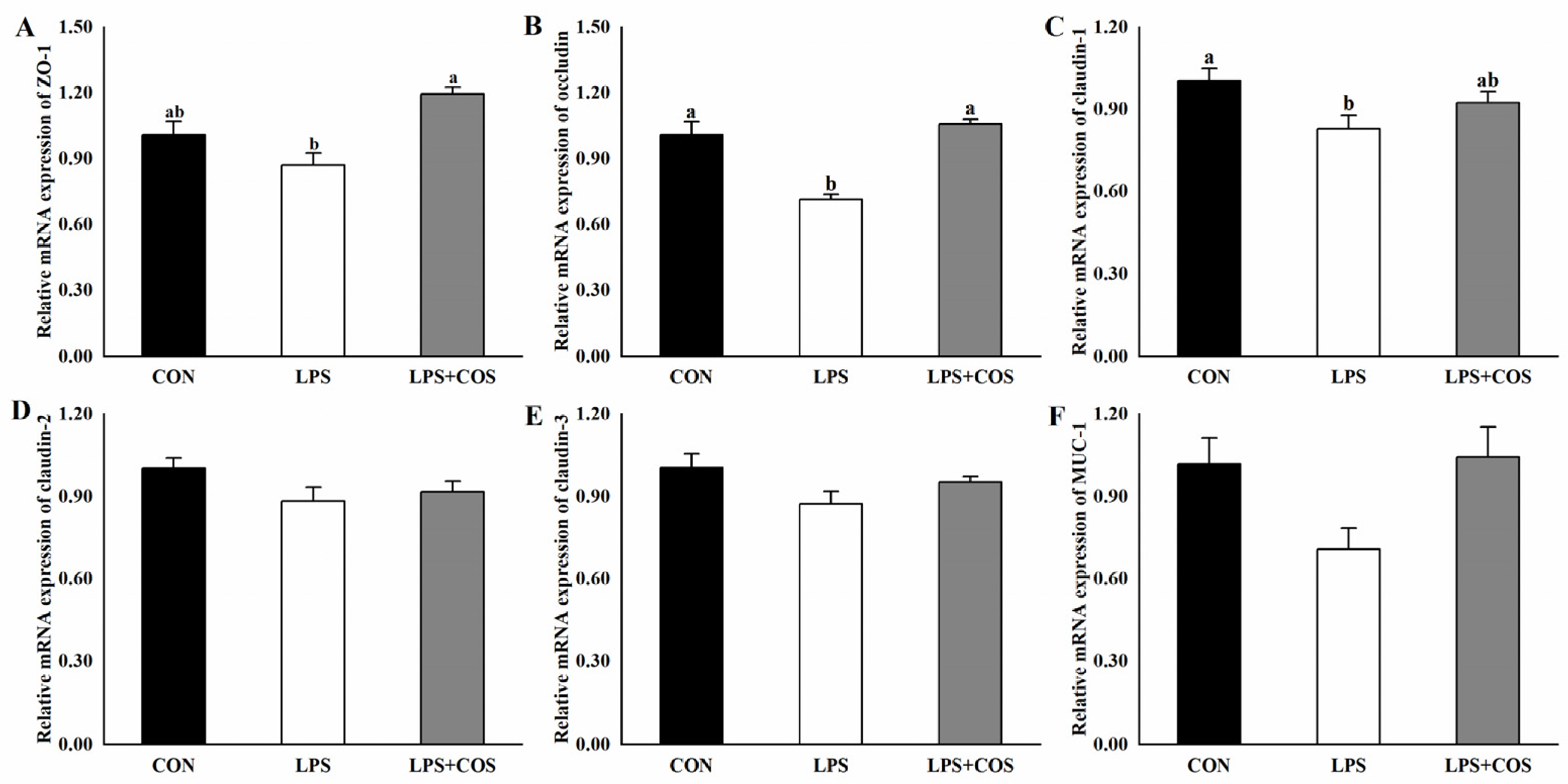
As shown in Figure 2, LPS exposure markedly reduced (p < 0.05) mRNA expression levels of occludin and claudin-1 when compared to the CON group (Figure 2B,C). Supplementation of COS effectively restored the mRNA expression of ZO-1 and occludin, with the levels being statistically different from those in the LPS group (p < 0.05). In contrast, the mRNA expression level of claudin-1 in the LPS + COS group was not significantly different from that in the CON or LPS group. Additionally, treatments did not affect mRNA expression of claudin-2, claudin-3 and MUC-1 (Figure 2D–F).
Figure 2.
Effects of chitooligosaccharide on intestinal barrier integrity in LPS-challenged broiler chickens. (A–F) Relative mRNA expression of ZO-1, occludin, claudin-1, claudin-2, claudin-3 and MUC-1 in jejunal tissues. CON, the unchallenged group, fed a basal diet; LPS, the LPS-challenged group, fed the same basal diet; LPS + COS, the LPS-challenged group, fed the basal diet supplemented with 300 mg/kg COS. All results are shown as mean ± SEM (n = 5). Bars without a common letter indicate significant differences (p < 0.05).
3.7. Cecal Microbiota
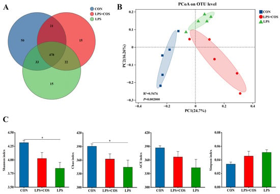
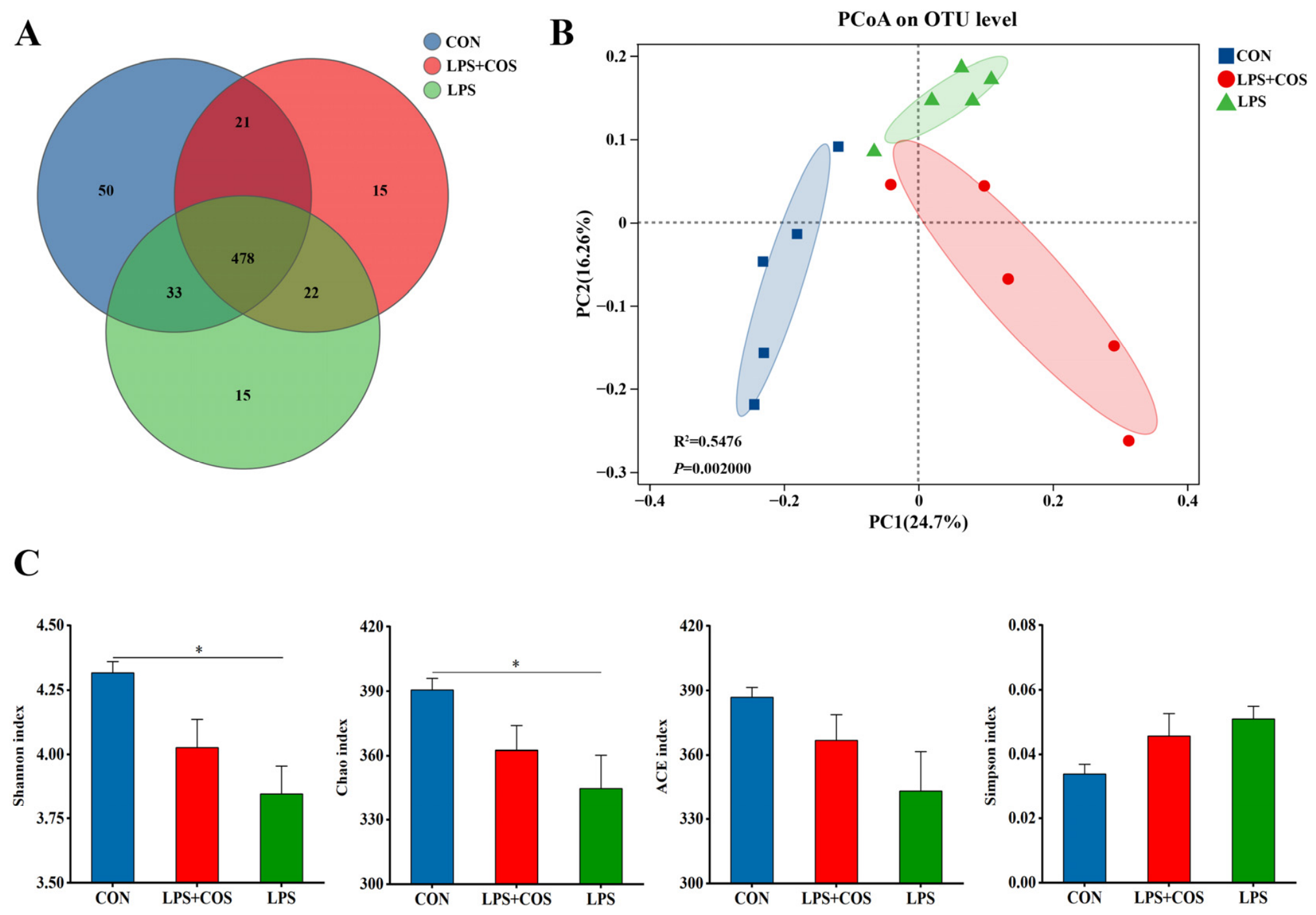
Effects of COS on the richness and diversity of cecal microbiota are shown in Figure 3. As shown in the Venn diagram, the numbers of unique OTUs detected in broilers of the CON, LPS and LPS + COS groups were 50, 15 and 15, respectively (Figure 3A). The results of PCoA demonstrated evident separation of samples across the three groups, reflecting marked shifts in the structure of gut microbial communities. (Figure 3B). Relative to the CON group, LPS administration markedly decreased (p < 0.05) the Shannon and Chao indices (Figure 3C). Nevertheless, the Shannon and Chao indices did not differ significantly between the CON and LPS + COS groups. In addition, the Ace and Simpson indices did not differ significantly among the three groups.
Figure 3.
Effects of chitooligosaccharide on the richness and diversity of cecal microbiota. (A) Venn diagram; (B) Principal coordinates analysis; (C) α-diversity. CON, the unchallenged group, fed a basal diet; LPS, the LPS-challenged group, fed the same basal diet; LPS + COS, the LPS-challenged group, fed the basal diet supplemented with 300 mg/kg COS. n = 5. * p < 0.05.
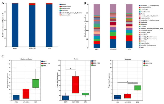
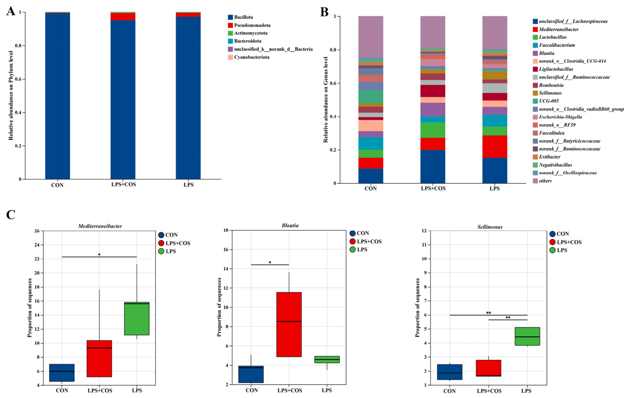
Changes in the composition of cecal microbiota in response to COS are presented in Figure 4. At the phylum level, Bacillota and Pseudomonadota constituted the predominant taxa, with a combined proportion exceeding 98%, accounting for more than 98% (Figure 4A). At the genus level, the top 15 genera were unclassified_f__Lachnospiraceae, Mediterraneibacter, Lactobacillus, Faecalibacterium, Blautia, norank_o__Clostridia_UCG-014, Ligilactobacillus, unclassified_f__Ruminococcaceae, Romboutsia, Sellimonas, UCG-005, norank_o__Clostridia_vadinBB60_group, Escherichia-Shigella, unclassified_o__RF39 and Faecalitalea (Figure 4B). Genus-level analysis of microbial composition revealed that LPS exposure significantly increased (p < 0.05) the relative abundance of Mediterraneibacter and Sellimonas relative to the CON group (Figure 4C). With prior COS supplementation, the relative abundance of Mediterraneibacter in the LPS + COS group returned to a level that was not significantly different from the CON group. Meanwhile, in comparison to the LPS group, COS supplementation markedly lowered (p < 0.05) the relative abundance of Sellimonas. Furthermore, inclusion of COS markedly elevated (p < 0.05) relative abundance of Blautia in comparison to the CON group.
Figure 4.
Effects of chitooligosaccharide on cecal microbiota composition in LPS-challenged broiler chickens. (A) Relative abundance on phylum level; (B) Relative abundance on genus level; (C) Changes in microbiota composition on genus level. CON, the unchallenged group, fed a basal diet; LPS, the LPS-challenged group, fed the same basal diet; LPS + COS, the LPS-challenged group, fed the basal diet supplemented with 300 mg/kg COS. n = 5. * p < 0.05, ** p < 0.01.
4. Discussion
Exposure to various stressors can exert detrimental effects on growth performance in broilers, which further impairs economic returns of production [33]. Therefore, mitigating such negative effects of stress is of great practical significance to broiler production. A previous study has shown that LPS challenge led to increased BW loss in layer chicks [34]. In the present study, similar effects were also observed in broiler chickens subjected to LPS challenge. The inhibitory effect of LPS on BW is mainly attributed to decreased appetite, intestinal injury and the redistribution of nutrients away from growth toward immunity [35]. In addition, our results demonstrated that dietary supplementation of COS mitigated the reduction in BW of LPS-challenged broilers, indicating that COS helps maintain BW in broilers under stress conditions. The alleviation of LPS-induced BW loss by COS may be associated with its protective effects against intestinal injury. Accordingly, we further investigated the effects of COS on the intestinal health of LPS-challenged broilers to elucidate the underlying mechanisms.
LPS is a potent immune-system activator that triggers inflammatory cells to secrete pro-inflammatory cytokines, thereby eliciting a strong immune response [36]. Multiple studies have shown that intraperitoneal injection of LPS can induce systemic inflammation in broiler chickens [9,37]. Consistent with previous findings, our results showed that LPS challenge markedly elevated serum and jejunal concentrations of proinflammatory cytokines TNF-α and IL-1β when compared with the CON group, indicating a robust inflammatory response upon LPS exposure. COS has been proven to alleviate stress-triggered inflammatory responses. For broilers subjected to heat stress, dietary COS supplementation markedly reduced the duodenal and jejunal IL-1β concentrations [22]. Additionally, COS effectively relieves LPS-induced inflammation in laying hens, as evidenced by the decreased levels of jejunal IFN-γ and IL-1β [23]. Consistent with the above reports, the present study revealed that dietary supplementation of COS significantly inhibited the LPS-induced increase in IL-1β concentration and promoted the secretion of anti-inflammatory cytokine IL-10 in the serum, indicating its anti-inflammatory potential against LPS-induced inflammation. Alleviated inflammatory responses may reduce nutrient allocation to immune reactions, allowing more nutrients to be directed toward growth and thereby mitigating the adverse effects of LPS on BW.
TLR4 is a transmembrane pattern recognition receptor of the innate immune system. It has been proven that the TLR4-dependent signaling pathway plays a pivotal role in LPS-triggered inflammatory responses [38]. Upon LPS exposure, TLR4-initiated signal transduction activates the NF-κB cascade, which consequently drives the secretion of multiple pro-inflammatory cytokines [39]. Given that COS attenuated the elevation of inflammatory cytokines induced by LPS, we further investigated the expression of TLR4 and NF-κB p65 to explore the underlying mechanism. As anticipated, the present results demonstrated that LPS stimulation upregulated jejunal mRNA expression of TLR4 and NF-κB p65, further verifying that LPS-triggered inflammatory cytokine secretion is mediated via the TLR4/NF-κB signaling pathway. Existing in vitro evidence indicates that COS attenuate LPS-induced inflammatory responses in IPEC-J2 cells through modulation of the TLR4/NF-κB signaling cascade [24]. Nevertheless, such findings have not yet been reported in LPS-challenged broilers in vivo. In the present study, supplementation of COS significantly suppressed LPS-triggered mRNA upregulation of TLR4, implying that the anti-inflammatory activity of COS in broilers is possibly linked to the inhibition of the TLR4 signaling cascade. Notably, these findings are limited to mRNA expression levels; further validation at the protein level is necessary to consolidate this regulatory mechanism.
Oxidative stress refers to a disrupted balance between reactive oxygen species generation and antioxidant defense capacity, resulting in cell damage [40]. Numerous studies have shown that LPS exposure can trigger oxidative stress in broilers [32,41]. Furthermore, oxidative stress plays a critical role in triggering and exacerbating pathological responses induced by LPS challenge [42]. Consistent with previous reports, our results also showed that LPS challenge induced oxidative stress in broilers, as indicated by decreased SOD and T-AOC activities in the serum and jejunum, and increased MDA concentration in the jejunum. In addition, dietary COS supplementation effectively reversed LPS-induced reductions in T-AOC in both serum and jejunum, and inhibited MDA accumulation in the jejunum, suggesting that COS exerts protective effects against LPS-triggered oxidative stress. Given that oxidative stress has been reported to elicit inflammatory responses that are predominantly mediated by the NF-κB signaling pathway [43], the attenuated oxidative stress by COS supplementation may contribute to the decreased inflammatory responses.
The integrity of the gut barrier is essential to prevent the translocation of pathogens and toxins, maintaining immune homeostasis and body health [44]. Accumulating evidence demonstrates that exposure to LPS disrupts intestinal barrier integrity and increases intestinal permeability [26]. DAO is an intracellular enzyme predominantly localized in small intestinal villus epithelial cells [37]. D-lactate is a metabolic product generated by indigenous commensal bacteria within the gastrointestinal tract [45], while endotoxin exists in the outer membranes of Gram-negative bacteria [46]. Elevated circulating levels of DAO, D-lactate, and endotoxin are well-recognized biomarkers reflecting impaired intestinal barrier integrity and increased intestinal permeability, as their translocation into the bloodstream occurs when the intestinal barrier is compromised [47]. The present results indicated that LPS challenge disrupted the intestinal barrier, as reflected by elevated serum DAO concentrations. Tight junctions are multiprotein complexes located at the apical portion of the lateral membranes of epithelial cells, and are crucial for maintaining intestinal barrier integrity [37]. Our results demonstrated that LPS injection markedly reduced the relative mRNA expression of occludin and claudin-1 in the jejunum. This finding was consistent with the serum results, further confirming detrimental impacts of LPS on gut barrier integrity. Favorable impacts of COS on gut barrier integrity have been documented in a previous study using an LPS-challenged laying hen model [23]. Similarly, the current study also found that COS upregulated relative mRNA expression of ZO-1 and occludin in LPS-challenged broilers, suggesting the protective role of COS in maintaining intestinal barrier function. Since barrier dysfunction is a key determinant of intestinal inflammation [48], the restoration of barrier function by COS supplementation may, at least in part, account for the alleviated inflammation. Notably, the present study also revealed that the LPS group had higher serum endotoxin concentration, while COS attenuated this increase in serum endotoxin concentration. Nevertheless, this rise in circulating endotoxin is likely derived mainly from the injected LPS, rather than translocation of endogenous gut-derived LPS. The reduction in serum LPS may be attributable to the capacity of COS to bind LPS. Accordingly, serum endotoxin concentration cannot be used as a direct biomarker of intestinal barrier permeability in this experimental model.
The intestinal microbiota, a complex microbial community colonizing in the gastrointestinal tract, exerts critical functions for sustaining host health [49]. Dietary intervention serves as a promising strategy to regulate the gut microbiota. Alpha diversity represents the microbial diversity within individual samples, commonly evaluated via indices including Shannon, Chao, Ace and Simpson. Higher α-diversity generally indicates a more complex and stable microbial community with greater resistance to invasion by exogenous pathogens [50]. The current results demonstrated that LPS exposure reduced α-diversity as reflected by lower Shannon and Chao indexes when compared with the CON group, indicating that LPS exposure induced a decline in both species richness and community diversity. Beta diversity describes the difference in species composition between communities [51]. The PCoA results revealed obvious separation of samples across groups, suggesting marked variations in intestinal microbial community structure. Differential analysis of microbial composition further revealed that a significant increase in the abundance of Mediterraneibacter and Sellimonas was observed in the LPS group relative to the CON group. It has been demonstrated that Mediterraneibacter exacerbates inflammatory responses by activating pro-inflammatory signaling pathways, thereby disrupting intestinal immune homeostasis [52]. Sellimonas has been reported to be elevated in depression and associated with inflammation [53,54]. The increased abundance of these bacteria indicated that LPS challenge disrupted the intestinal microbiota in broilers, potentially accounting for the intestinal inflammation in the LPS group. Notably, dietary COS supplementation decreased the relative abundance of Mediterraneibacter and Sellimonas to levels comparable to the CON group, suggesting its potential to reverse LPS-induced gut microbial dysbiosis. Furthermore, supplementation of COS also increased the relative abundance of Blautia compared with the CON group. Accumulating evidence indicates that Blautia exerts beneficial effects by attenuating inflammatory responses and maintaining intestinal barrier integrity [55,56]. Accordingly, the increased abundance of Blautia observed in the COS group may contribute to the attenuation of intestinal inflammation and maintenance of gut barrier homeostasis. However, it should be noted that these reported pro-inflammatory properties were largely characterized in different host systems, and direct extrapolation of these functional associations to broilers requires caution. Collectively, these findings evidenced that COS could ameliorate LPS-induced intestinal microbiota dysbiosis and regulate the structure and diversity of intestinal microbiota, thereby improving intestinal health.
This study has several limitations to be addressed in future research. First, the absence of a non-challenged COS treatment group prevents us from disentangling the intrinsic effect of COS and its interactive effect with LPS. A complete 2 × 2 factorial design with four groups (control, COS alone, LPS alone, LPS + COS) will be required in future research. Second, only a single COS dose and one sampling time point were applied, precluding analysis of dose-dependent effects and dynamic alterations. Third, transcriptional data (mRNA levels) were not validated by corresponding protein or functional measurements. Fourth, intraperitoneal COS administration does not simulate the oral route used in conventional animal feeding. Fifth, the limited sample size (n = 5 per group) may lower statistical power to identify subtle treatment responses. Finally, exclusive use of male birds restricts the generalizability of our findings to females, and sex-related differences require further investigation.
5. Conclusions
In conclusion, dietary COS supplementation can mitigate LPS-triggered inflammation, oxidative stress and microbiota dysbiosis, thus improving intestinal barrier function and attenuating acute BW loss in broilers. These findings suggest that COS is a potential natural feed additive for mitigating stress-induced intestinal injury and acute BW loss in broilers.
Author Contributions
Conceptualization, Y.H. and Q.S.; methodology, R.Y. and Q.S.; software, Q.S.; validation, Y.H. and H.L.; formal analysis, Q.S.; investigation, Y.H., R.Y., H.L., K.S. and Q.S.; resources, K.S. and H.L.; data curation, Q.S.; writing—original draft preparation, Y.H. and Q.S.; writing—review and editing, Y.H. and Q.S.; visualization, K.S.; supervision, R.Y. and Y.H.; project administration, Y.H.; funding acquisition, Q.S. All authors have read and agreed to the published version of the manuscript.
Funding
This research was funded by the Agricultural Science and Technology Innovation Project of Shandong Academy of Agricultural Sciences (CXGC2026C57).
Institutional Review Board Statement
The animal study protocol was approved by the Institutional Animal Care and Use Committee of Shandong Academy of Agricultural Sciences (protocol code SAAS-2024-G24). Approval Date: 3 March 2024.
Informed Consent Statement
Not applicable.
Data Availability Statement
The original contributions presented in this study are included in the article. Further inquiries can be directed to the corresponding author.
Conflicts of Interest
The authors declare no conflicts of interest.
References
- Averos, X.; Estevez, I. Meta-analysis of the effects of intensive rearing environments on the performance and welfare of broiler chickens. Poult. Sci. 2018, 97, 3767–3785. [Google Scholar] [CrossRef] [Scilit] [PubMed]
- Elwinger, K.; Fisher, C.; Jeroch, H.; Sauveur, B.; Tiller, H.; Whitehead, C.C. A brief history of poultry nutrition over the last hundred years. World’s Poult. Sci. J. 2016, 72, 701–720. [Google Scholar] [CrossRef] [Scilit]
- Hartcher, K.M.; Lum, H.K. Genetic selection of broilers and welfare consequences: A review. World’s Poult. Sci. J. 2020, 76, 154–167. [Google Scholar] [CrossRef] [Scilit]
- Dierick, E.; Hirvonen, O.P.; Haesebrouck, F.; Ducatelle, R.; Van Immerseel, F.; Goossens, E. Rapid growth predisposes broilers to necrotic enteritis. Avian Pathol. 2019, 48, 416–422. [Google Scholar] [CrossRef] [Scilit] [PubMed]
- Guaragni, A.; Boiago, M.M.; Bottari, N.B.; Morsch, V.M.; Lopes, T.F.; Schafer Da Silva, A. Feed supplementation with inulin on broiler performance and meat quality challenged with Clostridium perfringens: Infection and prebiotic impacts. Microb. Pathog. 2020, 139, 103889. [Google Scholar] [CrossRef] [Scilit] [PubMed]
- Jiang, S.Q.; Chen, Z.L.; Zhang, S.; Ye, J.L.; Wang, Y.B. Protective effects of protocatechuic acid on growth performance, intestinal barrier and antioxidant capacity in broilers challenged with lipopolysaccharide. Animal 2023, 17, 100693. [Google Scholar] [CrossRef] [Scilit] [PubMed]
- Zhang, H.; Chen, Y.; Chen, Y.; Li, Y.; Jia, P.; Ji, S.; Zhou, Y.; Wang, T. Dietary pterostilbene supplementation attenuates intestinal damage and immunological stress of broiler chickens challenged with lipopolysaccharide. J. Anim. Sci. 2020, 98, 1–9. [Google Scholar] [CrossRef] [Scilit] [PubMed]
- Kong, L.; Wang, Z.; Xiao, C.; Zhu, Q.; Song, Z. Glycerol monolaurate attenuated immunological stress and intestinal mucosal injury by regulating the gut microbiota and activating AMPK/Nrf2 signaling pathway in lipopolysaccharide-challenged broilers. Anim. Nutr. 2022, 10, 347–359. [Google Scholar] [CrossRef] [Scilit] [PubMed]
- Zha, P.; Liu, W.; Zhou, Y.; Chen, Y. Protective effects of chlorogenic acid on the intestinal barrier of broiler chickens: An immunological stress model study. Poult. Sci. 2024, 103, 103949. [Google Scholar] [CrossRef] [Scilit] [PubMed]
- Dibner, J.J.; Richards, J.D. Antibiotic growth promoters in agriculture: History and mode of action. Poult. Sci. 2005, 84, 634–643. [Google Scholar] [CrossRef] [Scilit] [PubMed]
- Fernández Miyakawa, M.E.; Casanova, N.A.; Kogut, M.H. How did antibiotic growth promoters increase growth and feed efficiency in poultry? Poult. Sci. 2024, 103, 103278. [Google Scholar] [CrossRef] [Scilit]
- Roth, N.; Kasbohrer, A.; Mayrhofer, S.; Zitz, U.; Hofacre, C.; Domig, K.J. The application of antibiotics in broiler production and the resulting antibiotic resistance in Escherichia coli: A global overview. Poult. Sci. 2019, 98, 1791–1804. [Google Scholar] [CrossRef] [Scilit] [PubMed]
- Yuan, X.; Zheng, J.; Jiao, S.; Cheng, G.; Feng, C.; Du, Y.; Liu, H. A review on the preparation of chitosan oligosaccharides and application to human health, animal husbandry and agricultural production. Carbohydr. Polym. 2019, 220, 60–70. [Google Scholar] [CrossRef] [Scilit] [PubMed]
- Naveed, M.; Phil, L.; Sohail, M.; Hasnat, M.; Baig, M.M.F.A.; Ihsan, A.U.; Shumzaid, M.; Kakar, M.U.; Mehmood Khan, T.; Akabar, M.; et al. Chitosan oligosaccharide (COS): An overview. Int. J. Biol. Macromol. 2019, 129, 827–843. [Google Scholar] [CrossRef] [Scilit] [PubMed]
- Chae, S.Y.; Jang, M.; Nah, J. Influence of molecular weight on oral absorption of water soluble chitosans. J. Control. Release 2005, 102, 383–394. [Google Scholar] [CrossRef] [Scilit] [PubMed]
- Mattaveewong, T.; Wongkrasant, P.; Chanchai, S.; Pichyangkura, R.; Chatsudthipong, V.; Muanprasat, C. Chitosan oligosaccharide suppresses tumor progression in a mouse model of colitis-associated colorectal cancer through ampk activation and suppression of NF-κB and mtor signaling. Carbohydr. Polym. 2016, 145, 30–36. [Google Scholar] [CrossRef] [Scilit] [PubMed]
- Sánchez, Á.; Mengíbar, M.; Rivera-Rodríguez, G.; Moerchbacher, B.; Acosta, N.; Heras, A. The effect of preparation processes on the physicochemical characteristics and antibacterial activity of chitooligosaccharides. Carbohydr. Polym. 2017, 157, 251–257. [Google Scholar] [CrossRef] [Scilit]
- Lan, R.; Chang, Q.; Wei, L.; Zhao, Z. The protect effects of chitosan oligosaccharides on intestinal integrity by regulating oxidative status and inflammation under oxidative stress. Mar. Drugs 2021, 19, 57. [Google Scholar] [CrossRef] [Scilit]
- Deng, X.; Ye, Z.; Cao, H.; Bai, Y.; Che, Q.; Guo, J.; Su, Z. Chitosan oligosaccharide ameliorated obesity by reducing endoplasmic reticulum stress in diet-induced obese rats. Food Funct. 2020, 11, 6285–6296. [Google Scholar] [CrossRef] [Scilit] [PubMed]
- Chang, L.; Ding, Y.; Wang, Y.; Song, Z.; Li, F.; He, X.; Zhang, H. Effects of different oligosaccharides on growth performance and intestinal function in broilers. Front. Vet. Sci. 2022, 9, 852545. [Google Scholar] [CrossRef] [Scilit] [PubMed]
- Lan, R.; Wu, F.; Wang, Y.; Lin, Z.; Wang, H.; Zhang, J.; Zhao, Z. Chitosan oligosaccharide improves intestinal function by promoting intestinal development, alleviating intestinal inflammatory response, and enhancing antioxidant capacity in broilers aged d 1 to 14. Poult. Sci. 2024, 103, 103381. [Google Scholar] [CrossRef] [Scilit] [PubMed]
- Lan, R.; Li, Y.; Chang, Q.; Zhao, Z. Dietary chitosan oligosaccharides alleviate heat stress-induced intestinal oxidative stress and inflammatory response in yellow-feather broilers. Poult. Sci. 2020, 99, 6745–6752. [Google Scholar] [CrossRef] [Scilit]
- Gu, Y.F.; Chen, Y.P.; Jin, R.; Wang, C.; Wen, C.; Zhou, Y.M. Dietary chitooligosaccharide supplementation alleviates intestinal barrier damage, and oxidative and immunological stress in lipopolysaccharide-challenged laying hens. Poult. Sci. 2022, 101, 101701. [Google Scholar] [CrossRef] [Scilit] [PubMed]
- Shi, L.; Fang, B.; Yong, Y.; Li, X.; Gong, D.; Li, J.; Yu, T.; Gooneratne, R.; Gao, Z.; Li, S.; et al. Chitosan oligosaccharide-mediated attenuation of LPS-induced inflammation in IPEC-J2 cells is related to the TLR4/NF-κB signaling pathway. Carbohydr. Polym. 2019, 219, 269–279. [Google Scholar] [CrossRef] [Scilit] [PubMed]
- Liu, J.; Kang, R.; Tang, D. Lipopolysaccharide delivery systems in innate immunity. Trends Immunol. 2024, 45, 274–287. [Google Scholar] [CrossRef] [Scilit]
- Tan, H.; Zhen, W.; Bai, D.; Liu, K.; He, X.; Ito, K.; Liu, Y.; Li, Y.; Zhang, Y.; Zhang, B.; et al. Effects of dietary chlorogenic acid on intestinal barrier function and the inflammatory response in broilers during lipopolysaccharide-induced immune stress. Poult. Sci. 2023, 102, 102623. [Google Scholar] [CrossRef] [Scilit]
- Liu, H.; Meng, H.; Du, M.; Lv, H.; Wang, Y.; Zhang, K. Chlorogenic acid ameliorates intestinal inflammation by inhibiting NF-κB and endoplasmic reticulum stress in lipopolysaccharide-challenged broilers. Poult. Sci. 2024, 103, 103586. [Google Scholar] [CrossRef] [Scilit] [PubMed]
- National Research Council. Nutrient Requirements of Poultry, 9th ed.; National Academies Press: Washington, DC, USA, 1994. [Google Scholar]
- Shang, Q. Inulin alleviates inflammatory response and gut barrier dysfunction via modulating microbiota in lipopolysaccharide-challenged broilers. Int. J. Biol. Macromol. 2024, 282, 137208. [Google Scholar] [CrossRef] [Scilit] [PubMed]
- Chen, J.Y.; Yu, Y.H. Bacillus subtilis-fermented products ameliorate the growth performance and alter cecal microbiota community in broilers under lipopolysaccharide challenge. Poult. Sci. 2021, 100, 875–886. [Google Scholar] [CrossRef] [Scilit]
- Shang, Q.; Li, H.; Han, Y. Protective effects of inulin against lipopolysaccharide-induced oxidative stress in broiler chickens. Front. Vet. Sci. 2025, 12, 1660935. [Google Scholar] [CrossRef] [Scilit] [PubMed]
- Livak, K.J.; Schmittgen, T.D. Analysis of relative gene expression data using real-time quantitative PCR and the 2−ΔΔCt method. Methods 2001, 25, 402–408. [Google Scholar] [CrossRef] [Scilit] [PubMed]
- Zhang, W.H.; Gao, F.; Zhu, Q.F.; Li, C.; Jiang, Y.; Dai, S.F.; Zhou, G.H. Dietary sodium butyrate alleviates the oxidative stress induced by corticosterone exposure and improves meat quality in broiler chickens. Poult. Sci. 2011, 90, 2592–2599. [Google Scholar] [CrossRef] [Scilit] [PubMed]
- Wils-Plotz, E.L.; Klasing, K.C. Effects of immunomodulatory nutrients on growth performance and immune-related gene expression in layer chicks challenged with lipopolysaccharide. Poult. Sci. 2017, 96, 548–555. [Google Scholar] [CrossRef] [Scilit] [PubMed]
- Han, H.; Zhang, J.; Chen, Y.; Shen, M.; Yan, E.; Wei, C.; Yu, C.; Zhang, L.; Wang, T. Dietary taurine supplementation attenuates lipopolysaccharide-induced inflammatory responses and oxidative stress of broiler chickens at an early age. J. Anim. Sci. 2020, 98, skaa311. [Google Scholar] [CrossRef] [Scilit] [PubMed]
- Gauthier, A.E.; Rotjan, R.D.; Kagan, J.C. Lipopolysaccharide detection by the innate immune system may be an uncommon defence strategy used in nature. Open Biol. 2022, 12, 220146. [Google Scholar] [CrossRef] [Scilit] [PubMed]
- Yang, L.; Tang, L.; Guo, S.; Wang, L.; Hou, Y. The effects of naringin on antioxidant function, intestinal barrier and immune response in broilers challenged with lipopolysaccharide. Animals 2025, 15, 3367. [Google Scholar] [CrossRef] [Scilit] [PubMed]
- Cirmi, S.; Maugeri, A.; Russo, C.; Musumeci, L.; Navarra, M.; Lombardo, G.E. Oleacein attenuates lipopolysaccharide-induced inflammation in THP-1-derived macrophages by the inhibition of TLR4/MyD88/NF-κB pathway. Int. J. Mol. Sci. 2022, 23, 1206. [Google Scholar] [CrossRef] [Scilit] [PubMed]
- Ali, W.; Choe, K.; Park, J.S.; Ahmad, R.; Park, H.Y.; Kang, M.H.; Park, T.J.; Kim, M.O. Kojic acid reverses LPS-induced neuroinflammation and cognitive impairment by regulating the TLR4/NF-κB signaling pathway. Front. Pharmacol. 2024, 15, 1443552. [Google Scholar] [CrossRef] [Scilit] [PubMed]
- Sies, H. Oxidative stress: Concept and some practical aspects. Antioxidants 2020, 9, 852. [Google Scholar] [CrossRef] [Scilit] [PubMed]
- Pang, X.; Miao, Z.; Dong, Y.; Cheng, H.; Xin, X.; Wu, Y.; Han, M.; Su, Y.; Yuan, J.; Shao, Y.; et al. Dietary methionine restriction alleviates oxidative stress and inflammatory responses in lipopolysaccharide-challenged broilers at early age. Front. Pharmacol. 2023, 14, 1120718. [Google Scholar] [CrossRef] [Scilit] [PubMed]
- Hu, W.; He, Z.; Du, L.; Zhang, L.; Li, J.; Ma, Y.; Bi, S. Biomarkers of oxidative stress in broiler chickens attacked by lipopolysaccharide: A systematic review and meta-analysis. Ecotoxicol. Environ. Saf. 2023, 266, 115606. [Google Scholar] [CrossRef] [Scilit] [PubMed]
- Lingappan, K. NF-κB in oxidative stress. Curr. Opin. Toxicol. 2018, 7, 81–86. [Google Scholar] [CrossRef] [Scilit] [PubMed]
- Neurath, M.F.; Artis, D.; Becker, C. The intestinal barrier: A pivotal role in health, inflammation, and cancer. Lancet Gastroenterol. Hepatol. 2025, 10, 573–592. [Google Scholar] [CrossRef] [Scilit] [PubMed]
- Yao, Y.; Yu, Y.; Wu, Y.; Lu, L.; Sheng, Z. Plasma D (-)-lactate as a new marker for diagnosis of acute intestinal injury following ischemia-reperfusion. World J. Gastroenterol. 1997, 3, 225–227. [Google Scholar] [CrossRef] [Scilit] [PubMed]
- Meng, Y.; Zhang, Y.; Liu, M.; Huang, Y.K.; Zhang, J.; Yao, Q.; Zhao, Y.L.; Xiong, J.J. Evaluating intestinal permeability by measuring plasma endotoxin and diamine oxidase in children with acute lymphoblastic leukemia treated with high-dose methotrexate. Anti-Cancer Agents Med. Chem. 2016, 16, 387–392. [Google Scholar] [CrossRef] [Scilit] [PubMed]
- Zhang, Q.; Gao, X.; Wu, J.; Chen, M. The correlation between endotoxin, d-lactate, and diamine oxidase with endoscopic activity in inflammatory bowel disease. Dis. Markers 2022, 2022, 9171436. [Google Scholar] [CrossRef] [Scilit] [PubMed]
- Wang, J.; Yan, J.; Shi, L.; Wang, Y.; Tian, X.; Qi, Y.; Li, G. Intestinal barrier function as a key determinant of inflammation and nutritional status in digestive surgery patients: A real-world study. Front. Nutr. 2025, 12, 1637877. [Google Scholar] [CrossRef] [Scilit] [PubMed]
- Perler, B.K.; Friedman, E.S.; Wu, G.D. The role of the gut microbiota in the relationship between diet and human health. Annu. Rev. Physiol. 2023, 85, 449–468. [Google Scholar] [CrossRef] [Scilit] [PubMed]
- Li, Z.; Zhou, J.; Liang, H.; Ye, L.; Lan, L.; Lu, F.; Wang, Q.; Lei, T.; Yang, X.; Cui, P.; et al. Differences in alpha diversity of gut microbiota in neurological diseases. Front. Neurosci. 2022, 16, 879318. [Google Scholar] [CrossRef] [Scilit] [PubMed]
- Su, X. Elucidating the beta-diversity of the microbiome: From global alignment to local alignment. mSystems 2021, 6, e00363-21. [Google Scholar] [CrossRef] [Scilit] [PubMed]
- Huang, H.J.; Liu, C.; Sun, X.W.; Wei, R.Q.; Liu, L.W.; Chen, H.Y.; Abdugheni, R.; Wang, C.Y.; Wang, X.M.; Jiang, H.; et al. The rheumatoid arthritis gut microbial biobank reveals core microbial species that associate and effect on host inflammation and autoimmune responses. IMETA 2024, 3, e242. [Google Scholar] [CrossRef] [Scilit] [PubMed]
- Shi, Y.; Liu, X.; Yang, Y.; Deng, H.; Zhang, J.; Shi, D.; Zhang, Y.; Cheng, Y.; Guo, Y.; Zhu, H.; et al. Human obese gut microbiota induces lipid metabolic disorder and hypophagia without weight gain in normocaloric mice. Sci. China Life Sci. 2026, 69, 3755–3758. [Google Scholar] [CrossRef] [Scilit] [PubMed]
- Radjabzadeh, D.; Bosch, J.A.; Uitterlinden, A.G.; Zwinderman, A.H.; Ikram, M.A.; van Meurs, J.B.J.; Luik, A.I.; Nieuwdorp, M.; Lok, A.; van Duijn, C.M.; et al. Gut microbiome-wide association study of depressive symptoms. Nat. Commun. 2022, 13, 7128. [Google Scholar] [CrossRef] [Scilit] [PubMed]
- Mao, B.; Guo, W.; Cui, S.; Zhang, Q.; Zhao, J.; Tang, X.; Zhang, H. Blautia producta displays potential probiotic properties against dextran sulfate sodium-induced colitis in mice. Food Sci. Hum. Wellness 2024, 13, 709–720. [Google Scholar] [CrossRef] [Scilit]
- Chen, Y.; Ma, Y.; Leng, Y.; Li, X.; Qin, S.; Li, X.; Zhang, Z.; Yu, H. The mitigative effects of blautia producta 1009924 on DSS-induced intestinal inflammation. Front. Microbiol. 2025, 16, 1560441. [Google Scholar] [CrossRef] [Scilit] [PubMed]

Disclaimer/Publisher’s Note: The statements, opinions and data contained in all publications are solely those of the individual author(s) and contributor(s) and not of MDPI and/or the editor(s). MDPI and/or the editor(s) disclaim responsibility for any injury to people or property resulting from any ideas, methods, instructions or products referred to in the content. |
© 2026 by the authors. Licensee MDPI, Basel, Switzerland. This article is an open access article distributed under the terms and conditions of the Creative Commons Attribution (CC BY) license.