


Simple Summary
Air may remain in the chest after lung surgery. In dogs, the thin tissue separating the right and left sides of the chest may allow air to pass between them. We describe a 10-year-old dog that underwent removal of the right cranial lung lobe and a nearby lymph node containing cancer cells. During surgery, an opening was created in the tissue separating the two sides of the chest and was subsequently closed. Air was then removed only from the right side. Although postoperative X-rays showed air in the chest, the dog initially had no significant breathing difficulties. The following day, the dog developed rapid and labored breathing. Blood was drained from the right side of the chest, but breathing difficulties persisted. A computed tomography scan revealed a large amount of air around the left lung. Removing approximately 95 mL of air from the left side was followed by rapid improvement in breathing. The exact source of the air could not be determined. This case highlights the importance of checking both sides of the chest and carefully planning postoperative drainage after surgery involving the tissue between the lungs.
Abstract
Postoperative pneumothorax is a recognized complication of lung lobectomy, and interpleural communication in dogs can make pleural air distribution and evacuation difficult to predict. A 10-year-old neutered male mixed-breed dog underwent right cranial lung lobectomy and excision of a metastatic cranial mediastinal lymph node. The excision created an approximately 2.5-cm full-thickness mediastinal defect through which the contralateral lung was visible; the defect was subsequently closed. At thoracic closure, pleural air was evacuated only from the right hemithorax. On postoperative day 1, tachypnea and increased respiratory effort developed, and a right-sided hemothorax was identified. After removal of 93 mL of hemorrhagic pleural fluid and initiation of whole-blood transfusion, respiratory abnormalities persisted. Contrast-enhanced computed tomography (CT) was performed to investigate the persistent respiratory abnormalities and a possible source of postoperative hemorrhage. CT demonstrated a large left-sided pneumothorax without active contrast extravasation or a definite bleeding source. Left thoracocentesis removed approximately 95 mL of air, with subsequent respiratory improvement. Temporary interpleural communication followed by repair and unilateral pleural evacuation may have contributed to contralateral air retention, although delayed bronchial stump or pulmonary parenchymal leakage could not be excluded. This case emphasizes bilateral pleural assessment and postoperative drainage planning after mediastinal manipulation or repair.
1. Introduction
Primary pulmonary neoplasia is uncommon in dogs, with pulmonary carcinomas representing the predominant tumor category [1,2]. Surgical resection is the treatment of choice for resectable primary pulmonary tumors [2]. Thoracic radiography is commonly used during initial assessment, whereas computed tomography (CT) provides more detailed evaluation of pulmonary lesions, additional pulmonary nodules, and intrathoracic lymph nodes [2,3].
Lung lobectomy is generally well tolerated [4], although perioperative complications may include hemorrhage and air leakage [5] as well as hemothorax and pneumothorax [6]. Postoperative tachypnea or increased respiratory effort therefore requires assessment of both pulmonary and pleural causes.
In dogs, the right and left pleural cavities may communicate through fenestrations in the caudal mediastinum [7,8]. However, the presence of such fenestrations does not necessarily imply unrestricted or clinically sufficient communication between the two pleural cavities; experimental observations indicate that pleural processes may remain unilateral when the mediastinum is functionally intact but can become bilateral after mechanical disruption [9]. Thus, the distribution of pleural air cannot be predicted solely from the side of thoracic entry. Cadaveric work further indicates that the effectiveness of pleural air evacuation may be influenced by both the degree of interpleural communication and the route of evacuation [10].
This report describes a clinically important left-sided pneumothorax after right cranial lung lobectomy and cranial mediastinal lymph node excision in a dog in which a full-thickness mediastinal defect was created and repaired. Rather than establishing a single causal mechanism, the case illustrates perioperative considerations associated with temporary interpleural communication, subsequent repair, unilateral pleural evacuation, postoperative drainage strategy, and assessment of both hemithoraces when respiratory abnormalities persist.
2. Case Description
A 10-year-old neutered male mixed-breed dog weighing 6.5 kg was presented to the Konkuk University Veterinary Medical Teaching Hospital for evaluation of an intermittent cough of approximately 2 years’ duration. There was no history of acute respiratory distress. The dog was bright, alert, and responsive at presentation, with a body condition score of 4/9. The respiratory rate was 30 breaths/min, and no adventitious lung sounds were detected on thoracic auscultation. A grade IV/VI systolic heart murmur was present. Myxomatous mitral valve disease had previously been diagnosed, and the dog had been receiving pimobendan (0.3 mg/kg, PO, q12h) and enalapril (0.5 mg/kg, PO, q12h). Echocardiography performed 3 years before presentation had documented mitral regurgitation, with a left atrial-to-aortic root ratio of 1.50 and a left ventricular internal diameter in diastole of 33.6 mm. At the current presentation, the cardiac silhouette was within normal limits on thoracic radiographs, with no radiographic evidence of congestive heart failure.
Initial thoracic radiographs revealed a focal, well-defined soft-tissue opacity in the right cranioventral thorax, with a pulmonary origin suspected. No evidence of pneumothorax or pleural effusion was identified.
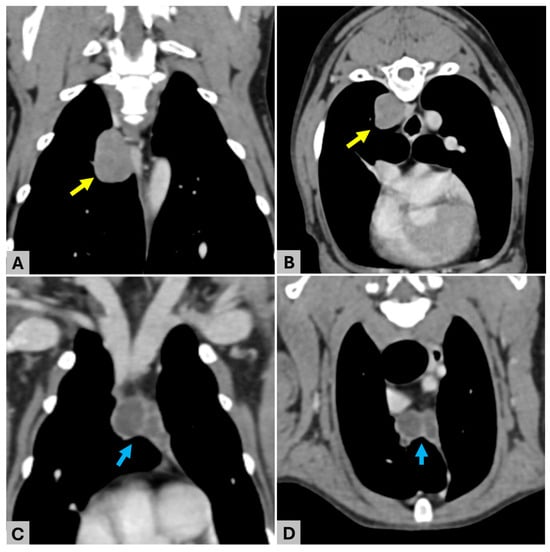
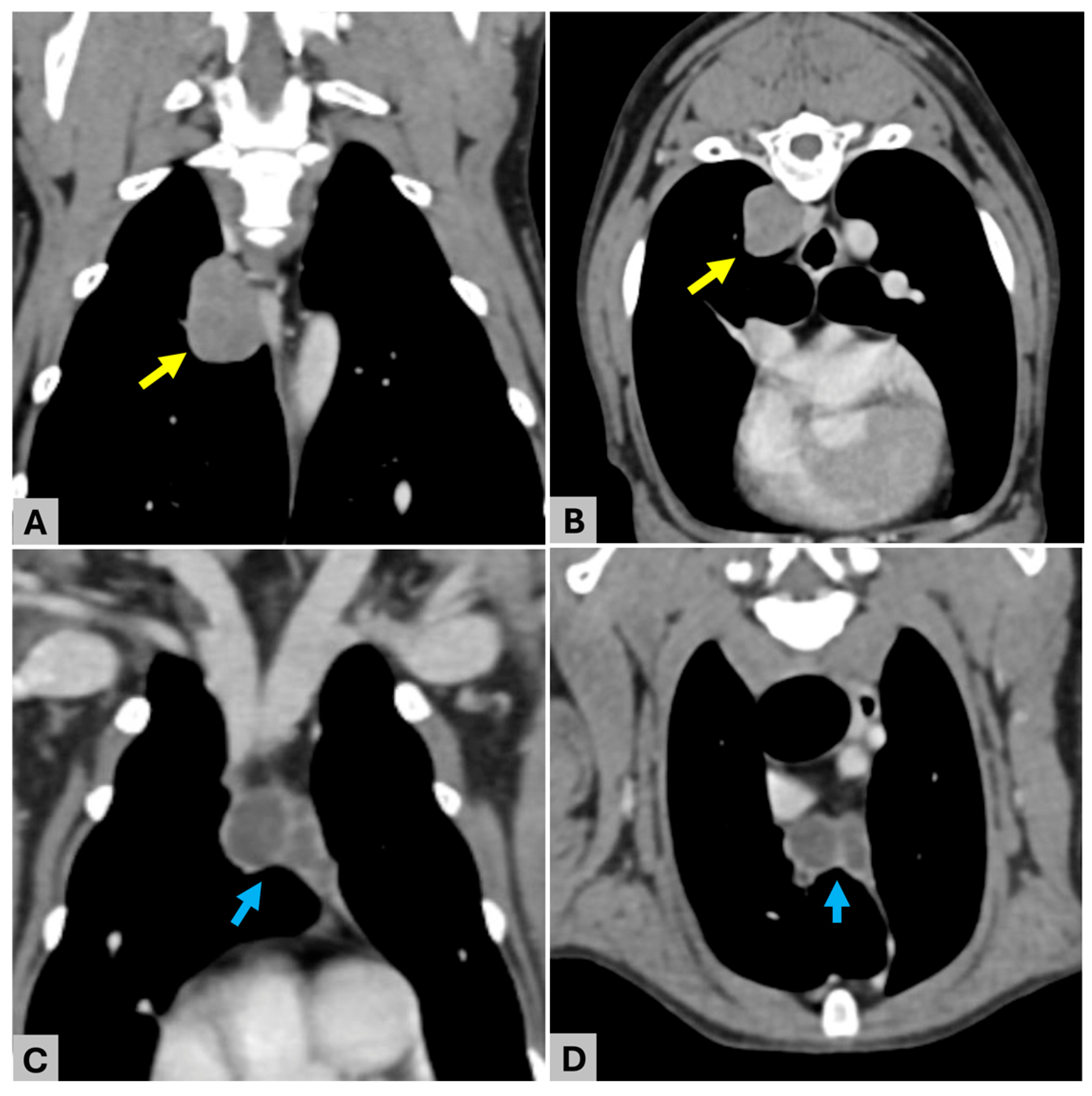
Contrast-enhanced whole-body CT was subsequently performed using a multidetector CT scanner (Aquilion Lightning; Canon Medical Systems, Otawara, Japan). Iohexol (Omnipaque 300; 300 mg I/mL; GE Healthcare, Chicago, IL, USA) was administered intravenously at 2.5 mL/kg (total volume, approximately 16.26 mL) at an injection rate of 2.3 mL/s. A well-defined pulmonary mass measuring approximately 23.0 × 14.2 × 19.6 mm was identified within the right cranial lung lobe at the level of T5–T6 (Figure 1A,B). Ground-glass opacity was also present within the right cranial lung lobe, and small air-attenuating pulmonary lesions consistent with bullae or blebs were identified in the right cranial and accessory lung lobes.
Figure 1.
Preoperative computed tomographic findings. (A) Dorsal and (B) transverse contrast-enhanced CT images showing a well-defined pulmonary mass within the right cranial lung lobe at the level of T5–T6; (C) dorsal and (D) transverse contrast-enhanced CT images showing two closely apposed nodular components within the cranial mediastinum at the level of T1–T2. Yellow arrows indicate the right cranial lung lobe mass, and blue arrows indicate the cranial mediastinal lesions.
Two closely apposed fluid-attenuating nodular components were identified within the cranial mediastinum at the level of T1–T2 (Figure 1C,D). They measured approximately 11.9 × 9.2 × 9.9 mm and 14.4 × 7.3 × 8.0 mm, respectively. Neither component demonstrated appreciable internal contrast enhancement, although peripheral rim enhancement was present.
No preoperative pneumothorax or pleural effusion was identified. No additional pulmonary nodules or CT evidence of distant metastasis was detected in the evaluated regions.
The imaging findings were considered most consistent with a primary right cranial pulmonary neoplasm with suspected cranial mediastinal lymph node involvement. Preoperative hematologic and biochemical testing revealed no clinically important abnormalities. Surgical excision was elected and performed 14 days after the initial diagnostic evaluation.
Cefazolin (22 mg/kg, IV) was administered before skin incision, and maropitant citrate (1 mg/kg, SC) was administered preoperatively. Premedication consisted of midazolam (0.2 mg/kg, IV) and fentanyl (2 µg/kg, IV). Anesthesia was induced with propofol administered intravenously to effect (total dose, 6 mg/kg), followed by endotracheal intubation, and was maintained with isoflurane in oxygen.
Pressure-controlled two-lung ventilation was used during anesthesia. Peak inspiratory pressure was maintained at approximately 10 cmH2O with positive end-expiratory pressure set at 5 cmH2O. Respiratory frequency was adjusted to maintain an end-tidal carbon dioxide concentration of 35–45 mmHg. One-lung ventilation and intrathoracic carbon dioxide insufflation were not used. No marked increase in airway pressure or other clinically relevant ventilation-associated abnormality was recorded.
The dog was positioned in left lateral recumbency. Because preoperative CT localized the cranial mediastinal lesion to approximately T1–T2 and both lesions were intended to be addressed through a single thoracotomy, a right third-intercostal-space approach was selected. An intercostal nerve block was performed with 2% lidocaine hydrochloride at a total dose of 2 mg/kg along the caudal margins of three consecutive ribs encompassing the third intercostal space.
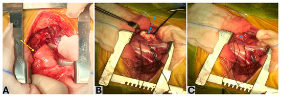
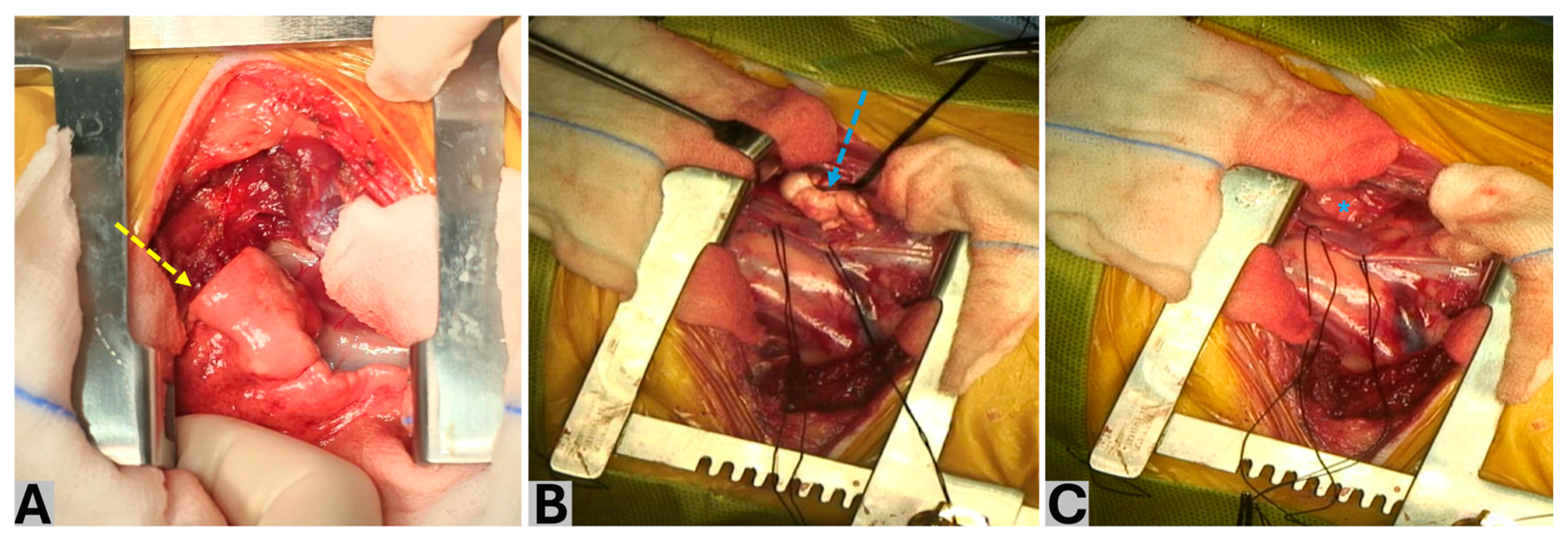
The right cranial lung lobe was isolated, and the pulmonary mass was identified (Figure 2A). Exposure of the pulmonary hilus through the third intercostal space was limited, and an unmeasured rim of proximal pulmonary parenchyma remained at the hilar stump. The pulmonary artery, pulmonary vein, and lobar bronchus were ligated en bloc using three 2-0 black silk ligatures: two ligatures were placed proximally on the patient side, and one ligature was placed distally on the specimen side. The hilar structures were transected with Metzenbaum scissors between the second and third ligatures, leaving two ligatures on the patient-side stump. Neither a surgical stapler nor a vessel-sealing device was used. A surgical stapler was not used because an appropriately sized device was unavailable for the patient. No active hemorrhage from the hilar stump was observed.
Figure 2.
Intraoperative findings. (A) Right cranial lung lobe containing the pulmonary mass before lobectomy (yellow dashed arrow); (B) cranial mediastinal lesion before excision (blue dashed arrow); (C) approximately 2.5-cm full-thickness mediastinal pleural defect identified after excision of the cranial mediastinal lesion (blue asterisk). The contralateral lung is directly visible through the defect, confirming temporary communication between the right and left pleural cavities.
The cranial mediastinal lesion was subsequently approached. On gross inspection, the cranial mediastinal pleura appeared intact before manipulation, and no pre-existing fenestration was identified. Although CT demonstrated two closely apposed nodular components, intraoperative examination revealed a single encapsulated lesion composed of two closely adherent nodular portions (Figure 2B). The vagus nerve was identified and gently retracted using 2-0 black silk to minimize the risk of iatrogenic injury during dissection. The lesion was gently separated from the surrounding tissues by blunt dissection using cotton-tipped applicators. Several vessels were present around the lesion, and localized bleeding encountered during dissection was controlled using monopolar electrosurgery.
During removal of the mediastinal lesion, full-thickness resection of cranial mediastinal tissue created an approximately 2.5-cm defect. The contralateral lung was directly visible through the defect, confirming direct interpleural communication at the surgical site (Figure 2C). Given the presence of the full-thickness defect, the surrounding vessels, and tissue manipulation associated with excision of the lesion, closure was elected to restore local tissue continuity and stability, with the potential for postoperative bleeding from the manipulated region also taken into consideration. The defect was closed using 3-0 polydioxanone sutures in a simple interrupted pattern.
After removal of both lesions, the right hemithorax was partially filled with warm sterile 0.9% sodium chloride solution, and the remaining lung lobes were reinflated. A saline bubble test was performed to assess the hilar/bronchial stump and remaining pulmonary parenchyma for air leakage, and no visible leakage was detected. The repaired mediastinal defect was separately inspected and appeared intact. No active intrathoracic hemorrhage was observed at that time. The saline was subsequently removed.
The thoracic wall was apposed with circumcostal sutures. Before subcutaneous and skin closure, an 18-gauge catheter was introduced into the right pleural cavity, and air was aspirated using a syringe and three-way stopcock until resistance to aspiration was encountered and no further air could be readily withdrawn. The catheter was removed, and the remaining soft tissue layers were closed routinely. The left pleural cavity was not separately evacuated. No air leakage was detected during the saline test, the hilar/bronchial stump and mediastinal repair appeared intact, no active hemorrhage was observed, the patient remained stable, and no further air could be readily aspirated from the right pleural cavity; therefore, a thoracostomy tube was not placed.
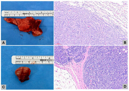
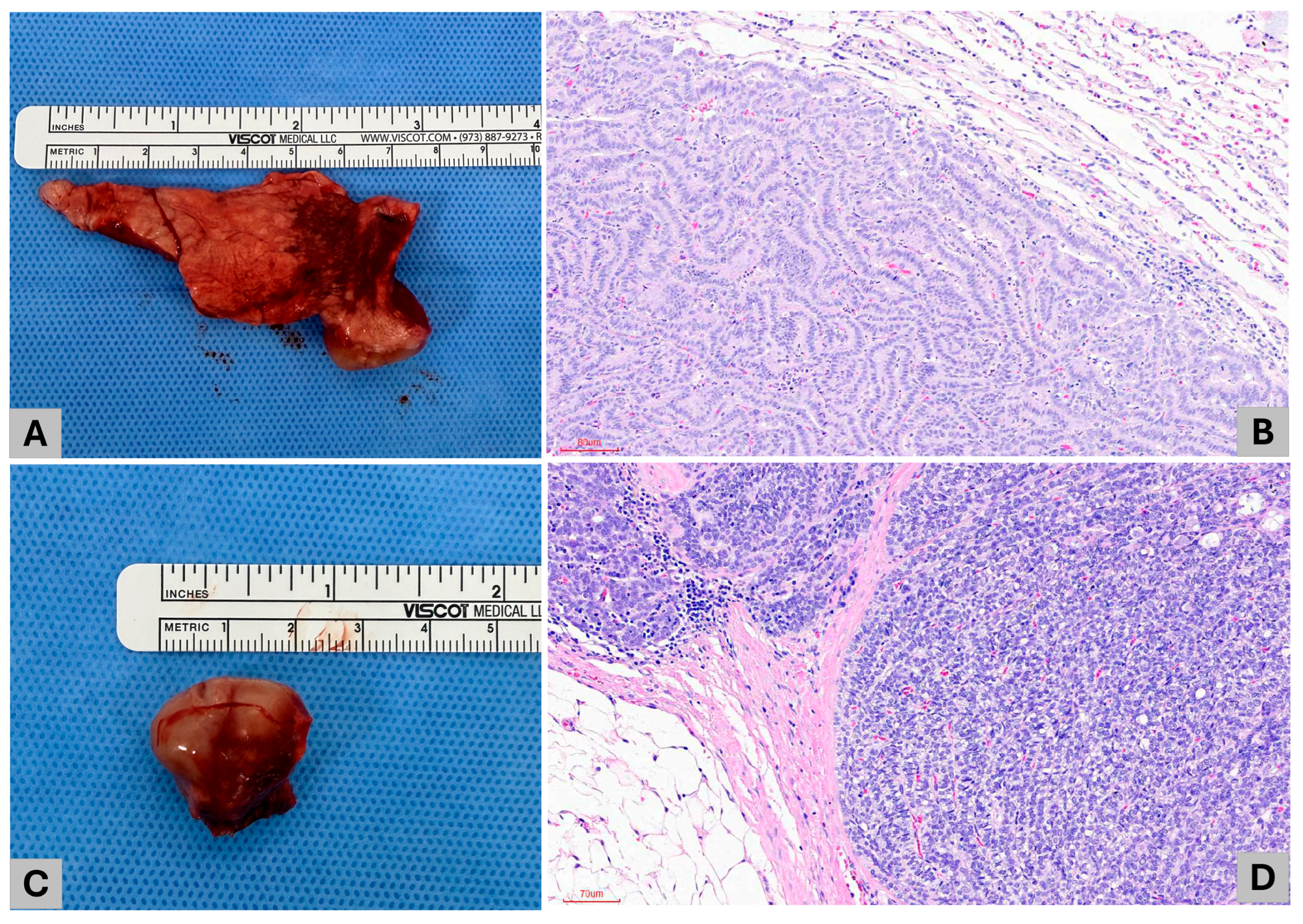
The pulmonary lesion and excised cranial mediastinal lesion were submitted to the Department of Veterinary Pathology, College of Veterinary Medicine, Konkuk University, for histopathological examination. The pulmonary lesion was diagnosed as pulmonary adenocarcinoma with invasion into adjacent pulmonary parenchyma and was considered histologic grade I by the pathologist according to a published canine pulmonary tumor grading scheme [11]. The neoplasm predominantly exhibited a tubulopapillary growth pattern and was composed mainly of cuboidal to columnar neoplastic epithelial cells with moderately eosinophilic to vacuolated cytoplasm and round to oval nuclei containing one to two distinct nucleoli. Nineteen mitotic figures were observed in 10 high-power fields (400×), and no tumor emboli were identified (Figure 3A,B). The excised cranial mediastinal lesion was confirmed histologically to be a lymph node. Normal nodal architecture was largely replaced by metastatic neoplastic epithelial cells arranged in tubular to solid patterns and morphologically consistent with the pulmonary primary, confirming cranial mediastinal lymph node metastasis (Figure 3C,D).
Figure 3.
Gross and histopathological findings. (A) Gross appearance of the resected right cranial lung lobe containing the pulmonary mass. (B) Histologic section of the pulmonary mass showing an infiltrative tubulopapillary proliferation of neoplastic epithelial cells, consistent with pulmonary adenocarcinoma (H&E). The tumor cells are predominantly cuboidal to columnar, with moderately eosinophilic to vacuolated cytoplasm and round to oval nuclei containing one to two distinct nucleoli; 19 mitotic figures were identified in 10 high-power fields, and no tumor emboli were observed. Scale bar = 80 μm. (C) Gross appearance of the excised cranial mediastinal lymph node. (D) Histologic section of the cranial mediastinal lymph node showing extensive replacement of normal nodal architecture by metastatic neoplastic epithelial cells arranged in tubular to solid patterns and morphologically consistent with the pulmonary primary (H&E). Scale bar = 70 μm.
Based on the primary tumor size, cranial mediastinal lymph node metastasis, and the absence of CT evidence of distant metastasis, the carcinoma was classified as T1N2M0 (T1, primary tumor ≤ 3 cm; N2, distant lymph node metastasis; M0, no evidence of distant metastasis) according to the canine lung carcinoma stage classification proposed by Lee et al. [12].
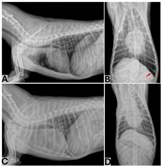
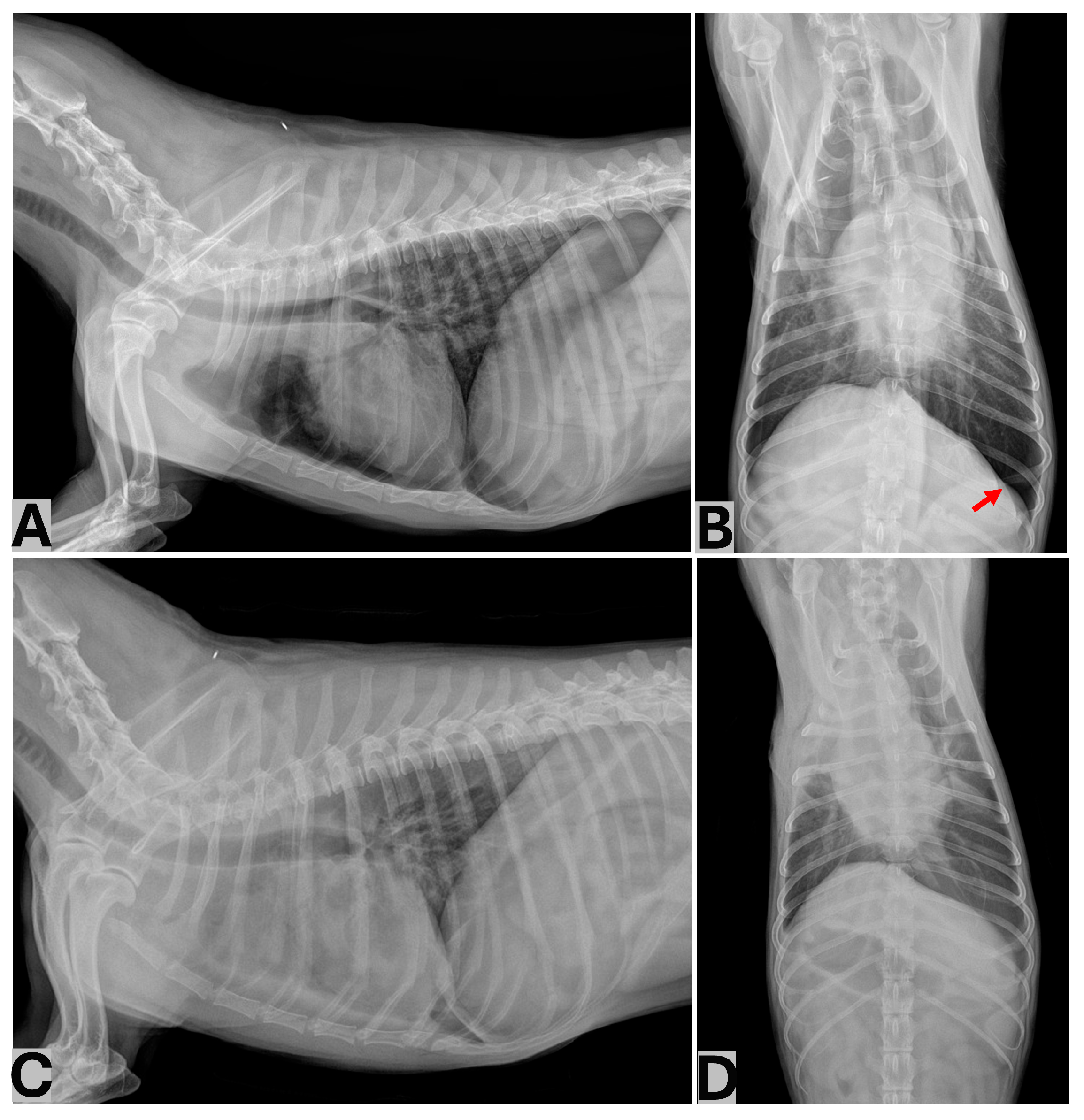
Recovery from anesthesia and extubation were uncomplicated. Thoracic radiographs obtained immediately after extubation demonstrated a mild pneumothorax, with pleural gas most evident in the left caudal pleural space, and no pleural effusion was identified (Figure 4A,B). During early postoperative recovery, the respiratory rate remained between 25 and 35 breaths/min, with no tachypnea or increased respiratory effort. Because the dog remained clinically stable and no progressive respiratory abnormality was observed, the pneumothorax was initially monitored.
Figure 4.
Postoperative thoracic radiographic changes before right-sided thoracocentesis. Right lateral (A) and ventrodorsal (B) thoracic radiographs obtained immediately after extubation demonstrate postoperative pneumothorax without pleural effusion. On the ventrodorsal projection, pleural gas is evident within the left hemithorax, confirming a left-sided pneumothorax (red arrow, (B)). Right lateral (C) and ventrodorsal (D) thoracic radiographs obtained on POD 1 before right-sided thoracocentesis demonstrate predominantly right-sided pleural effusion. The pleural gas visible immediately after surgery is less conspicuous in the presence of pleural fluid and other postoperative changes.
On the morning of postoperative day (POD) 1, the respiratory rate increased to 40–57 breaths/min and increased respiratory effort became evident. Thoracic radiographs demonstrated pleural effusion predominantly within the right hemithorax (Figure 4C,D). The pleural gas visible on the immediate postoperative study was less readily distinguishable in the presence of pleural fluid and other postoperative thoracic changes.
On POD 1, approximately 23 h after completion of surgery, ultrasound-guided right-sided thoracocentesis was performed in the caudal thorax with the dog in sternal recumbency and yielded 93 mL of grossly hemorrhagic fluid. Pleural fluid packed cell volume (PCV) was 26.5%, compared with a concurrent peripheral blood PCV of 27.3%, supporting a diagnosis of hemothorax. Thoracic radiographs obtained after drainage demonstrated a marked reduction in right-sided pleural fluid (Figure 5A,B).
Figure 5.
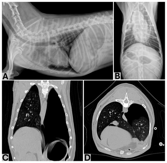
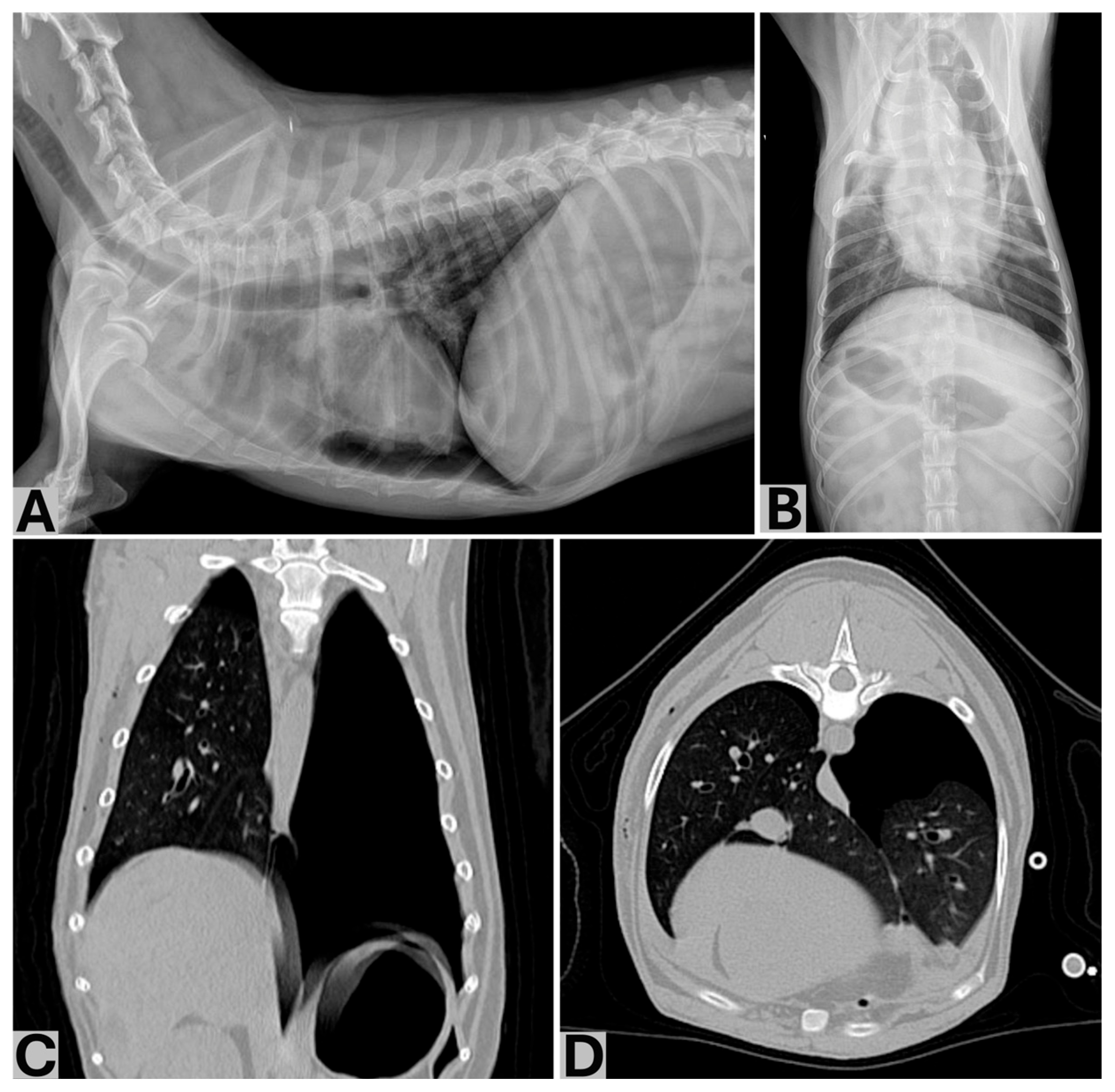
Identification of clinically important contralateral pneumothorax after drainage of the right-sided hemothorax. Right lateral (A) and ventrodorsal (B) thoracic radiographs obtained on POD 1 after ultrasound-guided drainage of 93 mL of hemorrhagic pleural fluid from the right hemithorax demonstrate a marked reduction in the right-sided pleural effusion. Thoracic CT images obtained later on POD 1 in the dorsal (C) and transverse (D) planes demonstrate a large volume of pleural gas within the left hemithorax and only a small volume of residual pleural fluid and gas on the right.
Following drainage of hemorrhagic pleural fluid from the right hemithorax on POD 1, whole-blood transfusion was initiated. Despite drainage of the right-sided hemothorax and initiation of transfusion, tachypnea and increased respiratory effort persisted. Because the right-sided hemothorax alone did not fully account for the persistent respiratory abnormalities, thoracic CT was performed later on POD 1 both to further evaluate the persistent respiratory abnormalities and to investigate a possible source of postoperative hemorrhage.
As the dog remained calm and cooperative, CT was performed without sedation or general anesthesia. Iohexol (Omnipaque 300; 300 mg I/mL) was administered intravenously at 2.5 mL/kg (total volume, approximately 16.26 mL) at an injection rate of 2.3 mL/s. Mild respiratory motion artifacts were present but did not compromise the diagnostic evaluation of the pleural cavities or the assessment for gross active contrast extravasation.
CT demonstrated only a small volume of residual pleural fluid and gas within the right hemithorax, whereas a large volume of pleural gas was present within the left hemithorax, contralateral to the thoracotomy and lobectomy site (Figure 5C,D). No active contrast extravasation or definite source of postoperative hemorrhage was identified. These findings indicated that active bleeding was not detectable at the time of CT, although previous or intermittent hemorrhage could not be excluded.
On POD 1, during the course of whole-blood transfusion, the transfusion was temporarily paused, and ultrasound-guided left-sided thoracocentesis was performed through the dorsal aspect of the caudal thorax. Approximately 95 mL of air was evacuated until resistance to aspiration was encountered and no further air could be readily withdrawn. The respiratory rate subsequently decreased from 48 to 28 breaths/min, with rapid improvement in respiratory effort, and later stabilized at 22 breaths/min at rest. The transfusion was then resumed and completed in the early hours of POD 2. A total of 180 mL of whole blood was administered in aliquots. Peripheral blood PCV increased from 27.3% on POD 1 to 33.1% on POD 2 and 39.5% on POD 3.
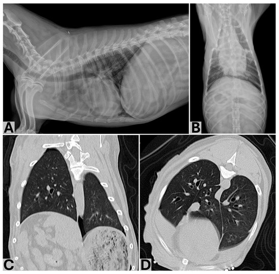
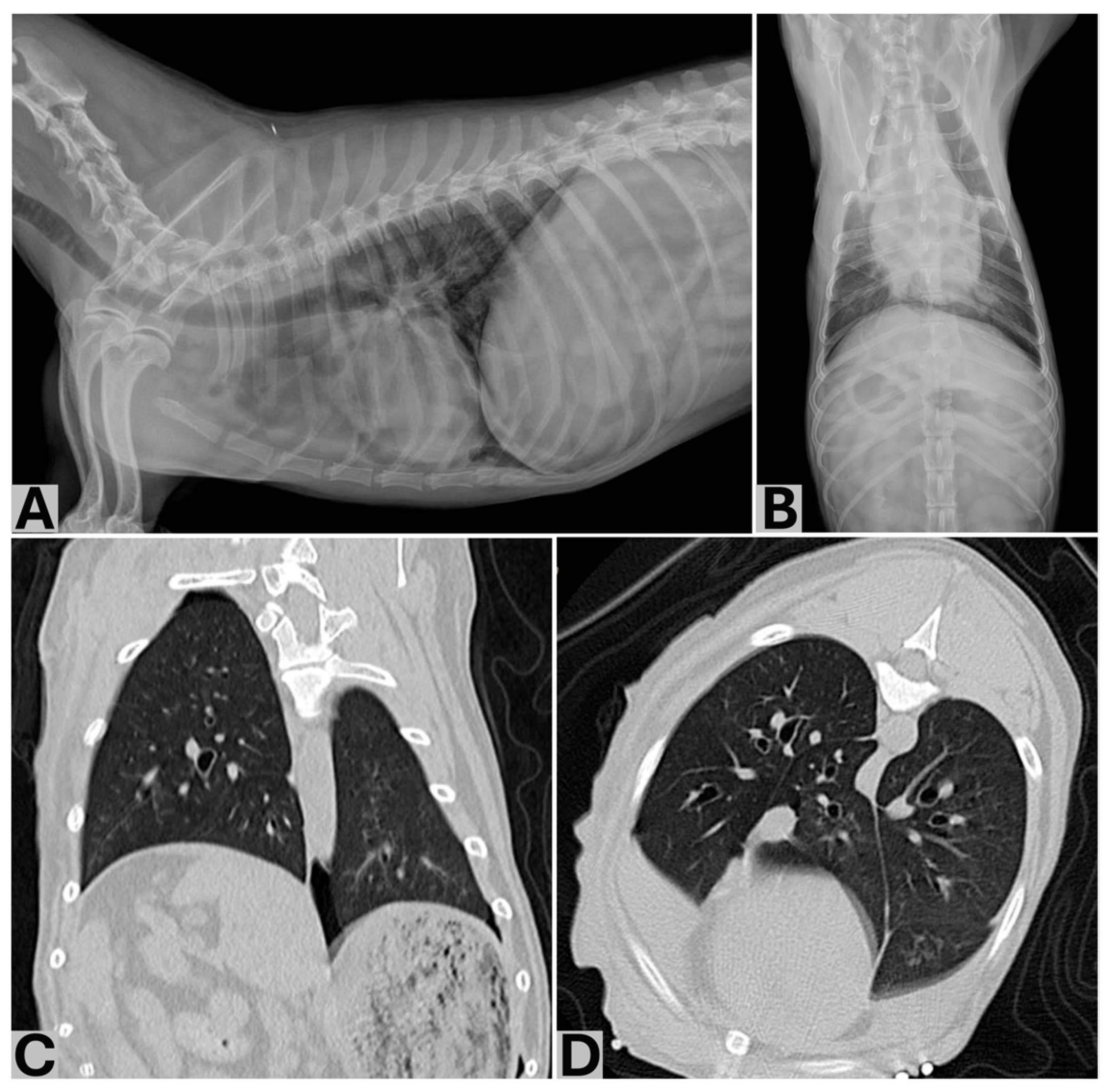
Follow-up non-contrast thoracic CT performed on POD 2 demonstrated resolution of the clinically important left-sided pneumothorax, without substantial reaccumulation of pleural gas or fluid (Figure 6C,D). No further thoracocentesis was required. Because no definite bleeding source had been identified and subsequent clinical and imaging findings showed no evidence of progressive pleural fluid accumulation, conservative management was continued; surgical re-exploration would have been considered if ongoing hemorrhage had been clinically suspected.
Figure 6.
Resolution of the contralateral pneumothorax after left thoracocentesis. Right lateral (A) and ventrodorsal (B) thoracic radiographs obtained after ultrasound-guided left-sided thoracocentesis and removal of approximately 95 mL of pleural air demonstrate marked improvement of the pneumothorax. Follow-up non-contrast thoracic CT images obtained on POD 2 in the dorsal (C) and transverse (D) planes demonstrate resolution of the previously identified large left-sided pneumothorax without substantial reaccumulation of pleural gas or fluid. The transverse CT image was obtained at approximately the same T7 level as Figure 5D.
Following left-sided thoracocentesis on POD 1, respiratory effort improved rapidly and remained normal thereafter, with no other clinically important respiratory abnormalities observed. The dog was discharged on POD 3 and was re-evaluated daily through POD 7. During this period, the thorax was reassessed with serial thoracic radiography, supplemented by thoracic ultrasonography on two occasions. No clinically important recurrence of pneumothorax or pleural effusion was identified.
Postoperative analgesia consisted of fentanyl administered as a continuous intravenous infusion at 5–6.5 µg/kg/h and gabapentin (20 mg/kg, PO, q8h), with additional supportive care provided as clinically indicated; the dog’s pre-existing cardiac medications were continued.
Follow-up thoracic CT on POD 86 showed no recurrent pneumothorax or pleural effusion. The dog remained clinically stable at POD 205.
3. Discussion
Postoperative pneumothorax is a recognized complication of lung lobectomy [6], and contralateral pleural air is anatomically plausible in dogs. Although fenestrations in the caudal mediastinum may permit interpleural communication [7,8], the degree of functional communication may vary. Experimental observations have shown that pleural disease may remain unilateral when the mediastinum is functionally intact but become bilateral following mechanical disruption [9]. The present case illustrates the potential challenges of pleural air evacuation following temporary surgical disruption and subsequent repair of the mediastinum. Despite evacuation of pleural air only from the right hemithorax, left-predominant pneumothorax was evident immediately after surgery, and a large left-sided pneumothorax was subsequently identified on POD 1. These findings emphasize the importance of assessing both pleural spaces after mediastinal manipulation.
The contribution of mediastinal defect closure to the postoperative pneumothorax cannot be determined from this case. Excision created an approximately 2.5-cm full-thickness defect through which the contralateral lung was directly visible. Given the surrounding vessels and tissue manipulation, closure was elected to restore local tissue continuity and stability, with potential postoperative bleeding also considered. Localized bleeding encountered during dissection had been controlled before thoracic closure. However, after the communication was repaired, right-sided evacuation could not be assumed to provide adequate evacuation of the left pleural cavity. In a study of 40 canine cadavers following diaphragmatic defect closure, Yoon et al. identified interpleural pressure differences after unilateral evacuation through a small caudal mediastinal puncture, whereas no significant differences were detected with a larger puncture or bilateral evacuation [10]. Mediastinal transection has also been described as a means of permitting bilateral drainage through a single pleural catheter [13]. In retrospect, maintaining or creating adequate interpleural communication, such as a ventral mediastinal window, might have facilitated bilateral evacuation.
The exact source and route of the postoperative pleural air remain uncertain. One plausible explanation is that air entered or redistributed into the left hemithorax while the surgically created communication was open and subsequently persisted after defect closure and right-sided evacuation. However, delayed leakage from the bronchial stump or adjacent pulmonary parenchyma cannot be excluded despite the negative intraoperative saline bubble test, which reflects only the conditions at surgery. Although the fourth or fifth intercostal space is generally recommended for access to the cranial lung lobe [6], the third intercostal space was selected to permit access to both the right cranial lung lobe and the cranial mediastinal lesion at T1–T2 through a single thoracotomy. This approach provided limited hilar exposure, leaving an unmeasured rim of proximal pulmonary parenchyma at the hilar stump and further precluding exclusion of delayed leakage. Small air-attenuating lesions consistent with bullae or blebs were also identified preoperatively in the right cranial and accessory lung lobes; rupture of a lesion in the remaining accessory lobe represents another possible source of pleural air. Additional possibilities include ventilation- or lung re-expansion-related air leakage and iatrogenic introduction of additional pleural air during postoperative thoracocentesis. Thus, temporary interpleural communication followed by repair and unilateral evacuation remains one plausible mechanism rather than a proven cause.
The clinical deterioration on POD 1 was likely multifactorial. Right-sided thoracocentesis yielded 93 mL of hemorrhagic fluid (14.3 mL/kg), with a pleural fluid PCV of 26.5% compared with a concurrent peripheral blood PCV of 27.3%, supporting clinically important intrathoracic blood loss. Pleural fluid accumulation, acute blood loss, and pleural gas may have collectively contributed to respiratory compromise. Persistent respiratory abnormalities despite drainage and initiation of transfusion prompted CT imaging, which demonstrated a large left-sided pneumothorax with only small amounts of residual pleural fluid and gas in the right hemithorax. No active contrast extravasation or definite bleeding source was identified. Potential hemorrhage sources included the hilar ligation region, cranial mediastinal dissection bed, and thoracotomy or intercostal region, although none could be confirmed. Because subsequent imaging showed no substantial pleural fluid reaccumulation and the patient remained clinically stable, surgical re-exploration was not pursued. Pleural gas was less conspicuous on POD 1 radiographs in the presence of pleural fluid and other postoperative changes. Previous canine studies suggest that CT or transthoracic ultrasonography may help identify radiographically inconspicuous pneumothorax, although these studies involved traumatic or experimentally induced rather than postoperative pneumothorax [14,15].
Following evacuation of approximately 95 mL of pleural air from the left hemithorax, the respiratory rate decreased from 48 to 28 breaths/min and respiratory effort improved rapidly, with the respiratory rate subsequently stabilizing at 22 breaths/min at rest. This temporal improvement is consistent with a clinically important contribution of the left-sided pneumothorax to the respiratory abnormalities. However, whole-blood transfusion, which had been initiated before thoracocentesis and was resumed afterward, may also have contributed to clinical stabilization. Therefore, the clinical improvement cannot be attributed exclusively to pleural air evacuation.
A thoracostomy tube was not placed for the following reasons: no preoperative pneumothorax or pleural effusion was identified, no ongoing air leakage or substantial pleural fluid accumulation was apparent at the conclusion of surgery, the hilar/bronchial stump and mediastinal repair appeared intact, and no further air could be readily aspirated from the right pleural cavity. In a retrospective study of dogs and cats without preoperative pneumothorax or pleural effusion that received postoperative thoracostomy tubes, the median time to re-establishment of negative thoracic pressure was 0.5 h, compared with a median tube maintenance duration of 8 h [16]. Although prolonged tube maintenance may not always be necessary in appropriately selected patients, this study did not evaluate routine omission of tube placement. In the present case, a thoracostomy tube could have allowed continued evacuation and quantification of pleural air and fluid from the operated hemithorax, but adequate contralateral drainage through a unilateral tube could not be assured after mediastinal repair. Postoperative drainage planning should therefore be individualized according to the clinical and imaging findings in both pleural cavities.
The subsequent clinical course was less consistent with a persistent high-volume air leak. Follow-up CT on POD 2 showed no substantial reaccumulation of pleural gas or fluid, and no further thoracocentesis was required. The dog was discharged on POD 3, with no clinically important recurrence identified during follow-up through POD 7. CT on POD 86 likewise showed no recurrence, and the dog remained clinically stable through POD 205. Although these findings are less consistent with persistent bronchial stump dehiscence or sustained high-volume pulmonary air leakage, they do not exclude transient leakage or distinguish among the proposed perioperative mechanisms.
Several limitations should be acknowledged. As a single clinical case, the proposed relationship among temporary interpleural communication, defect repair, unilateral evacuation, and contralateral air retention cannot be established. Although the contralateral lung was directly visible through the defect, air movement was not observed or quantified, and interpleural pressures were not measured. CT was not performed immediately after surgery, preventing precise assessment of the initial volume and distribution of pleural gas and its subsequent evolution before POD 1. The absence of a thoracostomy tube also precluded continuous quantification of postoperative pleural air and fluid. Concurrent hemothorax and whole-blood transfusion confounded interpretation of the response to left-sided thoracocentesis. Finally, the bleeding source remained unidentified, and the non-contrast CT performed on POD 2 could not assess active contrast extravasation. These limitations preclude definitive conclusions regarding the mechanism of contralateral pneumothorax.
4. Conclusions
This case illustrates the perioperative implications of altered interpleural communication during lung lobectomy combined with cranial mediastinal surgery. Temporary full-thickness communication between the pleural cavities, followed by mediastinal defect repair and unilateral pleural evacuation, may have contributed to contralateral pleural air retention, although the exact mechanism remains uncertain. Alternative causes, including delayed bronchial stump or pulmonary parenchymal leakage, could not be excluded. The postoperative respiratory deterioration was likely multifactorial, involving a clinically important right-sided hemothorax with associated acute blood loss and a concurrent left-sided pneumothorax. Following mediastinal manipulation or repair, evacuation of one hemithorax should not be assumed to ensure adequate drainage of the other. Persistent respiratory abnormalities despite treatment of the operated hemithorax warrant assessment of the contralateral pleural space, with further drainage guided by clinical and imaging findings.
Author Contributions
Conceptualization, B.-J.P. and H.-Y.K.; investigation, B.-J.P., S.P., S.L., C.P. and S.H.; data curation, B.-J.P., S.P. and S.L.; writing—original draft preparation, B.-J.P.; writing—review and editing, B.-J.P. and H.-Y.K.; visualization, B.-J.P.; supervision, H.-Y.K. All authors have read and agreed to the published version of the manuscript.
Funding
This research received no external funding.
Institutional Review Board Statement
Ethical review and approval were not required for this case report because all diagnostic and therapeutic procedures were performed as part of routine clinical care for a client-owned dog.
Informed Consent Statement
Written informed consent was obtained from the owner for publication of the clinical information and images included in this report.
Data Availability Statement
The original contributions presented in this study are included in the article. Further inquiries can be directed to the corresponding author.
Acknowledgments
During the preparation of this manuscript, the authors used ChatGPT (GPT-5, OpenAI) for language editing, clarity improvement, and manuscript organization. The authors reviewed and edited the output and take full responsibility for the content of this publication.
Conflicts of Interest
The authors declare no conflicts of interest.
Abbreviations
The following abbreviations are used in this manuscript:
| CT | Computed tomography |
| H&E J | Hematoxylin and eosin |
| IV | Intravenous |
| PCV | Packed cell volume |
| PO | Per os |
| POD | Postoperative day |
| SC | Subcutaneous |
References
- McPhetridge, J.B.; Scharf, V.F.; Regier, P.J.; Toth, D.; Lorange, M.; Tremolada, G.; Dornbusch, J.A.; Selmic, L.E.; Bae, S.; Townsend, K.L.; et al. Distribution of histopathologic types of primary pulmonary neoplasia in dogs and outcome of affected dogs: 340 cases (2010–2019). J. Am. Vet. Med. Assoc. 2022, 260, 234–243. [Google Scholar] [CrossRef] [Scilit]
- Rebhun, R.B.; Culp, W.T.N. Tumors of the respiratory system: Pulmonary neoplasia. In Withrow and MacEwen’s Small Animal Clinical Oncology, 5th ed.; Withrow, S.J., Vail, D.M., Page, R.L., Eds.; Elsevier: St. Louis, MO, USA, 2013; pp. 432–462. [Google Scholar]
- Paoloni, M.C.; Adams, W.M.; Dubielzig, R.R.; Kurzman, I.; Vail, D.M.; Hardie, R.J. Comparison of results of computed tomography and radiography with histopathologic findings in tracheobronchial lymph nodes in dogs with primary lung tumors: 14 cases (1999–2002). J. Am. Vet. Med. Assoc. 2006, 228, 1718–1722. [Google Scholar] [CrossRef] [Scilit]
- Bleakley, S.; Phipps, K.; Petrovsky, B.; Monnet, E. Median sternotomy versus intercostal thoracotomy for lung lobectomy: A comparison of short-term outcome in 134 dogs. Vet. Surg. 2018, 47, 104–113. [Google Scholar] [CrossRef] [Scilit]
- Sandoval, D.M.; Stobie, D.; Valenzano, D.M.; Zuendt, G.F.; Lopez, D.J. Short-term outcomes of dogs and cats undergoing lung lobectomy using either a self-ligating loop or a thoracoabdominal stapler. Vet. Surg. 2024, 53, 1287–1293. [Google Scholar] [CrossRef] [Scilit]
- Fossum, T.W. Small Animal Surgery, 5th ed.; Elsevier: Philadelphia, PA, USA, 2019. [Google Scholar]
- Baines, E.; Gendron, K. The mediastinum. In BSAVA Manual of Canine and Feline Thoracic Imaging, 2nd ed.; Schwarz, T., Scrivani, P., Eds.; British Small Animal Veterinary Association: Gloucester, UK, 2024; pp. 276–325. [Google Scholar] [CrossRef] [Scilit]
- Frame, M.; King, A. The pleural space. In BSAVA Manual of Canine and Feline Thoracic Imaging, 2nd ed.; Schwarz, T., Scrivani, P., Eds.; British Small Animal Veterinary Association: Gloucester, UK, 2024; pp. 326–355. [Google Scholar] [CrossRef] [Scilit]
- von Recum, A.F. The mediastinum and hemothorax, pyothorax, and pneumothorax in the dog. J. Am. Vet. Med. Assoc. 1977, 171, 531–533. [Google Scholar] [CrossRef] [Scilit]
- Yoon, H.-Y.; Mann, F.A.; Lee, S.; Jeong, S.-W. Comparison of techniques for transdiaphragmatic thoracic drainage after diaphragmatic defect closure in dogs: A cadaveric study. J. Vet. Sci. 2013, 14, 193–197. [Google Scholar] [CrossRef] [Scilit]
- Meuten, D.J. (Ed.) Tumors in Domestic Animals, 5th ed.; Wiley Blackwell: Ames, IA, USA, 2017; p. 970. [Google Scholar]
- Lee, B.M.; Clarke, D.; Watson, M.; Laver, T. Retrospective evaluation of a modified human lung cancer stage classification in dogs with surgically excised primary pulmonary carcinomas. Vet. Comp. Oncol. 2020, 18, 590–598. [Google Scholar] [CrossRef] [Scilit]
- Griffin, M.A.; Culp, W.T.N.; Buote, N.J. The role of minimally invasive surgery in oncology—Part 2: Thoracoscopy. Vet. Oncol. 2025, 2, 3. [Google Scholar] [CrossRef] [Scilit]
- Dancer, S.C.; Le Roux, C.; Fosgate, G.T.; Kirberger, R.M. Radiography is less sensitive relative to CT for detecting thoracic radiographic changes in dogs affected by blunt trauma secondary to a motor vehicle accident. Vet. Radiol. Ultrasound 2019, 60, 648–658. [Google Scholar] [CrossRef] [Scilit]
- Hwang, T.S.; Yoon, Y.M.; Jung, D.I.; Yeon, S.C.; Lee, H.C. Usefulness of transthoracic lung ultrasound for the diagnosis of mild pneumothorax. J. Vet. Sci. 2018, 19, 660–666. [Google Scholar] [CrossRef] [Scilit]
- DeRouen, A.; Waschak, M.; Stokes, R.; Maxwell, M. Duration of use of postoperative thoracostomy tubes in canines and felines without preoperative pneumothorax or pleural effusion. J. Am. Vet. Med. Assoc. 2026, 264, 1–5. [Google Scholar] [CrossRef] [Scilit]
Disclaimer/Publisher’s Note: The statements, opinions and data contained in all publications are solely those of the individual author(s) and contributor(s) and not of MDPI and/or the editor(s). MDPI and/or the editor(s) disclaim responsibility for any injury to people or property resulting from any ideas, methods, instructions or products referred to in the content. |
© 2026 by the authors. Licensee MDPI, Basel, Switzerland. This article is an open access article distributed under the terms and conditions of the Creative Commons Attribution (CC BY) license.