


Simple Summary
Feline chronic gingivostomatitis (FCGS) is a severe inflammatory disease of the oral cavity that causes pain, difficulty eating, and reduced quality of life. Dental extractions are considered the standard treatment, but little is known about how the oral tissues and local immune response change after surgery. This pilot prospective study followed five cats after treatment, evaluating their clinical and blood parameters, with three cats also being evaluated for histopathologic changes and local immune cell markers. Most cats showed clinical improvement after dental extractions. Reduced inflammation was observed in oral biopsies. Expression of B-cell markers in the affected tissues was reduced after dental extraction. However, changes in other immune markers varied among individuals. These findings provide preliminary information on the local tissue response following standard surgical treatment and improve our understanding of this chronic inflammatory disease.
Abstract
Feline chronic gingivostomatitis (FCGS) is a painful inflammatory disease of the oral cavity in which partial-mouth or full-mouth extraction (PME or FME) is considered the standard treatment. However, longitudinal information regarding histopathologic and local immunologic changes after surgery remains limited. The aim of this pilot prospective study was to assess the clinical, hematologic, biochemical, histopathologic, and immunologic evolution of cats with FCGS following PME. Five cats diagnosed with FCGS were enrolled and followed longitudinally. Clinical response was assessed in the five cats using the Stomatitis Disease Activity Index (SDAI), together with hematologic, biochemical, and retroviral testing. Biopsies of the caudal oral mucosa were collected from three cats before surgery and 30 days after surgery for histopathologic evaluation using a previously described inflammatory scoring system. Immunolabeling of CD20, FoxP3, and IL-17 was quantified by digital image analysis. Clinical scores improved after PME, although the magnitude of the response varied among individuals. Persistent non-regenerative anemia and marked monocytosis were observed in cats positive for feline leukemia virus and/or feline immunodeficiency virus. Hyperglobulinemia was the most common biochemical abnormality. Histopathologic examination revealed moderate-to-marked lymphoplasmacytic inflammation associated with epithelial hyperplasia, edema, and, in the most severe cases, spongiosis and necrosis. Sequential biopsies demonstrated reductions in histologic inflammatory scores in two of three cats. CD20 immunolabeling decreased post-PME, whereas FoxP3 and IL-17 expression showed marked interindividual variability. This pilot prospective study provides preliminary observations on tissue changes in the early stages following PME.
1. Introduction
Feline chronic gingivostomatitis (FCGS) is a painful, debilitating, and multifactorial inflammatory disease characterized by severe inflammation affecting the oral mucosa that markedly decreases the quality of life of affected cats [1,2,3,4,5]. Lesions may involve the gingival, alveolar, and buccolabial mucosae, and most characteristically, the caudal oral mucosa lateral to the palatoglossal folds [3,4,5]. Extraoral manifestations, including esophagitis and tracheobronchial inflammation, have also been described, further supporting the concept that FCGS is associated with widespread mucosal inflammation [6,7]. Histologically, the disease is characterized by a dense lymphoplasmacytic infiltrate accompanied by variable numbers of neutrophils and macrophages. Immunophenotypic studies have demonstrated the presence of multiple immune cell populations within affected tissues, including CD20+ B lymphocytes, CD3+ T cells (including CD4+ helper and CD8+ cytotoxic subsets), and FoxP3+ regulatory T cells [8,9]. More recently, transcriptomic and immunological studies have suggested that FCGS is characterized by persistent immune dysregulation involving alterations in T-cell function and inflammatory pathways, supporting the hypothesis that the disease represents an aberrant host response [2,8,10].
Surgical treatment through partial-mouth extraction (PME) or full-mouth extraction (FME) remains the current standard of care, although approximately 30% of cats exhibit partial clinical responses or remain refractory to treatment [1,11]. Previous investigations evaluating treatment efficacy have primarily focused on clinical outcomes [11,12,13,14], whereas more recent studies have explored alterations in the oral microbiome, systemic inflammatory biomarkers, and transcriptomic profiles associated with disease progression and treatment response [15,16,17,18,19]. In particular, transcriptomic analysis of circulating CD8+ T cells demonstrated an exhaustion-like phenotype and impaired mitochondrial bioenergetics, while cats evaluated after FME and mesenchymal stem cell (MSC) therapy showed partial recovery of these molecular signatures [19].
Despite these advances, relatively little is known regarding the evolution of the inflammatory infiltrate and local immune response within the oral mucosa following FME or PME. Most studies characterizing the oral immune microenvironment have focused either on disease pathogenesis or evolution in cats refractory to FME undergoing MSC therapy [2,8,20,21,22]. Although reductions in lymphoplasmacytic infiltration have been reported in treatment responders receiving cell-based therapies, longitudinal information describing the histopathologic and immunological changes after standard surgical treatment remains limited. Owing to the practical difficulties associated with obtaining sequential oral biopsies, the local tissue response to dental extractions has been insufficiently characterized.
The aim of the present prospective study was to describe the clinical, hematologic, biochemical, histopathologic, and local immunological changes that occur after PME in cats with FCGS.
2. Materials and Methods
2.1. Study Design
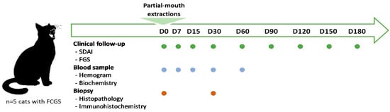
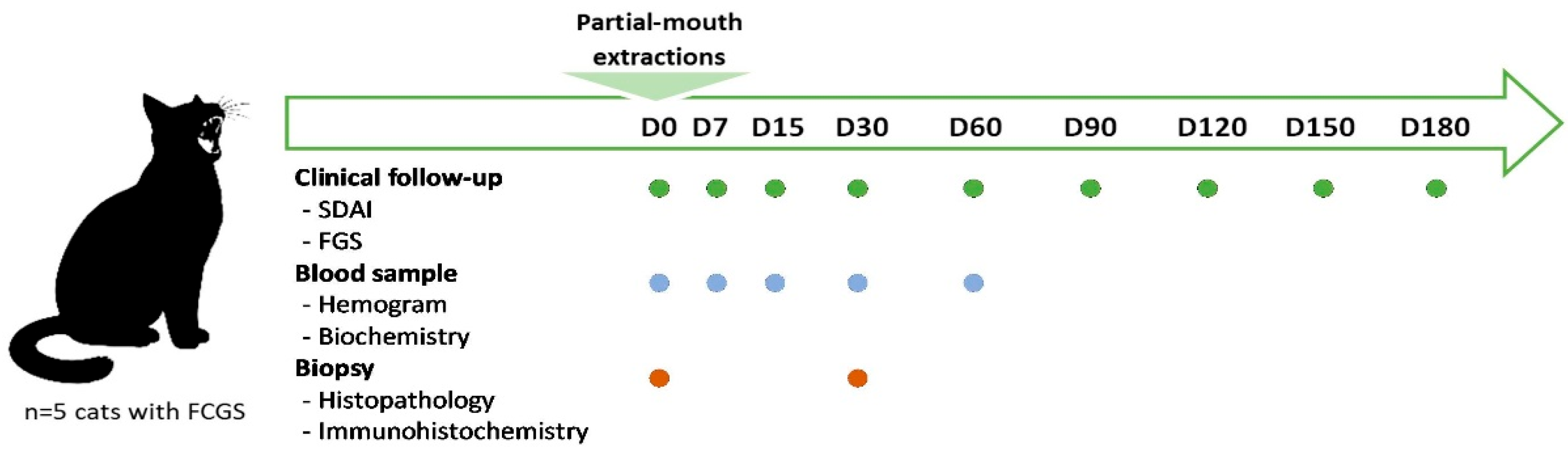
This pilot prospective longitudinal study was conducted to characterize the clinical, hematologic, biochemical, histopathologic, and local immunological changes that occur after PME in cats diagnosed with FCGS (Figure 1). The inclusion criteria were as follows: cats presenting inflammatory lesions compatible with FCGS, without oncologic or immune-mediated disease. Selected patients exhibited caudal oral mucositis and alveolar mucositis involving the premolar and/or molar regions, with no involvement of the canine or incisor regions. There were no restrictions regarding breed, age, body weight, or feline immunodeficiency virus (FIV) or feline leukemia virus (FeLV) status. Written informed consent was obtained from all owners before enrollment. Corticosteroid therapy, when present, was discontinued at least 20 days prior to surgery and was not permitted during the study period. No cat received a long-acting injectable glucocorticoid. NSAIDs, pregabalin, gabapentin, and opioids were allowed during the study period. Cats meeting the inclusion criteria underwent PME, comprising extraction of all the premolar and molar teeth. Before surgery, cats received antibiotics (amoxicillin–clavulanic acid (15 mg/kg BID) or spiramycin–metronidazole (75,000 IU/kg and 12.5 mg/kg, respectively)) and NSAIDs (meloxicam (0.1–0.02 mg/kg SID)). Antibiotics were maintained from 7 to 10 days after surgery. Intraoral radiographs were obtained before PME, but post-extraction dental radiographs were not routinely obtained; they were only performed when root fracture occurred to confirm complete root removal. Between 7 and 15 days after surgery, the cats received amoxicillin–clavulanic acid and meloxicam (dose reduced from 0.1 mg/kg to 0.03 mg/kg SID), as well as pregabalin (2 mg/kg SID or BID). The cats were evaluated on the day of PME (D0); at 7 (D7), 15 (D15), 30 (D30), and 60 (D60) days; and monthly thereafter until 6 months post-treatment (D180). Evaluations were performed without sedation at all time points, except at D0 and D30, when three cats were anesthetized for biopsy sampling.
Figure 1.
Overview of the study design.
2.2. Clinical Evaluation
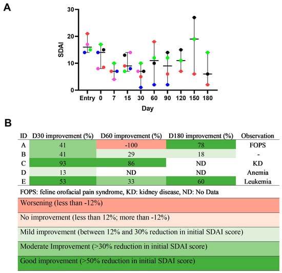
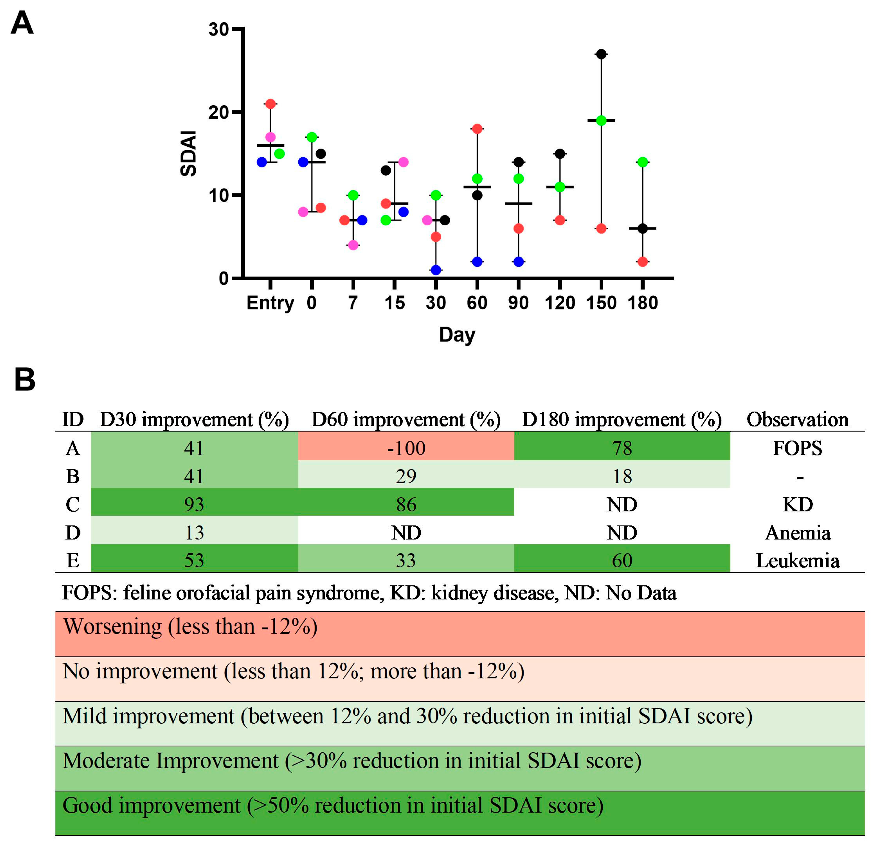
Clinical disease severity was evaluated using the Stomatitis Disease Activity Index (SDAI) scoring system, considering both investigator and owner evaluations, and ranged from 0 (healthy) to 30 (severe) [12,23]. Scoring was performed at every follow-up. Investigators scored the lesions (0–3, from No Lesion to Severe), and the cats’ owners scored the cat’s appetite, activity level, grooming behavior and perceived oral comfort. The percentage of SDAI improvement was calculated using the following formula: [(initial SDAI D0 − post-treatment follow-up SDAI)/initial SDAI D0] ×100; the threshold for improvement was >12% following previous studies [23]. A reduction of >50% compared to the initial SDAI score was considered good improvement, >30–50% was considered moderate improvement, 12–30% was considered mild improvement, −12 to <12% was considered no improvement, and less than −12% was considered worsening [23].
2.3. Blood Parameters
Complete blood counts (ProCyte One analyzer, Idexx, Maine, USA) and serum biochemical (CM320, Weiner lab, Buenos Aires, Argentina) analyses were performed at predetermined time points throughout follow-up. Patients were considered anemic when at least two of the following erythrocyte parameters were below the established reference ranges for the species: total red blood cell count, hematocrit, and/or hemoglobin concentration. Anemia was classified as regenerative or non-regenerative according to the absolute reticulocyte count [24]. Retroviral infection status was determined using commercially available point-of-care assays for FeLV antigen and FIV antibodies (FASTest FIV/FeLV, Megacor Diagnostik GmbH, Hörbranz, Germany).
2.4. Evaluation of Local Inflammation
A 0.5 cm full-thickness biopsy was collected under general anesthesia from the caudal oral mucosa lateral to the palatoglossal folds of three cats at D0 and D30. The biopsy sample was collected from one side of the oral cavity at D0 and from the contralateral side at D30. Histopathologic and immunohistochemical (IHC) analyses were performed on formalin-fixed, paraffin-embedded specimens. Tissue samples were fixed in 4% buffered formaldehyde, processed, and sectioned into 4 μm thick sections. To assess inflammation, the sections were stained with hematoxylin and eosin (H&E). The degree of inflammation was evaluated using a scoring system for the severity of the microscopic inflammatory changes (Grades 0–3, from normal to severe) [9]. Histological images were visualized and acquired using a transmitted light microscope (Olympus BX41, Olympus Corporation, Tokyo, Japon) equipped with a high-resolution digital camera and capture software (AmScope MU853B USB 3.0 high-speed digital camera, CA, USA). The sections were also scanned using a whole-slide scanner (Motic EasyScan One scanner, Motic China Group Co., Ltd., Xiamen, China) at a focal plane with default settings and a magnification of 400× (image resolution: 0.25 µm/pixel) and visualized using a viewer (Motic ‘DSAssistant’, Motic VM V1 Viewer 2.0, version 2019-08-02, China) and scanning software (Motic ‘EasyScanner’, Hong Kong, China). Histopathologic evaluation was performed by four observers blinded to the patient and sampling time point, with final scores assigned by consensus.
To assess the type of cellular infiltrate, expression of CD20, FoxP3, and IL-17 antigens was evaluated. For this, tissue sections were deparaffinized with xylene and rehydrated through a graded ethanol series. A 3% hydrogen peroxide blocking solution was applied, followed by heat-induced antigen retrieval in a steamer, with slides submerged in 0.01 M citrate buffer (pH 6) in a polypropylene Coplin jar (Deltalab, Barcelona, Spain) for 30 min. IHC conditions were optimized using feline lymph nodes and caudal oral mucosa as positive controls. Negative controls were performed by replacing the primary antibody with phosphate-buffered saline (PBS). Isotype controls were not performed. Manufacturer recommendations were followed with minor modifications. The primary antibodies used in this study are listed in Table 1. The selected CD20 antibody [25] and FoxP3 clone (221D) [26] have been used in feline tissues. IHC detection of IL-17A has previously been reported in other feline tissues using a different antibody [27]; however, this does not constitute validation of the antibody used in the present study for feline tissue. Each primary antibody was applied to the tissue, which was then incubated overnight at 4 °C in a humid chamber. The detection system (Mach 4 Universal HRP-Polymer Detection System, Biocare Medical, CA, USA) and streptavidin–peroxidase were applied. Labeling was visualized using DAB solution for 3–5 min, and then the sections were counterstained with Mayer’s hematoxylin, dehydrated, and mounted. Histological images were acquired as previously described.
Table 1.
Details of antibodies for immunohistochemical labeling.
Immunolabeled areas were quantified using digital image analysis software, allowing for objective assessment of marker expression and minimizing observer-related bias. Analysis of whole-slide images was performed collaboratively by four observers blinded to the patient and sampling time point using Motic SlideViewer software (Motic VM V1 Viewer 1.0) to ensure consistency in interpretation. Immunolabeled cells were evaluated in five non-overlapping high-power fields (400×) [28] selected from representative areas of the inflammatory infiltrate after examination of the entire section at low magnification. Necrotic areas were excluded from the evaluation due to their potential to produce nonspecific staining and false-positive results, as were regions with prominent vascularization, severe spongiosis, or technical artifacts related to tissue processing, sectioning, or staining. Areas with technical staining artifacts that prevented reliable discrimination of specific DAB signals from the background were excluded. Quantification was conducted using FIJI (ImageJ 2.14.0 1.54p, NIH, MD, USA). A color threshold was applied to isolate the specific chromogenic signal and distin guish it from the background. The thresholded image was then binarized to convert the selected signal into a binary format. The area occupied by positive staining was subsequently quantified and expressed as a percentage of the total image area. All images were processed using identical parameters for brightness, contrast, and thresholding to ensure consistency across samples.
2.5. Descriptive Analysis
The data are presented descriptively as individual values for each cat and time point. No inferential statistical analyses were performed.
3. Results
3.1. Patient Characteristics
Five client-owned mixed-breed cats with a mean age of 7 years were included. Four had the ulcerative form of FCGS, and one had the proliferative form. Cats were housed indoors or had a mixed indoor–outdoor lifestyle and lived with a mean of two additional cats per household. Individual patient characteristics and FIV/FeLV status are shown in Table A1 and Table A2, respectively.
3.2. Clinical Evaluation
Clinical response after PME varied among individuals; however, all cats exhibited reductions in SDAI scores by day 30 compared with baseline (Figure 2). The degree of improvement differed among cats. At D0, cats showed a mean SDAI score of 12.6. At D30, two showed good improvement, two showed moderate improvement, and one showed mild improvement. Three cats were followed up to D180, with two of them showing good improvement and one showing mild improvement at this time point. Cat E showed good improvement in SDAI score but died due to complications associated with FeLV (lymphoblastic leukemia) after the study period. Cat C was withdrawn after D90 due to kidney disease, although it showed an improvement in SDAI score after PME. Cat D was lost to follow-up after D30. Cat A received amitriptyline for feline orofacial pain syndrome (FOPS) control from D60 to D120 and then CBD oil afterwards, achieving full remission by D180. Cat C received omeprazole.
Figure 2.
(A) Stomatitis Disease Activity Index (SDAI) for each cat and timepoint. Each colored dot represents a single feline patient, coded as follows: A (red), B (green), C (blue), D (pink), and E (black). The horizontal black line indicates the median, and the whiskers indicate the minimum and maximum values. ND: no data. (B) Percentage change in SDAI over time and additional observations for each cat. The color scale represents clinical changes, with green indicating improvement and red indicating worsening.
3.3. Blood Parameters
Three of the five cats showed anemia at baseline (Table A2, Figure S1). Persistent non-regenerative anemia was observed in cats D and E, both of which were positive for FeLV and/or FIV. In contrast, transient regenerative anemia was documented in cat C during follow-up. Monocytosis was particularly pronounced in cats D and E. Eosinophilia and increased ALP were observed in cat B at D15, which declined by D60, coinciding with the detection of endoparasites (Dipylidium spp.), after which the cat received anthelmintic treatment.
Hyperglobulinemia represented the most consistent biochemical abnormality and was present in all cats at one or more time points (Figure S2). Hypoalbuminemia was observed in two of five cats at D0, which subsequently normalized in all but one cat. Additionally, one cat had elevated serum urea and creatinine levels and consequently received treatment for kidney disease.
3.4. Histopathological Findings
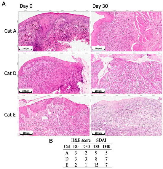
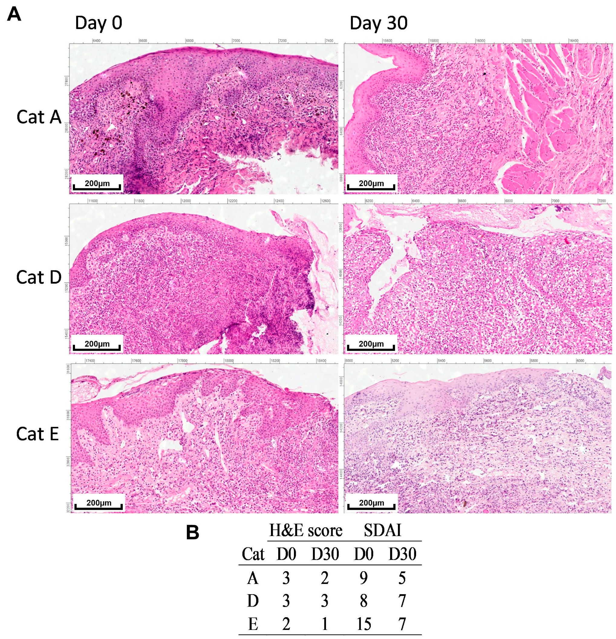
Oral biopsies were collected from three FeLV-positive cats (A, D, and E) at D0 and D30 (Figure 3A). Cat B did not attend the scheduled D30 visit under conditions suitable for general anesthesia, and cat C was not biopsied because elevated serum urea and creatinine concentrations were detected. Considering the potential anesthetic risk, general anesthesia for biopsy collection was not performed in the interest of this cat’s welfare. Tissue sections exhibited epithelial hyperplasia with abundant cellular debris in the most severe cases. Moderate-to-marked lymphoplasmacytic infiltrate was consistently noted, primarily localized within the submucosa. Mott cells were occasionally observed within the infiltrate. Neutrophils were identified in the submucosa and perivascular regions, particularly in areas displaying edema and necrosis. In samples graded as most severe, there was evident spongiosis, interstitial edema, and dense inflammatory infiltrate encasing muscle fibers. Histopathologic examination of the D0 and D30 oral biopsies from cat E showed chronic inflammatory changes without evidence of neoplastic infiltration. Lymphoblastic leukemia was diagnosed after completion of the study period, on day 200.
Figure 3.
Inflammatory changes in oral biopsies pre- and post-PME. (A) Microscopic images of oral biopsies (H&E, 100×, scale bar = 200 μm). (B) Score for severity of microscopic inflammatory changes and SDAI scores.
Two cats exhibited a reduction in severity of microscopic inflammatory changes accompanied by clinical improvement (decrease in SDAI) (Figure 3B), whereas one cat showed an unchanged inflammatory score and minimal improvement in SDAI, and was positive for FIV and FeLV.
3.5. Immunohistochemical Findings
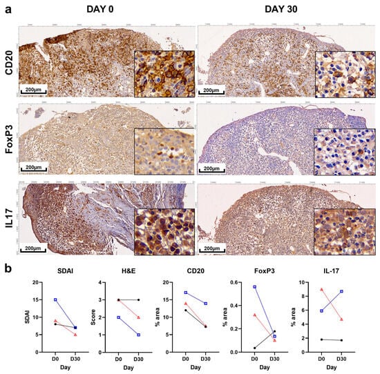
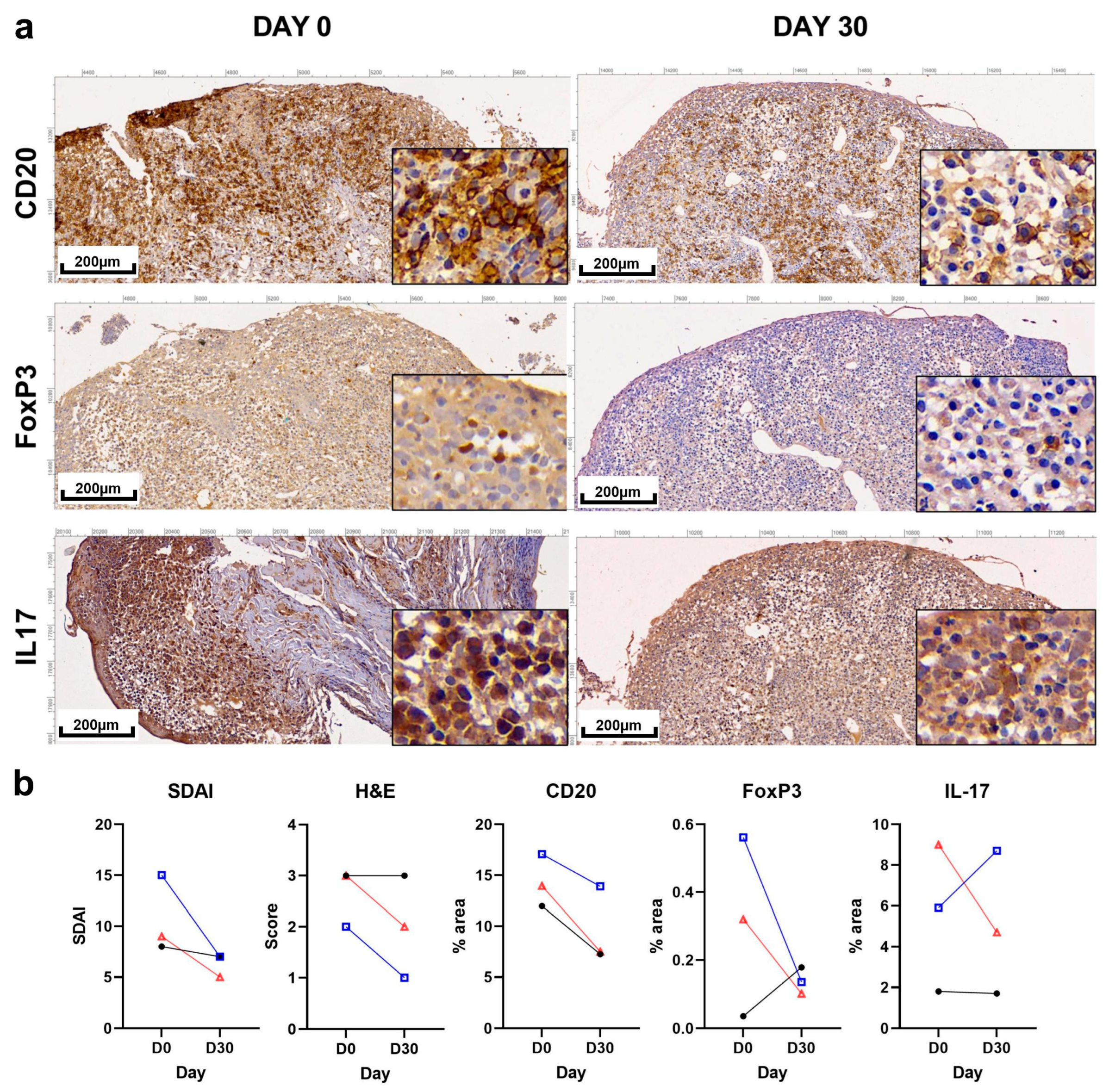
IHC analysis revealed distinct labeling patterns for CD20, FoxP3, and IL-17 in sequential biopsies from three cats (Figure 4a). CD20 labeling was intense and predominantly concentrated in the lamina propria, with infiltration of the submucosa. Both nuclear and cytoplasmic FoxP3 labeling was observed in a few cells within the lamina propria, mainly located near the epithelium in areas showing inflammation or edema. IL-17 labeling was detected in the cytoplasm and interstitium in the epithelium, lamina propria, and submucosa.
Figure 4.
Immunohistochemistry images. (a) Illustrative microscopic images (100×) of immunohistochemical staining for CD20, FoxP3, and IL-17 in oral mucosa biopsies at days 0 and 30 from cat A. Insets (400×) show higher-magnification details of immunolabeling. (b) Individual SDAI scores, severity of microscopic inflammatory scores (H&E), and mean percentage of immunolabeled area for each marker. Each symbol/line represents a single feline patient, coded as follows: A (red), D (black), and E (blue).
Quantitative analysis of immunolabeled areas demonstrated reductions in CD20 expression in all cats following PME. The magnitude of these changes varied among individuals, with decreases of 46.3%, 39.4%, and 18.6% in cats A, D, and E, respectively. Cats A and E also showed a decrease in histologic inflammatory score and clinical improvement. In contrast, FoxP3 and IL-17 immunolabeling patterns showed marked interindividual variability. FoxP3-positive areas decreased in two cats that also exhibited a reduction in both SDAI and histologic inflammatory scores, but one cat showed an increase in FoxP3 labeling area while its histologic inflammation score remained unchanged, and it showed minimal clinical improvement. IL-17 labeling decreased in two cats and increased in one cat. Individual SDAI scores, histopathologic inflammatory scores, and quantitative immunohistochemical measurements are summarized in Figure 4b.
4. Discussion
The present pilot prospective study provides longitudinal information regarding the clinical, histopathologic, and local immunological changes that occur following PME in cats with FCGS. Traditionally, patients require medical management for more than 14 days following dental extraction [11]. The cats showed clinical improvement 1 month post-PME, though the degree varied over time and between cats as measured by SDAI. The clinical response after dental extractions may continue to evolve over several months, and early evaluations may not reflect the final outcome. Improvement or clinical resolution has been reported six months after surgery [11,29]. Therefore, although the cats were followed for up to 180 days, this study primarily aimed to characterize early post-PME changes, particularly during the first 30 days. Previous reports using different scoring systems observed that 52% of cats showed clinical improvement 38 days after surgical treatment [29], while more recent studies have reported significant improvement at 60 days [18].
In this small cohort, the cats positive for retroviral infections exhibited the most pronounced hematologic abnormalities. The main hematologic finding was anemia, observed predominantly in cats positive for FIV and/or FeLV. These findings are consistent with recent reports of anemia in 18% of FeLV and 12% of FIV-positive cats, as well as leukemia in 16% and 3% of these populations, respectively [30]. The same study reported FCGS in 11% of FeLV-positive cats and 24% of FIV-positive cats. Among the three FeLV-positive cats, cats D and E developed clinically relevant hematologic complications during follow-up. Despite FeLV positivity, cats A and E showed clinical improvement (improved SDAI score). FeLV infection has been associated with a 7.5-fold higher likelihood of poor clinical response after dental extraction [13]. In this study, eosinophilia and increased ALP were observed in one patient at D15 post-PME, possibly associated with its concurrent Dipylidium spp. infection. Although eosinophilia is not commonly reported in cats with FCGS, a retrospective study documented peripheral eosinophilia in approximately 25% of cases [31], highlighting the importance of investigating concurrent infections.
The main biochemical finding at D0 was hyperglobulinemia, consistent with previous reports describing chronic antigenic stimulation and systemic inflammatory activation in cats with FCGS [5,32]. However, no changes were observed post-PME. Recent studies suggested that dental extraction does not contribute to a significant decrease in alpha-1 acid glycoprotein levels at D60, indicating the maintenance of systemic inflammation despite clinical improvement [18]. In contrast, our results showed a correction of serum albumin levels 15 days after PME in all but one cat. The post-surgical increase in serum albumin levels may reflect a reduction in systemic inflammation following PME, as albumin is a prominent negative acute-phase protein in cats, and decreased concentrations are observed in chronic inflammatory diseases [33]. These contrasting results emphasize the need for further investigation into the effects of PME on the cat immune system.
In this study, one cat showed increased urea and creatinine levels compatible with chronic kidney disease (CKD). Previous reports found that approximately 10% of cats with FCGS presented with CKD [34]. Later reviews highlighted the importance of screening for concurrent systemic disorders, as comorbidities such as CKD may influence both clinical severity and treatment response [4,5]. A recent cross-sectional analysis comparing cats with caudal stomatitis and healthy controls revealed mild but significant differences in biochemical markers, such as lower creatinine and higher phosphorus levels, without a higher prevalence of CKD [35]. However, previous studies found that 26.5% of cats showed serum urea concentrations above the upper reference limit [31]. These conflicting results indicate that we should consider monitoring renal function biomarkers in cats with FCGS.
The histologic findings showed the classical lymphoplasmacytic and neutrophilic infiltrate mentioned in previous reports [5,9,32,36]. Area-based quantification was used to provide a standardized assessment of the overall extent of immunolabeling within the inflammatory infiltrate, particularly in regions with dense or partially confluent staining where reliable identification of individual cell boundaries was not always possible. The same approach was applied consistently across all markers. The results showed a reduction in the histologic inflammatory infiltrate score in two cats and a reduction in CD20 immunolabeling in three cats. However, CD20 immunolabeling decreased to different degrees in the three cats, irrespective of their SDAI response, which raises the question of whether it reflects the removal of the extraction-related antigenic stimulus rather than control of the disease. Interestingly, the changes in FoxP3 and IL-17 immunolabeling were heterogeneous: two cats exhibiting histologic improvement showed reductions in FoxP3-positive areas, while the cat with persistent severe histologic inflammation demonstrated increased FoxP3 immunolabeling despite minimal clinical improvement. Although the biological significance of this observation remains uncertain and any interpretation is necessarily speculative, decreased FoxP3 immunolabeling could be consistent with reduced antigenic stimulation following PME, whereas increased FoxP3 immunolabeling could reflect a compensatory response to persistent inflammation. Furthermore, since IHC detection provides expression and morphologic information, it does not allow for the assessment of cellular functionality. IL17-A showed marked interindividual variability. Previous transcriptomic studies on FCGS suggest that signaling pathways related to the IL-17 family may be involved in the local immune response [2,8,10]. However, these findings are not directly comparable with the immunohistochemical observations of the present study. Elevated pro-inflammatory cytokines, such as IL-6, TNF-α, and IFN-γ, have been documented in FCGS and constitute part of an inflammatory milieu that promotes the differentiation and function of Th17 cells [10,32]. The detection of these cytokines using immunolabeling techniques has not been performed in caudal oral mucosa. Although IHC detection of IL-17A has previously been reported in other feline tissues using a different antibody [27], this does not constitute validation of the antibody used in the present study for feline tissue. Therefore, the IL-17A IHC findings should be interpreted with caution.
The main limitations of this pilot study include the small sample size and the availability of paired histopathologic and IHC samples from only three cats. Because paired sections were not always processed within the same IHC run, between-run variability in chromogen development and staining intensity cannot be excluded. Sequential biopsies were obtained from contralateral sides at D0 and D30 to avoid healing tissue; therefore, site-related differences in tissue composition may have influenced the area-based IHC measurements. Interobserver agreement was not formally assessed, although final histopathologic scores were assigned by consensus among the four blinded observers. Furthermore, it should be noted that, by chance, the three cats biopsied were FeLV-positive, which may negatively affect the lymphocyte populations and the clinical response following dental extraction [13]. Another limitation of this pilot study is that feline calicivirus (FCV) was not tested because a validated FCV assay was not available at our institution. Meloxicam was administered during the early postoperative period and discontinued before the D30 biopsy; nevertheless, a potential influence on inflammatory or immune-related markers at D30 cannot be completely excluded. Because this study involved client-owned cats, concomitant medical treatment could not be withheld when clinically indicated, particularly for pain management and animal welfare. In addition, one cat developed FOPS during follow-up and subsequently received additional medication that could have potentially influenced the immune-related outcomes in a manner that was not controlled for by the study design. However, these additional treatments were initiated after the D30 biopsy and therefore should not affect the early histopathologic and IHC comparisons between D0 and D30. Nevertheless, their potential influence should be considered when interpreting the later clinical and immunologic findings.
A limited number of studies have reported clinical follow-up after PME in cats with FCGS. Notably, studies on cell therapy have provided valuable insight into the immunological evolution of cats with refractory FCGS treated with MSC [20,21,22]. However, these findings should not be directly extrapolated to cats undergoing PME because MSC therapy represents an active immunomodulatory intervention, whereas PME primarily reduces local antigenic stimulation. This lack of comparable data also highlights the relevance of investigating immunologic changes following the recommended standard-of-care surgical treatment. To the best of our knowledge, the present pilot study is among the few that prospectively evaluated the longitudinal clinical, histopathological, and local immunological changes that occur after PME. A better understanding of the immune changes induced by PME could provide critical insights into the pathophysiology of the disease and help explain the heterogeneity in clinical outcomes. Larger longitudinal studies are needed to determine whether specific immunological patterns are associated with treatment outcomes.
5. Conclusions
Clinical SDAI scores decreased after PME in the five cats, although the magnitude and duration of improvement varied among individuals. In the three FeLV-positive cats with paired biopsies, CD20 immunolabeling decreased irrespective of the clinical and histologic responses, whereas FoxP3 and IL-17 levels showed variable changes. These findings represent preliminary descriptive observations of early tissue changes following PME and should be interpreted considering the small biopsy subset and the methodological limitations of this study.
Supplementary Materials
The following supporting information can be downloaded at: https://www.mdpi.com/article/10.3390/vetsci13101038/s1, Figure S1. This figure displays individual data points for key hematological parameters: red blood cells, hemoglobin, hematocrit, leukocytes, lymphocytes, neutrophils, monocytes, and eosinophils. Measurements were conducted at specified time points from day 0 to day 60. Each individual colored dot represents a single feline patient, coded as follows: A (Red), B (Green), C (Blue), D (Pink), and E (Black). Error bars indicate the standard deviation (SD) of the mean, and the dashed horizontal lines denote the established reference range for each respective parameter. Figure S2. This figure displays individual data points for key biochemistry parameters: serum globulin, albumin, total proteins, alkaline phosphatase (ALP), urea and creatinine. Measurements were conducted at specified time points from Day 0 to Day 60. Each individual colored dot represents a single feline patient, coded as follows: A (Red), B (Green), C (Blue), D (Pink), and E (Black). Error bars indicate the standard deviation (SD) of the mean, and the dashed horizontal lines denote the established reference range for each respective parameter.
Author Contributions
Conceptualization, A.A. and K.Y.; methodology, A.A., G.T. and K.Y.; validation, B.V., G.T., A.W., J.M.V. and K.Y.; formal analysis, A.A., L.T., B.V. and K.Y.; investigation, A.A., L.T., B.V., G.T., M.M., J.E., M.N.G. and K.Y.; resources, B.V., G.T., J.E., J.M.V. and K.Y.; data curation, A.A., L.T. and M.M.; writing—original draft preparation, A.A., L.T., B.V., G.T., M.M., J.E. and M.N.G.; writing—review and editing, A.W., J.M.V. and K.Y.; visualization, A.A., J.M.V. and K.Y.; supervision, G.T., A.W., J.M.V. and K.Y.; project administration, A.A. and K.Y.; funding acquisition, A.A., B.V., J.M.V. and K.Y. All authors have read and agreed to the published version of the manuscript.
Funding
This research was funded by FMV-ANII FMV_3_2022_1_172705 (Fondo María Viñas–Agencia Nacional de Investigación e Innovación) and CAP-Udelar posgraduate internships (Comisión Académica de Posgrado–Universidad de la República, Uruguay).
Institutional Review Board Statement
This study was approved by the Institutional Animal Care and Use Committee of Faculty of Veterinary (CEUAFVET approved protocol code 1460) approved on 30 March 2022.
Data Availability Statement
The original contributions presented in this study are included in the article/Supplementary Material. Further inquiries can be directed to the corresponding author.
Acknowledgments
The authors thank every owner of cats who participated in this study. The authors used ChatGPT (OpenAI, GPT-5.5) to assist with English-language editing and improvement of scientific writing. All suggested revisions were critically reviewed and approved by the authors.
Conflicts of Interest
The authors declare no conflicts of interest.
Appendix A
Table A1.
Characteristics of cats with feline chronic gingivostomatitis.
Table A2.
Anemia status over time for each feline patient.
References
- Lobprise, H.; St Denis, K.; Anderson, J.G.; Hoyer, N.; Fiani, N.; Yaroslav, J. 2025 FelineVMA Feline Oral Health and Dental Care Guidelines. J. Feline Med. Surg. 2025, 27, 1098612X251398793. [Google Scholar] [CrossRef] [Scilit] [PubMed]
- Soltero-Rivera, M.; Shaw, C.; Arzi, B.; Lommer, M.; Weimer, B.C. Feline Chronic Gingivostomatitis Diagnosis and Treatment through Transcriptomic Insights. Pathogens 2024, 13, 192. [Google Scholar] [CrossRef] [Scilit] [PubMed]
- Winer, J.N.; Arzi, B.; Verstraete, F.J.M. Therapeutic Management of Feline Chronic Gingivostomatitis: A Systematic Review of the Literature. Front. Vet. Sci. 2016, 3, 54. [Google Scholar] [CrossRef] [Scilit] [PubMed]
- Lee, D.B.; Verstraete, F.J.M.; Arzi, B. An Update on Feline Chronic Gingivostomatitis. Vet. Clin. N. Am. Small Anim. Pract. 2020, 50, 973–982. [Google Scholar] [CrossRef] [Scilit] [PubMed]
- Soltero-Rivera, M.; Goldschmidt, S.; Arzi, B. Feline Chronic Gingivostomatitis: Current Concepts in Clinical Management. J. Feline Med. Surg. 2023, 25, 1098612X231186834. [Google Scholar] [CrossRef] [Scilit] [PubMed]
- Kouki, M.I.; Papadimitriou, S.A.; Psalla, D.; Kolokotronis, A.; Rallis, T.S. Chronic Gingivostomatitis with Esophagitis in Cats. J. Vet. Intern. Med. 2017, 31, 1673–1679. [Google Scholar] [CrossRef] [Scilit] [PubMed]
- Lorida, O.; Konstantinidis, A.; Brellou, G.D.; Koutouzidou, G.; Papadopoulou, P.; Matiakis, A.; Adamama-Moraitou, K.K.; Papadimitriou, S. Clinical and Histopathological Investigation of the Possible Occurrence of Tracheobronchial Disease in Cats with Chronic Gingivostomatitis. Front. Vet. Sci. 2025, 12, 1624016. [Google Scholar] [CrossRef] [Scilit] [PubMed]
- Vapniarsky, N.; Simpson, D.L.; Arzi, B.; Taechangam, N.; Walker, N.J.; Garrity, C.; Bulkeley, E.; Borjesson, D.L. Histological, Immunological, and Genetic Analysis of Feline Chronic Gingivostomatitis. Front. Vet. Sci. 2020, 7, 310. [Google Scholar] [CrossRef] [Scilit] [PubMed]
- Harley, R.; Gruffydd-Jones, T.J.; Day, M.J. Immunohistochemical Characterization of Oral Mucosal Lesions in Cats with Chronic Gingivostomatitis. J. Comp. Pathol. 2011, 144, 239–250. [Google Scholar] [CrossRef] [Scilit] [PubMed]
- Peralta, S.; Grenier, J.K.; Webb, S.M.; Miller, A.D.; Miranda, I.C.; Parker, J.S.L. Transcriptomic Signatures of Feline Chronic Gingivostomatitis Are Influenced by Upregulated IL6. Sci. Rep. 2023, 13, 13437. [Google Scholar] [CrossRef] [Scilit] [PubMed]
- Jennings, M.W.; Lewis, J.R.; Soltero-Rivera, M.M.; Brown, D.C.; Reiter, A.M. Effect of Tooth Extraction on Stomatitis in Cats: 95 Cases (2000–2013). J. Am. Vet. Med. Assoc. 2015, 246, 654–660. [Google Scholar] [CrossRef] [Scilit] [PubMed]
- Lommer, M.J. Efficacy of Cyclosporine for Chronic, Refractory Stomatitis in Cats: A Randomized, Placebo-Controlled, Double-Blinded Clinical Study. J. Vet. Dent. 2013, 30, 8–17. [Google Scholar] [CrossRef] [Scilit] [PubMed]
- Silva, M.; Fernandes, M.; Fialho, M.; Mestrinho, L. A Case Series Analysis of Dental Extractions’ Outcome in Cats with Chronic Gingivostomatitis Carrying Retroviral Disease. Animals 2021, 11, 3306. [Google Scholar] [CrossRef] [Scilit] [PubMed]
- Coelho, J.C.; Duarte, N.; Bento da Silva, A.; Bronze, M.d.R.; Mestrinho, L.A. Placebo-Controlled Trial of Daily Oral Cannabidiol as Adjunctive Treatment for Cats with Chronic Gingivostomatitis. Animals 2023, 13, 2716. [Google Scholar] [CrossRef] [Scilit] [PubMed]
- Shaw, C.A.; Soltero-Rivera, M.; Profeta, R.; Weimer, B.C. Case Report: Inflammation-Driven Species-Level Shifts in the Oral Microbiome of Refractory Feline Chronic Gingivostomatitis. Bacteria 2025, 4, 1. [Google Scholar] [CrossRef] [Scilit]
- Dai, P.; Yang, M.; Du, J.; Wang, K.; Chen, R.; Feng, X.; Chen, C.; Zhang, X. Epidemiological Investigation of Feline Chronic Gingivostomatitis and Its Relationship with Oral Microbiota in Xi’an, China. Front. Vet. Sci. 2024, 11, 1418101. [Google Scholar] [CrossRef] [Scilit] [PubMed]
- Tetlow, E.R.; Wentlent, L.A.; Carney, P.C.; Byron, M.J.; Fiani, N.; Wright, A.L.; Chrostek, E.; Drozd, M.E.; Winokur, C.E.; Davis, E.M.; et al. Serum Protein Alterations Characterize Feline Chronic Gingivostomatitis: A Case-Control Study of Electrophoresis, Immunoglobulin, and Cytokine Profiles. Am. J. Vet. Res. 2026, 87, 1–10. [Google Scholar] [CrossRef] [Scilit] [PubMed]
- Mestrinho, L.A.; Rosa, R.; Ramalho, P.; Branco, V.; Iglésias, L.; Pissarra, H.; Duarte, A.; Niza, M. A Pilot Study to Evaluate the Serum Alpha-1 Acid Glycoprotein Response in Cats Suffering from Feline Chronic Gingivostomatitis. BMC Vet. Res. 2020, 16, 390. [Google Scholar] [CrossRef] [Scilit] [PubMed]
- Soltero-Rivera, M.; Wanakumjorn, P.; Chen, Y.; Barnum, S.; Charpentier, L.D.C.; Arzi, B.; Arzi, N.V.; Kol, A. Transcriptomic Evidence of CD8+ T-Cell Exhaustion-like Phenotype and Mitochondrial Impairment Underlying Immune Dysregulation in Feline Chronic Gingivostomatitis. Sci. Rep. 2026, 16, 20821. [Google Scholar] [CrossRef] [Scilit] [PubMed]
- Arzi, B.; Clark, K.C.; Sundaram, A.; Spriet, M.; Verstraete, F.J.M.; Walker, N.J.; Loscar, M.R.; Fazel, N.; Murphy, W.J.; Vapniarsky, N.; et al. Therapeutic Efficacy of Fresh, Allogeneic Mesenchymal Stem Cells for Severe Refractory Feline Chronic Gingivostomatitis. Stem Cells Transl. Med. 2017, 6, 1710–1722. [Google Scholar] [CrossRef] [Scilit] [PubMed]
- Arzi, B.; Mills-Ko, E.; Verstraete, F.J.M.; Kol, A.; Walker, N.J.; Badgley, M.R.; Fazel, N.; Murphy, W.J.; Vapniarsky, N.; Borjesson, D.L. Therapeutic Efficacy of Fresh, Autologous Mesenchymal Stem Cells for Severe Refractory Gingivostomatitis in Cats. Stem Cells Transl. Med. 2016, 5, 75–86. [Google Scholar] [CrossRef] [Scilit] [PubMed]
- Arzi, B.; Peralta, S.; Fiani, N.; Vapniarsky, N.; Taechangam, N.; Delatorre, U.; Clark, K.C.; Walker, N.J.; Loscar, M.R.; Lommer, M.J.; et al. A Multicenter Experience Using Adipose-Derived Mesenchymal Stem Cell Therapy for Cats with Chronic, Non-Responsive Gingivostomatitis. Stem Cell Res. Ther. 2020, 11, 115. [Google Scholar] [CrossRef] [Scilit] [PubMed]
- Febre, M.; Saulnier, N.; Roux, P.; Boutoille, F.; Girard, N.; Robert, C.; Rakic, R.; Rosset, E.; Maddens, S.; Sas, V. Placenta-Derived Mesenchymal Stromal Cells as a Treatment for Refractory Chronic Gingivostomatitis in Cats: Eight Cases (2018). J. Small Anim. Pract. 2023, 64, 296–305. [Google Scholar] [CrossRef] [Scilit] [PubMed]
- Tvedten, H. Laboratory and clinical diagnosis of anemia. In Schalm’s Veterinary Hematology, 6th ed.; Weiss, D.J., Wardrop, K.J., Eds.; Wiley-Blackwell: Ames, IA, USA, 2010; pp. 152–161. [Google Scholar]
- Quintavalla, F.; Di Lecce, R.; Carlini, D.; Zanfabro, M.; Cantoni, A.M. Multifocal cutaneous non-epitheliotropic B-cell lymphoma in a cat. JFMS Open Rep. 2020, 6, 2055116920972077. [Google Scholar] [CrossRef] [Scilit] [PubMed]
- Banham, A.H.; Lyne, L.; Scase, T.J.; Blacklaws, B.A. Monoclonal antibodies raised to the human FOXP3 protein can be used effectively for detecting Foxp3+ T cells in other mammalian species. Vet. Immunol. Immunopathol. 2009, 127, 376–381. [Google Scholar] [CrossRef] [Scilit] [PubMed]
- Crossfield, L.; Fortuna, L.; Carling, R.; Scurrell, E.; Matas Riera, M.; Chang, Y.M.; Dawson, C.; Pittaway, R.; Yeung, I.Y.; Thaung, C.; et al. Immunohistochemical characterization of feline lymphoplasmacytic anterior uveitis. Vet. Ophthalmol. 2019, 22, 206–212. [Google Scholar] [CrossRef] [Scilit] [PubMed]
- Fontes, A.C.; Vieira, M.C.; Oliveira, M.; Lourenço, L.; Viegas, C.; Faísca, P.; Seixas, F.; Requicha, J.F.; Pires, M.A. Feline calicivirus and natural killer cells: A study of its relationship in chronic gingivostomatitis. Vet. World 2023, 16, 1708–1713. [Google Scholar] [CrossRef] [Scilit] [PubMed]
- Druet, I.; Hennet, P. Relationship between Feline Calicivirus Load, Oral Lesions, and Outcome in Feline Chronic Gingivostomatitis (Caudal Stomatitis): Retrospective Study in 104 Cats. Front. Vet. Sci. 2017, 4, 209. [Google Scholar] [CrossRef] [Scilit] [PubMed]
- de Almeida, P.M.; Belas, A.; Nogueira, P.; Meneses, A.; de Oliveira, J.T.; Viegas, C. Prevalence of Feline Leukemia Virus Infection and Associated Diseases in a Portuguese Domestic Cat Population: A 4.5-Year Cross-Sectional Study. PLoS ONE 2026, 21, e0339172. [Google Scholar] [CrossRef] [Scilit] [PubMed]
- Haidary, H.M.; Radzi, R.; Aslam, M.W.; Lau, S.F.; Kamal, F.M.; Radzali, A.R. Retrospective Study of Clinical Manifestations and Multiple Treatment Outcomes in 57 Cats Diagnosed with Feline Chronic Gingivostomatitis. J. Anim. Health Prod. 2022, 10, 51–59. [Google Scholar] [CrossRef] [Scilit]
- Lopes, R.S.; Carvalho, P.P.; Pires, M.A.; Rodrigues-Santos, P.; Costa, E.; Requicha, J.F. Local and Systemic Immunological Response in Feline Chronic Gingivostomatitis: A Critical Review. Front. Immunol. 2025, 16, 1572631. [Google Scholar] [CrossRef] [Scilit] [PubMed]
- Fong, K.Y.M.; Oikonomidis, I.L.; Leong, D.; Lo, G.; Heal, J.; Woods, G. Hypoalbuminaemia and Its Association with Disease and Clinical Outcomes in Cats. J. Small Anim. Pract. 2024, 65, 721–729. [Google Scholar] [CrossRef] [Scilit] [PubMed]
- Hennet, P.R.; Camy, G.A.L.; McGahie, D.M.; Albouy, M.V. Comparative Efficacy of a Recombinant Feline Interferon Omega in Refractory Cases of Calicivirus-Positive Cats with Caudal Stomatitis: A Randomised, Multi-Centre, Controlled, Double-Blind Study in 39 Cats. J. Feline Med. Surg. 2011, 13, 577–587. [Google Scholar] [CrossRef] [Scilit] [PubMed]
- Vetter, C.A.M.; Lourenço, B.N.; Meindl, A.G.; Mees, A.; Stone, A.; Farnsworth, R.; Everett, S.E.; Gonzalez, S.E.; Roth, I.G.; Schmiedt, C. Cross-Sectional Characterization of Renal Function in Cats with Caudal Stomatitis. J. Feline Med. Surg. 2023, 25, 1098612X231179883. [Google Scholar] [CrossRef] [Scilit] [PubMed]
- Rolim, V.M.; Pavarini, S.P.; Campos, F.S.; Pignone, V.; Faraco, C.; Muccillo, M.d.S.; Roehe, P.M.; da Costa, F.V.A.; Driemeier, D. Clinical, Pathological, Immunohistochemical and Molecular Characterization of Feline Chronic Gingivostomatitis. J. Feline Med. Surg. 2017, 19, 403–409. [Google Scholar] [CrossRef] [Scilit] [PubMed]

Disclaimer/Publisher’s Note: The statements, opinions and data contained in all publications are solely those of the individual author(s) and contributor(s) and not of MDPI and/or the editor(s). MDPI and/or the editor(s) disclaim responsibility for any injury to people or property resulting from any ideas, methods, instructions or products referred to in the content. |
© 2026 by the authors. Licensee MDPI, Basel, Switzerland. This article is an open access article distributed under the terms and conditions of the Creative Commons Attribution (CC BY) license.