


Simple Summary
Klebsiella pneumoniae is an opportunistic bacterium that can colonize the respiratory tract of cattle and has been isolated from calves with bovine respiratory disease (BRD). We collected deep nasal swabs once from 517 calves with BRD on 38 farms in Henan, China, over six years (2021–2026). K. pneumoniae was isolated from 16.8% of the samples. The isolates showed high genetic diversity (34 sequence types) and widespread multidrug resistance, especially to β-lactams, sulfonamides, and quinolones. Of the isolates, 96.6% were multidrug-resistant. Many resistance and virulence-associated genes were detected, but classical hypervirulence markers were absent. Because no healthy control calves or causality experiments were included, these findings do not establish K. pneumoniae as the causative agent of BRD. Prudent antibiotic use and ongoing surveillance are essential to control this pathogen and protect cattle health.
Abstract
Bovine respiratory disease (BRD) is a major cause of morbidity and mortality in cattle, and Klebsiella pneumoniae (K. pneumoniae) has emerged as an opportunistic pathogen that can colonize the bovine respiratory tract. However, epidemiological data on K. pneumoniae from cattle with BRD in Henan Province, central China, remain scarce. This study aimed to investigate the isolation rate, antimicrobial resistance profiles, resistance genes, virulence-associated genes, and genetic diversity of K. pneumoniae from BRD-affected calves in Henan. From 2021 to 2026, deep nasal swabs were collected once from 517 calves with clinical signs of BRD (Wisconsin score ≥5) across 38 farms in 13 cities over six years. A total of 87 K. pneumoniae isolates were obtained, giving an overall isolation rate of 16.8% (87/517), ranging from 13.8% to 22.6% across five geographic regions. Antimicrobial susceptibility was determined by disk diffusion using CLSI VET01S and CLSI M100-S26 criteria for 15 antimicrobials belonging to six classes. Resistance and virulence-associated genes were screened by PCR, and multilocus sequence typing (MLST) was performed. MLST revealed 34 sequence types (STs), with ST950 being the most prevalent (6.9%), followed by ST262, ST187, and ST306. Antimicrobial resistance was highest against ampicillin (94.3%), sulfisoxazole (79.3%), cefoxitin (73.6%), cefazolin (70.1%), and enrofloxacin (67.8%). Multidrug resistance (resistance to ≥3 classes) was observed in 96.6% (84/87; 95% CI 90.3–98.8%) of isolates. The most common resistance genes were blaTEM (72.4%), sul1 (39.1%), blaSHV (37.9%), and qnrB (14.9%). After Firth’s penalized logistic regression with farm-level cluster-robust standard errors and Benjamini–Hochberg FDR correction, no resistance gene was significantly associated with phenotypic resistance (all q > 0.05). Virulence-associated genes fimH, mrkD, uge, wabG, entB, iutA, and ureA were highly prevalent (>90%), whereas hypervirulence markers (rmpA and rmpA2) were absent. The most common virulence-associated gene combination (eight genes) was present in 35.6% of isolates. These findings indicate high genetic diversity, widespread multidrug resistance, and a broad virulence-associated gene repertoire among K. pneumoniae isolated from cattle with BRD in Henan. Because no healthy comparison group or etiological confirmation was included, the study does not establish K. pneumoniae as the causative agent of BRD. The high resistance rates to β-lactams, sulfonamides, and quinolones highlight the need for prudent antimicrobial use. Florfenicol showed the lowest in vitro resistance rate among the tested agents; however, clinical efficacy, treatment outcome, and safety were not evaluated. Continuous surveillance and molecular monitoring are essential to manage this pathogen in cattle populations.
1. Introduction
Bovine respiratory disease (BRD), also known as bovine bronchopneumonia, is a major cause of morbidity and mortality in intensively managed feedlot and dairy calf production systems, although estimates vary by production system, age, management, and geographic region [1,2,3,4]. BRD is a multifactorial complex involving management stress, infectious agents, and antimicrobial exposure, and the bacterial pathogens routinely isolated are frequently opportunistic [5,6,7,8]. Recent studies in intensively managed beef cattle, such as Masebo et al. (2024), have documented a high burden of respiratory disease, extensive antimicrobial treatment, and persistent pulmonary lesions at slaughter, emphasizing the difficulties associated with antimicrobial management of BRD [9]. Longitudinal surveillance studies, such as Dini et al. (2024), further illustrate how infectious exposure can change over time in relation to farm management and biosecurity conditions [10].
Klebsiella pneumoniae (K. pneumoniae) is a Gram-negative opportunistic pathogen that can colonize the respiratory or intestinal tracts of both humans and animals [11,12]. It has been associated with a range of infectious diseases, including mastitis, pneumonia, bacteremia, liver abscess, urinary tract infection, arthritis, and meningitis, thereby posing a threat to human health and livestock husbandry [11]. K. pneumoniae isolates in cattle have been reported in many countries [13,14,15]. In China, numerous studies have demonstrated that K. pneumoniae can infect dairy cows and cause mastitis [16,17,18]. Some studies have suggested that K. pneumoniae is associated with BRD in regions such as Hebei province and Chongqing City, China [6,19]. To the best of our knowledge, only one study has reported the isolation of K. pneumoniae from cows in Henan Province in 2017, but that paper did not report the health status of the cows or the sample types [20]. Therefore, the epidemiological patterns of K. pneumoniae in cattle herds in Henan in recent years remain unclear. Considering that Henan borders Hebei Province and more than 3,500,000 cattle have been kept in Henan province in central China [21], it is essential to characterize K. pneumoniae isolates obtained from cattle with BRD.
Numerous studies have examined the diversity of K. pneumoniae in both humans and animals. Multilocus sequence typing (MLST) assigns sequence types (STs) on the basis of allelic profiles of seven housekeeping loci; isolates with identical allelic profiles are assigned the same ST [22]. In humans in China, many STs have been identified by MLST among K. pneumoniae isolates [23,24]. Another investigation identified two STs using MLST in pigs in China [25]. These findings suggest that genetic diversity among animal K. pneumoniae is widespread. To our knowledge, no studies have yet explored the predominant STs of K. pneumoniae strains associated with BRD in Henan province, China, leaving a gap in the available molecular epidemiological data.
Antimicrobial therapy continues to be the cornerstone for both the prevention and treatment of BRD. In China, a wide range of antimicrobials have been approved for this indication, including β-lactams, tetracyclines, sulfonamides, quinolones, and aminoglycosides. Nevertheless, the extensive use of these agents has fostered the emergence of multidrug resistance through selection of acquired resistance genes, chromosomal mutations, mobile genetic elements, and clonal dissemination, progressively diminishing their clinical efficacy [6,19,26,27]. Although nationwide programs for the monitoring and surveillance of antimicrobial resistance in animals have been in place in China for many years, information on antibiotic resistance in K. pneumoniae derived from cattle remains scarce. The resistance mechanisms of K. pneumoniae are tightly linked to specific resistance genes; for instance, blaIMP, blaVIM, blaOXA-48, blaNDM, blaKPC, blaDHA, blaFOX, blaCTX-M-2, blaSHV, and blaTEM are regarded as the major genes underlying β-lactam resistance [28,29], while sul1 and sul2 mediate resistance to sulfonamides [29]. The potential transfer of these resistance determinants to other bacteria, including those in the human respiratory microbiota, is a public-health concern [30]; however, the present study did not investigate transmission.
Virulence-associated genes serve as the essential determinants of bacterial pathogenicity. Typically, K. pneumoniae possesses a range of virulence factors—such as fimbriae, capsular polysaccharides, lipopolysaccharides, and siderophores—that are encoded by various virulence-associated genes, including fimH, rmpA, rmpA2, uge, wabG, iutA, and iroN [31]. According to previous reports, the expression of these virulence-associated genes is closely linked to the infectivity and pathogenicity of K. pneumoniae [29]. However, detection of a virulence-associated gene by PCR does not demonstrate its expression or establish pathogenicity.
To the best of our knowledge, no prior research has investigated the antimicrobial susceptibility and genetic diversity of K. pneumoniae originating from cattle with BRD in Henan province, China. The objective of this study was to characterize K. pneumoniae isolates obtained from calves with BRD in Henan Province, including isolation rate, antimicrobial susceptibility, resistance genes, virulence-associated genes, and MLST sequence types.
2. Materials and Methods
2.1. Sample Collection, Bacterial Isolation, and Identification
From 2021 to 2026, a total of 517 calves with clinical BRD were enrolled in this study. The calves ranged in age from 1 to 6 months (median 3 months; IQR 2–4 months). The sex distribution was 270 males (52.2%) and 247 females (47.8%). Production types included intensive (10 farms), semi-intensive (17 farms), and backyard (11 farms) systems. Information on antimicrobial exposure before sampling was recorded: 14 farms (36.8%) reported antimicrobial use within 14 days before sampling, 22 farms (57.9%) reported no use, and 2 farms (5.3%) had unknown status (Table S1). Farms were selected according to the following criteria: ≥20 calves, a history of BRD, and owner consent. Within each farm, calves were selected if they had a Wisconsin score ≥5, had not been sampled previously, and had no other obvious systemic disease.
According to the Wisconsin Calf Respiratory Scoring System [32], all calves presented with clinical signs corresponding to a total score of ≥5, including fever, cough, nasal discharge, ocular discharge, and ear droop. Clinical scoring was performed by trained field veterinarians. The same scoring sheet, threshold, and procedures were used throughout the six-year period. Inter-observer standardization was performed before each sampling season. No clinically healthy control calves were included in this study; this is a major limitation and is discussed below.
These calves were distributed across 38 farms in 13 cities of Henan Province. Sampling was performed annually from 2021 to 2026. The specific sampling periods, numbers of farms and cities, sample sizes, positive counts, and isolation rates for each year are summarized in Table 1 and Table S1. No calf was sampled more than once; repeated sampling of the same animal was excluded (Table 1 and Table S1). Samples were collected from deep nasal swabs. Following collection, samples were placed in sterile tubes, kept on ice, and transported to the laboratory for analysis.
Table 1.
Nasal swab samples collected from calves with BRD on cattle farms in five regions of Henan province from 2021 to 2026.
For bacterial identification, samples were directly streaked onto MacConkey agar without enrichment (Beijing Solarbio Science & Technology, China) and incubated at 37 °C for 18–24 h. Up to five presumptive K. pneumoniae colonies per sample—small, pink, with smooth, moist surfaces and well-defined edges—were subcultured and identified by Gram staining and biochemical identification. Mixed cultures were purified by repeated streaking. Suspected K. pneumoniae isolates were confirmed by PCR detection using khe species-specific primers [28]. Briefly, genomic DNA was extracted using a commercial kit (TransGen Biotech, China) per the manufacturer’s instructions, dissolved in 100 µL of ultrapure water, and stored at −70 °C. Five randomly selected khe-positive isolates were sequenced to confirm the specificity of the khe PCR. All remaining isolates were identified by khe PCR following presumptive biochemical identification. One confirmed isolate per sample was stored for further analysis, using K. pneumoniae ATCC700603 for quality control. Selecting only one isolate per sample may underestimate within-host strain diversity; this limitation is acknowledged in the Discussion. The isolation rate was calculated as (number of positive cases/total samples) × 100%.
2.2. MLST Analysis of K. pneumoniae
MLST analyses of K. pneumoniae isolates were carried out following a previously described protocol [22]. Primers targeting seven housekeeping genes (rpoB, gapA, mdh, pgi, phoE, infB, and tonB) were used for PCR amplification [22]. The amplification was conducted with an annealing temperature of 50 °C for all genes, except for gapA (60 °C) and tonB (45 °C). Purified PCR products were subjected to sequencing. Sequence quality was assessed using chromatograms; low-quality ends were trimmed. Alleles and STs were assigned using the online tool at https://bigsdb.pasteur.fr/klebsiella/ [22] (accessed on 18 July 2026). All allelic profiles corresponded to previously assigned STs; no novel alleles or STs were identified.
2.3. Antimicrobial Susceptibility Testing
The antibiotic resistance patterns of each positive K. pneumoniae isolate from the samples (selected and stored in Section 2.1) were determined by disk diffusion methods according to Clinical and Laboratory Standards Institute (CLSI) guidelines (CLSI VET01S and CLSI M100-S26) [33,34]. The inhibition zones were measured and recorded. For the interpretation of inhibition zones, the antibiotic standards already included in CLSI VET01S Table 2A should be used preferentially [33]; for those not included in CLSI VET01S, refer to the standards in CLSI M100-S26 Table 2A. The exact zone diameter breakpoints (S/I/R, in mm) for every antimicrobial tested are listed in Table 2 [34].
Table 2.
Antimicrobial agents, disk contents, interpretive criteria, sources, and resistance profiles of 87 K. pneumoniae isolates.
Resistance to 15 antimicrobials belonging to six antimicrobial categories were investigated in the drug susceptibility test. Macrolides (erythromycin and azithromycin) were not included because members of the family Enterobacteriaceae, including K. pneumoniae, are intrinsically resistant to macrolides. Antimicrobials were selected because they are commonly used for BRD treatment in the region, based on information provided by farm owners. No quantitative farm-specific antimicrobial use data were systematically collected; therefore, relationships between use and resistance could not be analyzed. For each isolate–antimicrobial combination, disk diffusion was performed in three independent cultures. Discordant classifications were resolved by a fourth independent test, and the majority result was recorded. K. pneumoniae ATCC700603 and Escherichia coli ATCC 25922 were served as quality control strains. Isolates exhibiting resistance to three or more antimicrobial categories were classified as multidrug resistant (MDR) [35].
2.4. Detection of Resistance Genes
Genomic DNA was extracted from the bacterial isolates as described in the identification section. Conventional PCR was used to detect antibiotic resistance genes. Based on the categories of antimicrobials tested and the resistance genes most frequently reported in K. pneumoniae from China [19,28,31], we targeted the following: β-lactam resistance genes (blaVIM, blaSHV, blaTEM, blaCTX-M-2) [29,36,37]; sulfonamide resistance gene (sul1, sul2, and sul3) [38]; tetracycline resistance genes (tetA, tetB, tetC) [39,40,41]; phenicol resistance gene (catI, cmlA, floR) [42,43]; aminoglycoside resistance gene (aadA1, aacC1, and aacC2) [40,44]; and quinolone resistance genes (oqxA, aac(6′)-Ib-cr, qnrA, qnrB, qnrS, and gyrA) [20,45,46,47,48]. Primer details are listed in Table S2.
PCR was performed using an EasyTaq® PCR SuperMix kit (TransGen, China). Each 20 µL reaction contained 10 µL of 2× SuperMix, 0.4 µM of each primer, and 20 ng of template DNA. The cycling protocol consisted of an initial denaturation at 94 °C for 5 min, followed by 30 cycles of 94 °C for 30 s, annealing at a primer-specific temperature for 30 s (see Table S2), 72 °C for 30 s, and a final extension at 72 °C for 10 min. Gene-positive reference strains and well-characterized clinical isolates carrying blaTEM, blaSHV, sul1, sul2, tetC, aadA1, qnrB, and other targets were used as positive controls. No-template controls were included in every run. PCR products were separated by electrophoresis on a 2% agarose gel (Solarbio, China) at 120 V for 60 min.
2.5. Detection of Virulence-Associated Genes
A panel of virulence-associated genes was screened by PCR. Targeted genes included those fimbriae synthesis-related genes (fimH, mrkD) [49,50]; lipopolysaccharide-related genes (uge, wabG) [49,51]; iron uptake system genes (entB, iutA, iroN, kfu) [29,52,53]; urease-related genes (ureA, allS) [49,54]; tellurite resistance gene (terB) [55]; Hemolysin gene (hly) [29]; and capsular polysaccharide synthesis and synthesis regulation related gene (rmpA and rmpA2) [55]. K. pneumoniae ATCC 43816 and K. pneumoniae NTUH-K2044 were used as positive controls for virulence-associated gene PCR assays. A no-template control was included in every PCR run. The primers, corresponding product sizes, and annealing temperatures for all virulence genes are listed in Table S3. The PCR protocols were consistent with those described in Section 2.4.
2.6. Statistical Analyses
Isolation rates are reported with 95% confidence intervals. Farm-level clustering was accounted for using mixed-effects logistic regression with farm as a random intercept, or cluster-robust standard errors where appropriate. To compare isolation rates across years, we used a chi-square test for heterogeneity and pairwise proportion tests with Bonferroni adjustment. A trend test was not performed because the objective was to assess whether isolation rates differed among years rather than to test for a linear trend. For genotype–phenotype analyses, 2 × 2 contingency tables were constructed for each gene–antimicrobial class pair. An isolate was considered resistant to a class if it was resistant to at least one agent within that class. Because several gene–class pairs exhibited complete or quasi-complete separation, Firth’s penalized logistic regression was used to obtain finite coefficient estimates. Farm-level clustering was accounted for by computing cluster-robust standard errors from the score function, with farm as the clustering variable. Odds ratios (ORs) with 95% confidence intervals (CIs) were derived from the Firth model. Cluster-adjusted p-values were obtained from the cluster-robust standard errors. Benjamini–Hochberg false discovery rate (FDR)-adjusted q-values were calculated based on the cluster-adjusted p-values. Associations with q < 0.05 were considered statistically significant. For gene–class pairs with complete separation or sparse data, ORs and cluster-adjusted p-values could not be estimated and were reported as NE (not estimable). Because the number of isolates for each ST was too small to support a robust difference analysis, this study did not perform significance testing on the character differences among different STs of K. pneumoniae. All analyses were performed using R version 4.5.2 (R Foundation for Statistical Computing, Vienna, Austria).
3. Results
3.1. Isolation and Identification of K. pneumoniae
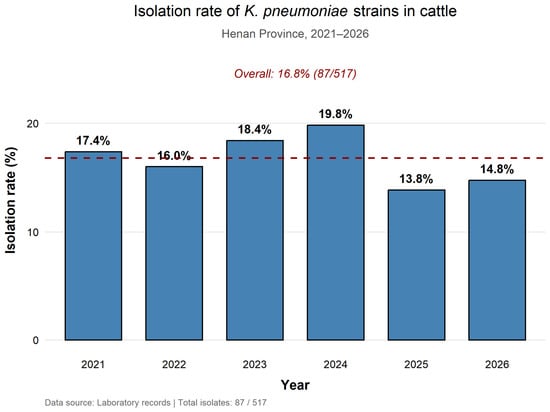
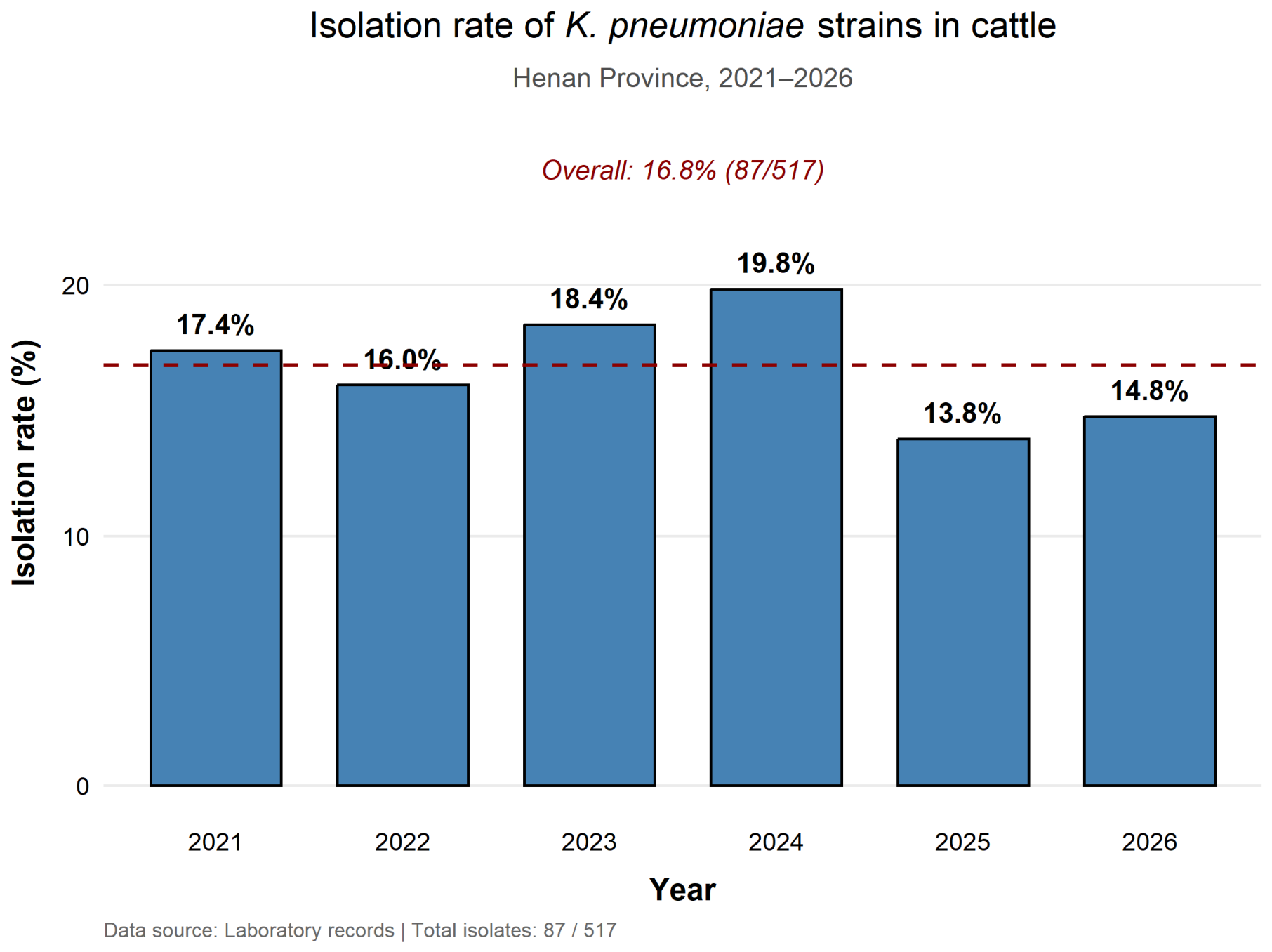
In this study, 87 out of 517 deep nasal swab samples yielded bacterial isolates that were confirmed as K. pneumoniae through Gram staining and species-specific PCR, resulting in an overall isolation rate of 16.8%. The annual sample sizes and positive counts are presented in Figure 1 and Table S1. Briefly, the numbers of samples collected per year were 46 (2021), 50 (2022), 114 (2023), 116 (2024), 130 (2025), and 61 (2026). The overall isolation rate was 16.8% (87/517; 95% CI 13.9–20.3). Annual isolation rates with 95% confidence intervals were 17.4% (9.1–30.7) in 2021, 16.0% (8.3–28.5) in 2022, 18.4% (12.4–26.5) in 2023, 19.8% (13.6–28.0) in 2024, 13.8% (8.9–20.8) in 2025, and 14.8% (8.0–25.7) in 2026. (Figure 1). Geographically, isolation rates varied across five regions of Henan province, with east Henan showing the numerically highest rate (22.6%, 14/62; 95% CI: 13.8–35.5), followed by west (20.8%, 11/53; 95% CI: 11.9–34.4), central (16.8%, 16/95; 95% CI: 10.5–26.1), south (15.5%, 34/220; 95% CI: 11.2–20.9), and north (13.8%, 12/87; 95% CI: 8.0–22.7). However, the 95% confidence intervals for all regions overlapped substantially, and the regional differences were not statistically significant after accounting for farm-level clustering in a mixed-effects logistic regression model (farm random intercept variance = 0.42; ICC = 0.11; likelihood ratio test for the random effect: p = 0.18). These regional estimates had unequal denominators and overlapping 95% confidence intervals after accounting for farm clustering. A Pearson chi-square test for heterogeneity yielded χ2 = 1.78, df = 5, p = 0.879, indicating no statistically significant difference in isolation rates among the six years (2021–2026). This test assessed heterogeneity across years and was not a trend test. All Bonferroni-adjusted p-values were ≥0.99, confirming no significant pairwise differences between any two years. At the city level, the numerically highest and lowest isolation rates were observed in Shangqiu (25.0%, 3/12; 95% CI: 8.7–53.1) and Puyang (9.1%, 1/11; 95% CI: 1.6–37.7), respectively; however, these estimates were based on very small denominators and the confidence intervals were extremely wide, precluding any meaningful comparison. A summary of all isolates is provided in Table 1 and Table S1.
Figure 1.
Isolation rate of K. pneumoniae strains in cattle from 2021 to 2026 in Henan province.
3.2. MLST Sequence Type Distribution
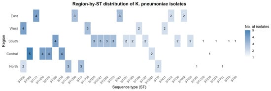
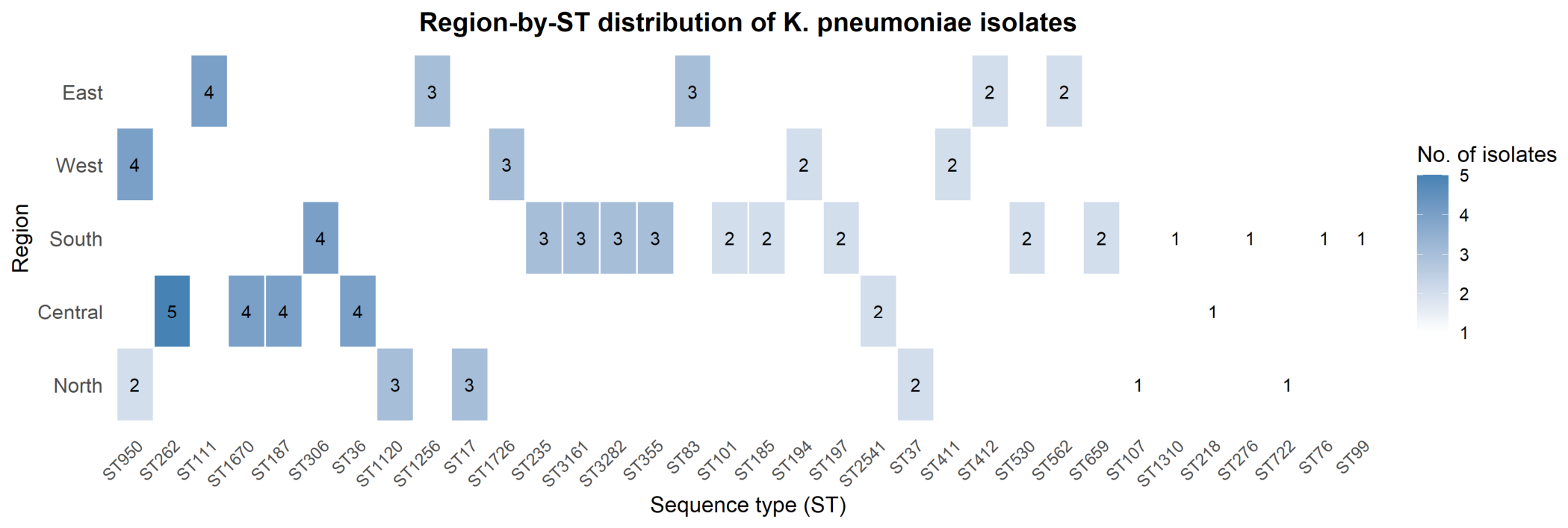
A total of 87 K. pneumoniae isolates recovered from nasal swabs of cattle with bovine respiratory disease (BRD) across five regions of Henan province (North, Central, South, West, East) were characterized by multilocus sequence typing (MLST). The region-by-ST heatmap (Figure 2) shows the distribution of each ST across the five geographic regions. The analysis revealed 34 distinct STs, indicating considerable genetic diversity (Figure 2). The most prevalent ST was ST950, identified in northern (n = 2) and western (n = 4) regions, accounting for six isolates (6.9% of typed strains). ST262 (n = 5) and ST187 (n = 4) were exclusively found in the central region, while ST306 (n = 4) was restricted to the south. ST111 (n = 4) and ST412 (n = 2) were observed only in the east, and ST1726 (n = 3) only in the west. Several other STs, including ST1670, ST36, ST2541, and ST3282, were present in low frequencies (1–4 isolates each), with distinct geographical clustering. These findings indicate a heterogeneous distribution of K. pneumoniae STs across Henan, with certain STs showing regional clustering. Whether this reflects local transmission dynamics or host adaptation remains a hypothesis because MLST has limited discriminatory resolution and no phylogenomic or epidemiological linkage analysis was performed.
Figure 2.
Region-by-ST distribution of 87 K. pneumoniae isolates from calves with BRD in five regions of Henan Province, China.
The heatmap shows the number of isolates for each sequence type (ST) across five geographic regions (North, Central, South, West, East). STs are ordered by total isolate count (descending). Cells are coloured according to the number of isolates, with the exact count shown in each cell.
3.3. Antimicrobial Susceptibility
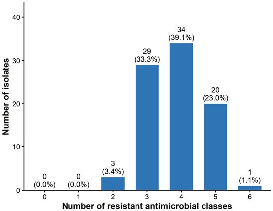
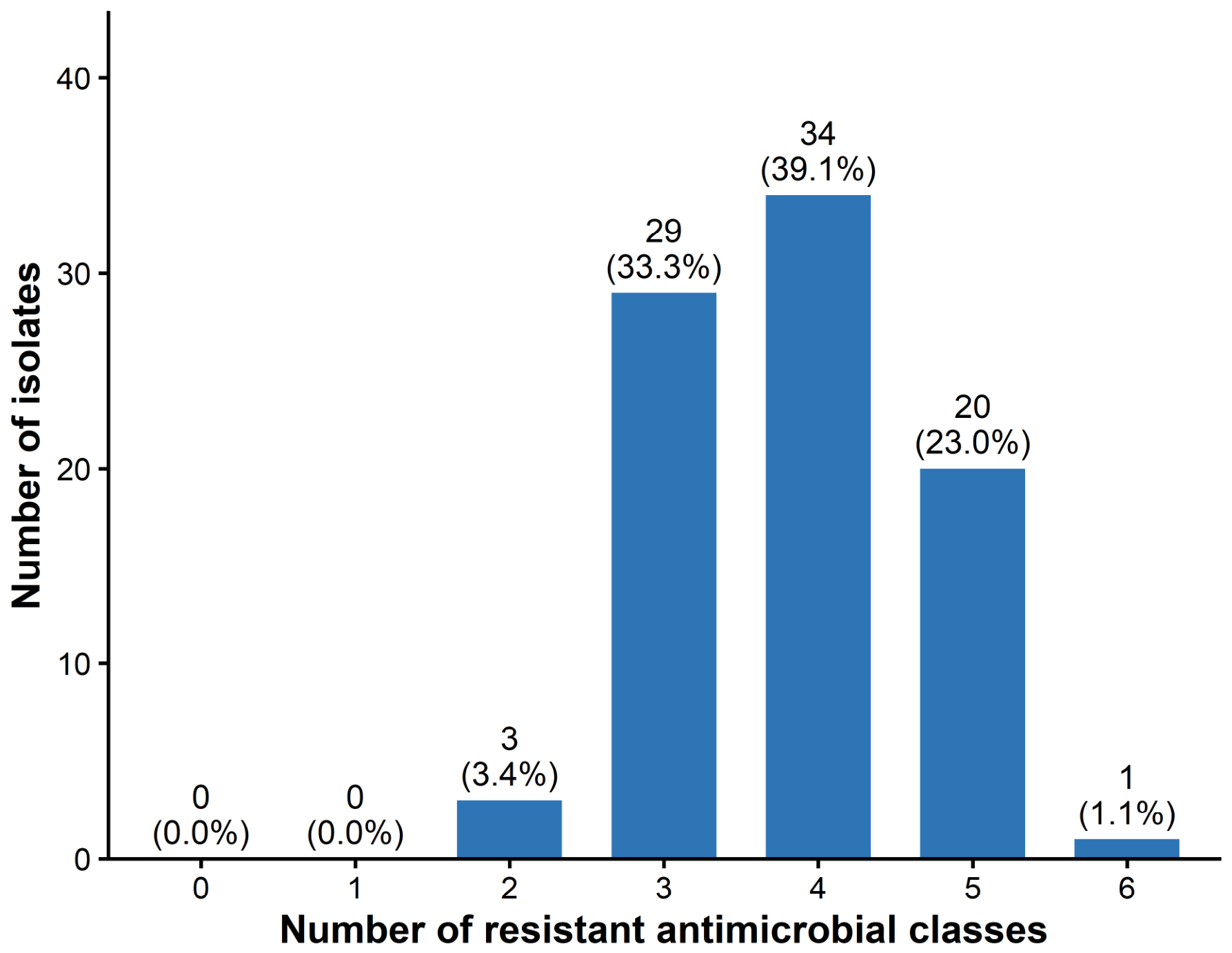
The 87 K. pneumoniae strains were classified as susceptible, intermediate, or resistant to 15 antibiotics across six classes. As presented in Table 2, isolates from different regions exhibited varying resistance profiles. The antibiotic resistance rates for agents with CLSI interpretive criteria, in descending order, were: ampicillin (94.3%), sulfisoxazole (79.3%), cefoxitin (73.6%), cefazolin (70.1%), enrofloxacin (67.8%), cefixime (62.1%), levofloxacin (59.8%), amoxicillin-clavulanate (59.8%), tetracycline (55.2%), trimethoprim-sulfamethoxazole (46.0%), kanamycin (40.2%), amikacin (36.8%), doxycycline (34.5%), gentamicin (25.3%), and florfenicol (11.5%). MDR was defined as resistance to at least one agent in ≥3 of the six classes: β-lactams, sulfonamides, tetracyclines, phenicols, aminoglycosides, and quinolones. Intermediate isolates were classified as non-resistant. Among the 87 isolates, 96.6% (84/87; 95% CI 90.3–98.8%) were MDR. The distribution of the number of resistant classes is shown in Figure 3. Specifically, 3 isolates (3.4%) were resistant to 2 classes, 29 (33.3%) to 3 classes, 34 (39.1%) to 4 classes, 20 (23.0%) to 5 classes, and 1 (1.1%) to 6 classes; no isolate was susceptible to all classes or resistant to only 1 class.
Figure 3.
Distribution of the number of antimicrobial classes to which the 87 K. pneumoniae isolates were resistant. The bar chart shows the number of isolates (y-axis) resistant to 0, 1, 2, 3, 4, 5, or 6 antimicrobial classes (x-axis).
For geographical analysis, isolate-level class resistance—defined as resistance to at least one agent within a class—was summarized for the five regions (Table 3). β-lactam resistance ranged from 92.9% to 100.0%, sulfonamide resistance from 87.5% to 92.9%, tetracycline resistance from 54.5% to 64.3%, phenicol resistance from 8.3% to 14.3%, aminoglycoside resistance from 50.0% to 54.5%, and quinolone resistance from 72.7% to 78.6%. Overall resistance was highest for β-lactams (98.9%) and sulfonamides (89.7%), followed by quinolones (74.7%), tetracyclines (58.6%), aminoglycosides (51.7%), and phenicols (11.5%). The numerically lower resistance proportions observed in North Henan for some classes should be interpreted as descriptive only; no formal statistical comparison was performed because of unequal sample sizes across regions and farm-level clustering.
Table 3.
Isolate-level class resistance of K. pneumoniae isolates from five regions of Henan Province, China.
3.4. Detection of Antimicrobial Resistance Genes
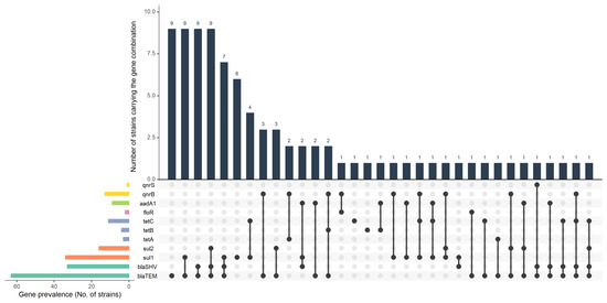
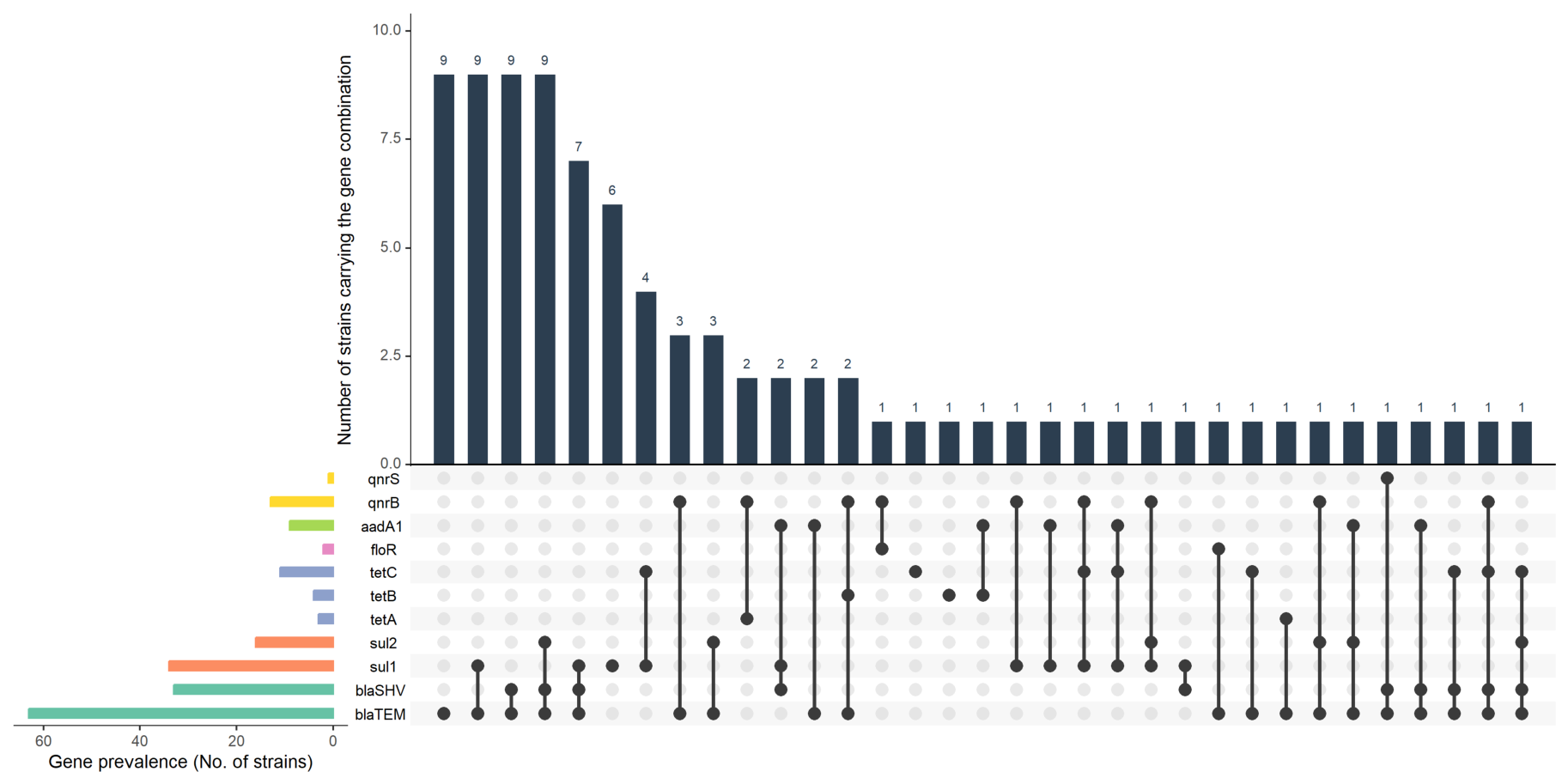
A total of 22 antimicrobial resistance genes, representing six distinct drug classes, were screened via PCR, leading to the identification of 11 genes across these classes. Regarding β-lactam resistance, blaTEM was the most prevalent gene (72.4%), followed by blaSHV (37.9%). Notably, blaVIM and blaCTX-M-2 were not detected. For sulfonamide resistance, sul1 (39.1%) was more common than sul2 (18.4%). Among tetracycline resistance genes, tetC (12.6%) was detected more frequently than tetB (4.6%) and tetA (3.4%). The only phenicol resistance gene identified was floR (2.3%). The aminoglycoside resistance gene (aadA1) was found in 10.3% of isolates. Among quinolone resistance determinants, qnrB and qnrS were detected at rates of 14.9% and 1.1%, respectively, while other resistance genes were not found (Figure 4).
Figure 4.
The distribution of antibiotic resistance genes among 87 K. pneumoniae strains.
As detailed in Figure 4 and Table S4, 17 strains harbored only a single resistance gene, while the remaining 70 carried two or more. The most prevalent multi-gene patterns were blaTEM + blaSHV, blaTEM + blaSHV + sul2, and blaTEM + sul1, each occurring in 10.34% of strains. This was followed by the blaTEM + blaSHV + sul1 pattern (8.05%), and then blaTEM + sul2 and blaTEM + qnrB (both 3.45%). Four other patterns (aadA1 + blaSHV + sul1, qnrB + tetA, aadA1 + blaTEM, and blaTEM + qnrB + tetB) were each found in two strains (2.3%). The remaining strains exhibited unique patterns.
Associations between resistance genes and phenotypic resistance were assessed at the antimicrobial class level using 2 × 2 contingency tables (Table 4). After FDR correction, no gene was significantly associated with phenotypic resistance (all q > 0.05). The strongest nominal association was observed between sul1 and sulfonamide resistance (OR = 4.17, 95% CI 0.89–19.61; cluster-adjusted p = 0.070; q = 0.422), which did not remain significant after FDR correction. For gene–class pairs with one-sided complete separation or sparse data (blaSHV, blaTEM, tetA, floR, and qnrS), ORs and cluster-adjusted p-values could not be estimated and are reported as NE. No other gene–class pair reached statistical significance.
Table 4.
Genotype–phenotype associations for antimicrobial resistance in K. pneumoniae isolates.
3.5. Detection of Virulence-Associated Genes
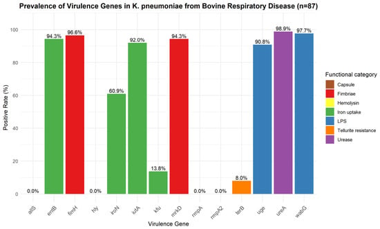
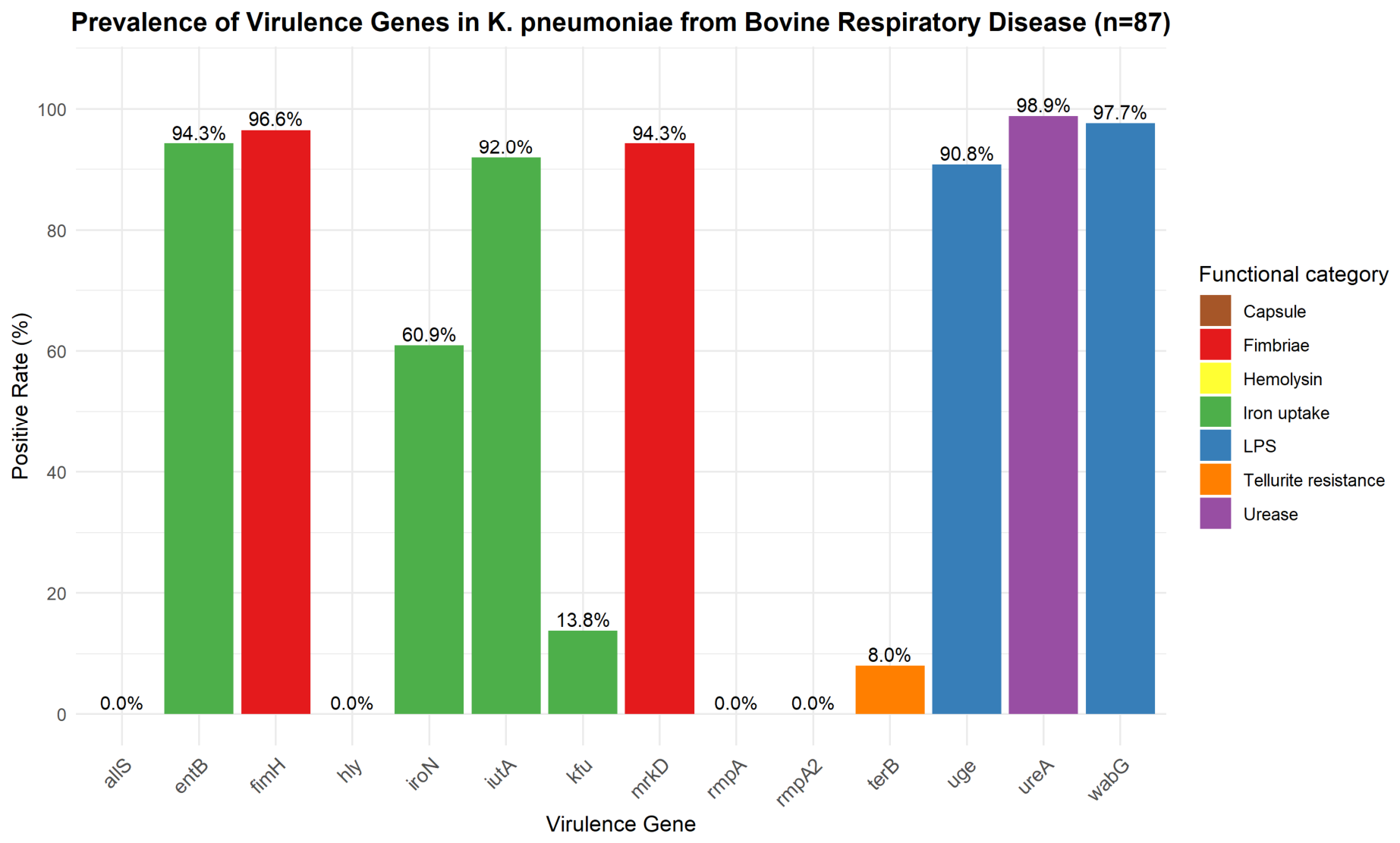
The detailed status of virulence-associated genes carried by 87 K. pneumoniae strains is shown in Figure 5. Among the fimbriae synthesis-related genes, fimH and mrkD were detected in 84 (96.6%) and 82 (94.3%) strains, respectively. For lipopolysaccharide-related genes, uge was present in 79 strains (90.8%), while wabG was found in 85 strains (97.7%). Regarding iron uptake system genes, entB and iutA were identified in 82 (94.3%) and 80 (92.0%) strains, respectively; iroN was positive in 53 strains (60.9%), and kfu was present in only 12 strains (13.8%). The urease-related gene ureA was detected in 86 strains (98.9%), whereas allS was not detected in any strain (0%). The tellurite resistance gene terB was found in 8 strains (9.2%). Notably, the hemolysin gene (hly), the capsular polysaccharide synthesis gene and the regulation-related genes (rmpA and rmpA2) were absent in all 87 isolates (0%). Because positive controls confirmed assay functionality, these genes were not detected in the tested isolates; however, this does not exclude the presence of other hypervirulence-associated determinants. PCR detection indicates carriage only and does not demonstrate expression or functional virulence.
Figure 5.
The distribution of virulence-associated genes among 87 K. pneumoniae strains.
According to the statistics of multiple virulence-associated gene carriers, all 87 isolates carried at least six virulence-associated genes. The most prevalent combination was fimH + mrkD + uge + wabG + entB + iutA + iroN + ureA (eight genes), which was observed in 31 isolates (35.6%). The second most common profile was fimH + mrkD + uge + wabG + entB + iutA + ureA (seven genes, lacking iroN), present in 24 isolates (27.6%). A smaller proportion of isolates (9.2%, 8/87) carried the eight-gene combination together with terB. The least frequent profiles among those with at least six genes were unique patterns carried by single isolates (e.g., ST99 with fimH-negative but positive for mrkD, uge, wabG, entB, iutA, iroN), each accounting for only 1.1% (1/87). These results indicate that multiple virulence-associated gene carriage is common in bovine respiratory K. pneumoniae isolates from Henan, with a dominant profile comprising fimbrial, lipopolysaccharide, iron uptake, and urease genes.
4. Discussion
In this study, the isolation rate of K. pneumoniae was 16.8%. This rate is higher than reports from dairy cows with BRD in Hebei (37/316, 11.7%) [6] and cattle with BRD in Chongqing (33/213, 15.5%) [19] in China, but lower than the isolation rate from pneumonic lung tissues, and tracheal swabs were collected from Aceh cattle in Indonesia (38/61,62.3%) [14]. These comparisons should be interpreted with caution because nasal swabs from live BRD calves cannot be directly compared with pneumonic lung tissue or tracheal samples without considering differences in diagnostic sensitivity, colonization, disease severity, and sampling context. K. pneumoniae is an opportunistic pathogen that can colonize the upper respiratory tract of healthy cattle. Because only nasal swabs were collected, this study cannot distinguish colonization from infection or determine whether K. pneumoniae was the sole causative agent of BRD. The Wisconsin Calf Respiratory Scoring System is a field screening tool, not a diagnostic gold standard. Coinfection with Mannheimia haemolytica, Pasteurella multocida, Mycoplasma bovis, or other BRD pathogens was not assessed. Therefore, the role of K. pneumoniae as a sole or contributory pathogen could not be determined by this method. Geographically, the isolation rates were numerically lower in North (13.8%) and South (15.5%) Henan than in East (22.6%) and West (20.8%) Henan. However, the 95% confidence intervals for all regional estimates overlapped substantially, and after accounting for farm-level clustering in a mixed-effects logistic regression model, no regional difference was statistically significant. The intraclass correlation coefficient was 0.11, indicating that a modest proportion of the total variation in isolation status was attributable to differences between farms. This clustering is biologically plausible, as calves within the same farm share management practices, housing conditions, and antimicrobial exposure histories. The regional comparisons in this study should therefore be interpreted with caution because the sample sizes varied considerably across regions (range: 53–220 samples) and the study was not powered to detect small regional differences. The absence of statistically significant regional variation does not necessarily indicate that true regional differences are absent; rather, it may reflect limited statistical power. Future studies with balanced sampling across regions and larger sample sizes are needed to rigorously evaluate geographic patterns. Although isolation rates fluctuated numerically across years, no statistically significant temporal trend was detected (Pearson chi-square test for heterogeneity: χ2 = 1.78, df = 5, p = 0.879). The isolation rate remained above 10.0% in all years, indicating that K. pneumoniae was consistently present in calves with BRD in Henan Province. However, because no trend test was performed and no significant annual difference was found, the data should not be interpreted as evidence of a declining or increasing temporal trend.
In this study, we isolated 87 K. pneumoniae strains from cattle with pneumonia, which were classified into 34 STs. ST950 was the most frequently identified ST; ST950 has been associated with hypervirulent K. pneumoniae (hvKP) in previous human studies [56]. However, ST designation alone is insufficient to infer hypervirulence for these bovine isolates, and hypervirulence markers were absent in our collection. Other notable STs included ST262 (5.7%, 5/87), ST306 (4.6%, 4/87), ST187 (4.6%, 4/87), and others. These STs have also been reported in other studies at low detection rates [6,20]. Notably, ST262 was previously isolated and reported from sheep in Henan Province [20]. Given that some of the cattle farms in this study were backyard farms where cattle and sheep shared grazing land, the shared ST262 between cattle and sheep suggests a hypothesis of possible cross-species transmission; however, MLST has limited discriminatory power, and whole-genome sequencing with epidemiological linkage is required to test this hypothesis. Moreover, isolates from cattle exhibited a high diversity of STs. A survey identified 19 STs among 37 K. pneumoniae strains from cattle with BRD, with ST43 as the predominant type (29.7%) in Hebei province, China [6]. In Chongqing city, 7 STs were identified among 33 K. pneumoniae strains from cattle with BRD, with ST218 being the most common (57.6%) [19]. K. pneumoniae can infect not only cattle, causing pneumonia, but also the mammary glands of dairy cows, leading to mastitis. Moreover, the STs of mastitis-associated strains also display diversity. One study across seven provinces found 42 STs among 108 K. pneumoniae isolates, with ST1049 as the major type (11.1%) [17]. Additionally, an investigation in Hubei province detected 100 STs among 239 K. pneumoniae isolates, with ST2854 as the dominant type (12.6%) [28]. However, among these three predominant types found in these studies, only ST218 was detected in our study, at a rate of 1.1%. In USA, 23 STs were detected in 29 K. pneumoniae strains, including ST101 (13.0%), ST114 (13.0%), and ST112 (13.0%) [57]; only ST101 was observed in the present study. Comparing these findings, integrated surveillance systems are needed for monitoring K. pneumoniae across animal, human, and environmental compartments.
In China, antibiotics remain the primary approach for treating bacteria-associated BRD. However, recent reports have shown an increasing trend of bacterial resistance to antimicrobial agents among K. pneumoniae isolates [6,19]. Antimicrobial susceptibility testing is essential to guide the selection of appropriate drugs for infection treatment. In this study, the highest resistance was observed against β-lactams, which is consistent with findings from previous reports on K. pneumoniae isolates from dairy cows with BRD [6] or with mastitis in China [28]. Additionally, sulfonamides resistance ranged from 87.5% to 92.9% across regions, while resistance to quinolones ranged from 72.7% to 78.6% across regions. These rates are comparable to or exceed those reported for bovine K. pneumoniae in China [6,17]. This trend is not surprising, as sulfonamides and quinolones are among the most frequently used antibiotics in dairy animals in China [58]. The high resistance rates may be associated with antimicrobial selection pressure; however, quantitative antimicrobial-use data were not collected, and this mechanism cannot be established from the present data. Their extensive use likely contributes to the rising resistance in K. pneumoniae. Moreover, the resistance rates of the isolates to the remaining three antimicrobial classes were lower than those for the three classes mentioned above, with phenicols showing the lowest resistance rate. These results highlight the urgent need for prudent antimicrobial use in treating cattle K. pneumoniae. The challenges of antimicrobial management in BRD are further illustrated by Masebo et al. (2024), who investigated BRD in fattening bulls under commercial conditions and reported a high disease burden, extensive antimicrobial treatment, and persistent pulmonary lesions at slaughter [9]. These observations reinforce the need for antimicrobial stewardship and susceptibility-guided treatment in cattle production. Therefore, the clinical use of β-lactams, sulfonamides, and quinolones should be minimized in Henan province unless supported by susceptibility testing. Florfenicol showed the lowest resistance rate among the tested agents. However, in vitro susceptibility alone cannot establish clinical preference, and treatment decisions should be guided by susceptibility testing, clinical outcome data, and antimicrobial stewardship.
β-lactamase-mediated hydrolysis is a primary mechanism of β-lactam resistance, thereby rendering these drugs ineffective [59]. In this study, blaTEM was detected in 72.4% of K. pneumoniae isolates, lower than the rates anged from 87.5% to 92.9% reported (92.6%) in China [28] and in South Africa (85.5%) [60]. Sulfonamide resistance mediated by the sul gene has become widespread globally, particularly in K. pneumoniae from dairy cattle with mastitis, where resistance to sulfonamide antibiotics is pronounced [61]. In this study, the detection rates of sul1 and sul2 were 39.1% and 18.4%, respectively, consistent with previously reported data on K. pneumoniae isolated from mastitis cases in China [17]. Generally, tetA, tetB, and tetC are the most common tetracycline resistance genes in K. pneumoniae of animal origin, and these genes are part of the small nonconjugative transposons in plasmids [62]. Among the isolates in this study, the detection rates of these three genes were 3.4%, 4.6%, and 12.6%, respectively, which were higher than the rates in earlier studies in dairy cattle [28]. The only phenicol resistance gene identified was floR (2.3%), lower than the rates in cattle in China [28] and Egypt [63]. Resistance to aminoglycosides is conferred by genes encoding aminoglycoside-modifying enzymes, such as the aadA1 gene [64]. Quinolone resistance is associated with plasmid-mediated quinolone resistance genes, such as qnrB and qnrS genes [15,65]. As in this study, these resistance genes have also been detected in isolates from cows in China with varying prevalence rates [6,17,44]. After FDR correction for multiple comparisons, no gene was significantly associated with phenotypic resistance. hese results show profiles different from those in previous work, where correlations of some genes have been observed in K. pneumoniae isolates from dairy cow, chicken, sheep, and pig in Xinjiang in China [66]. The wide confidence intervals and lack of significant associations likely reflect the limited number of isolates (n = 87), the class-level resistance definition, the presence of other resistance mechanisms not captured by the PCR panel, and farm-level clustering. These findings highlight the complexity of genotype–phenotype relationships and the need for whole-genome sequencing to fully characterize resistance mechanisms.
The pathogenicity of K. pneumoniae is critically dependent on virulence factors. In the present study, genes linked to hypervirulence—rmpA, rmpA2, and hly—were not detected. However, absence of the screened markers alone cannot exclude hypervirulence, and carriage of several common virulence-associated genes cannot establish pathogenic potential. Functional assays, capsule typing, siderophore quantification, biofilm assays, infection models, or genomic characterization would be needed for stronger conclusions. Additionally, PCR analysis revealed that fimbria-associated genes (fimH, mrkD), iron-acquisition system genes (iutA, iroN, entB), urease gene (ureA), and lipopolysaccharide-related genes (uge, wabG) were broadly distributed among the isolates, consistent with previous reports which have also been reported in isolates from cows in many provinces in China [17,28]. These findings offer preliminary elucidation of the pathogenic mechanisms of K. pneumoniae, which may involve: assembling fimbriae to attach to host cell surfaces or to form biofilms that promote virulence; secreting siderophores to capture host iron for metabolism and enhanced virulence; employing capsular polysaccharides to evade serum-mediated killing by phagocytes and to dampen host immunity; and organizing lipopolysaccharide into complexes on the bacterial surface, enabling the pathogen to escape or resist killing by the host’s innate immune system [67,68]. In addition, Wan et al. investigated similar virulence-associated genes in K. pneumoniae strains obtained from pigs in Northwest China [25]. Another study by Hou et al. (2024) also identified comparable virulence-associated genes in K. pneumoniae isolated from dairy cows, chicken, sheep, and pigs in Xinjiang, China [66]. These studies suggest that virulence-associated genes in K. pneumoniae may exhibit similarities and broad distribution among strains from different host origins. Whether this is related to the transmission of K. pneumoniae across different hosts remains unclear and warrants further investigation.
Future whole-genome sequencing studies are warranted to further elucidate resistance mechanisms, transmission pathways, and host-adaptation signatures of this pathogen in cattle. Longitudinal surveillance is also valuable in intensive cattle production. Dini et al. (2024) showed in a longitudinal study of fattening beef cattle that infectious exposure can change over time in relation to farm management and biosecurity conditions [10]. Although that study concerned Toxoplasma gondii rather than K. pneumoniae or BRD, it illustrates the broader value of longitudinal monitoring for understanding infectious disease dynamics in intensively managed cattle.
This study has several important limitations. First, no clinically healthy control calves were included; therefore, the isolation rate among BRD-positive calves cannot establish an association between K. pneumoniae carriage and BRD. Second, only deep nasal swabs were collected, and no lung histopathology, pathogen load quantification, or challenge–re-isolation experiments were performed; thus, the study cannot distinguish colonization from infection or determine whether K. pneumoniae was the sole causative agent. Third, coinfection with other core BRD pathogens such as Mannheimia haemolytica, Pasteurella multocida, and Mycoplasma bovis was not assessed. Fourth, only one confirmed K. pneumoniae isolate per nasal swab was stored and characterized. Because a single sample may contain multiple strains with different STs, this approach may underestimate within-host strain diversity and population heterogeneity. Fifth, farm-level clustering was present and was accounted for where possible, but some analyses remain descriptive.
5. Conclusions
This study provides the first comprehensive molecular and phenotypic characterization of K. pneumoniae isolates from cattle with BRD in Henan Province, China, covering a six-year period (2021–2026). K. pneumoniae was isolated from 16.8% of sampled calves with BRD. Because no healthy comparison group or etiological confirmation was included, this study cannot establish K. pneumoniae as a causative agent of BRD; the organism may be a colonizer, an opportunistic pathogen, or a component of a polymicrobial infection. The 87 isolates were assigned to 34 diverse STs, indicating substantial genetic heterogeneity. The shared ST262 with sheep suggests a hypothesis of possible cross-species transmission that requires whole-genome sequencing and epidemiological linkage for confirmation. Notably, 96.6% of isolates were multidrug-resistant, with extremely high resistance to β-lactams, sulfonamides, and quinolones. After FDR correction, no resistance gene was significantly associated with phenotypic resistance. Multiple virulence-associated gene carriage is common in bovine respiratory K. pneumoniae isolates from Henan, with a dominant profile comprising fimbrial, lipopolysaccharide, iron uptake, and urease genes. However, carriage of these genes does not establish pathogenic potential. The dominant virulence-associated gene profile (fimH + mrkD + uge + wabG + entB + iutA + iroN + ureA) was found in over one-third of isolates, but gene prevalence alone does not establish expression, immunogenicity, essentiality, or protective potential, and these findings should not be presented as a basis for vaccine or therapeutic target development without further functional studies. Antimicrobial stewardship, susceptibility-guided treatment, and whole-genome sequencing are warranted to resolve K. pneumoniae strain relatedness, characterize resistance determinants, investigate mobile genetic elements, and provide stronger evidence regarding possible transmission between farms, regions, and animal species.
Supplementary Materials
The following supporting information can be downloaded at: https://www.mdpi.com/article/10.3390/vetsci13101029/s1, Table S1: Annual sampling and isolation of K. pneumoniae from calves with BRD in Henan Province, China, 2021–2026. Table S2: Primers of resistance genes. Table S3: Primers of virulence-associated genes. Table S4: Antimicrobial resistance genes distributions among 87 strains.
Author Contributions
H.S. and Y.B.: idea and concept. H.Z. and Y.X.: writing and editing of the manuscript. H.S. and Y.B.: editing of the manuscript and funding. T.X., S.Y. and Z.C.: revision of the manuscript. H.Z., Y.X., T.X., S.Y. and Z.C.: sample collection, detection, sequencing, and analysis of data. All authors have read and agreed to the published version of the manuscript.
Funding
The Special Fund for Henan Agriculture Research System (HARS-22-13-G3), the Natural Science Foundation of Henan province (Grant no. 242300421334), the National Natural Science Foundation of China (Grant no. 31902263), and the Nanyang Normal University (Grant no. 241344) supported this study.
Institutional Review Board Statement
All methods were carried out in accordance with Chinese Law for the Care and Use of Animals. The research protocol was approved by the Animal Welfare and Ethics Committee of Nanyang Normal University (approval no. 202119; approved on 8 January 2021).
Informed Consent Statement
Written informed consent was obtained from the owners of all animals included in the study.
Data Availability Statement
The original contributions presented in this study are included in the article/Supplementary Material. Further inquiries can be directed to the corresponding authors.
Conflicts of Interest
The authors declare that no competing financial interests and no conflicts of interest exist.
References
- Taylor, J.D.; Fulton, R.W.; Lehenbauer, T.W.; Step, D.L.; Confer, A.W. The epidemiology of bovine respiratory disease: What is the evidence for predisposing factors? Can. Vet. J. 2010, 51, 1095–1102. [Google Scholar] [PubMed]
- Dubrovsky, S.A.; Van Eenennaam, A.L.; Aly, S.S.; Karle, B.M.; Rossitto, P.V.; Overton, M.W.; Lehenbauer, T.W.; Fadel, J.G. Preweaning cost of bovine respiratory disease (BRD) and cost-benefit of implementation of preventative measures in calves on California dairies: The BRD 10K study. J. Dairy Sci. 2020, 103, 1583–1597. [Google Scholar] [CrossRef] [Scilit] [PubMed]
- Timsit, E.; Holman, D.B.; Hallewell, J.; Alexander, T.W. The nasopharyngeal microbiota in feedlot cattle and its role in respiratory health. Anim. Front. 2016, 6, 44–50. [Google Scholar] [CrossRef] [Scilit]
- Edwards, T.A. Control Methods for Bovine Respiratory Disease for Feedlot Cattle. Vet. Clin. N. Am. Food Anim. Pract. 2010, 26, 273–284. [Google Scholar] [CrossRef] [Scilit] [PubMed]
- Centeno-Martinez, R.E.; Glidden, N.; Mohan, S.; Davidson, J.L.; Fernández-Juricic, E.; Boerman, J.P.; Schoonmaker, J.; Pillai, D.; Koziol, J.; Ault, A.; et al. Identification of bovine respiratory disease through the nasal microbiome. Anim. Microbiome 2022, 4, 15. [Google Scholar] [CrossRef] [Scilit] [PubMed]
- Jia, L.; Zhang, G.; Li, X.; Wang, H.; Zhao, J.; Huang, X.; Ba, X.; Shen, Q.; Ma, Y. ESBL-producing Klebsiella Pneumoniae isolated from dairy cows with pneumonia: Insight into their epidemiology, genetic features and antimicrobial resistance profiles. Microb. Pathog. 2025, 207, 107909. [Google Scholar] [CrossRef] [Scilit] [PubMed]
- Wang, Z.; Duan, L.; Liu, F.; Hu, Y.; Leng, C.; Kan, Y.; Yao, L.; Shi, H. First report of Enterobacter hormaechei with respiratory disease in calves. BMC Vet. Res. 2020, 16, 1. [Google Scholar] [CrossRef] [Scilit] [PubMed]
- Zeineldin, M.; A. Elolimy, A.; Barakat, R. Meta-analysis of bovine respiratory microbiota: Link between respiratory microbiota and bovine respiratory health. FEMS Microbiol. Ecol. 2020, 96, fiaa127. [Google Scholar] [CrossRef] [Scilit] [PubMed]
- Masebo, N.T.; Marliani, G.; Shannon Del Re, F.; Abram, L.; Cavallini, D.; Di Pietro, M.; Beltrame, A.; Schiavon, E.; Bolcato, M.; Hernandez Bermudez, J.; et al. Evaluation of antimicrobial and non-steroidal anti-inflammatory treatments for BRD on health and welfare in fattening bulls: A cross-sectional study. Vet. Q. 2024, 44, 1–11. [Google Scholar] [CrossRef] [Scilit] [PubMed]
- Dini, F.M.; Jacinto, J.G.P.; Cavallini, D.; Beltrame, A.; Del Re, F.S.; Abram, L.; Gentile, A.; Galuppi, R. Observational longitudinal study on Toxoplasma gondii infection in fattening beef cattle: Serology and associated haematological findings. Parasitol. Res. 2024, 123, 169. [Google Scholar] [CrossRef] [Scilit] [PubMed]
- Hu, Y.; Anes, J.; Devineau, S.; Fanning, S. Klebsiella pneumoniae: Prevalence, reservoirs, antimicrobial resistance, pathogenicity, and infection: A hitherto unrecognized zoonotic bacterium. Foodborne Pathog. Dis. 2021, 18, 63–84. [Google Scholar] [CrossRef] [Scilit] [PubMed]
- Nguyen, T.N.T.; Howells, G.; Short, F.L. How Klebsiella pneumoniae controls its virulence. PLoS Pathog. 2025, 21, e1013499. [Google Scholar] [CrossRef] [Scilit] [PubMed]
- Rissetti, R.M.; de Paula, C.L.; Yamada, A.Y.; Bertani, A.M.J.; Costa, A.B.N.; Sacchi, C.T.; Campos, K.R.; Guerra, S.T.; Guimarães, F.F.; Joaquim, S.F.; et al. Detection of multiple clones and extended-spectrum β-lactamase production among Klebsiella pneumoniae complex isolates from the milk of cows with different severity scores of clinical mastitis. Lett. Appl. Microbiol. 2025, 78, ovaf118. [Google Scholar] [CrossRef] [Scilit] [PubMed]
- Darniati, D.; Handharyani, E.; Setiyaningsih, S. Molecular analysis of Klebsiella pneumoniae serotypes and virulence genes isolated from pneumonic Aceh cattle. Open Vet. J. 2025, 15, 3737–3745. [Google Scholar] [CrossRef] [Scilit] [PubMed]
- Bandyopadhyay, S.; Bhattacharyya, D.; Samanta, I.; Banerjee, J.; Habib, M.; Dutta, T.K.; Dutt, T. Characterization of multidrug-resistant biofilm-producing Escherichia coli and Klebsiella pneumoniae in healthy cattle and cattle with diarrhea. Microb. Drug Resist. 2021, 27, 1457–1469. [Google Scholar] [CrossRef] [Scilit] [PubMed]
- Cai, K.; Xu, M.; Liu, L.; Zhao, H. Molecular Epidemiology and Antimicrobial Resistance of Klebsiella pneumoniae strains isolated from dairy cows in Xinjiang, China. Vet. Med. Sci. 2025, 11, e70120. [Google Scholar] [CrossRef] [Scilit] [PubMed]
- Wang, J.; Li, J.; Ji, X.; Zhang, L.; Wang, R.; Wang, H.; He, T. Antimicrobial resistance and molecular epidemiology of Klebsiella pneumoniae isolated from bovine mastitis in seven provinces in China. BMC Microbiol. 2025, 25, 407. [Google Scholar] [CrossRef] [Scilit] [PubMed]
- Wusiman, M.; Zuo, J.; Yu, Y.; Lv, Z.; Wang, M.; Nie, L.; Zhang, X.; Wu, J.; Wu, Z.; Jiang, W.; et al. Molecular characterization of Klebsiella pneumoniae in clinical bovine mastitis in 14 provinces in China. Vet. Res. Commun. 2024, 49, 18. [Google Scholar] [CrossRef] [Scilit] [PubMed]
- Cheng, F.; Li, Z.; Lan, S.; Liu, W.; Li, X.; Zhou, Z.; Song, Z.; Wu, J.; Zhang, M.; Shan, W. Characterization of Klebsiella pneumoniae associated with cattle infections in southwest China using multi-locus sequence typing (MLST), antibiotic resistance and virulence-associated gene profile analysis. Braz. J. Microbiol. 2018, 49, 93–100. [Google Scholar] [CrossRef] [Scilit] [PubMed]
- Yang, F.; Deng, B.; Liao, W.; Wang, P.; Chen, P.; Wei, J. High rate of multiresistant Klebsiella pneumoniae from human and animal origin. Infect. Drug Resist. 2019, 12, 2729–2737. [Google Scholar] [CrossRef] [Scilit] [PubMed]
- Luo, J.; Shi, H.; Wang, J.; Zhang, F.; Zhen, H.; Ji, Z.; Liu, Y.; Li, C.; Wang, P. A summary of the development of chinese dairy goat industry-development trends and characteristics. China Dairy Cattle 2019, 36, 1–11. [Google Scholar] [CrossRef]
- Diancourt, L.; Passet, V.; Verhoef, J.; Grimont, P.A.; Brisse, S. Multilocus sequence typing of Klebsiella pneumoniae nosocomial isolates. J. Clin. Microbiol. 2005, 43, 4178–4182. [Google Scholar] [CrossRef] [Scilit] [PubMed]
- Shi, H.; Tu, Y.; Li, H.; Gao, H.; Wang, F.; Zhang, W.; Jiang, M.; Sun, Q.; Bao, Z.; Yang, X.; et al. Molecular characteristics and antimicrobial susceptibility of carbapenem-resistant Klebsiella pneumoniae in a multicenter study in Ningbo, China. Front. Microbiol. 2025, 16, 1628592. [Google Scholar] [CrossRef] [Scilit] [PubMed]
- Wang, X.; Wang, J.; Jiang, X.; Huang, Z.; Huang, L.; Wei, Q.; Zhang, L. Molecular epidemiological analysis and research on resistance and virulence of carbapenem-resistant Klebsiella pneumoniae in a tertiary hospital from 2016 to 2023. BMC Microbiol. 2025, 25, 217. [Google Scholar] [CrossRef] [Scilit] [PubMed]
- Wan, S.H.; Li, N.; Zheng, P.; Li, Y.; Liang, Y.; Qu, Y. Virulence characteristics and antibiotic resistance analysis of Klebsiella pneumoniae isolated from pig farms in Xinjiang, China: Revealing potential zoonotic risks. Porc. Health Manag. 2025, 11, 25. [Google Scholar] [CrossRef] [Scilit] [PubMed]
- Bao, R.; Ma, Z.; Stanford, K.; McAllister, T.A.; Niu, Y.D. Antimicrobial activities of α-helix and β-sheet peptides against the major bovine respiratory disease agent, Mannheimia haemolytica. Int. J. Mol. Sci. 2024, 25, 4164. [Google Scholar] [CrossRef] [Scilit] [PubMed]
- Kong, L.C.; Wang, Z.; Wang, Y.M.; Dong, W.L.; Jia, B.Y.; Gao, D.; Jiang, X.Y.; Ma, H.X. Antimicrobial susceptibility and molecular typing of Pasteurella multocida isolated from six provinces in China. Trop. Anim. Health Prod. 2019, 51, 987–992. [Google Scholar] [CrossRef] [Scilit] [PubMed]
- Wu, X.; Liu, J.; Feng, J.; Shabbir, M.A.B.; Feng, Y.; Guo, R.; Zhou, M.; Hou, S.; Wang, G.; Hao, H.; et al. Epidemiology, environmental risks, virulence, and resistance determinants of Klebsiella pneumoniae from dairy cows in Hubei, China. Front. Microbiol. 2022, 13, 858799. [Google Scholar] [CrossRef] [Scilit] [PubMed]
- Candan, E.D.; Aksöz, N. Klebsiella pneumoniae: Characteristics of carbapenem resistance and virulence factors. Acta Biochim. Pol. 2015, 62, 867–874. [Google Scholar] [CrossRef] [Scilit] [PubMed]
- Guo, Y.; Zhou, H.; Qin, L.; Pang, Z.; Qin, T.; Ren, H.; Pan, Z.; Zhou, J. Frequency, antimicrobial resistance and genetic diversity of Klebsiella pneumoniae in food samples. PLoS ONE 2016, 11, e0153561. [Google Scholar] [CrossRef] [Scilit] [PubMed]
- Remya, P.A.; Shanthi, M.; Sekar, U. Characterisation of virulence genes associated with pathogenicity in Klebsiella pneumoniae. Indian. J. Med. Microbiol. 2019, 37, 210–218. [Google Scholar] [CrossRef] [Scilit] [PubMed]
- McGuirk, S.M. Disease management of dairy calves and heifers. Vet. Clin. N. Am. Food Anim. Pract. 2008, 24, 139–153. [Google Scholar] [CrossRef] [Scilit] [PubMed]
- CLSI. Performance Standards for Antimicrobial Disk and Dilution Susceptibility Tests for Bacteria Isolated From Animals, 7th ed.; CLSI Supplement VET01S; CLSI: Wayne, PA, USA, 2024. [Google Scholar]
- CLSI. Performance Standards for Antimicrobial Susceptibility Testing, 29th ed.; CLSI M100-S26; CLSI: Wayne, PA, USA, 2016. [Google Scholar]
- Moodley, A.; Stegger, M.; Bagcigil, A.F.; Baptiste, K.E.; Loeffler, A.; Lloyd, D.H.; Williams, N.J.; Leonard, N.; Abbott, Y.; Skov, R.; et al. spa typing of methicillin-resistant Staphylococcus aureus isolated from domestic animals and veterinary staff in the UK and Ireland. J. Antimicrob. Chemother. 2006, 58, 1118–1123. [Google Scholar] [CrossRef] [Scilit] [PubMed]
- Andrade, L.N.; Novais, Â.; Stegani, L.M.M.; Ferreira, J.C.; Rodrigues, C.; Darini, A.L.C.; Peixe, L. Virulence genes, capsular and plasmid types of multidrug-resistant CTX-M(-2, -8, -15) and KPC-2-producing Klebsiella pneumoniae isolates from four major hospitals in Brazil. Diagn. Microbiol. Infect. Dis. 2018, 91, 164–168. [Google Scholar] [CrossRef] [Scilit] [PubMed]
- Zhang, Y.; Zhao, C.; Wang, Q.; Wang, X.; Chen, H.; Li, H.; Zhang, F.; Li, S.; Wang, R.; Wang, H. High prevalence of hypervirulent Klebsiella pneumoniae infection in China: Geographic distribution, clinical characteristics, and antimicrobial resistance. Antimicrob. Agents Chemother. 2016, 60, 6115–6120. [Google Scholar] [CrossRef] [Scilit] [PubMed]
- Fazel, F.; Jamshidi, A.; Khoramian, B. Phenotypic and genotypic study on antimicrobial resistance patterns of E. coli isolates from bovine mastitis. Microb. Pathog. 2019, 132, 355–361. [Google Scholar] [CrossRef] [Scilit] [PubMed]
- Chiu, S.K.; Huang, L.Y.; Chen, H.; Tsai, Y.K.; Liou, C.H.; Lin, J.C.; Siu, L.K.; Chang, F.Y.; Yeh, K.M. Roles of ramR and tet(A) mutations in conferring tigecycline resistance in carbapenem-resistant Klebsiella pneumoniae clinical isolates. Antimicrob. Agents Chemother. 2017, 61, e00391. [Google Scholar] [CrossRef] [Scilit] [PubMed]
- Tabaran, A.; Mihaiu, M.; Tăbăran, F.; Colobatiu, L.; Reget, O.; Borzan, M.M.; Dan, S.D. First study on characterization of virulence and antibiotic resistance genes in verotoxigenic and enterotoxigenic E. coli isolated from raw milk and unpasteurized traditional cheeses in Romania. Folia Microbiol. 2017, 62, 145–150. [Google Scholar] [CrossRef] [Scilit] [PubMed]
- Maynard, C.; Bekal, S.; Sanschagrin, F.; Levesque, R.C.; Brousseau, R.; Masson, L.; Larivière, S.; Harel, J. Heterogeneity among virulence and antimicrobial resistance gene profiles of extraintestinal Escherichia coli isolates of animal and human origin. J. Clin. Microbiol. 2004, 42, 5444–5452. [Google Scholar] [CrossRef] [Scilit] [PubMed]
- Travis, R.M.; Gyles, C.L.; Reid-Smith, R.; Poppe, C.; McEwen, S.A.; Friendship, R.; Janecko, N.; Boerlin, P. Chloramphenicol and kanamycin resistance among porcine Escherichia coli in Ontario. J. Antimicrob. Chemother. 2006, 58, 173–177. [Google Scholar] [CrossRef] [Scilit] [PubMed]
- Maynard, C.; Fairbrother, J.M.; Bekal, S.; Sanschagrin, F.; Levesque, R.C.; Brousseau, R.; Masson, L.; Larivière, S.; Harel, J. Antimicrobial resistance genes in enterotoxigenic Escherichia coli O149:K91 isolates obtained over a 23-year period from pigs. Antimicrob. Agents Chemother. 2003, 47, 3214–3221. [Google Scholar] [CrossRef] [Scilit] [PubMed]
- Al-Marzooq, F.; Ngeow, Y.F.; Tay, S.T. Emergence of Klebsiella pneumoniae producing dual carbapenemases (NDM-1 and OXA-232) and 16S rRNA methylase (armA) isolated from a Malaysian patient returning from India. Int. J. Antimicrob. Agents 2015, 45, 445–446. [Google Scholar] [CrossRef] [Scilit] [PubMed]
- Rodríguez-Martínez, J.M.; Díaz de Alba, P.; Briales, A.; Machuca, J.; Lossa, M.; Fernández-Cuenca, F.; Rodríguez Baño, J.; Martínez-Martínez, L.; Pascual, Á. Contribution of OqxAB efflux pumps to quinolone resistance in extended-spectrum-β-lactamase-producing Klebsiella pneumoniae. J. Antimicrob. Chemother. 2013, 68, 68–73. [Google Scholar] [CrossRef] [Scilit] [PubMed]
- Park, C.H.; Robicsek, A.; Jacoby, G.A.; Sahm, D.; Hooper, D.C. Prevalence in the United States of aac(6′)-Ib-cr encoding a ciprofloxacin-modifying enzyme. Antimicrob. Agents Chemother. 2006, 50, 3953–3955. [Google Scholar] [CrossRef] [Scilit] [PubMed]
- Shin, J.H.; Jung, H.J.; Lee, J.Y.; Kim, H.R.; Lee, J.N.; Chang, C.L. High rates of plasmid-mediated quinolone resistance QnrB variants among ciprofloxacin-resistant Escherichia coli and Klebsiella pneumoniae from urinary tract infections in Korea. Microb. Drug Resist. 2008, 14, 221–226. [Google Scholar] [CrossRef] [Scilit] [PubMed]
- Ko, W.C.; Hsueh, P.R. Increasing extended-spectrum beta-lactamase production and quinolone resistance among gram-negative bacilli causing intra-abdominal infections in the Asia/Pacific region: Data from the smart study 2002–2006. J. Infect. 2009, 59, 95–103. [Google Scholar] [CrossRef] [Scilit] [PubMed]
- XU, S.; Yang, s.; Weng, S.; Chen, C.; Chen, S.; Zhang, W.; Jing, J. Distribution of serotypes and virulence genes in hypervirulent Klebsiella pneumoniae and exploration of molecular markers. J. Microbes Infect. 2019, 14, 338–344. [Google Scholar]
- Lan, Y.; Zhou, M.; Jian, Z.; Yan, Q.; Wang, S.; Liu, W. Prevalence of pks gene cluster and characteristics of Klebsiella pneumoniae-induced bloodstream infections. J. Clin. Lab. Anal. 2019, 33, e22838. [Google Scholar] [CrossRef] [Scilit] [PubMed]
- Osman, K.M.; Hassan, H.M.; Orabi, A.; Abdelhafez, A.S. Phenotypic, antimicrobial susceptibility profile and virulence factors of Klebsiella pneumoniae isolated from buffalo and cow mastitic milk. Pathog. Glob. Health 2014, 108, 191–199. [Google Scholar] [CrossRef] [Scilit] [PubMed]
- Sivaraman, G.K.; Sudha, S.; Muneeb, K.H.; Shome, B.; Holmes, M.; Cole, J. Molecular assessment of antimicrobial resistance and virulence in multi drug resistant ESBL-producing Escherichia coli and Klebsiella pneumoniae from food fishes, Assam, India. Microb. Pathog. 2020, 149, 104581. [Google Scholar] [CrossRef] [Scilit] [PubMed]
- Rodriguez-Siek, K.E.; Giddings, C.W.; Doetkott, C.; Johnson, T.J.; Nolan, L.K. Characterizing the APEC pathotype. Vet. Res. 2005, 36, 241–256. [Google Scholar] [CrossRef] [Scilit] [PubMed]
- Brisse, S.; Fevre, C.; Passet, V.; Issenhuth-Jeanjean, S.; Tournebize, R.; Diancourt, L.; Grimont, P. Virulent clones of Klebsiella pneumoniae: Identification and evolutionary scenario based on genomic and phenotypic characterization. PLoS ONE 2009, 4, e4982. [Google Scholar] [CrossRef] [Scilit] [PubMed]
- Harada, S.; Doi, Y. Hypervirulent Klebsiella pneumoniae: A Call for Consensus Definition and International Collaboration. J. Clin. Microbiol. 2018, 56, e00959. [Google Scholar] [CrossRef] [Scilit] [PubMed]
- Xu, Z.; Xiong, Y.; Duan, X.; Han, J.; Xiang, X.; Han, R.; Zhang, S. Molecular and virulence differences of Klebsiella pneumoniae isolated from blood. Front. Microbiol. 2025, 16, 1650010. [Google Scholar] [CrossRef] [Scilit] [PubMed]
- Paulin-Curlee, G.G.; Singer, R.S.; Sreevatsan, S.; Isaacson, R.; Reneau, J.; Foster, D.; Bey, R. Genetic diversity of mastitis-associated Klebsiella pneumoniae in dairy cows. J. Dairy Sci. 2007, 90, 3681–3689. [Google Scholar] [CrossRef] [Scilit] [PubMed]
- Zhao, Y.; Gong, Y.; Kong, L.; Gao, D. Isolation, identification and drug resistance analysis of pathogen inducing pneumonia in calf. Jilin Anim. Husb. Vet. Med. 2012, 33, 7–9. [Google Scholar]
- Pitout, J.D.D.; Laupland, K.B. Extended-spectrum β-lactamase-producing Enterobacteriaceae: An emerging public-health concern. Lancet Infect. Dis. 2008, 8, 159–166. [Google Scholar] [CrossRef] [Scilit] [PubMed]
- Montso, K.P.; Dlamini, S.B.; Kumar, A.; Ateba, C.N. Antimicrobial resistance factors of extended-spectrum beta-lactamases producing Escherichia coli and Klebsiella pneumoniae isolated from cattle farms and raw beef in north-west province, South Africa. BioMed. Res. Int. 2019, 2019, 4318306. [Google Scholar] [CrossRef] [Scilit] [PubMed]
- Poirel, L.; Madec, J.-Y.; Lupo, A.; Schink, A.-K.; Kieffer, N.; Nordmann, P.; Schwarz, S. Antimicrobial resistance in Escherichia coli. Microbiol. Spectr. 2018, 6, 1128. [Google Scholar] [CrossRef] [Scilit] [PubMed]
- Chalmers, R.; Sewitz, S.; Lipkow, K.; Crellin, P. Complete nucleotide sequence of Tn10. J. Bacteriol. 2000, 182, 2970–2972. [Google Scholar] [CrossRef] [Scilit] [PubMed]
- Ahmed, A.M.; Shimamoto, T. Molecular characterization of antimicrobial resistance in Gram-negative bacteria isolated from bovine mastitis in Egypt. Microbiol. Immunol. 2011, 55, 318–327. [Google Scholar] [CrossRef] [Scilit] [PubMed]
- Vo, A.T.; van Duijkeren, E.; Fluit, A.C.; Gaastra, W. Characteristics of extended-spectrum cephalosporin-resistant Escherichia coli and Klebsiella pneumoniae isolates from horses. Vet. Microbiol. 2007, 124, 248–255. [Google Scholar] [CrossRef] [Scilit] [PubMed]
- Koovapra, S.; Bandyopadhyay, S.; Das, G.; Bhattacharyya, D.; Banerjee, J.; Mahanti, A.; Samanta, I.; Nanda, P.K.; Kumar, A.; Mukherjee, R.; et al. Molecular signature of extended spectrum β-lactamase producing Klebsiella pneumoniae isolated from bovine milk in eastern and north-eastern India. Infect. Genet. Evol. J. Mol. Epidemiol. Evol. Genet. Infect. Dis. 2016, 44, 395–402. [Google Scholar] [CrossRef] [Scilit] [PubMed]
- Hou, G.; Ahmad, S.; Li, Y.; Yan, D.; Yang, S.; Chen, S.; Qiu, Z.; Yu, X.; Li, N.; Li, Y.; et al. Epidemiological, virulence, and antibiotic resistance analysis of Klebsiella pneumoniae, a major source of threat to livestock and poultry in some regions of Xinjiang, China. Animals 2024, 14, 1433. [Google Scholar] [CrossRef] [Scilit] [PubMed]
- Li, B.; Zhao, Y.; Liu, C.; Chen, Z.; Zhou, D. Molecular Pathogenesis of Klebsiella pneumoniae. Future Microbiol. 2014, 9, 1071–1081. [Google Scholar] [CrossRef] [Scilit] [PubMed]
- Liu, Z.; Gu, Y.; Li, X.; Liu, Y.; Ye, Y.; Guan, S.; Li, J. Identification and characterization of NDM-1-producing hypervirulent (hypermucoviscous) Klebsiella pneumoniae in China. Ann. Lab. Med. 2019, 39, 167–175. [Google Scholar] [CrossRef] [Scilit] [PubMed]

Disclaimer/Publisher’s Note: The statements, opinions and data contained in all publications are solely those of the individual author(s) and contributor(s) and not of MDPI and/or the editor(s). MDPI and/or the editor(s) disclaim responsibility for any injury to people or property resulting from any ideas, methods, instructions or products referred to in the content. |
© 2026 by the authors. Licensee MDPI, Basel, Switzerland. This article is an open access article distributed under the terms and conditions of the Creative Commons Attribution (CC BY) license.