


Abstract
Pulque is a traditional Mexican beverage produced through the fermentation of agave sap (aguamiel). Its primary sensory properties are attributed to the fermentative activity of Saccharomyces cerevisiae, Leuconostoc mesenteroides, and Zymomonas mobilis. However, the overproliferation of these microorganisms results in an extremely short shelf life, which hinders its commercialization. Tomatillo accrescent calyx extract (TACE) shows potential as a food preservative due to its high physalin content. Therefore, this study aimed to evaluate the effect of adding microencapsulated TACE on the shelf life and organoleptic properties of pulque. The extract demonstrated effective antimicrobial activity against L. mesenteroides, Z. mobilis, and S. cerevisiae, successfully delaying further fermentation. Additionally, the addition of TACE prevented an excessive increase in acidity, maintaining values suitable for consumption for up to 15 days, in accordance with regulatory standards, while the viscosity and alcohol content were not negatively affected. These findings suggest that TACE has significant potential for preserving both the microbiological and sensory quality of pulque.
1. Introduction
Pulque is an alcoholic beverage, produced by the fermentation of agave (maguey) sap (aguamiel), with significant cultural heritage in Mexico. Pulque is characterized by its foaminess, whitish appearance, viscous consistency, and an alcohol content in the range of 4 to 9% v/v [1]. The aguamiel used for pulque production is extracted from specific agave species, including A. atrovirens, A. mapisaga and A. salmiana. The pulque production process involves harvesting sap by making a moderate cut in the center of the plant and scraping the stem. This sap is then fermented at room temperature by microorganisms such as Zymomonas mobilis and lactic acid bacteria. To achieve sweeter, less acidic flavors, aguamiel is boiled in some regions before inoculation. The activity of microorganisms in aguamiel influences its flavor and physicochemical properties [2]. The main obstacle in the commercialization of pulque is its short shelf life, up to 3 days even when refrigerated, attributed to the high microbial population present in the beverage. Additionally, the preservation methods used to extend the shelf life of pulque have been shown to reduce its sensory and probiotic properties [3].
During the fermentation of agave sap, the main microorganisms that ultimately determine pulque quality include Saccharomyces cerevisiae and Zymomonas mobilis, which produce ethanol, and Leuconostoc mesenteroides, which produce dextrans, fructans, and exopolysaccharides. However, the overwhelming population of these microorganisms can decrease its shelf life by reducing the pH, increasing the alcohol content, and generating unpleasant sensory properties (color, odor, taste) [4].
L. mesenteroides is the predominant species of lactic acid bacteria (LAB) from pulque. This microorganism is an obligate heterofermentative bacterium that exclusively uses the phosphoketolase-dependent metabolic route to ferment sugars into lactic acid, acetic acid, and ethanol. L. mesenteroides lacks the enzyme fructose-1,6-diphosphate aldolase, which is involved in the oxidation of glucose to obtain lactic acid through the Embden–Meyerhof–Parnas pathway; consequently, it does not perform a strictly homofermentative metabolism but instead follows a heterofermentative route. As a result, in addition to lactic acid, it produces ethanol, acetic acid, and CO2, metabolites that contribute to the characteristic acidity, slight effervescence, sensory complexity, and microbial balance during pulque fermentation [5]. L. mesenteroides, through the action of enzymes such as invertase and dextransucrase, can produce dextran from carbon sources such as sucrose. Dextran is a polysaccharide formed by glucose units linked by α-1,6 bonds, with branches linked by α-1,3 bonds, with a molecular weight of approximately 1000 to 20,000 Da and a muciform consistency [6].
Z. mobilis, is a Gram-negative, facultative anaerobic bacterium that degrades glucose by Entner–Doudoroff route (ED). This process employs the enzymes pyruvate decarboxylase and alcohol dehydrogenase to convert glucose into ethanol and CO2. In glucose-rich media, Z. mobilis also produces glycerol, succinate, acetate, lactate, acetoin, and butanediol. Furthermore, compared to S. cerevisiae, Z. mobilis exhibits a higher specific cell surface area and a faster glucose consumption rate, resulting in higher ethanol yields [7]. The metabolites produced by Z. mobilis depend on the presence or absence of oxygen during fermentation. In the presence of oxygen, it produces ethanol, acetaldehyde, acetoin, and acetate, whereas in the absence of oxygen, it focuses on ethanol production [8].
The tomatillo (Physalis ixocarpa) is a fruit native to Mexico with a spherical shape, smooth surface, green color, and a characteristic husk known as an accrescent calyx [9]. It is a recognized source of antioxidants (anthocyanins, phenolic compounds, flavonoids, and tannins), as well as of β-carotene and lutein in low concentrations. The crude extract of tomatillo’s accrescent calyx contains physalins, which exhibit antimicrobial activity against Staphylococcus aureus, Escherichia coli, LAB, and Pseudomonas aeruginosa, among others [10]. Physalins are steroidal secondary metabolites classified as secosteroids and are characteristic of the genus Physalis. Physalins are withanolide-type compounds (a group of naturally occurring oxygenated steroids), widely recognized for their diverse pharmacological properties. They are classified into two main subclasses according to their molecular configuration: physalins characterized by the formation of an acetal bridge through the linkage of carbon 14 to carbon 17 via an oxygen atom; and neophysalins, in which carbon 14 is directly linked to carbon 16, while esterification between carbons 15 and 17 results in the formation of a lactone [11].
One of the disadvantages of extracts is the rapid degradation of their bioactive compounds, driven by exposure to light, oxygen, and high temperatures—factors that catalyze their oxidation and decomposition [12]. Therefore, alternatives have been sought to increase the stability of these compounds, with encapsulation among the most widely used. This technique confines the extract within a polymer matrix, creating a safe microatmosphere. This strategy not only stabilizes the compounds against environmental stress but also allows for their controlled release [13].
Spray drying encapsulation is one of the most widely used techniques in the food industry due to its efficiency, low cost, and ease of scalability. This process involves atomizing a solution containing the bioactive compound and an encapsulating agent, forming dry particles that protect the compound from adverse environmental conditions [14]. This technique is effective for incorporating bioactive compounds into complex matrices, such as fermentation systems, as it protects their biological activity and enhances their interaction with the microorganisms present. This prolongs their effect, reduces their degradation, and contributes to the stability of the final product, which is especially relevant in foods with high microbial activity [15]. This work aimed to evaluate the potential of a preservation method for pulque based on the addition of microencapsulated P. ixocarpa calyx extract, by monitoring its effect on the beverage’s shelf life.
2. Materials and Methods
2.1. Materials
Tomatillo accrescent calyx (TAC) was purchased at the Aculco de Espinoza market in the State of Mexico; only samples exhibiting a firm texture and characteristic glossy appearance were selected. Modified starch (sodium octenyl succinate E1450, Ingredion, San Juan del Río, Querétaro, Mexico), glucose (Meyer, Mexico City, Mexico), yeast extract, casein peptone, sodium citrate (all from Merk, St. Louis, MO, USA), crystal sucrose (J.T. Baker, Phillipsburg, NJ, USA), and bacteriological agar (Becton Dickinson, Franklin Lakes NJ, USA) were also obtained. The microbial strains L. mesenteroides MCDO523 and Saccharomyces cerevisiae P530 were provided by the Food Biotechnology Laboratory of the Autonomous University of Querétaro (Santiago de Queretaro, Queretaro, Mexico). Zymomonas mobilis was donated by the Department of Cellular Engineering and Biocatalysis of Biotechnology Institute, Cuernavaca, Morelos, Mexico. Fresh pulque samples were provided by the community of Texcatepec, Chilcuautla, Hidalgo, Mexico.
2.2. Methods
2.2.1. TAC Preparation
The accreting tomato calyx was dried in an oven (Felisa FE-293, Jalisco, Mexico) at 40 °C for 48 h. Once dried, the material was ground using a coffee grinder (Krups Epices GX4100, Solingen, Germany), and the particle size was homogenized to 0.420 mm by sieving the powder through a #40 sieve (Advantech, Taipei, Taiwan).
2.2.2. TAC Extract Obtainment and Characterization
The TAC extract (TACE) was obtained according to the methodology of Huang et al. [16], with modifications. The TAC powder (8 g) was suspended in 200 mL of ethanol (96%; v/v) and stirred on a hot plate (Yellow MAG HS7, Staufen, Germany) for 1 h at room temperature. The suspension was transferred into conical centrifuge tubes (50 mL) and sonicated (Bransonic 5510R-DTH Ultrasonicator, St. Louis, MO, USA) for 45 min at room temperature. Next, the suspension was centrifuged at 10,000 rpm for 10 min (Eppendorf 5810R, Hamburg, Germany), and the supernatant was decanted. Finally, the solvent was removed by using a rotary evaporator (Büchi, Flawil, Switzerland) at 39 °C, under vacuum.
For TACE characterization, HPLC, FT-IR and UV-Vis analyses were conducted. The HPLC analysis was performed using an HPCL1260 Agilent Instrument (Santa Clara, CA, USA) equipped with an Agilent Eclipse XDB-C18 column (250 mm × 4.6 mm × 5 µm) using acetonitrile and 0.2% phosphoric acid in aqueous solution as mobile phases. The gradient profile was set at 20–23% acetonitrile from 0 to 13 min, 23–31% from 13 to 37 min, 31–23% from 37 to 42 min, and 23–20% from 42 to 45 min. The process was maintained at 30 °C with a flow rate of 1 mL min−1 and an injection volume of 20 µL [17,18]. For FT-IR analysis, the TACE spectrum was obtained in the wavenumber range of 600–4000 cm−1 using a PerkinElmer Spectrum 100 FT-IR spectrometer equipped with an ATR accessory (Waltham, MA, USA) [19]. Finally, the UV-Vis spectrum was recorded by measuring the TACE absorbance with a Genesys 10S UV-Vis spectrophotometer (Thermo Scientific, Waltham, MA, USA), across a wavelength range of 200 to 800 nm.
2.2.3. Microencapsulation of TACE
The microencapsulation of TACE was carried out according to the procedure proposed by Bernal et al. [20], with some modifications. Modified starch was dispersed in distilled water (28.6% w/w) and stirred overnight at 50 °C. Subsequently, the TACE was added to the starch solution to a final concentration of 10% (v/v), and the solution was spray-dried using a Büchi B290 (Flawil, Switzerland) with an inlet temperature of 190 °C and an outlet temperature of 100 °C. The product was collected in an airtight container and stored at room temperature.
2.2.4. Effect of Microencapsulation on TACE Antimicrobial Activity
To assess the effect of TACE microencapsulation on antimicrobial activity against pulque microorganisms (S. cerevisiae, L. mesenteroides, and Z. mobilis), a one-factor experimental design was used, with the TACE state (free or microencapsulated) as the factor. For instance, a suspension of each tested microorganism was added with free or microencapsulated TACE until a concentration of 3% (v/v or w/v, respectively), and the optical density at 600 nm was monitored every hour using a UV-Vis spectrophotometer (Thermo Scientific Genesys 10S, Waltham, MA, USA) until the stationary phase was reached [21]. The media used for S. cerevisiae, L. mesenteroides and Z. mobilis culture were YPD (20 g L−1 peptone, 20 g L−1 glucose and 10 g L−1 yeast), Mayeux (10 g L−1 peptone, 5 g L−1 yeast extract, 100 g L−1 sucrose, 1 g L−1 sodium citrate and 5 g L−1 glucose) and Zymomonas medium (20 g L−1 glucose, 10 g L−1 peptone and 10 g L−1 yeast extract), respectively.
2.2.5. Minimum Inhibitory Concentration (MIC) Assay of Microencapsulated TACE
For the MIC assay, the microencapsulated TACE was added to an inoculated media (106 CFU/mL) and the optical density was monitored as described in the previous section. The concentrations tested were 2.5, 3.0, and 3.5% (w/v) against S. cerevisiae, L. mesenteroides, and Z. mobilis. Differences in the antimicrobial effects of tested concentrations were assessed by calculating the doubling time (Td) and specific growth rate (μ) using the Monod model and GraphPad Prism 5.0 software [22,23] (San Diego, CA, USA).
2.2.6. Effect of Microencapsulated Extract on the Shelf Life of Pulque
Freshly prepared pulque samples were collected and microencapsulated TACE was added at different concentrations (2.5, 3.0 and 3.5%, w/v) to evaluate its effect on the shelf life of pulque by measuring its physical and chemical properties at defined time intervals (0, 5, 10, and 15 days) while stored at 4–8 °C.
Titratable Acidity
The acidity of pulque was determined using the AOAC 942.15 method. Pulque samples (20 mL) were collected and brought to a final volume of 100 mL with distilled water. The sample was titrated with 0.1 M NaOH, with phenolphthalein as the indicator; the acidity was calculated using Equation (1).
where VNaOH is the volume of NaOH, N is the NaOH normality, Peq is the milliequivalent of lactic acid (0.090), and A is the aliquot taken from the sample.
Viscosity
Viscosity was determined using an analog rotational viscometer (NDJ-4, China). For this test, 45 mL of pulque, with and without TAC extract, were placed in 50 mL conical tubes. The test was performed at 60 rpm for 1 min. Shear rate, shear stress, and apparent viscosity were obtained using TRIOS 4 software (v4.1.1.33073, TA Instruments, New Castle, DE, USA) by fitting the flow curves to the Casson model (Equation (2)).
where σ is the shear stress (Pa), σ0 is the yield strength (Pa), γ is the shear rate (s−1), and η is the apparent viscosity of the fluid.
Infrared Spectroscopy
The chemical interactions among the compounds present in pulque were monitored during the shelf-life study using IR spectroscopy. Spectrum 100 FT-IR spectrometer with an ATR accessory, PerkinElmer Inc., Waltham, MA, USA). The analysis was performed between 600 and 4000 cm−1 at a resolution of 1 cm−1 [19].
2.2.7. Statistical Analysis
All experiments were performed in triplicate, and the results are expressed as the mean ± standard deviation. The normality of the data distribution was assessed using the Shapiro–Wilk test, while homoscedasticity was verified by Bartlett’s test. A one-way analysis of variance (ANOVA) was performed to evaluate significant differences between groups. Post hoc comparisons were conducted using Tukey’s test (p < 0.05). All statistical analyses were performed using GraphPad Prism 5.0 software (San Diego, CA, USA).
3. Results and Discussion
3.1. TACE Characterization
3.1.1. TACE Composition
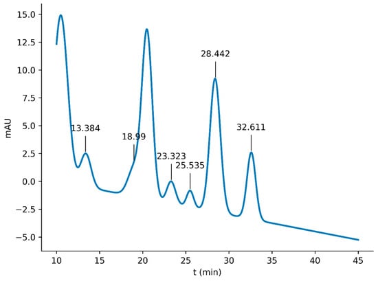
The presence of physalins in the extract obtained from the growing calyx was evaluated by HPLC (Figure 1). The chromatogram revealed characteristic peaks corresponding to specific physalin signals at 20.649 min (physalin A), 25.535 min (physalin D), 28.442 min (physalin H), and 32.611 min (physalin O) [24]. Similar results were obtained by Xu et al. [25], who reported retention times of 6.7 min for luteoside, 21.9 min for luteolin, 27.9 min for Physalin A, 32.2 min for Physalin P, and 33.1 min for Physalin O.
Figure 1.
Physalis ixocarpa extract HPLC-chromatogram.
Table 1 shows the peaks, retention times, areas, and heights obtained by HPLC of the green tomato (Physalis ixocarpa) calyx extract. The first peaks, corresponding to retention times of 9.93 to 23.32 min, represent the most polar or intermediate compounds, which, according to Cao et al. [26] in a study on withanolides in species of the genus Physalis, could be flavonoids or phenolic acids, eluting in the early and intermediate regions of the C18 column. The peaks observed at retention times of 25.53 min to 32.61 min correspond to hydrophobic compounds, which, in previous studies, have been associated with the elution times of physalins from the calyx of Physalis alkekengi, and therefore could correspond to physalins B, D, F, or G identified by Huang et al. [16]. The chemical structure of physalins and other withanolides from Physalis features a highly oxidized steroidal core with various functional modifications, which explains their high hydrophobicity and tendency to elute in the late region of HPLC chromatograms [27], as shown in Figure 1.
Table 1.
Data obtained from HPLC of the accrescent calyx extract of Physalis.
According to the results obtained, peaks 1 and 4 have the largest percentage of area, at 41.99% and 20.60%, respectively; this region is associated with the presence of flavonoids, while the peaks with retention times associated with physalins represent 23.54% of the composition.
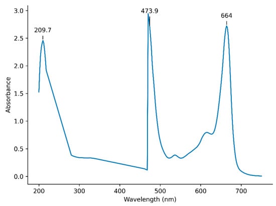
Figure 2 shows the UV-Vis spectrum of the TAC extract, with a maximum absorption at 209.7 nm, characteristic of the α-lactone β-unsaturated system present in withanolides and physalins. Figure 3 presents the FTIR spectrum of the same extract, showing peaks at 3424 cm−1 and 1646 cm−1, which are typical of conjugated hydroxyl and carbonyl groups characteristic of quercetin and kaempferol. The signal observed at 1009 cm−1 is related to C-O stretching of glycosidic bonds, which are present in the structures of physalins and flavonoids [28,29].
Figure 2.
UV-VIS spectrogram of Physalis ixocarpa accrescent calyx extract.
Figure 3.
FTIR spectrogram of Physalis ixocarpa accrescent calyx extract.
The IR spectrum (Figure 3) shows absorption bands at 3424 and 3208 cm−1, corresponding to stretching vibrations of the –OH bonds characteristic of phenolic compounds. The presence of this bond is reinforced by the signal observed in the 3000 cm−1 region. A peak at 1403 cm−1 is also identified, associated with C–H bond bending in alkane groups. Finally, the signal at 1009 cm−1 is attributed to vibrations of the flavonoid backbone, complemented by the band at 1161 cm−1, which represents C–O bond stretching [30,31].
3.1.2. TACE Antimicrobial Activity
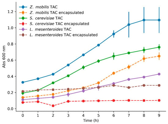
Figure 4 shows a comparison of free and encapsulated TACE antimicrobial effects against Z. mobilis, S. cerevisiae, and L. mesenteroides. The results indicate that Z. mobilis exhibited the greatest resistance, followed by S. cerevisiae, while L. mesenteroides was the most sensitive to antimicrobial treatment. These observations agree with the findings of Helvaci et al. [10], who reported that physalin D exhibits greater antimicrobial activity against Gram-positive than against Gram-negative bacteria. This difference may be attributed to the outer membrane of Gram-negative bacteria, which is composed of lipoproteins, porins, and lipopolysaccharides, thereby conferring greater resistance to antimicrobial agents [15,32].
Figure 4.
Growth kinetics of free (F) and encapsulated (E) TAC extract against Zymomonas mobilis, Saccharomyces cerevisiae, and Leuconostoc mesenteroides.
The encapsulation of TACE improves its antimicrobial activity against all tested microorganisms. Notably, encapsulated TACE exhibited a complete inhibitory effect against S. cerevisiae and L. mesenteroides. This enhancement in antimicrobial efficacy can be attributed to the protective layer, which isolates the TACE from degrading environmental factors such as light and oxygen. This protection enables controlled, prolonged release, thereby extending the TACE inhibitory effect. Furthermore, encapsulation masks potentially undesirable sensory characteristics, allowing for more neutral integration into food. Together, these properties enhance the stability and antimicrobial efficacy of the TACE, optimizing its potential use for food preservation [33].
3.1.3. Encapsulated TACE Minimum Inhibitory Concentration
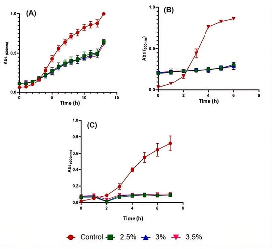
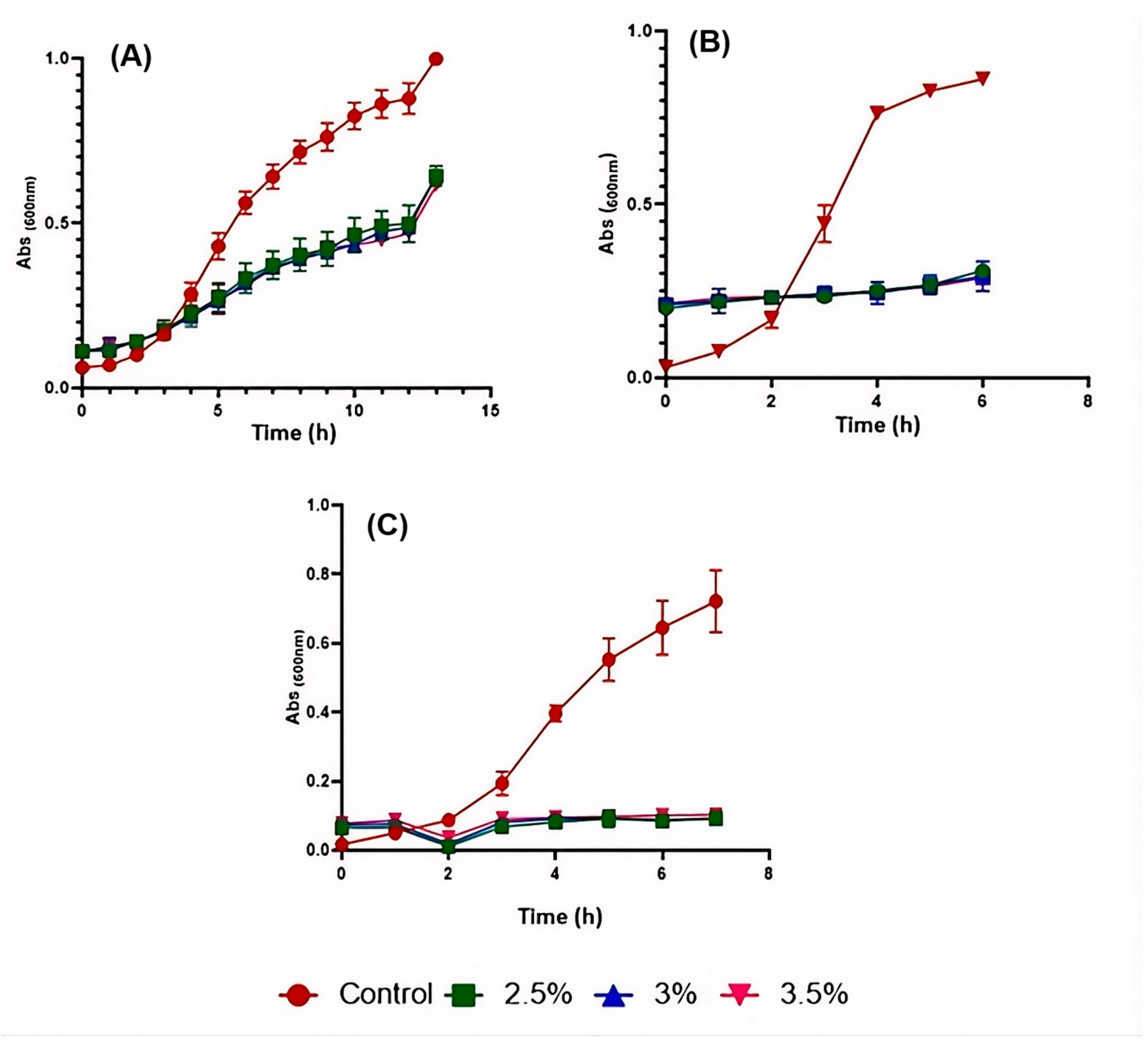
The results of MIC assay for the three tested microorganisms are shown in Figure 5. As can be seen for Z. mobilis (Figure 5A), microbial growth decreased; however, it was not fully inhibited even at the maximum concentration of TACE. In contrast, S. cerevisae (Figure 5B) and L. mesenteroides (Figure 5C) were completely inhibited by the addition of 2.5% (w/v) encapsulated TACE.
Figure 5.
Growth kinetics. (A) Z. mobilis, (B) S. cerevisiae, (C) L. mesenteroides.
The effect of the encapsulated TACE on the kinetic parameters of the tested microorganisms is shown in Table 2. The results show that TACE significantly decreases the specific growth rate of all three microorganisms. L. mesenteroides and S. cerevisiae exhibit the greatest effect of TAC extract, with doubling times 5 and 6 times longer, respectively, compared to the control. This effect on kinetic parameters could be due to the lipophilic, polyoxyfunctional structure of the TACE.
Table 2.
Kinetic growth parameters in the presence of TACE.
The lipophilic nature of the physalins present in the TACE may enable them to integrate into the bacterial membrane, thereby altering membrane structure and affecting bacterial growth. In the case of yeasts, Torabzadeh et al. [34] report that physalins have a limited-spectrum antifungal effect, requiring higher concentrations to inhibit the growth of fungi and yeasts than bacteria. Previous studies report that the antimicrobial activity of physalins depends on their structure and type [35]. Therefore, for future studies, it will be crucial to identify the specific physalin type used and to evaluate its effects on different microorganisms with greater precision.
3.1.4. Pulque Characterization
Pulque characterization was carried out on samples with TAC extract and samples without TAC extract (control) at different storage times (0, 5, 10, and 15 days).
Acidity
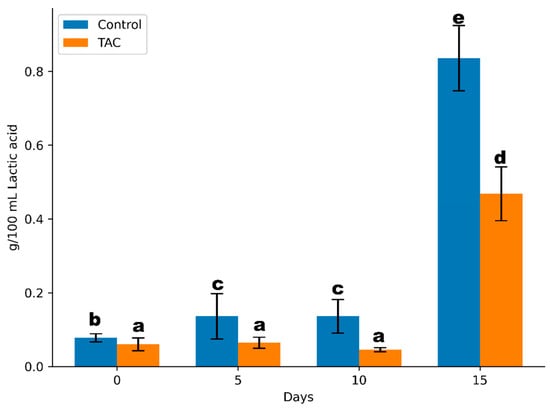
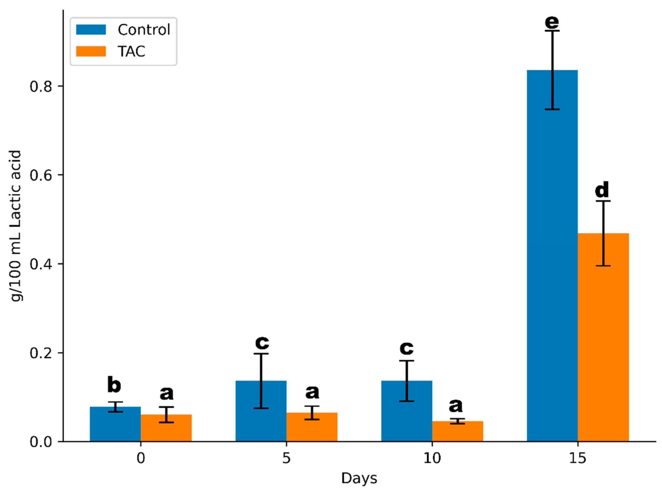
The effect of microencapsulated TAC extract on pulque was evaluated at different storage times, with pulque without TAC extract as a control. Figure 6 shows that the acidity of pulque increases with storage time. The addition of TAC extract significantly decreases the acidity compared to the pulque sample without TAC extract.
Figure 6.
Pulque acidity samples at different storage times. Columns with the same letters do not show a significant difference (p < 0.05).
The increase in pulque acidity during storage is attributable to lactic acid bacteria, which ferment glucose to produce lactic acid [5]. TAC extract reduced the growth of the main microorganisms responsible for pulque fermentation, resulting in lower acidity in samples treated with TAC extract. However, since they do not completely inhibit microbial growth, the treated pulque also becomes acidic.
According to the Regulations for the Sanitary Control of Pulque [36], pulque must have a total acidity (as lactic acid) of 400–700 mg/100 mL to be considered suitable for public consumption. It can be observed that, after 15 days of storage, pulque without TAC extract no longer complies with this regulation, whereas pulque with TAC extract showed an acidity of 450 mg/100 mL and is suitable for public consumption according to this parameter.
Viscosity
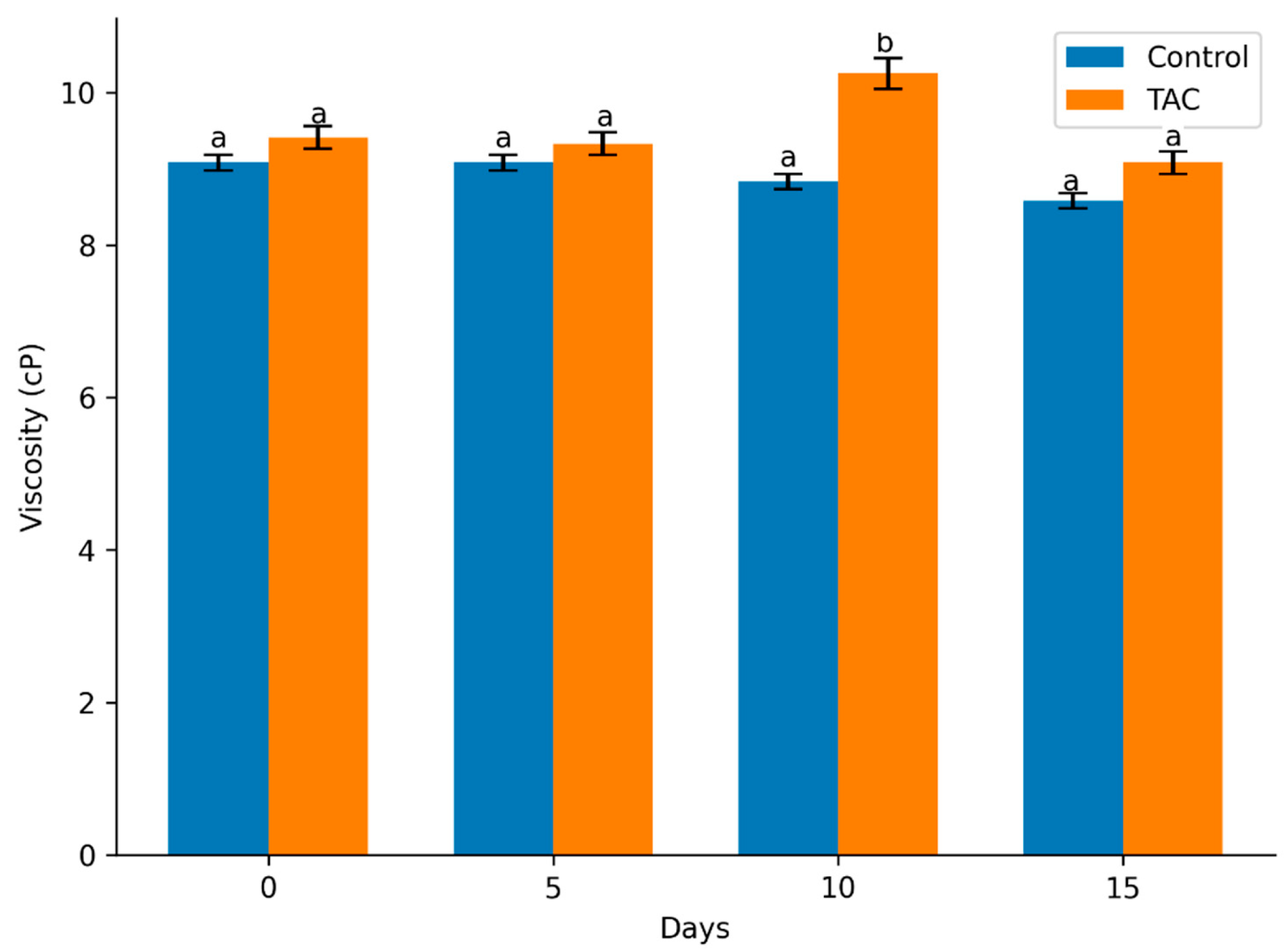
The quality of traditional pulque is defined by specific sensory indicators, including moderate viscosity due to dextran production by L. mesenteroides [37]. Figure 7 presents the viscosity of pulque with and without TACE at different storage times. Given that TACE exhibits antimicrobial activity against L. mesenteroides, TACE-containing samples were initially expected to show lower viscosity than the control. However, the samples treated with the extract maintained higher viscosity. Notably, the fact that TACE does not diminish this property is beneficial, as viscosity is a critical and characteristic sensory attribute of pulque [38]. Although a significant decrease in viscosity was observed during storage relative to day zero, the TACE-treated samples effectively preserved the beverage’s desired texture.
Figure 7.
Viscosity of pulque samples at different storage times. Columns with the same letter show no significant difference (p < 0.05).
The increase in viscosity in samples containing TACE could be attributed to the extract’s structure and its interactions with the microorganisms present. Although TAC extract affects the growth of L. mesenteroides, its impact on Z. mobilis is less pronounced. Z. mobilis is known to contribute to the viscosity of pulque by producing microbial levan, an enzyme that synthesizes levan, a high-molecular-weight fructose polymer that increases viscosity [39]. The presence and activity of Z. mobilis in samples containing TAC extracts may be facilitated by the unique structure of these molecules. TAC extract, with its large, rigid steroid nucleus and derivatizable functional groups, could interact to enhance levan production or modify viscosity. This interaction between the structure of the microencapsulated TAC extract and the microbial components could explain the increased viscosity observed in the samples containing the extract, despite its antimicrobial effect on L. mesenteroides. However, there are no significant differences in the viscosity of pulque with and without TAC [40].
Infrared Spectroscopy
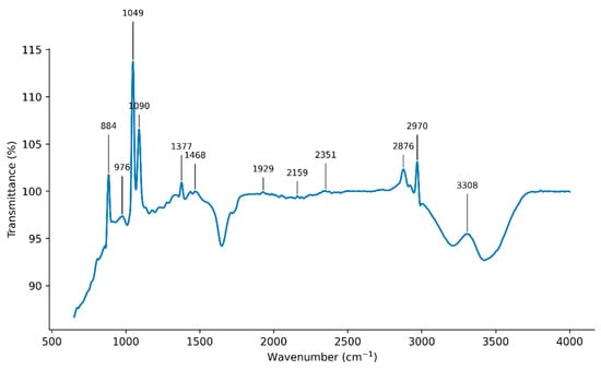
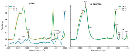
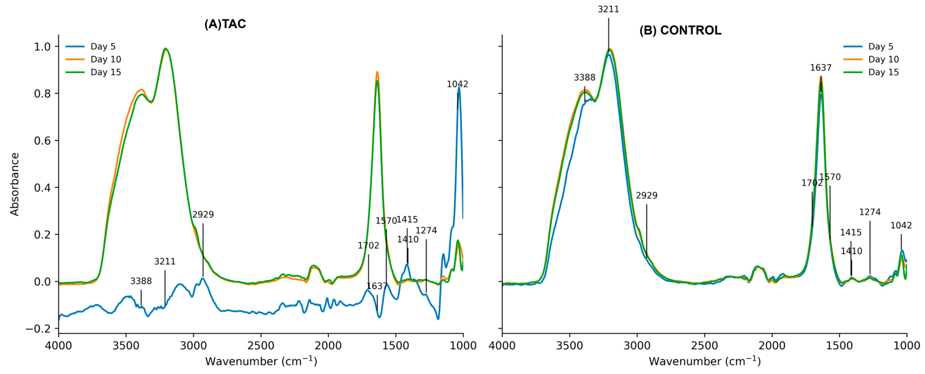
Infrared spectroscopy was used to assess the effect of the TAC extract on the shelf life of pulque (Figure 8). On day 5, the sample treated with TAC extract exhibits a distinct spectrum compared with the other samples. The sample treated with TAC extract at 5 days shows signals in 1702, 1570, and 1042 cm−1. The signal at 1042 cm−1 could be attributed to C-O-C groups, characteristic of sugars. The signals at 1702 cm−1 and 1570 cm−1 are characteristic of carbonyl groups and may indicate the presence of lactic acid. In the remaining samples, a band at 1637 cm−1, characteristic of organic acids, is observed, suggesting the production of lactic acid [41].
Figure 8.
IR spectrogram of pulque samples with and without TAC extract at different storage times.
The spectrograms show signals at 3388.3 cm−1 and 3211 cm−1, characteristic of alcohols and corresponding to O-H stretching vibrations. It is worth noting that in this region, a greater number of hydrogen bonds are observed, indicating more concentrated alcohols. In Figure 6, the peaks of the pulque samples without TAC extract at 5 days have similar intensities to those of samples with longer storage times. Conversely, in the 5-day TAC extract-treated sample, these peaks are less intense, indicating a delay in alcohol production relative to the other samples. Although TAC extract slowed fermentation, by the end of the storage period, the peak intensities were similar in samples treated with TAC extract and in untreated samples [42].
4. Limitations
Pulque is a traditional Mexican beverage produced from the aguamiel of various agave species. The samples used in this study were cultivated in the community of Texactepec, in the municipality of Chilcuautla, Hidalgo. Therefore, the variety and even the region where the pulque is produced must be considered. Another consideration is that the TAC extract was added immediately after scraping the agave. Furthermore, the agave is typically scraped twice daily (approximately 06:00 and 18:00 h); the samples collected were from the morning scraping period.
To identify the TAC compounds responsible for extending the shelf life of pulque, their purification and subsequent characterization are necessary. Once these compounds are identified, their antimicrobial activity should be evaluated against microorganisms typically present in pulque. However, it is important to consider that the microbial diversity of this beverage is broader than that analyzed in this study; therefore, future work should include a wider range of microorganisms. In addition, it would be valuable to investigate changes in the microbial load of pulque throughout its shelf life in the presence of TAC. Although this study focuses on Physalis ixocarpa, the preservative potential of extracts from other plant sources, such as golden berry, bell pepper, and tomato, could also be explored, given their similar compositional profiles. In the present study, the shelf life of TACE-treated pulque was assessed through physicochemical and microbiological parameters. While these indicators provide quantitative evidence of stability, a formal sensory evaluation using a trained panel was not performed. Therefore, further studies incorporating organoleptic analysis are necessary to confirm consumer acceptance.
5. Conclusions
In this study, encapsulated TAC extract demonstrated greater antimicrobial activity than free TAC extract, effectively extending the shelf life of pulque. Based on acidity and alcohol parameters, samples treated with encapsulated TAC extract maintained significant stability for up to 15 days of storage, while the control sample (without TAC extract) showed considerable changes starting on day 5. These results suggest that TAC extract have a relevant effect on microbial growth, viscosity, and the pulque shelf life. The TAC extract demonstrated effective antimicrobial activity, particularly against Leuconostoc mesenteroides, with a lesser effect against Zymomonas mobilis and Saccharomyces cerevisiae.
The reduction in titratable is directly linked to the fermentative microorganism’s inhibition, while their lipophilic structure and ability to interact with other compounds contribute to increased viscosity in treated samples, which could benefit the texture and consistency of pulque during storage. The reduction in acidity and the increase in viscosity promote pulque stability, suggesting that TAC extract could extend its shelf life by enhancing stability and slowing deterioration. The ability of TAC extract to modify microbial activity, increase viscosity, and stabilize the product during storage suggests it could be a potentially valuable additive for the pulque industry. This approach not only optimizes the fermentation process but also significantly improves the product’s durability, facilitating broader commercialization without compromising its traditional sensory identity.
Author Contributions
Conceptualization, M.E.-G., A.A.-R. and J.Á.G.-A.; methodology, M.E.-G., A.A.-R., J.Á.G.-A., A.M.-O., R.E.V.-P. and D.D.-S.; validation, H.M.H.-H., J.C.-G. and L.G.A.-G.; formal analysis, M.E.-G., A.A.-R., J.Á.G.-A. and J.C.-G.; investigation M.E.-G., D.D.-S., J.C.-G., A.M.-O. and J.Á.G.-A.; resources, M.E.-G., A.A.-R. and J.Á.G.-A.; data curation, H.M.H.-H., J.C.-G., A.A.-R. and L.G.A.-G.; writing—original draft preparation, M.E.-G., R.E.V.-P., A.M.-O. and D.D.-S.; writing—review and editing, M.E.-G., J.Á.G.-A. and A.M.-O.; visualization, L.G.A.-G. and H.M.H.-H.; supervision, M.E.-G. and J.Á.G.-A.; project administration, M.E.-G.; funding acquisition, M.E.-G., A.A.-R. and J.Á.G.-A. All authors have read and agreed to the published version of the manuscript.
Funding
The authors would like to thank the “Chemistry is all of us FCQ202121” Fund program of the Autonomous University of Querétaro.
Institutional Review Board Statement
Not applicable.
Informed Consent Statement
Not applicable.
Data Availability Statement
The original contributions presented in this study are included in the article. Further inquiries can be directed to the corresponding authors.
Acknowledgments
The authors wish to express their gratitude to the Autonomous University of Querétaro for providing the facilities and materials necessary to carry out this project. They also thank the communities of Aculco de Espinoza (State of Mexico) and Texcatepec (Chilcuautla, Hidalgo) for providing the tomato calyx and pulque samples, respectively. Special thanks are due to Galdina Olguín Sánchez and Calixto Serrano Pérez for their assistance in scraping the maguey plants from which the samples were taken.
Conflicts of Interest
The authors declare no conflicts of interest.
References
- Rocha-Arriaga, C.; Espinal-Centeno, A.; Martinez-Sánchez, S.; Caballero-Pérez, J.; Alcaraz, L.D.; Cruz-Ramírez, A. Deep Microbial Community Profiling along the Fermentation Process of Pulque, a Biocultural Resource of Mexico. Microbiol. Res. 2020, 241, 126593. [Google Scholar] [CrossRef] [Scilit] [PubMed]
- IntechOpen. Traditional Fermented Beverages in México. Available online: https://www.intechopen.com/chapters/89485 (accessed on 2 April 2026).
- Alcántara-Zavala, A.E.; de Dios Figueroa-Cárdenas, J.; Pérez-Robles, J.F.; Arámbula-Villa, G.; Miranda-Castilleja, D.E. Thermosonication as an Alternative Method for Processing, Extending the Shelf Life, and Conserving the Quality of Pulque: A Non-Dairy Mexican Fermented Beverage. Ultrason. Sonochem. 2021, 70, 105290. [Google Scholar] [CrossRef] [Scilit] [PubMed]
- Alcántara-Zavala, A.E.; de Dios Figueroa-Cárdenas, J.; Morales-Sánchez, E.; Aldrete-Tapia, J.A.; Arvizu-Medrano, S.M.; Martínez-Flores, H.E. Application of Ohmic Heating to Extend Shelf Life and Retain the Physicochemical, Microbiological, and Sensory Properties of Pulque. Food Bioprod. Process. 2019, 118, 139–148. [Google Scholar] [CrossRef] [Scilit]
- Álvarez-Ríos, G.D.; Figueredo-Urbina, C.J.; Casas, A. Physical, Chemical, and Microbiological Characteristics of Pulque: Management of a Fermented Beverage in Michoacán, Mexico. Foods 2020, 9, 361. [Google Scholar] [CrossRef] [Scilit]
- Cruz-Noriega, M.D.L.; Rojas-Flores, S.; Benites, S.M.; Álvarez, M.A.Q.; García, N.M.O.; Yupanqui, M.R. Use of Leuconostoc mesenteroides to Produce a Dextran Bioflocculant. Environ. Res. Eng. Manag. 2022, 78, 38–45. [Google Scholar] [CrossRef] [Scilit]
- Xia, J.; Yang, Y.; Liu, C.-G.; Yang, S.; Bai, F.-W. Engineering Zymomonas mobilis for Robust Cellulosic Ethanol Production. Trends Biotechnol. 2019, 37, 960–972. [Google Scholar] [CrossRef] [Scilit]
- Boismier, E.C.; Aboulnaga, E.A.; TerAvest, M.A. Zymomonas mobilis: Bringing an Ancient Human Tool into the Genomic Era. Curr. Opin. Biotechnol. 2025, 92, 103257. [Google Scholar] [CrossRef] [Scilit]
- González-Pérez, J.E.; Guerrero-Beltrán, J.Á. Tomatillo or Husk Tomato (Physalis philadelphica and Physalis ixocarpa): A Review. Sci. Hortic. 2021, 288, 110306. [Google Scholar] [CrossRef] [Scilit]
- Helvacı, S.; Kökdil, G.; Kawai, M.; Duran, N.; Duran, G.; Güvenç, A. Antimicrobial Activity of the Extracts and Physalin D from Physalis alkekengi and Evaluation of Antioxidant Potential of Physalin D. Pharm. Biol. 2010, 48, 142–150. [Google Scholar] [CrossRef] [Scilit]
- Meira, C.S.; Soares, J.W.C.; dos Reis, B.P.Z.C.; Pacheco, L.V.; Santos, I.P.; Silva, D.K.C.; de Lacerda, J.C.; Daltro, S.R.T.; Guimarães, E.T.; Soares, M.B.P. Therapeutic Applications of Physalins: Powerful Natural Weapons. Front. Pharmacol. 2022, 13, 864714. [Google Scholar] [CrossRef] [Scilit]
- Riley, S.; Lale, A.; Nguyen, V.; Xi, H.; Wilkinson, K.; Searle, I.R.; Fisk, I. Volatile Profiles of Commercial Vetch Prepared via Different Processing Methods. Food Chem. 2022, 395, 133569. [Google Scholar] [CrossRef] [Scilit] [PubMed]
- Zabot, G.L.; Schaefer Rodrigues, F.; Polano Ody, L.; Vinícius Tres, M.; Herrera, E.; Palacin, H.; Córdova-Ramos, J.S.; Best, I.; Olivera-Montenegro, L. Encapsulation of Bioactive Compounds for Food and Agricultural Applications. Polymers 2022, 14, 4194. [Google Scholar] [CrossRef] [Scilit] [PubMed]
- Gharsallaoui, A.; Roudaut, G.; Chambin, O.; Voilley, A.; Saurel, R. Applications of Spray-Drying in Microencapsulation of Food Ingredients: An Overview. Food Res. Int. 2007, 40, 1107–1121. [Google Scholar] [CrossRef] [Scilit]
- Eghbal, N.; Liao, W.; Dumas, E.; Azabou, S.; Dantigny, P.; Gharsallaoui, A. Microencapsulation of Natural Food Antimicrobials: Methods and Applications. Appl. Sci. 2022, 12, 3837. [Google Scholar] [CrossRef] [Scilit]
- Huang, C.; Xu, Q.; Chen, C.; Song, C.; Xu, Y.; Xiang, Y.; Feng, Y.; Ouyang, H.; Zhang, Y.; Jiang, H. The Rapid Discovery and Identification of Physalins in the Calyx of Physalis alkekengi L. Var. Franchetii (Mast.) Makino Using Ultra-High Performance Liquid Chromatography–Quadrupole Time of Flight Tandem Mass Spectrometry Together with a Novel Three-Step Data Mining Strategy. J. Chromatogr. A 2014, 1361, 139–152. [Google Scholar] [CrossRef] [Scilit]
- Nguyen, K.-N.H.; Tran, L.H.; Nguyen, N.-V.T.; Duong, N.T.; Dai, X.-T.T.; Le, C.-T.T.; Nguyen, K.T. New QuEChERS Method for Quantification of Physalin B and D in Physalis angulata L. in Vietnam. Pharmacia 2022, 69, 883–890. [Google Scholar] [CrossRef] [Scilit]
- Dai, J.; Mumper, R.J. Plant Phenolics: Extraction, Analysis and Their Antioxidant and Anticancer Properties. Molecules 2010, 15, 7313–7352. [Google Scholar] [CrossRef] [Scilit]
- Zeinalipour-Yazdi, C.D.; Loizidou, E.Z. An Experimental FTIR-ATR and Computational Study of H-Bonding in Ethanol/Water Mixtures. Chem. Phys. 2021, 550, 111295. [Google Scholar] [CrossRef] [Scilit]
- Bernal, C.A.; Ramos, F.A.; Baena, Y. Dry powder formulation from Physalis peruviana L. fruits extract with antidiabetic activity formulated via co-spray drying. Int. J. Appl. Pharm. 2019, 11, 109–117. [Google Scholar] [CrossRef] [Scilit]
- Abdollahzadeh, E.; Nematollahi, A.; Hosseini, H. Composition of Antimicrobial Edible Films and Methods for Assessing Their Antimicrobial Activity: A Review. Trends Food Sci. Technol. 2021, 110, 291–303. [Google Scholar] [CrossRef] [Scilit]
- Saucedo-Gutierrez, J.J.; Escamilla-García, M.; Amaro-Reyes, A.; Carrillo-Garmendia, A.; Madrigal-Pérez, L.A.; Regalado-González, C.; Granados-Arvizu, J.Á. Differential Impacts of Furfural and Acetic Acid on the Bioenergetics and Fermentation Performance of Scheffersomyces stipitis. Fungal Genet. Biol. 2024, 174, 103914. [Google Scholar] [CrossRef] [Scilit]
- Olivares-Marin, I.K.; Madrigal-Perez, L.A.; Canizal-Garcia, M.; García-Almendárez, B.E.; González-Hernández, J.C.; Regalado-Gonzalez, C. Interactions between Carbon and Nitrogen Sources Depend on RIM15 and Determine Fermentative or Respiratory Growth in Saccharomyces Cerevisiae. Appl. Microbiol. Biotechnol. 2018, 102, 4535–4548. [Google Scholar] [CrossRef] [Scilit] [PubMed]
- Zheng, Y.; Luan, L.; Chen, Y.; Ren, Y.; Wu, Y. Characterization of Physalins and Fingerprint Analysis for the Quality Evaluation of Physalis alkekengi L. Var. Franchetii by Ultra-Performance Liquid Chromatography Combined with Diode Array Detection and Electrospray Ionization Tandem Mass Spectrometry. J. Pharm. Biomed. Anal. 2012, 71, 54–62. [Google Scholar] [CrossRef] [Scilit] [PubMed]
- Xu, B. High-Performance Liquid Chromatography (HPLC) Determination of Five Active Ingredients in the Calyces of Physalis alkekengi L. Var. Franchetii (Mast.) Mskino. J. Med. Plants Res. 2012, 6, 2438–2442. [Google Scholar] [CrossRef] [Scilit]
- Cao, C.-M.; Kindscher, K.; Gallagher, R.J.; Zhang, H.; Timmermann, B.N. Analysis of Major Withanolides in Physalis longifolia Nutt. by HPLC-PDA. J. Chromatogr. Sci. 2015, 53, 1044–1047. [Google Scholar] [CrossRef] [Scilit]
- Huang, M.; He, J.-X.; Hu, H.-X.; Zhang, K.; Wang, X.-N.; Zhao, B.-B.; Lou, H.-X.; Ren, D.-M.; Shen, T. Withanolides from the Genus Physalis: A Review on Their Phytochemical and Pharmacological Aspects. J. Pharm. Pharmacol. 2020, 72, 649–669. [Google Scholar] [CrossRef] [Scilit]
- Li, X.; Zhao, J.; Yang, M.; Liu, Y.; Li, Z.; Li, R.; Li, X.; Li, N.; Xu, Q.; Khan, I.A.; et al. Physalins and Withanolides from the Fruits of Physalis alkekengi L. Var. Franchetii (Mast.) Makino and the Inhibitory Activities against Human Tumor Cells. Phytochem. Lett. 2014, 10, 95–100. [Google Scholar] [CrossRef] [Scilit]
- Cobaleda-Velasco, M.; Almaraz-Abarca, N.; Alanis-Bañuelos, R.E.; Uribe-Soto, J.N.; González-Valdez, L.S.; Muñoz-Hernández, G.; Zaca-Morán, O.; Rojas-López, M. Rapid Determination of Phenolics, Flavonoids, and Antioxidant Properties of Physalis ixocarpa Brot. Ex Hornem. and Physalis angulata L. by Infrared Spectroscopy and Partial Least Squares. Anal. Lett. 2018, 51, 523–536. [Google Scholar] [CrossRef] [Scilit]
- Isaq, M.; Somu, P.; Acharya, D.; Gomez, L.A.; Thathapudi, J.J.; Ramachandra, Y.L.; Rudraiah, S.B.; Ravi, P.; Rai, P.S.; Rosalin, R.; et al. Phytochemical Screening and Bioactivity Studies of Endophytes Cladosporium sp. Isolated from the Endangered Plant Vateria indica Using In Silico and In Vitro Analysis. Appl. Biochem. Biotechnol. 2022, 194, 4546–4569. [Google Scholar] [CrossRef] [Scilit]
- Kashyap, S.; Agarwal, G.; Kumar, M.; Kant Awasthi, K. Extraction of Bioactive Compounds from Withania somnifera Using Hydrothermal Technique. Mat. Today Proc. 2022, 69, 1596–1599. [Google Scholar] [CrossRef] [Scilit]
- MacDermott-Opeskin, H.I.; Gupta, V.; O’Mara, M.L. Lipid-Mediated Antimicrobial Resistance: A Phantom Menace or a New Hope? Biophys. Rev. 2022, 14, 145–162. [Google Scholar] [CrossRef] [Scilit]
- Januário, A.H.; Filho, E.R.; Pietro, R.C.L.R.; Kashima, S.; Sato, D.N.; França, S.C. Antimycobacterial Physalins from Physalis angulata L. (Solanaceae). Phytother. Res. 2002, 16, 445–448. [Google Scholar] [CrossRef] [Scilit]
- Torabzadeh, P.; Panahi, P. Evaluation of Antifungal Activity of Physalis alkekengi L. Extracts on Microsporum canis, Candida albicans, Trichophyton mentagrophytes and Nocardia asteroids. Middle East J. Sci. Res. 2013, 13, 926–929. [Google Scholar] [CrossRef]
- Wu, J.; Zhao, J.; Zhang, T.; Gu, Y.; Khan, I.A.; Zou, Z.; Xu, Q. Naturally Occurring Physalins from the Genus Physalis: A Review. Phytochem. 2021, 191, 112925. [Google Scholar] [CrossRef] [Scilit]
- Legislación Sanitaria. Regulations for the Sanitary Control of Pulque; Salud Publica de Mexico: Cuernavaca, Mexico, 1981; Volume 23, pp. 515–524. [Google Scholar]
- Bautista-Moreno, F.A.; Giles-Gómez, M.; Silva-Chávez, M.A.; Severiano-Pérez, P.; Rodríguez-Alegría, M.E.; Hernández-Chávez, G.; Bolívar, F.; Escalante, A. Design of a Minimal Starter Culture for Controlled Fermentation of Maguey Sap: Microbial and Sensory Comparison with Traditional Pulque. Food Biosci. 2025, 74, 107880. [Google Scholar] [CrossRef] [Scilit]
- Escalante, A.; López Soto, D.R.; Velázquez Gutiérrez, J.E.; Giles-Gómez, M.; Bolívar, F.; López-Munguía, A. Pulque, a Traditional Mexican Alcoholic Fermented Beverage: Historical, Microbiological, and Technical Aspects. Front. Microbiol. 2016, 7, 1026. [Google Scholar] [CrossRef] [Scilit]
- Escalante, A.; Gomez, M.; Flores, G.; Matus, V.; Moreno-Terrazas, R.; Lopez Munguia, A.; Lappe-Oliveras, P. Pulque Fermentation. In Handbook of Plant-Based Fermented Food Beverage Technology, 2nd ed.; CRC Press: Boca Raton, FL, USA, 2012; pp. 691–706. [Google Scholar]
- Svobodová, H.; Noponen, V.; Kolehmainen, E.; Sievänen, E. Recent Advances in Steroidal Supramolecular Gels. RSC Adv. 2012, 2, 4985–5007. [Google Scholar] [CrossRef] [Scilit]
- Silverstein, R.M.; Webster, F.X.; Kiemle, D.J.; Bryce, D.L. Spectrometric Identification of Organic Compounds, 8th ed.; John Wiley and Sons: Hoboken, NJ, USA, 2014. [Google Scholar]
- Peña, P.P.; Bretado, M.Á.E.; Lozano, M.A.G.; Castro, R.H.L.; Ramírez, D.M.N. Espectroscopía de Infrarrojo; Universidad Juárez del Estado de Durango: Durango, Mexico, 2023. [Google Scholar]

Disclaimer/Publisher’s Note: The statements, opinions and data contained in all publications are solely those of the individual author(s) and contributor(s) and not of MDPI and/or the editor(s). MDPI and/or the editor(s) disclaim responsibility for any injury to people or property resulting from any ideas, methods, instructions or products referred to in the content. |
© 2026 by the authors. Licensee MDPI, Basel, Switzerland. This article is an open access article distributed under the terms and conditions of the Creative Commons Attribution (CC BY) license.