


Abstract
Alzheimer’s disease (AD) is a progressive neurodegenerative disorder with a long preclinical phase, making early biomarker discovery essential for timely diagnosis, risk stratification, and intervention. Hearing loss has increasingly been recognized as a clinically important and potentially modifiable factor associated with cognitive decline and dementia risk. Beyond peripheral auditory impairment, accumulating evidence suggests that central auditory dysfunction, altered auditory processing, and electrophysiological abnormalities may emerge during the course of AD and related cognitive disorders. Auditory electrophysiological signals, including electroencephalography (EEG), auditory event-related potentials (ERP), auditory evoked cortical potentials (AECP), auditory brainstem response (ABR) recordings, cochlear microphonics (CM), and distortion product otoacoustic emission (DPOAE) measures, provide objective and repeatable measurements of peripheral and central auditory pathway function. However, conventional analysis based on peak latency, amplitude, spectral power, and manually engineered features may not fully capture subtle disease-related alterations distributed across time, frequency, and waveform morphology. Artificial intelligence (AI) offers new opportunities to extract high-dimensional and physiologically informative representations from auditory and neural signals. Machine learning (ML), deep learning (DL), self-supervised learning (SSL), explainable artificial intelligence (XAI), and multimodal modeling can support data-driven biomarker discovery, especially in settings where labeled datasets are limited and electrophysiological changes are subtle. This critical narrative review synthesizes current knowledge at the intersection of hearing loss, AD, auditory electrophysiology, and AI. We discuss traditional signal processing, supervised ML, deep neural networks, self-supervised representation learning, explainable models, and multimodal integration strategies. We also identify major methodological limitations, including small sample sizes, heterogeneous acquisition protocols, insufficient external validation, limited interpretability, data leakage risks, and challenges in translating animal-model findings to human disease. Finally, we propose future directions for trustworthy, interpretable, and multimodal AI systems capable of advancing auditory electrophysiological biomarkers for AD and hearing-related neurodegeneration.
1. Introduction
Alzheimer’s disease (AD) is a major neurodegenerative disorder characterized by progressive cognitive decline, synaptic dysfunction, neural network disruption, and eventual loss of independence. Although amyloid beta and tau pathology remain central biological features of the disease, there is increasing recognition that AD affects multiple functional systems long before advanced dementia becomes clinically evident. This has intensified interest in early, non-invasive, scalable, and physiologically meaningful biomarkers that can detect subtle neural alterations before irreversible cognitive deterioration occurs. Artificial intelligence (AI) has become increasingly important in this context because it can identify complex patterns from high-dimensional biomedical data, including neuroimaging, electrophysiology, clinical variables, sensor measurements, and multimodal biomarker profiles [1,2].
Hearing loss has emerged as one of the most important modifiable factors associated with dementia risk. Large epidemiological studies have reported an association between hearing impairment and incident dementia, and the 2024 Lancet Commission on dementia prevention, intervention, and care emphasized hearing loss as a major potentially modifiable risk factor for dementia [3,4]. The relationship between hearing loss and AD is likely multifactorial. Proposed mechanisms include reduced auditory input, increased cognitive load, social isolation, changes in brain structure and function, shared vascular or neurodegenerative pathways, and altered central auditory processing. Recent clinical evidence also suggests that hearing intervention may provide cognitive benefit in older adults at increased risk for cognitive decline, although effects may vary across populations and baseline risk profiles [5,6]. These findings support the need to study the auditory system not only as a sensory pathway but also as a window into brain aging and neurodegenerative vulnerability.
Auditory dysfunction in AD is not limited to peripheral hearing loss. Central auditory processing deficits, altered sensory gating, impaired temporal processing, and abnormal auditory event-related responses have been reported in individuals with AD, mild cognitive impairment (MCI), and related at-risk conditions. Auditory evoked potentials and event-related potentials (ERP), including components such as P50, N100, P200, mismatch negativity, and P300, have been investigated as candidate neurophysiological biomarkers of cognitive impairment and disease progression [7,8]. These signals are attractive because they are objective, repeatable, relatively inexpensive, and capable of capturing dynamic neural responses to sensory and cognitive events. In animal models, auditory brainstem responses (ABR), auditory evoked cortical potentials (AECP), cochlear microphonics (CM), and distortion product otoacoustic emissions (DPOAE) can further provide mechanistic insight into peripheral and central auditory pathway dysfunction.
Despite this promise, auditory electrophysiological biomarkers remain underused in AD research compared with structural magnetic resonance imaging (MRI), positron emission tomography (PET), cerebrospinal fluid markers, and emerging blood-based biomarkers. One reason is that electrophysiological signals are complex, noisy, time-varying, and sensitive to acquisition protocols, preprocessing decisions, stimulus parameters, electrode placement, anesthesia conditions in animal studies, and participant state in human studies. Traditional analysis often focuses on manually selected features, such as peak amplitude, peak latency, interpeak intervals, spectral power, or time–frequency summaries. Although these features are physiologically interpretable, they may fail to capture subtle multiscale waveform alterations associated with early neurodegeneration.
AI provides a complementary approach for extracting latent structure from auditory and neural signals. Classical machine learning (ML) methods, including support vector machines (SVM), random forests, gradient boosting models, and logistic regression, can classify disease states using engineered electrophysiological features. Deep learning (DL) methods, including convolutional neural networks (CNN), recurrent neural networks (RNN), autoencoders, and transformer-based architectures, can learn hierarchical representations directly from raw or minimally processed signals. More recently, self-supervised learning (SSL) has become especially relevant for biomedical electrophysiology because large volumes of unlabeled signal data may be available, whereas expert labels are often limited, expensive, or inconsistent. Methods such as contrastive learning, redundancy-reduction learning, masked signal modeling, and temporal representation learning may enable models to learn useful signal embeddings without requiring dense clinical labels.
However, the application of AI to auditory electrophysiological biomarkers also raises important methodological concerns. Many studies in biomedical AI rely on small datasets, single-center cohorts, incomplete external validation, and inconsistent preprocessing pipelines. In AD classification, DL studies may report high accuracy but remain vulnerable to data leakage, subject-level split errors, overfitting, and limited generalizability [9]. Interpretability is another major concern. A model that accurately classifies AD or hearing-loss-related risk is insufficient unless its predictions can be linked to meaningful physiological features, such as waveform latency shifts, altered oscillatory dynamics, impaired auditory encoding, or structural changes in auditory pathways. Explainable artificial intelligence (XAI) methods, including feature importance analysis, saliency mapping, SHAP-based interpretation, and Integrated Gradients, can help connect model decisions to biologically plausible signal characteristics, but these methods require careful validation.
This topic is inherently interdisciplinary and requires integration of several scientific perspectives. From a medical and otolaryngology perspective, hearing loss, central auditory dysfunction, and AD-related cognitive decline are clinically important conditions that require objective and translatable biomarkers. From a physiology and pathophysiology perspective, auditory brainstem, cochlear, cortical, and cognitive responses provide measurable information about neural timing, sensory encoding, cochlear integrity, and network dysfunction. From a computer science and information-technology perspective, ML, DL, SSL, XAI, and multimodal modeling provide computational tools for analyzing high-dimensional electrophysiological signals. From a mathematical and quantitative perspective, signal processing, feature extraction, model validation, uncertainty estimation, and reproducibility are essential for reliable biomarker development. From a chemistry and molecular-biology perspective, AD-related pathology, oxidative stress, inflammation, synaptic dysfunction, and cochlear or neural stress provide mechanistic context for interpreting auditory electrophysiological changes. Finally, from a broader acoustic and biological-signal perspective, auditory electrophysiology can be viewed as a complex time-series signal shaped by stimulus properties, biological variability, environmental noise, and recording conditions. Integrating these perspectives strengthens the reasoning for a critical review focused on AI-based auditory electrophysiological biomarkers.
Building on this interdisciplinary framework, this critical narrative review examines the emerging intersection of AI, auditory electrophysiology, hearing loss, and AD biomarker discovery. The review has four main goals. First, it summarizes the biological and clinical connections between hearing loss, auditory dysfunction, and AD. Second, it discusses auditory electrophysiological signals as candidate biomarkers across peripheral and central auditory pathways. Third, it critically evaluates how traditional signal processing, ML, DL, SSL, and XAI can be applied to auditory and neural signals. Fourth, it identifies current limitations and proposes future directions for developing trustworthy, interpretable, and multimodal AI frameworks for auditory biomarker discovery in AD and hearing-related neurodegeneration.
2. Literature Selection Strategy for Critical Narrative Review
This work was designed as a critical narrative review rather than a systematic review, scoping review, or meta-analysis. The purpose was not to exhaustively identify every available publication or quantitatively pool effect sizes, but to critically synthesize literature relevant to the intersection of AI, auditory electrophysiology, hearing loss, AD, and biomarker discovery. This approach is appropriate for an interdisciplinary and emerging topic in which evidence is distributed across multiple fields, including neurodegeneration, audiology, electrophysiology, biomedical signal processing, ML, DL, and XAI.
To improve transparency, relevant literature was identified through targeted searches of major biomedical, engineering, and interdisciplinary databases, including PubMed/MEDLINE, Web of Science, Scopus, Google Scholar, and IEEE Xplore. Searches were performed using combinations of terms related to AD, dementia, MCI, hearing loss, auditory dysfunction, auditory evoked potentials, ABR, AECP, ERP, electroencephalography (EEG), CM, DPOAE, auditory steady-state response (ASSR), ML, DL, SSL, XAI, multimodal AI, and biomarker discovery. The literature search was updated during manuscript revision to include recent and methodologically relevant studies available up to the final revision stage.
The literature selection was guided by conceptual relevance, scientific quality, methodological importance, and contribution to the development of future AI-based auditory biomarker frameworks. Priority was given to peer-reviewed articles, systematic reviews, meta-analyses, clinical trials, methodological papers, and highly relevant experimental studies. The reviewed literature included studies on hearing loss and dementia risk, auditory dysfunction in AD, auditory evoked potentials and related electrophysiological biomarkers, ML and DL for neurodegenerative disease classification, SSL for biomedical signals, XAI, and multimodal biomarker integration.
Relevant literature was identified using combinations of keywords related to four major domains. The first domain included AD, dementia, MCI, cognitive decline, neurodegeneration, and biomarker discovery. The second domain included hearing loss, auditory dysfunction, central auditory processing, auditory evoked potentials, auditory ERP, ABR, AECP, EEG, CM, DPOAE, and ASSR. The third domain included AI, ML, DL, CNN, RNN, transformers, autoencoders, SSL, contrastive learning, representation learning, and foundation models. The fourth domain included XAI, interpretability, SHAP, saliency mapping, Integrated Gradients, multimodal learning, data integration, and translational biomarker development.
Studies were considered for inclusion when they were directly relevant to one or more of the following themes: the association between hearing loss and cognitive decline or dementia; auditory dysfunction in AD or MCI; auditory electrophysiological measures as candidate biomarkers; signal-processing methods for auditory or neural electrophysiology; ML, DL, SSL, or XAI applied to biomedical signals; and multimodal biomarker discovery in neurodegenerative disease. Priority was given to peer-reviewed original studies, systematic reviews, meta-analyses, clinical trials, preclinical studies, methodological papers, and highly relevant review articles. Studies were excluded or de-emphasized when they were not related to AD, cognitive decline, hearing loss, auditory electrophysiology, neurodegeneration, or biomedical AI; when they focused only on unrelated engineering applications; when they lacked sufficient methodological relevance; or when they provided only general commentary without substantive scientific or methodological contribution.
Study selection was performed in a staged manner. First, titles and abstracts were screened for relevance to the review scope. Second, full texts of potentially relevant articles were examined to determine their contribution to the biological, electrophysiological, computational, or translational themes of the review. Third, selected studies were organized according to their main role in the manuscript, including background evidence, auditory biomarker evidence, signal-processing methods, AI methodology, interpretability, multimodal integration, methodological limitations, and future directions. Because the purpose of this work was critical synthesis rather than exhaustive evidence mapping, representative studies were selected to illustrate major concepts, methodological strengths, limitations, and emerging opportunities.
Because this review is narrative and critical in design, and because its purpose was conceptual and methodological synthesis rather than exhaustive evidence mapping, formal PRISMA-based screening, duplicate independent screening, risk-of-bias scoring, and quantitative evidence synthesis were not performed. Nevertheless, representative quantitative details, including sample size and reported model-performance information, were extracted for selected AI studies when available and summarized in Table 2 to improve transparency. PRISMA 2020 is primarily intended for systematic reviews, while PRISMA-ScR was developed for scoping reviews; therefore, these reporting frameworks were not applied as formal methodological requirements in the present work [10,11]. Nevertheless, the review was structured to maintain transparency by clearly defining the conceptual scope, major literature domains, and rationale for including representative studies. This strategy is also consistent with quality principles for narrative reviews, including justification of the article’s importance, clear aims, appropriate referencing, scientific reasoning, and balanced presentation of evidence [12].
The included literature was interpreted with emphasis on methodological strengths and limitations rather than simple description. Particular attention was given to whether studies used appropriate validation strategies, avoided subject-level data leakage, addressed small sample size and class imbalance, reported clinically or physiologically meaningful outcomes, and provided interpretable model outputs. Studies reporting high classification performance without external validation, clear preprocessing descriptions, or physiological interpretation were considered cautiously. Conversely, studies that combined rigorous experimental design, transparent computational methods, interpretable features, and clinically relevant conclusions were emphasized as stronger evidence.
This literature selection strategy supports the central aim of the review: to develop a critical and forward-looking synthesis of how AI can advance auditory electrophysiological biomarkers for AD and hearing-loss-related neurodegeneration. Rather than treating AI as a purely predictive tool, the review emphasizes its potential role in discovering physiologically meaningful signal representations, improving early biomarker sensitivity, supporting multimodal integration, and guiding translational research across animal models and human clinical populations.
3. Hearing Loss and Alzheimer’s Disease: Biological and Clinical Connections
Hearing loss is increasingly recognized as an important factor in brain aging, cognitive decline, and dementia risk. Traditionally, hearing impairment was viewed mainly as a sensory disorder involving reduced sound detection or impaired communication. However, accumulating epidemiological, clinical, neurophysiological, and mechanistic evidence indicates that hearing loss may also be closely connected with cognitive function, neural network integrity, and neurodegenerative vulnerability [13,14,15,16]. This connection is especially relevant for AD because auditory dysfunction may occur before severe cognitive impairment becomes clinically obvious, making the auditory system a potentially valuable window for early biomarker discovery.
3.1. Human Clinical and Epidemiological Evidence
Large population-based studies have shown that hearing impairment is associated with increased risk of incident dementia and cognitive decline. One landmark longitudinal study reported that hearing loss was independently associated with incident dementia, including AD, even after adjustment for several demographic and clinical factors [3]. A later systematic review and meta-analysis further supported the association of age-related hearing loss with cognitive decline, cognitive impairment, and dementia [13]. The 2024 Lancet Commission on dementia prevention, intervention, and care also emphasized hearing loss as one of the most important potentially modifiable risk factors for dementia [4]. This does not mean that hearing loss directly causes AD in every individual. Rather, it suggests that hearing impairment may contribute to cognitive vulnerability, interact with existing brain pathology, or serve as an early marker of broader neural dysfunction. The major conceptual pathways linking hearing loss, central auditory dysfunction, cognitive load, social isolation, shared biological mechanisms, and AD-related neurodegeneration are summarized in Figure 1. Figure 1 illustrates that hearing loss and AD-related vulnerability may be connected through several interacting pathways, including degraded auditory input, increased cognitive load, reduced cognitive reserve, sensory deprivation, cortical reorganization, central auditory dysfunction, communication difficulty, social isolation, reduced cognitive engagement, and shared aging-related biological mechanisms such as inflammation, vascular injury, neural stress, and cochlear stress. These pathways should be interpreted as complementary and interacting mechanisms rather than as a single linear causal pathway.
Figure 1.
Conceptual link between hearing loss, auditory dysfunction, and Alzheimer’s disease. The schematic summarizes interacting biological and clinical pathways that may connect hearing loss, central auditory dysfunction, cognitive decline, and Alzheimer’s disease-related vulnerability. Abbreviations: ABR, auditory brainstem response; AECP, auditory evoked cortical potential; ASSR, auditory steady-state response; CM, cochlear microphonic; DPOAE, distortion product otoacoustic emission; EEG, electroencephalography; ERP, event-related potential.
Several mechanisms have been proposed to explain the relationship between hearing loss and AD. One major explanation is the cognitive load hypothesis. When auditory input is degraded, the brain must allocate greater cognitive resources to decode speech and environmental sounds. Over time, this increased listening effort may reduce the resources available for memory, attention, and executive function. In older adults, this added cognitive burden may be especially important because neural reserve and compensatory capacity are already reduced. Therefore, even moderate hearing loss may increase the functional strain on cognitive systems that are vulnerable in AD [14].
A second explanation is the sensory deprivation hypothesis. Reduced auditory stimulation may lead to long-term changes in auditory cortex, association cortex, and broader neural networks involved in perception, attention, and memory. Persistent reduction in sensory input can contribute to cortical reorganization, decreased neural engagement, and altered connectivity between auditory and cognitive networks. In this framework, hearing loss is not only a peripheral problem but also a driver of central nervous system adaptation. These central changes may interact with AD pathology and accelerate cognitive decline in susceptible individuals [14,16].
A third explanation involves social isolation and communication difficulty. Hearing impairment can reduce participation in conversation, social activities, and cognitively stimulating environments. Social isolation and reduced cognitive engagement are themselves associated with dementia risk. Therefore, hearing loss may indirectly contribute to cognitive decline by reducing social interaction, increasing loneliness, and limiting environmental stimulation. This pathway is clinically important because it suggests that hearing rehabilitation, communication support, and environmental interventions may help reduce some downstream effects of hearing impairment [15,16].
A fourth possibility is that hearing loss and AD share common biological or pathological mechanisms. These may include vascular injury, inflammation, oxidative stress, mitochondrial dysfunction, synaptic degeneration, altered neural excitability, or age-related structural changes in the brain. In this view, hearing loss may not simply cause cognitive decline but may reflect broader biological aging processes that also increase AD vulnerability. This is important for biomarker research because auditory dysfunction may provide measurable evidence of systemic or neural changes occurring before advanced dementia [14,16].
Central auditory processing is particularly relevant to AD. Peripheral hearing loss refers mainly to impairment in the ear or auditory nerve that reduces the detection or transmission of sound. In contrast, central auditory dysfunction refers to impaired processing of auditory information within the brainstem, thalamus, auditory cortex, and higher-order cortical networks. Patients may have difficulty understanding speech in noise, detecting temporal changes, processing complex sounds, or integrating auditory information with attention and memory. These central auditory deficits may be closely related to cognitive impairment and may occur even when peripheral hearing thresholds do not fully explain the listening difficulties [17].
AD affects distributed neural networks, including regions involved in memory, attention, sensory integration, and cortical processing. Although the hippocampus and medial temporal lobe are strongly emphasized in AD, sensory and association cortices can also show functional and structural alterations. Auditory processing requires coordinated activity across peripheral receptors, brainstem nuclei, thalamocortical pathways, primary auditory cortex, association cortex, frontal attention networks, and memory-related systems. Because of this distributed organization, auditory electrophysiological responses may be sensitive to both peripheral auditory dysfunction and central neurodegenerative changes [7,8,17].
The relationship between hearing intervention and cognitive outcomes is an important area of active research. The ACHIEVE randomized trial investigated whether hearing intervention could reduce cognitive decline in older adults with hearing loss [5]. Although the overall results showed differential effects across study populations, secondary analyses suggested that individuals at greater baseline risk for cognitive decline may benefit more from hearing intervention [6]. These findings are relevant for biomarker development because they suggest that hearing-related measures may help identify subgroups of older adults who are more vulnerable to cognitive decline or more likely to benefit from intervention.
3.2. Preclinical Animal Evidence and Mechanistic Relevance
Preclinical rodent models provide complementary mechanistic evidence because they allow controlled assessment of genotype, age, auditory physiology, and tissue-level pathology. In amyloid precursor protein/presenilin 1 (APP/PS1) and five familial Alzheimer’s disease mutations mouse model (5xFAD) mouse models, ABR, AECP, CM, and DPOAE measurements can be linked with auditory pathway dysfunction and AD-related pathology. These animal studies should not be interpreted as direct clinical diagnostic evidence in humans. Rather, they provide mechanistic support for the hypothesis that auditory dysfunction may appear early in AD-related neurodegeneration and may help identify physiological features that can later be tested in human cohorts.
Together, human clinical studies and preclinical animal models provide complementary but distinct forms of evidence: human studies establish clinical and epidemiological relevance, whereas animal studies support mechanistic investigation and biological validation.
From a biomarker perspective, the connection between hearing loss and AD supports the use of auditory electrophysiological signals as objective measures of auditory pathway integrity and neural processing. Behavioral hearing tests provide important clinical information, but they may be influenced by attention, motivation, language ability, and cognitive status. Importantly, evidence suggests that the association between hearing and cognition may be detectable even at subclinical levels of hearing loss, before conventional clinical thresholds are reached [18]. Electrophysiological measures can provide more direct physiological information about auditory encoding, neural timing, cortical responsiveness, and sensory-cognitive processing. This is particularly important in AD research, where subtle neural dysfunction may precede clear behavioral symptoms.
AI can further strengthen this area by detecting complex patterns in auditory and neural signals that may not be captured by conventional analysis. For example, disease-related changes may appear as small shifts in waveform morphology, altered latency-amplitude relationships, reduced signal consistency, changes in time–frequency structure, or distributed changes across multiple auditory responses. These patterns may be difficult to detect using single handcrafted features but may be identifiable using ML, DL, or self-supervised representation learning. Therefore, the biological and clinical connection between hearing loss and AD provides the scientific foundation for AI-based auditory electrophysiological biomarker discovery.
In summary, hearing loss and AD are connected through multiple overlapping pathways, including increased cognitive load, reduced sensory input, social isolation, central auditory dysfunction, shared biological mechanisms, and altered neural network function [14,15,16,17]. This relationship does not imply a simple one-directional causal pathway, but it strongly supports the auditory system as a meaningful target for biomarker research. Auditory electrophysiological signals are especially promising because they provide objective, time-resolved measurements of peripheral and central auditory function. When combined with AI, these signals may help reveal early neurophysiological changes associated with AD, hearing-loss-related cognitive vulnerability, and broader neurodegenerative processes.
4. Auditory Electrophysiological Signals as Candidate Biomarkers
Auditory electrophysiological signals provide objective measurements of peripheral, brainstem, subcortical, cortical, and cognitive auditory processing. Unlike behavioral hearing tests, these signals can measure neural and cochlear responses with high temporal resolution and can be collected using controlled acoustic stimulation. This makes them especially valuable for AD and hearing-loss research, where subtle alterations in auditory processing may occur before severe cognitive impairment or clinically obvious communication difficulty. Recent reviews have emphasized auditory evoked potentials as non-invasive and repeatable methods with potential value for AD detection and adjunctive diagnosis [8].
Auditory electrophysiological biomarkers are attractive because they capture different levels of the auditory pathway. Some signals reflect peripheral cochlear function, whereas others reflect auditory nerve, brainstem, midbrain, thalamocortical, cortical, or higher-order cognitive processing. This layered organization is important because AD and hearing loss may affect the auditory system at multiple levels. A single behavioral audiogram may not reveal whether dysfunction arises from outer hair cells, auditory nerve synchrony, brainstem conduction, cortical sensory encoding, or attention-dependent auditory processing. In contrast, combining multiple electrophysiological measures can provide a more complete physiological profile of auditory function. The major auditory and electrophysiological signals relevant to AD, hearing loss, and AI-based biomarker discovery are summarized in Table 1. These signals provide complementary information across peripheral, brainstem, cortical, and cognitive levels of auditory processing. EEG, AECP, ABR, ERP, CM, DPOAE, and ASSR may support different forms of biomarker analysis, including waveform classification, feature-based ML, time–frequency modeling, connectivity analysis, and multimodal integration.
4.1. Human Auditory Electrophysiological Evidence
In human studies, auditory electrophysiological evidence has most often focused on cortical and cognitive responses such as auditory event-related potentials, EEG, P300, mismatch negativity, and related auditory evoked potentials. ABR, DPOAE, and other peripheral or brainstem measures may also provide useful information, but their direct clinical use as AD biomarkers remains less established than their use in auditory and hearing assessment. The ABR is generated within the first several milliseconds after sound stimulation and reflects synchronized neural activity along the auditory nerve and brainstem auditory pathway. In both human and animal studies, ABR analysis commonly includes hearing thresholds, Wave I amplitude and latency, later wave amplitudes, interpeak intervals, and central gain-related ratios. These features are useful because they provide information about peripheral sensitivity, auditory nerve output, neural conduction, and brainstem-level auditory processing.
4.2. Preclinical Rodent Auditory Electrophysiological Evidence
In AD mouse models, ABR-based studies have reported hearing loss and altered auditory pathway responses, supporting the possibility that ABR features may serve as early physiological indicators of auditory dysfunction in neurodegenerative disease [19]. Liu et al. reported that APP/PS1 mice showed ABR-detected hearing loss as early as two months in the high-frequency region, suggesting that auditory dysfunction may appear early in this model [19].
ABR measures may also reveal central auditory alterations that are not explained only by peripheral hearing loss. In 5xFAD, increased central auditory gain was reported before ABR Wave I amplitude reduction and before hearing loss became evident, suggesting that central auditory dysfunction may precede some peripheral auditory changes in this model [20]. The same study reported amyloid plaque deposition in auditory cortical and subcortical regions and concluded that altered ABR-related central gain could be a candidate early biomarker of AD-related auditory dysfunction [20]. These findings are particularly relevant for AI because central gain, waveform ratios, interpeak timing, and distributed waveform morphology may be difficult to interpret using isolated manual features alone.
A second important group of signals includes AECP and auditory ERP measures. These responses occur later than ABR components and reflect cortical sensory processing, attention, memory updating, stimulus discrimination, and cognitive evaluation. Common auditory event-related components include P50, N100, P200, mismatch negativity, and P300. These components have been studied in AD, MCI, and subjective cognitive decline because they may reflect altered sensory gating, auditory discrimination, attention, and cognitive processing. A systematic review and meta-analysis reported that auditory ERP have been proposed as non-invasive, cost-effective, and efficient biomarkers for AD and related risk states [7].
Among auditory ERP, P300 has received substantial attention. P300 latency is generally interpreted as a marker of cognitive processing speed and stimulus evaluation, whereas P300 amplitude is often associated with attention and working-memory-related processing. In AD and MCI, delayed P300 latency and reduced amplitude have frequently been reported, although results vary across tasks, participant characteristics, and recording protocols. Olichney et al. reviewed the use of the P300 ERP as a cognitive biomarker in neurodegenerative disease drug development, highlighting its relevance for tracking cognitive function and treatment response [21]. For AI-based biomarker research, P300 and related components are useful because they link electrophysiological waveform features to cognitive processes that are directly relevant to AD.
Mismatch negativity is another important auditory ERP. It reflects automatic detection of a deviant auditory stimulus within a sequence of repeated standard stimuli. Because mismatch negativity can be elicited without requiring an active behavioral response, it may be useful in populations with cognitive impairment or reduced task compliance. In the AD context, mismatch negativity may provide information about auditory sensory memory, prediction error, and pre-attentive auditory discrimination. These properties make it a candidate signal for AI models designed to detect subtle abnormalities in auditory cortical processing.
Peripheral auditory measures are also relevant. DPOAE is generated by outer hair cell activity in the cochlea and provides an objective measure of cochlear function. DPOAEs are important because they can identify cochlear dysfunction even when conventional audiometry is limited or when subtle cochlear changes are not easily detected behaviorally. A recent study reported that cochlear function measured using DPOAEs, rather than conventional audiometry alone, was associated with cognitive impairment and brain atrophy in older adults with normal hearing or mild hearing loss [22]. This finding supports the broader idea that peripheral cochlear measures may contribute to cognitive-risk assessment and may complement central electrophysiological biomarkers.
CM provides another peripheral electrophysiological measure. It primarily reflects receptor potentials from cochlear hair cells and can be useful for assessing cochlear integrity and differentiating sensory from neural components of auditory dysfunction. Although CM has been less extensively studied in AD than ABR or cortical ERP, it may be useful in multimodal auditory biomarker frameworks, especially when combined with ABR, DPOAE, and cortical responses. In animal models, CM can help separate peripheral cochlear dysfunction from central auditory pathway abnormalities.
ASSRs represent phase-locked neural activity to periodic auditory stimulation. They can be used to assess frequency-specific auditory function and cortical or subcortical synchronization. These responses are relevant for AD research because neural synchrony, oscillatory activity, and sensory entrainment are increasingly studied in relation to cognitive decline and neurodegeneration. Although ASSRs are not yet as established as P300 or ABR for AD biomarker research, they may become important in future AI-based analyses of auditory network function, particularly when combined with time–frequency and representation-learning approaches.
EEG provides a broader framework for measuring auditory and cognitive brain activity. EEG can capture spontaneous oscillations, stimulus-evoked responses, ERP, phase synchronization, connectivity measures, and time–frequency dynamics. In AD, EEG studies often report slowing of background rhythms, altered connectivity, reduced complexity, and abnormal event-related responses. When auditory stimulation is used, EEG can provide both sensory and cognitive readouts. This makes auditory EEG especially suitable for AI-based analysis because it contains rich temporal, spectral, spatial, and event-related information.
4.3. Cross-Species Implications for Biomarker Discovery
Human and animal evidence should be interpreted as complementary rather than interchangeable. Human studies provide direct clinical relevance for cognitive decline, MCI, dementia risk, and AD-related outcomes. Preclinical rodent studies provide mechanistic control, longitudinal access, and opportunities for histological validation, but they require careful translation because of species differences in auditory anatomy, stimulus frequency range, anesthesia, recording protocols, and disease biology. Therefore, AI-based auditory biomarker discovery should distinguish between clinical prediction in human cohorts and mechanistic biomarker exploration in animal models.
The main advantage of these auditory electrophysiological signals is that they provide complementary information. ABR is useful for early auditory nerve and brainstem function; DPOAE and CM reflect peripheral cochlear physiology; AECP and ERP reflect cortical and cognitive processing; and EEG-based time–frequency measures capture distributed neural dynamics. For AD and hearing-loss research, no single signal is likely to be sufficient. Instead, a multilevel biomarker strategy may be needed, combining peripheral, brainstem, cortical, and cognitive measures.
AI is well suited for this multilevel biomarker setting. Traditional analysis often reduces auditory signals to a small number of features, such as peak latency, peak amplitude, threshold, or spectral power. These features are useful and interpretable, but they may ignore complex waveform morphology, nonlinear temporal structure, stimulus-specific differences, and interactions across signal types. ML and DL methods can integrate many features simultaneously, while SSL can learn latent representations from unlabeled waveforms. XAI can then help identify which waveform regions, frequency bands, or electrophysiological components contribute most strongly to disease-related predictions.
In brief, auditory electrophysiological signals provide a biologically meaningful and technically rich foundation for AD and hearing-loss biomarker discovery. ABR, AECP, auditory ERP, EEG, DPOAE, CM, and ASSR measure different aspects of the auditory system and its interaction with cognition. Their high temporal resolution, objective nature, and compatibility with both animal and human studies make them especially suitable for AI-based analysis. The challenge is to determine how these signals can be processed, modeled, interpreted, and validated in a way that supports reliable and translatable biomarker discovery.
Table 1.
Auditory and electrophysiological signals relevant to Alzheimer’s disease, hearing loss, and artificial intelligence-based biomarker discovery. The table summarizes major signal types, physiological levels, typical measurements, relevance to Alzheimer’s disease and hearing-loss research, and potential artificial intelligence-based applications.
5. Traditional Signal Processing for Auditory and Neural Signals
Traditional signal processing remains the foundation of auditory electrophysiological analysis. Before AI models can be applied reliably, auditory and neural signals must be acquired, cleaned, segmented, normalized, and transformed into physiologically meaningful representations. Conventional signal processing methods are valuable because they are transparent, interpretable, and closely connected to auditory neurophysiology. In AD and hearing-loss research, these methods provide the baseline framework for measuring latency shifts, amplitude reductions, waveform morphology, spectral slowing, neural synchrony, and signal complexity. Recent work has also emphasized the value of accessible neurophysiological biomarkers for predicting AD progression [23], while ABR-based analysis has been reported to detect Alzheimer-like functional decline in 5xFAD mice [30].
A typical electrophysiological signal-processing pipeline begins with preprocessing. Raw auditory and neural recordings often contain baseline drift, line noise, muscle artifacts, movement artifacts, electrode noise, stimulus artifacts, and trial-to-trial variability. Common preprocessing steps include band-pass filtering, notch filtering, artifact rejection, baseline correction, epoching around stimulus onset, averaging across trials, amplitude normalization, and quality-control inspection [24,25]. These steps are especially important for auditory evoked responses because many biologically meaningful components are small in amplitude and time-locked to acoustic stimulation. Poor preprocessing can distort peak latency, reduce amplitude reliability, introduce artificial group differences, or remove disease-relevant signal characteristics.
For ABR analysis, the most common traditional features include hearing threshold, Wave I to Wave V peak latency, peak amplitude, interpeak interval, waveform reproducibility, and amplitude ratios reflecting peripheral and central auditory pathway function [29]. Wave I is often interpreted as a marker of auditory nerve output, whereas later waves reflect brainstem-level auditory processing. Interpeak intervals can provide information about neural conduction timing along the auditory pathway. In animal models of AD, ABR features are particularly useful because they can provide objective measurements of auditory pathway function across age, genotype, stimulus frequency, and sound intensity. Recent 5xFAD mouse evidence further supports the relevance of ABR-derived measurements for identifying Alzheimer-like functional decline [30]. These conventional ABR features remain highly interpretable and can serve as candidate inputs for ML models.
AECP and auditory ERP are commonly analyzed using peak-based and component-based measurements. Components such as P50, N100, P200, mismatch negativity, and P300 are often quantified using peak amplitude, peak latency, mean amplitude within a predefined time window, area under the waveform, or difference waves between standard and deviant stimuli. In AD and MCI, delayed P300 latency has often been reported, whereas amplitude findings may be more variable across studies [7,21,31]. This makes latency-based features particularly relevant for cognitive electrophysiology, although amplitude, morphology, and task-related modulation may provide additional information.
Spectral analysis is another important traditional approach. In EEG and related cortical recordings, frequency-domain analysis can quantify power within canonical frequency bands, including delta, theta, alpha, beta, and gamma. AD has frequently been associated with slowing of EEG activity, often reflected by increased low-frequency power and reduced higher-frequency activity. Power spectral density analysis, relative band power, peak frequency, and spectral ratios can therefore provide useful indicators of altered neural dynamics [24,25,26]. In addition, coherence and phase-based measures can be used to estimate functional connectivity between brain regions. These measures are relevant because AD is not only a local pathological process but also a disorder of distributed neural networks.
Time–frequency analysis extends traditional spectral analysis by examining how frequency content changes over time. This is particularly important for auditory evoked responses because acoustic stimulation produces transient neural activity that may vary across milliseconds to seconds. Short-time Fourier transform, wavelet transform, and related time–frequency methods can reveal stimulus-locked changes in oscillatory power, phase consistency, and event-related synchronization or desynchronization [24,27]. These methods can capture information that may be missed by simple peak amplitude or latency analysis. For example, two waveforms may have similar peak latencies but differ in their transient gamma-band response, theta-band synchronization, or post-stimulus oscillatory recovery.
Signal complexity measures have also been used in neural signal analysis. Entropy, fractal dimension, Hjorth parameters, Lempel–Ziv complexity, and related nonlinear features can quantify irregularity, variability, and dynamical structure in electrophysiological recordings. In AD research, reduced complexity and altered entropy have been investigated as potential indicators of impaired neural dynamics [28]. These measures are attractive because neurodegenerative disease may reduce the richness and adaptability of neural activity. However, complexity features can be sensitive to noise, recording duration, preprocessing choices, and parameter selection; therefore, they require careful interpretation.
Connectivity analysis is another conventional approach relevant to AD. EEG connectivity measures may include coherence, phase-locking value, phase-lag index, synchronization likelihood, mutual information, and graph-theoretic network metrics. These methods aim to quantify interactions between brain regions or electrodes. Because AD affects distributed brain networks, connectivity features may provide complementary information beyond local power or event-related amplitude measures [26]. However, connectivity measures are sensitive to volume conduction, reference choice, signal length, frequency-band definition, and artifact contamination. For auditory biomarker research, connectivity analysis may be especially useful when auditory stimulation is combined with multichannel EEG recording.
Feature engineering remains a major strength of traditional signal processing. Domain-informed features can be designed to reflect specific physiological processes, such as auditory nerve synchrony, brainstem conduction, cortical sensory encoding, cognitive stimulus evaluation, cochlear outer hair cell function, and oscillatory network activity. Examples include ABR Wave I amplitude, Wave V latency, P300 latency, mismatch negativity amplitude, DPOAE amplitude, spectral centroid, band-limited EEG power, signal-to-noise ratio, baseline noise, waveform energy, and trial-to-trial variability [24,25,29]. These features are interpretable and can help connect computational models to biological mechanisms.
However, traditional signal processing also has important limitations. First, handcrafted features may capture only a small portion of the information present in the full waveform. Disease-related changes may be distributed across subtle temporal patterns, waveform morphology, frequency interactions, or multichannel relationships that are difficult to summarize manually. Second, feature extraction often depends on predefined time windows, frequency bands, and peak-detection rules. These choices may vary across studies, reducing reproducibility. Third, manual or semi-automatic peak detection can be difficult in noisy recordings, low-amplitude responses, or disease groups with altered waveform morphology. Fourth, conventional features may not generalize well across species, devices, stimulus protocols, laboratories, or clinical populations.
Another challenge is that traditional signal processing often treats each feature independently. For example, ABR latency, ERP amplitude, and EEG spectral power may be analyzed separately, even though disease-related auditory dysfunction may involve interactions across peripheral, brainstem, cortical, and cognitive levels. AD and hearing loss are both heterogeneous conditions, and their electrophysiological signatures may not be captured by any single feature. This creates a strong rationale for multivariate and AI-based approaches that can combine multiple signal features, waveform segments, time–frequency representations, and modalities into a unified predictive or representational framework. A reliable auditory electrophysiological AI workflow should begin with controlled signal acquisition and rigorous preprocessing, followed by physiologically meaningful feature extraction or representation learning. Model development should be coupled with subject-level validation, external testing where possible, and explainability analyses to determine whether predictions are driven by biologically plausible waveform regions, frequency bands, or signal features. An end-to-end workflow linking auditory signal acquisition, preprocessing, feature extraction, AI-based modeling, validation, interpretation, and biomarker discovery is illustrated in Figure 2.
Figure 2.
End-to-end artificial intelligence workflow for auditory electrophysiological biomarker discovery. The workflow links signal acquisition, preprocessing, feature extraction, artificial intelligence modeling, validation, interpretation, and biomarker output. Abbreviations: ABR, auditory brainstem response; AECP, auditory evoked cortical potential; AI, artificial intelligence; ASSR, auditory steady-state response; CM, cochlear microphonic; DL, deep learning; DPOAE, distortion product otoacoustic emission; EEG, electroencephalography; ERP, event-related potential; ML, machine learning; SHAP, SHapley Additive exPlanations; SSL, self-supervised learning.
Traditional signal processing should therefore not be viewed as obsolete. Instead, it provides the physiological foundation upon which AI methods should be built. Carefully extracted signal-processing features can improve model interpretability, reduce data requirements, guide feature selection, support quality control, and provide biologically meaningful validation for DL outputs [24,25,29]. Conversely, AI methods can extend traditional analysis by detecting higher-dimensional patterns that are difficult to define manually. The most promising future approaches are likely to combine both strategies: physiologically grounded signal processing for transparency and AI for representation learning, pattern discovery, and multimodal integration.
To sum up, traditional signal processing provides essential tools for analyzing auditory and neural signals in AD and hearing-loss research. Peak latency, amplitude, interpeak interval, spectral power, time–frequency structure, complexity, connectivity, and waveform morphology remain important candidate biomarkers [23,24,25,26,27,28,29,30,31]. These features are interpretable and biologically meaningful, but they may be insufficient to capture subtle, distributed, and nonlinear disease-related changes. This limitation motivates the transition from conventional feature-based analysis toward ML, DL, and SSL frameworks that can integrate richer information from auditory electrophysiological recordings.
6. Machine Learning for Auditory and Neurodegenerative Biomarker Discovery
ML provides an important bridge between traditional signal processing and modern DL. In the context of AD, hearing loss, and auditory electrophysiology, ML methods can use structured features extracted from electrophysiological signals, clinical variables, imaging measurements, genetic information, or multimodal biomarker profiles to support classification, prediction, risk stratification, and feature discovery. Unlike purely descriptive signal analysis, ML aims to identify reproducible patterns that distinguish disease states, predict future decline, or reveal biologically meaningful features that may not be obvious through manual inspection alone. Recent systematic reviews have emphasized the growing role of ML in the early detection and screening of neurodegenerative and neurocognitive disorders, including AD and MCI [32]. The major AI methodological families relevant to electrophysiological signal analysis, including classical ML, DL, SSL, XAI, and multimodal AI, are organized in Figure 3. These methodological families are complementary rather than mutually exclusive. Classical ML is often useful for engineered and physiologically interpretable features, DL can learn hierarchical representations from raw or transformed signals, SSL can use unlabeled data to learn reusable embeddings, XAI can connect predictions with physiological interpretation, and multimodal AI can integrate electrophysiology with imaging, histology, behavioral, and clinical variables. A clear distinction should be made between prediction and biomarker discovery. Prediction-oriented models aim to classify disease status, estimate risk, or forecast progression in new subjects. Their primary evaluation criteria include discrimination, calibration, sensitivity, specificity, and generalizability to independent cohorts. In contrast, biomarker-discovery-oriented models aim to identify physiologically meaningful signal features, waveform regions, frequency bands, or multimodal patterns that may reflect underlying auditory, neural, cochlear, or cognitive mechanisms. Their value depends not only on predictive performance but also on biological plausibility, reproducibility, interpretability, and validation against independent physiological, clinical, imaging, histological, or longitudinal evidence. Therefore, supervised diagnostic classification and exploratory biomarker discovery should be interpreted as related but distinct objectives. To reduce overlap among the methodological sections, Section 6 focuses mainly on feature-based and classical ML approaches, whereas Section 7 and Section 8 focus on representation learning through DL and SSL, respectively.
Figure 3.
Taxonomy of artificial intelligence methods for electrophysiological signal analysis. The taxonomy organizes classical machine learning, deep learning, self-supervised learning, explainable artificial intelligence, and multimodal artificial intelligence approaches relevant to auditory biomarker discovery. Abbreviations: AI, artificial intelligence; CNN, convolutional neural network; LSTM, long short-term memory; ML, machine learning; RNN, recurrent neural network; SHAP, SHapley Additive exPlanations; SimCLR, Simple Framework for Contrastive Learning of Visual Representations; SVM, support vector machine.
In AD research, ML has been applied to many biomarker sources, including structural MRI, PET, cerebrospinal fluid markers, blood biomarkers, neuropsychological scores, genomics, EEG, and multimodal datasets. A major advantage of ML is that it can integrate heterogeneous variables into a single predictive framework. For example, clinical variables, biochemical markers, cognitive scores, and electrophysiological features can be combined to improve disease classification or identify individuals at higher risk of cognitive decline. Chang et al. showed that ML combined with novel biomarkers and multiple variables may improve sensitivity and specificity for AD diagnosis [33]. This supports the broader idea that ML can help transform isolated measurements into clinically useful biomarker signatures.
For auditory and neural electrophysiological signals, ML usually begins with feature extraction. Instead of directly using raw signals, many classical ML pipelines first compute handcrafted features such as peak latency, peak amplitude, spectral power, coherence, entropy, Hjorth parameters, time–frequency coefficients, wavelet features, functional connectivity measures, and signal complexity. These features are then used as inputs to classifiers or regression models. This approach is attractive because the features can often be linked to physiology. For example, ABR peak latency may reflect neural conduction timing, P300 latency may reflect cognitive processing speed, spectral slowing may reflect altered cortical dynamics, and entropy may reflect changes in neural complexity.
Feature selection is a central issue in electrophysiology-based ML. Auditory and EEG datasets may contain hundreds or thousands of candidate features, but sample sizes are often small. If irrelevant or redundant features are included, models may overfit and produce misleadingly high performance. Trambaiolli et al. showed that feature selection before EEG classification can support AD diagnosis and help identify relevant features consistent with clinical knowledge [34]. This is particularly important for auditory biomarker discovery because interpretable features may be more useful than black-box predictions when the goal is to understand disease-related changes in auditory physiology.
ML studies using EEG have provided useful but mixed evidence for AD and MCI classification. Resting-state EEG is attractive because it is relatively low-cost, portable, and non-invasive. However, EEG classification performance can vary depending on the disease stage, feature type, preprocessing pipeline, number of electrodes, recording protocol, and validation strategy. Farina et al. compared resting-state EEG and structural MRI for classifying AD and MCI and reported that structural MRI outperformed EEG for AD classification, while both EEG and MRI showed only moderate performance for distinguishing MCI from healthy aging [35]. This type of result is important because it shows that ML should be evaluated critically rather than assumed to be clinically useful based only on high accuracy values from small datasets.
ABR analysis is another promising area for ML. ABR interpretation often requires expert visual assessment of waveform morphology, peak presence, peak latency, amplitude, reproducibility, and threshold. This process can be subjective, time-consuming, and sensitive to reader expertise. ML can support more objective ABR interpretation by identifying waveform features associated with normal or abnormal auditory pathway function. Wimalarathna et al. compared several ML models for classifying ABR recordings from children with auditory processing disorder and reported that ML could automate ABR waveform analysis, with XGBoost identified as a robust model in their study [36]. In a related preclinical direction, Ceriani et al. used ABR data and ML to predict early hallmarks of progressive age-related hearing loss in mice, showing that subtle ABR features may contain information not easily recognized by manual inspection [37]. These studies are highly relevant for AI-based auditory biomarker discovery because they show that ABR waveforms can be modeled computationally for objective and early detection tasks.
ML can also integrate resting-state EEG and auditory ERP features. This is important because AD affects both spontaneous neural activity and stimulus-evoked sensory-cognitive processing. Resting-state EEG features may capture baseline neural slowing, connectivity disruption, or reduced complexity, whereas auditory ERP features may capture sensory gating, auditory discrimination, attention, and cognitive evaluation. Deng et al. investigated ML models using integrated resting-state EEG and auditory ERP characteristics and reported that multimodal EEG and ERP features could enhance diagnostic performance for AD and cognitive impairment [38]. This type of approach is directly relevant to the present review because it supports the idea that combined electrophysiological features may be more informative than a single isolated signal. Because the included studies differ substantially in design, modality, population, and computational objective, formal quantitative pooling was not appropriate for this critical narrative review. However, to improve transparency, Table 2 reports evidence type, population, sample size, reported quantitative information where available, computational approach, main contribution, and key limitation of representative studies. In the surrounding text and table entries, studies are interpreted according to whether their primary contribution is diagnostic prediction, methodological demonstration, or exploratory biomarker discovery.
Table 2.
Representative artificial intelligence studies for Alzheimer’s disease, auditory electrophysiology, and neurodegenerative biomarker discovery. The table summarizes selected studies by evidence type, modality, artificial intelligence approach, sample size, reported quantitative information, contribution, and key limitation.
Commonly used classical ML algorithms include SVMs, random forests, gradient boosting, XGBoost, logistic regression, naive Bayes, k-nearest neighbors, decision trees, and ensemble learning. SVMs are often useful for small to moderate-sized biomedical datasets because they can handle high-dimensional feature spaces and nonlinear decision boundaries through kernel functions [39]. Random forests use ensembles of decision trees to improve robustness and reduce overfitting compared with a single decision tree [40]. XGBoost and related gradient boosting methods are especially popular in biomedical tabular-data modeling because they can handle nonlinear feature interactions, missing values, class imbalance strategies, and feature-importance estimation [41]. These models are attractive for electrophysiological biomarker discovery because they can work with engineered signal features and can often be interpreted more directly than large deep neural networks.
Despite these advantages, ML models in AD and auditory electrophysiology face several limitations. Small sample size is one of the most common problems. Many studies include limited numbers of participants, animals, or recordings, which increases the risk of unstable model performance. Another major issue is data leakage. If multiple recordings from the same subject are split across both training and test sets, the model may learn subject-specific patterns rather than disease-related biomarkers. This can lead to inflated performance estimates and poor generalization to new subjects [9]. For electrophysiological studies, leakage can also occur if trial-level, segment-level, ear-level, or stimulus-level data from the same subject are not carefully grouped during cross-validation.
Validation strategy is therefore critical. A reliable ML pipeline should use subject-level splitting, appropriate cross-validation, external test sets when possible, and careful reporting of sensitivity, specificity, balanced accuracy, receiver operating characteristic curves, precision–recall curves, and calibration. In imbalanced datasets, accuracy alone can be misleading because a model may perform well for the majority class while failing to identify minority or early-stage disease cases. For AD biomarker discovery, sensitivity to early or mild impairment is particularly important. A clinically meaningful model should not only separate advanced AD from healthy controls but also detect subtle changes in preclinical or MCI stages.
Interpretability is another major requirement. In auditory biomarker discovery, an ML model should ideally identify which signal features drive the classification. For example, it should be possible to determine whether predictions are influenced by ABR Wave I amplitude, Wave V latency, P300 latency, theta-band power, auditory ERP amplitude, connectivity features, or waveform complexity. Feature importance analysis can help connect model outputs to biological mechanisms, but it must be interpreted carefully because importance values can be affected by feature correlation, model instability, and preprocessing choices. For this reason, ML should be integrated with domain knowledge from audiology, electrophysiology, neuroscience, and AD biology.
ML also provides an opportunity to study disease heterogeneity. AD and hearing loss are both heterogeneous conditions. Patients may differ in age, sex, disease stage, comorbidities, hearing thresholds, auditory processing ability, cognitive reserve, genetic risk, and biomarker status. Similarly, animal models differ in genotype, age, sex, auditory phenotype, and pathology. ML can help identify subgroups, stratify risk, and discover feature patterns associated with different disease trajectories. However, subgroup discovery requires sufficiently large and well-characterized datasets; otherwise, apparent clusters may reflect noise, batch effects, or recording-site differences rather than true biological subtypes.
For auditory electrophysiological biomarker discovery, the most useful ML strategy may be a hybrid approach. Traditional signal processing can provide physiologically meaningful features, while ML can combine these features into predictive models. For example, an ABR-based model could integrate thresholds, Wave I amplitude, Wave V latency, interpeak intervals, time–frequency coefficients, and waveform reproducibility. An auditory ERP model could integrate P50, N100, P200, mismatch negativity, P300, spectral power, and connectivity measures. A multimodal model could combine ABR, DPOAE, CM, EEG, auditory ERP, cognitive, imaging, and histological features. Such hybrid models may be more interpretable and clinically translatable than models that rely only on raw data without physiological grounding.
In conclusion, classical ML provides a useful framework for transforming physiologically interpretable auditory and neurophysiological features into candidate biomarkers for AD and hearing-loss-related neurodegeneration. Its main value lies in combining handcrafted features, clinical variables, and multimodal measurements in relatively small datasets. However, because classical ML remains dependent on predefined features and careful validation, it should be viewed as complementary to, rather than a replacement for, DL approaches that can learn richer representations from raw or minimally processed electrophysiological signals.
7. Deep Learning for Electrophysiological Signal Analysis
DL extends classical ML by allowing models to learn hierarchical representations directly from data. Therefore, this section avoids repeating the feature-based ML discussion and instead emphasizes how deep neural networks can learn temporal, spectral, spatial, and nonlinear structure directly from electrophysiological recordings. Instead of depending entirely on manually engineered features, deep neural networks can learn multiple levels of abstraction from raw signals, processed waveforms, spectrograms, time–frequency maps, or multichannel electrophysiological recordings. This is particularly relevant for auditory and neural signals because disease-related changes may be distributed across waveform morphology, temporal dynamics, frequency structure, spatial relationships, and nonlinear interactions that are difficult to define manually. DL has become an important methodological direction in biomedical AI because it can model complex data patterns when sufficient data, appropriate validation, and careful regularization are available [42].
Electrophysiological signals such as EEG, auditory ERP, AECP, and ABR recordings are well suited for DL analysis because they contain rich temporal information. Reviews of DL-based EEG analysis have shown that neural networks have been applied to multiple EEG-related tasks, including seizure detection, sleep staging, brain–computer interfaces, cognitive state classification, affective monitoring, and neurological disease classification [43,44]. Although many of these applications are outside AD, they provide methodological foundations that can be adapted to auditory biomarker discovery. These foundations include one-dimensional convolutional networks for waveform analysis, two-dimensional convolutional networks for time–frequency images, recurrent models for sequential dependencies, autoencoders for latent representation learning, and attention-based models for identifying informative time segments or channels.
CNNs are among the most widely used DL architectures for electrophysiological signal analysis. In EEG and auditory signal modeling, convolutional filters can learn local temporal patterns, oscillatory motifs, waveform edges, phase-related structures, and frequency-sensitive features. One-dimensional CNNs can be applied directly to raw or preprocessed waveforms, whereas two-dimensional CNNs can be applied to spectrograms, scalograms, or topographic EEG maps. Schirrmeister et al. demonstrated that deep CNNs can decode EEG signals and, when combined with visualization techniques, may help reveal learned signal features relevant to brain mapping [45]. This is important for auditory biomarker research because convolutional models can potentially detect subtle changes in ABR peaks, auditory cortical waveform morphology, or EEG time–frequency patterns without requiring all features to be manually specified in advance.
For one-dimensional microvolt-scale auditory signals such as ABR, CNN architectures require several signal-specific adaptations. First, the input should usually be represented as a one-dimensional waveform with dimensions corresponding to channels and time samples rather than as a natural image. One-dimensional convolutional kernels can then be designed to capture short-latency waveform morphology, including peak shape, peak timing, interpeak intervals, and stimulus-locked temporal patterns. Second, because ABR amplitudes are small and sensitive to noise, preprocessing and scaling steps such as band-pass filtering, baseline correction, artifact rejection, averaging, amplitude normalization, and signal-to-noise quality control are essential before model training. Third, kernel size, stride, pooling, and dilation should be selected according to the temporal resolution of the signal so that clinically relevant waveform components are not removed or blurred. Excessive pooling may discard narrow ABR peaks, whereas appropriately chosen dilated or multiscale convolutions may help capture both early and later waveform components. Finally, CNN outputs should be interpreted with waveform-level attribution methods to confirm that the model relies on physiologically plausible ABR regions rather than baseline noise, stimulus artifacts, or recording-system differences.
In AD and MCI research, DL has been increasingly applied to EEG signals. A recent comprehensive review of DL methods for EEG-based AD and MCI detection reported that many studies used CNNs, recurrent networks, hybrid architectures, and time–frequency representations for classification tasks [46]. These studies suggest that DL may improve automated detection of neurodegenerative disease patterns from EEG, but they also highlight recurring limitations such as small datasets, heterogeneous preprocessing, inconsistent validation, and limited clinical generalizability. Therefore, DL should be viewed as promising but not automatically superior to simpler approaches unless evaluated using rigorous subject-level validation and external testing.
RNN and long short-term memory (LSTM) networks are also relevant for electrophysiological signal analysis. Unlike convolutional networks, which emphasize local pattern extraction, recurrent models are designed to capture sequential dependencies over time. LSTM networks were introduced to address limitations of standard RNN in learning long-range temporal dependencies [47]. For auditory evoked responses, this property may be useful because early and late waveform components are temporally related. For example, ABR Wave I abnormalities may interact with later brainstem responses, and early cortical sensory responses may influence later cognitive components such as P300. However, recurrent models can be computationally expensive and may overfit when training data are limited.
Transformer-based architectures represent another emerging direction. Transformers use attention mechanisms to model relationships between different parts of a sequence without relying solely on recurrence or convolution [48]. In electrophysiological analysis, attention mechanisms may help identify which time points, channels, frequency bands, or stimulus-response segments contribute most strongly to classification or prediction. This is highly relevant for auditory biomarker discovery because different disease-related signatures may appear at different time scales. For example, ABR abnormalities may occur within milliseconds after stimulation, whereas cortical auditory responses and cognitive ERP may occur hundreds of milliseconds later. A transformer or attention-based model could, in principle, learn how to weight these temporally distributed features.
Transformer models also require adaptation before they can be applied reliably to one-dimensional auditory electrophysiological signals. Because transformers operate on tokens, ABR or AECP waveforms can be divided into short temporal patches, overlapping windows, learned embeddings, or physiologically defined segments corresponding to expected response windows. Positional encoding is needed so that the model preserves the temporal order of early and late auditory components. For ABR analysis, tokenization should preserve millisecond-scale timing because small latency shifts may be physiologically meaningful. Hybrid convolutional-transformer models may be useful because convolutional layers can first extract local waveform features, while transformer layers can model longer-range relationships across peaks, channels, ears, stimulus intensities, or repeated trials. However, transformers are data-intensive and may overfit small electrophysiological datasets; therefore, model size, regularization, augmentation, and subject-level validation must be carefully controlled.
Autoencoders provide another useful DL approach, especially when labeled data are limited. An autoencoder learns to compress input data into a lower-dimensional latent representation and then reconstruct the original input from that representation [49]. In auditory and neural signal analysis, autoencoders can be used for denoising, dimensionality reduction, anomaly detection, and unsupervised feature learning. For AD and hearing-loss research, autoencoders may help learn compact representations of ABR, AECP, EEG, or DPOAE signals without requiring explicit diagnostic labels. These latent representations can later be used for clustering, visualization, downstream classification, or comparison across age, genotype, disease stage, or treatment groups.
DL can also support end-to-end modeling. In an end-to-end framework, the model receives raw or minimally processed electrophysiological signals and learns task-relevant features automatically. This differs from traditional pipelines in which researchers first extract predefined features such as latency, amplitude, spectral power, or entropy. End-to-end learning may reveal signal patterns that are difficult to capture manually, including nonlinear morphology changes, weak distributed effects, or interactions across multiple channels and stimulus conditions. However, end-to-end learning also requires caution. Without careful quality control, the model may learn artifacts, recording-site differences, device effects, subject identity, or preprocessing-related biases instead of biologically meaningful disease markers.
A major challenge in DL for auditory and neurodegenerative biomarker discovery is sample size. Deep neural networks often contain many trainable parameters, and small biomedical datasets can lead to severe overfitting. This is especially problematic in AD, MCI, and animal-model studies, where the number of subjects may be limited. Segmenting long electrophysiological recordings into many short windows can artificially increase the number of samples, but this strategy can create data leakage if segments from the same subject appear in both training and testing sets. Therefore, DL studies should use subject-level splitting, group-aware cross-validation, and external validation whenever possible [9].
Regularization is essential when applying DL to small electrophysiological datasets. Common strategies include dropout, weight decay, early stopping, data augmentation, batch normalization, model-size reduction, and transfer learning. Dropout was introduced as a method to reduce overfitting by randomly dropping neural units during training, which discourages excessive co-adaptation among features [50]. For auditory electrophysiology, data augmentation may include mild temporal shifting, amplitude scaling, additive noise, time masking, frequency masking, cropping, and channel dropout. However, augmentations must preserve physiological meaning. Excessive augmentation may distort ABR peaks, ERP latencies, or spectral signatures and may reduce biological validity.
Another important issue is input representation. DL models can receive electrophysiological data in several forms. Raw waveform input preserves the full temporal structure but may be sensitive to noise and require larger datasets. Handcrafted feature vectors reduce dimensionality and improve interpretability but may limit the model’s ability to discover new patterns. Time–frequency images, such as spectrograms or wavelet scalograms, allow the use of two-dimensional convolutional networks and may reveal transient oscillatory changes. Multichannel topographic maps can preserve spatial information from EEG electrodes. For auditory biomarker discovery, the best input representation may depend on the signal type. ABR may benefit from high-resolution waveform modeling, while EEG and auditory cortical responses may benefit from combined waveform, spectral, and time–frequency representations.
For ABR and related short-latency auditory responses, the input representation should preserve microvolt-scale amplitude information and millisecond-level temporal structure. Raw or minimally processed waveforms may be suitable when sufficient data and strong quality control are available, whereas averaged waveforms, normalized waveforms, derivative features, multi-intensity response stacks, or time–frequency representations may be preferable in smaller datasets. When multiple stimulus intensities, frequencies, ears, or repeated trials are available, these dimensions can be represented as separate input channels or as structured multi-view inputs. This allows the model to learn relationships across stimulus conditions while still preserving subject-level independence during validation.
Interpretability remains a critical concern. DL models are often criticized as black boxes, especially in biomedical applications where clinical or physiological explanation is necessary. For auditory electrophysiological biomarkers, it is not enough to classify AD or hearing-loss-related risk with high accuracy. The model should also help identify whether predictions are driven by meaningful signal features, such as delayed ABR waves, reduced cortical response amplitude, altered P300 latency, abnormal gamma-band activity, reduced waveform reproducibility, or impaired auditory network synchrony. Visualization methods, saliency maps, layer-wise relevance analysis, Integrated Gradients, attention weights, and occlusion analysis can help interpret DL models, but these methods must be used carefully because they can be sensitive to preprocessing and model architecture.
DL also has strong potential for multimodal auditory biomarker discovery. A model could integrate ABR waveforms, DPOAE measures, CM, AECP, EEG rhythms, imaging features, histological measurements, behavioral outcomes, and clinical variables. Such integration may be especially valuable because AD and hearing loss involve multiple biological levels. Peripheral cochlear dysfunction, auditory nerve synchrony, brainstem gain, cortical sensory processing, cognitive evaluation, and neurodegenerative pathology may all contribute to the observed phenotype. DL models can, in principle, learn interactions across these levels, but multimodal learning requires carefully harmonized datasets and transparent validation.
In animal-model research, DL can be useful for detecting early auditory abnormalities before advanced pathology or overt behavioral impairment. Mouse models allow controlled recording conditions, genetic consistency, longitudinal follow-up, histological validation, and invasive tissue-level analysis. These strengths make animal studies valuable for developing and testing AI-based auditory biomarkers. However, translation from animal models to humans remains challenging. Differences in auditory anatomy, stimulus protocols, anesthesia, recording equipment, disease mechanisms, and behavioral outcomes must be considered. DL models trained only on animal data should therefore be interpreted as mechanistic or preclinical tools rather than direct clinical diagnostic systems.
In brief, DL provides a powerful framework for learning complex patterns from auditory and neural signals, including waveform morphology, time–frequency structure, and multichannel temporal relationships. Its value is greatest when conventional handcrafted features are insufficient, but its use in AD and auditory biomarker discovery requires rigorous validation, careful regularization, and physiological interpretability. These requirements motivate SSL as a complementary strategy for learning useful representations from unlabeled biomedical signals.
8. Self-Supervised Learning for Unlabeled Biomedical Signals
SSL is emerging as a highly relevant approach for biomedical signal analysis because it reduces dependence on large manually labeled datasets. In contrast to Section 7, which focuses on supervised DL, this section focuses on pretraining strategies that learn reusable signal representations before downstream diagnostic or biomarker tasks are defined. In supervised learning, each training sample requires a corresponding label, such as healthy control, AD, MCI, hearing loss, or disease stage. In contrast, SSL learns useful representations from the structure of the data itself by creating pretext tasks from unlabeled samples. This is particularly important for auditory electrophysiology and neurodegenerative disease research because expert labels are often expensive, subjective, incomplete, or unavailable, whereas large volumes of raw or partially annotated signals may be available. Recent work on time-series SSL has emphasized that SSL can improve representation learning and reduce reliance on labeled data across classification, forecasting, anomaly detection, and clustering tasks [51].
The central idea of SSL is to train a model to learn meaningful signal representations before using those representations for a downstream task. For example, the model may learn to identify whether two augmented versions of the same signal belong together, reconstruct masked portions of a signal, predict future temporal segments, distinguish local from global temporal context, or reduce redundancy between different views of the same input. After this pretraining stage, the learned encoder can be used for classification, clustering, visualization, retrieval, anomaly detection, or risk prediction. In auditory electrophysiological biomarker discovery, this approach is attractive because ABR, AECP, EEG, ERP, DPOAE, CM, and ASSR recordings often contain rich physiological structure even when disease labels are limited.
Contrastive learning is one of the most widely used SSL strategies. In contrastive learning, the model learns to bring representations of similar samples closer together while pushing representations of dissimilar samples apart. SimCLR is a representative contrastive learning framework that uses augmented views of the same sample as positive pairs and other samples in the batch as negative pairs [52]. In electrophysiological signal analysis, this principle can be adapted by creating two physiologically plausible augmented versions of the same auditory waveform, such as mild noise addition, temporal shifting, amplitude scaling, local cropping, time masking, or channel dropout. The goal is for the model to learn signal features that remain stable across harmless transformations while ignoring irrelevant noise.
Redundancy-reduction methods provide another important SSL direction. Barlow Twins learns representations by making the cross-correlation matrix between two distorted views of the same sample close to the identity matrix [53]. This encourages the model to make paired views similar while reducing redundancy across embedding dimensions. For auditory electrophysiology, redundancy-reduction learning may be useful because different channels, ears, stimulus types, and waveform regions may carry overlapping but non-identical information. A redundancy-reduction objective can encourage the encoder to preserve meaningful auditory information while avoiding collapsed or repetitive representations.
Masked modeling is also relevant for electrophysiological signals. Masked autoencoders were originally developed for scalable visual representation learning by masking portions of an input and training the model to reconstruct the missing content [54]. The same general principle can be adapted to time-series and biomedical signals. For example, a model could mask segments of an ABR, AECP, EEG, or ERP waveform and learn to reconstruct the missing temporal region from the surrounding context. This type of learning may encourage the model to capture waveform morphology, latency structure, oscillatory dynamics, and stimulus-locked temporal dependencies.
Predictive SSL methods are especially suitable for time-series signals. Contrastive predictive coding learns representations by predicting future information in latent space [55]. TS2Vec extends this idea to universal time-series representation learning by using hierarchical contrastive learning over contextual views [56]. These approaches are highly relevant to auditory and neural signals because electrophysiological recordings are inherently temporal. Early waveform segments may contain information about later responses, and local temporal features may be related to global response morphology. For example, early ABR components may be related to later brainstem waveforms, and early cortical sensory responses may influence later ERP components associated with attention or cognitive evaluation.
SSL has also been directly explored in EEG analysis. Banville et al. showed that SSL can uncover structure in clinical EEG signals by learning representations from multivariate time-series data without requiring conventional manual labels [57]. This is important for the present review because EEG and auditory electrophysiological recordings share several methodological challenges, including high dimensionality, noise, subject variability, temporal dependence, limited labels, and heterogeneous acquisition protocols. These similarities suggest that SSL methods developed for EEG may be adapted to auditory evoked responses and auditory biomarker discovery.
A comparative summary of ML, DL, and SSL methods relevant to auditory electrophysiological signal analysis is shown in Table 3. The methods in Table 3 differ in their data requirements, interpretability, and suitability for different signal representations. Classical ML is often appropriate for smaller datasets with handcrafted features, DL may be useful for waveform and time–frequency representation learning, and SSL may be valuable when unlabeled auditory or neural recordings are available. This comparison highlights that classical ML is often more interpretable when using handcrafted features, DL can learn hierarchical representations from raw or transformed signals, and SSL can exploit unlabeled data to learn reusable embeddings for downstream biomarker discovery.
Table 3.
Machine learning, deep learning, and self-supervised learning methods relevant to auditory electrophysiological signal analysis. The table compares representative methods, input types, modeling principles, strengths, and limitations for auditory and neural electrophysiological data.
Foundation-model approaches represent a longer-term extension of SSL. Foundation models are trained on broad datasets at scale and can be adapted to many downstream tasks [58]. In auditory electrophysiology, a future foundation model could be pretrained on large collections of ABR, AECP, EEG, ERP, ASSR, DPOAE, and CM recordings from different laboratories, species, disease stages, and stimulus protocols. The pretrained model could then be fine-tuned for specific tasks such as AD classification, hearing-loss prediction, age-stratified biomarker discovery, genotype classification in animal models, or treatment-response monitoring. However, this direction requires large harmonized datasets, careful metadata standards, privacy-aware data sharing, external validation, and transparent reporting.
The major advantage of SSL for auditory biomarker discovery is that it can use unlabeled or weakly labeled data. This is important because animal and human electrophysiological studies often produce many recordings but relatively few clinically definitive labels. For example, multiple waveforms may be collected from each mouse or participant across stimulus types, ears, intensities, frequencies, and time points. Traditional supervised learning may use only the final disease label, whereas SSL can learn from the structure of all available recordings. This may improve data efficiency and support downstream classification with fewer labels.
Another strength of SSL is its compatibility with multimodal learning. Auditory electrophysiological signals can be paired with histology, confocal microscopy, behavioral tests, cognitive scores, imaging, genetics, or clinical variables. SSL could learn aligned representations across modalities by encouraging signals from the same subject, animal, time point, or disease stage to share a common latent structure. For example, an auditory waveform encoder could be trained together with an imaging encoder or histology encoder to learn multimodal embeddings that reflect both functional and structural disease features. This is highly relevant for AD because no single modality fully captures the disease process.
Despite its promise, SSL has several methodological challenges. The first challenge is augmentation design. In computer vision, augmentations such as cropping, color distortion, and flipping are commonly used. In electrophysiology, augmentations must be physiologically appropriate. A small temporal shift may be acceptable, but excessive shifting could distort latency-based biomarkers. Mild noise addition may improve robustness, but excessive noise could erase low-amplitude components. Time masking may improve representation learning, but masking critical ABR peaks or ERP components could reduce biological validity. Therefore, augmentation design should be guided by auditory physiology, signal-processing knowledge, and the downstream biomarker question.
The second challenge is avoiding subject-level leakage. SSL models are often trained on many segments or augmented views from the same subject. If recordings from the same subject are distributed across training and testing sets, the model may learn subject identity rather than disease-relevant physiology. This problem is especially important in auditory electrophysiology, where multiple ears, stimuli, intensities, channels, and repeated trials may come from the same animal or participant. Therefore, SSL studies should use subject-level or animal-level splits and should clearly separate pretraining, validation, and downstream evaluation datasets [9].
The third challenge is interpretation. SSL embeddings can improve classification or clustering, but their biological meaning is not always obvious. In auditory biomarker research, it is important to determine whether the learned representation reflects meaningful physiology, such as ABR threshold elevation, Wave I amplitude reduction, central gain alteration, P300 latency delay, reduced DPOAE amplitude, altered cortical synchrony, and disease-related waveform morphology. Embedding visualization, feature attribution, nearest-neighbor analysis, waveform reconstruction, and correlation with known physiological markers can help interpret SSL representations. However, interpretation should be validated against independent biological or clinical evidence whenever possible.
The fourth challenge is evaluation. SSL models should not be judged only by visually appealing clusters or high classification accuracy on a small internal test set. Stronger evaluation should include downstream classification, regression, clustering, external validation, age-stratified analysis, robustness to acquisition variability, and comparison with traditional features. For AD and hearing-loss research, the most clinically meaningful test is whether SSL representations improve early-stage detection, predict progression, identify biologically meaningful subgroups, or generalize to independent cohorts.
In short, SSL is a promising strategy for learning reusable representations from unlabeled auditory and neural signals. Contrastive learning, redundancy reduction, masked modeling, predictive learning, and time-series SSL may be adapted to ABR, AECP, EEG, ERP, DPOAE, CM, and ASSR recordings. However, these approaches require physiologically valid augmentation, subject-level evaluation, and biological interpretation before they can support reliable biomarker discovery.
9. Explainable Artificial Intelligence and Physiological Interpretability
XAI is essential for translating computational models from predictive tools into scientifically meaningful biomarker discovery systems. In biomedical research, especially in AD and auditory electrophysiology, model performance alone is not sufficient. A model that classifies disease status with high accuracy is more useful when its decisions can be connected to measurable physiology, such as ABR latency shifts, reduced Wave I amplitude, altered central auditory gain, delayed P300 latency, reduced DPOAE amplitude, abnormal oscillatory activity, or disrupted auditory network synchrony. XAI methods aim to make model behavior more transparent, interpretable, and useful for scientific reasoning [59,60].
The need for explainability is particularly strong in auditory electrophysiological biomarker discovery because the scientific goal is not only classification but also mechanistic understanding. In prediction-oriented studies, explainability can help determine whether a model’s decision is based on plausible signal features rather than artifacts or confounders. In biomarker-discovery-oriented studies, explainability has an additional role: it helps generate biologically testable hypotheses about which electrophysiological features may reflect disease-related auditory or neural dysfunction. In AD and hearing-loss research, an AI model should ideally answer two questions: whether a signal pattern separates disease from control, and why that signal pattern is biologically meaningful. For example, if a model classifies a mouse or participant as AD-like, the explanation should help determine whether the decision was driven by early brainstem response abnormalities, cortical auditory processing deficits, spectral slowing, reduced waveform complexity, or non-physiological artifacts. Without such interpretation, the model may remain a black-box classifier rather than a biomarker discovery tool.
A useful distinction can be made between interpretability and explainability. Interpretability usually refers to the degree to which a human can understand the internal logic of a model or the relationship between input features and output predictions. Explainability often refers to post hoc methods that explain the predictions of more complex models after training. In medical AI, Holzinger et al. introduced the concept of causability, which refers to the extent to which an explanation is useful and understandable for human experts in a specific domain [61]. This distinction is important for auditory biomarker research because a technically correct explanation may still be scientifically weak if it cannot be interpreted by audiologists, neuroscientists, neurologists, or electrophysiologists.
In some high-stakes settings, inherently interpretable models may be preferable to complex black-box models. Rudin argued that for high-stakes decisions, researchers should not automatically rely on black-box models followed by post hoc explanations when interpretable models can achieve comparable performance [62]. This perspective is highly relevant to AD and hearing-loss biomarker development. If a logistic regression model, decision tree, generalized additive model, or sparse rule-based model using physiologically meaningful features performs similarly to a deep neural network, the simpler model may be more suitable for early translational research. However, DL may still be valuable when disease-relevant patterns are distributed across complex waveform morphology or multichannel temporal structure.
Model-agnostic explanation methods are useful because they can be applied to different types of ML models. Local interpretable model-agnostic explanations (LIME) explain individual predictions by fitting a simpler interpretable model around the local neighborhood of a specific prediction [63]. In auditory electrophysiology, LIME-like approaches could help explain why a specific subject or animal was classified as high risk by identifying influential features such as ABR threshold, P300 latency, theta-band power, or DPOAE amplitude. However, local explanations can be unstable if small perturbations of the input lead to different explanations, so they should be interpreted cautiously and validated across repeated analyses.
SHapley Additive exPlanations (SHAP) is another widely used explanation framework. SHAP is based on Shapley values from cooperative game theory and assigns each feature an importance value for a particular prediction [64]. SHAP is especially useful for structured feature-based models, including random forests, gradient boosting, and XGBoost. In auditory biomarker research, SHAP can help rank the contribution of features such as ABR Wave I amplitude, Wave V latency, interpeak interval, ERP amplitude, P300 latency, spectral power, entropy, and connectivity. Tree-based SHAP approaches can also support local and global understanding of nonlinear models and feature interactions [65]. This is useful when the goal is to identify which auditory or neurophysiological features contribute most consistently to disease classification.
Gradient-based attribution methods are particularly relevant for DL models trained on raw or minimally processed signals. Integrated Gradients attributes a neural network’s prediction to input features by accumulating gradients along a path from a baseline input to the actual input [66]. For auditory electrophysiological signals, Integrated Gradients could identify which time regions of an ABR, AECP, EEG, or ERP waveform contributed most strongly to a model’s prediction. For example, attribution could reveal whether classification depends on the early ABR Wave I region, later brainstem waves, cortical N100/P200 components, or the P300 time window. This type of analysis can connect DL outputs to physiologically meaningful temporal windows.
Class activation mapping methods can also support interpretability, especially when CNNs are used. Gradient-weighted class activation mapping (Grad-CAM) was developed to highlight important regions used by CNNs for prediction [67]. Although originally developed for images, the same general idea can be adapted to time–frequency maps, spectrograms, scalograms, or topographic EEG representations. In auditory biomarker discovery, Grad-CAM-like methods could identify which time–frequency regions or waveform segments are most important for distinguishing AD, MCI, hearing loss, or genotype groups in animal models.
For auditory electrophysiology, physiological interpretability should be evaluated at multiple levels. At the waveform level, explanations should identify meaningful temporal regions, such as ABR waves, auditory cortical components, or ERP windows. At the frequency level, explanations should identify relevant oscillatory bands, such as theta, alpha, beta, or gamma. At the channel level, explanations should identify whether specific electrodes, ears, stimulus conditions, or recording channels contribute more strongly to the prediction. At the subject level, explanations should clarify whether a prediction is driven by consistent disease-related physiology or by outlier noise, artifact, or acquisition variability.
The quality of an explanation should not be judged only by visual appearance. A heatmap or feature-importance plot may look convincing but still fail to reflect the true model decision process. Interpretability methods should be assessed for stability, biological plausibility, consistency across cross-validation folds, agreement with known auditory physiology, and robustness to preprocessing choices. Doshi-Velez and Kim emphasized the need for a rigorous science of interpretable ML, including careful evaluation of whether explanations are meaningful and useful for the intended task [68]. For biomarker discovery, this means that explanations should be tested against independent physiological measures, external cohorts, histological findings, or known disease mechanisms whenever possible.
Explainability can also help detect methodological errors. If a model appears to classify AD from auditory waveforms but its explanation focuses on baseline noise, edge artifacts, recording duration, stimulus artifacts, or equipment-related patterns, then the model may be learning confounders rather than disease biology. This is particularly important in electrophysiological studies because recordings can differ by laboratory, device, electrode placement, stimulus calibration, anesthesia condition, preprocessing pipeline, or subject identity. XAI can therefore function as a quality-control tool, helping researchers identify data leakage, batch effects, artifacts, and non-physiological shortcuts.
In SSL, interpretability becomes even more important because learned embeddings may not correspond directly to predefined physiological features. A self-supervised model may produce strong clustering or downstream classification performance, but the biological meaning of the embedding may remain unclear. To address this, SSL representations should be compared with known auditory features, such as ABR threshold, Wave I amplitude, central gain ratio, P300 latency, DPOAE amplitude, spectral power, and waveform reproducibility. Correlation analysis, nearest-neighbor waveform inspection, attribution analysis, and embedding-feature association can help determine whether SSL embeddings reflect meaningful physiology or hidden confounders.
XAI is also important for multimodal biomarker discovery. A model that integrates ABR, AECP, EEG, DPOAE, CM, imaging, histology, behavioral outcomes, and clinical variables should provide interpretable evidence about which modalities drive predictions. For example, the model should indicate whether classification is driven mainly by peripheral cochlear dysfunction, brainstem auditory timing, cortical auditory processing, cognitive ERP, structural imaging markers, or histological pathology. Such multimodal explanations can help researchers understand whether auditory electrophysiological biomarkers provide independent information or mainly duplicate information from other modalities.
For animal-to-human translation, explainability can help identify conserved and non-conserved biomarkers. If an AI model in mouse data emphasizes ABR latency, central gain, or auditory cortical response morphology, researchers can test whether analogous features appear in human data. Conversely, if the model depends on features that are specific to a mouse recording protocol, anesthesia condition, or genotype-specific artifact, translation may be limited. Therefore, XAI can guide the selection of biomarkers that are more likely to generalize across species, recording systems, and clinical settings.
In short, XAI is central to the development of trustworthy auditory electrophysiological biomarkers for AD and hearing loss. Methods such as LIME, SHAP, Integrated Gradients, Grad-CAM, feature-importance analysis, saliency mapping, and interpretable model design can help connect AI predictions to physiological mechanisms. However, explanations must be evaluated critically and should not be treated as proof of biological causality. The strongest biomarker studies will combine predictive performance, subject-level validation, external testing, physiological plausibility, and independent biological confirmation. In this way, XAI can transform auditory electrophysiological modeling from black-box classification into interpretable and mechanistically informative biomarker discovery.
10. Multimodal Artificial Intelligence for Auditory Biomarker Discovery
Multimodal AI aims to integrate complementary information from different biological, physiological, behavioral, and clinical data sources into a unified analytical framework. This approach is especially relevant for AD because the disease cannot be fully characterized by a single measurement. The National Institute on Aging–Alzheimer’s Association (NIA-AA) Research Framework emphasizes that AD can be biologically defined using biomarker evidence related to amyloid deposition, tau pathology, and neurodegeneration [69]. Large initiatives such as the Alzheimer’s Disease Neuroimaging Initiative (ADNI) have also demonstrated the value of combining longitudinal clinical, imaging, genetic, cognitive, and biofluid biomarker data for AD research [70]. For auditory biomarker discovery, this same principle can be extended by integrating auditory electrophysiology with imaging, histology, behavioral data, and clinical variables.
In multimodal ML, different data sources are treated as complementary views of the same biological or clinical state. Baltrušaitis et al. described core challenges in multimodal ML, including representation, translation, alignment, fusion, and co-learning [71]. These challenges are directly relevant to auditory and neurodegenerative biomarker discovery. For example, ABR may measure auditory nerve and brainstem function, AECP and ERP may capture cortical sensory and cognitive processing, DPOAE and CM may reflect cochlear physiology, MRI and PET may capture structural and molecular brain changes, and histology may provide cellular-level validation. Integrating these modalities may provide a more complete representation of disease than any single modality alone.
A future multimodal AI framework for auditory biomarker discovery is illustrated in Figure 4. This framework emphasizes integration of auditory electrophysiology, peripheral auditory measures, imaging, histology, behavioral and clinical variables, and molecular or genetic data. Such multimodal integration requires harmonization, quality control, subject-level alignment, explainability, biological validation, and longitudinal modeling before it can support reliable biomarker discovery or translation. In this framework, multiple input modalities are collected from the same animal, participant, or cohort. These may include auditory electrophysiological signals such as ABR, AECP, ERP, EEG, and ASSR; peripheral auditory measures such as DPOAE, CM, and hearing thresholds; imaging and histological data such as confocal microscopy, MRI, and PET; behavioral and clinical data such as cognitive testing, hearing assessment, and demographic variables; and molecular or genetic data such as pathology, risk markers, and genotype. These inputs must be harmonized, quality-controlled, aligned at the subject level, integrated through multimodal AI fusion, interpreted using XAI, and validated biologically and externally.
Figure 4.
Future multimodal artificial intelligence framework for auditory biomarker discovery. The framework illustrates how auditory, imaging, histological, behavioral, clinical, molecular, and genetic data may be integrated for interpretable Alzheimer’s disease biomarker research. Abbreviations: ABR, auditory brainstem response; AECP, auditory evoked cortical potential; AI, artificial intelligence; ASSR, auditory steady-state response; CM, cochlear microphonic; DPOAE, distortion product otoacoustic emission; EEG, electroencephalography; ERP, event-related potential; MRI, magnetic resonance imaging; PET, positron emission tomography.
Multimodal DL has already shown promise in AD assessment. Qiu et al. developed a multimodal DL framework that accepted flexible combinations of routinely collected clinical information, including demographics, medical history, neuropsychological testing, neuroimaging, and functional assessments, for dementia assessment [72]. Similarly, Huang et al. proposed a multi-modality 3D CNN that integrated T1-weighted MRI and FDG-PET information for AD diagnosis and prognosis [73]. These studies support the broader principle that combining modalities can improve disease characterization when the modalities provide complementary information.
More recent AI-based fusion approaches have moved toward scalable biomarker estimation. Jasodanand et al. developed a multimodal computational framework integrating data from multiple cohorts to estimate amyloid and tau PET profiles using more accessible neurological assessments [74]. This type of approach is important because PET imaging and cerebrospinal fluid testing are powerful but expensive or invasive. If multimodal AI can estimate molecular or pathological risk using more accessible data, it may support screening, trial enrichment, and risk stratification. For auditory biomarker research, a similar strategy could test whether electrophysiological and hearing-related signals provide useful information about AD biology, either independently or in combination with imaging and clinical measures.
Multimodal AI can be implemented at several levels. In early fusion, raw or preprocessed features from different modalities are concatenated before model training. This approach is simple but may be sensitive to differences in scale, missing data, and modality imbalance. In intermediate fusion, separate encoders first learn modality-specific representations, which are then combined into a shared latent space. This approach is attractive for auditory biomarker discovery because waveform encoders, imaging encoders, histology encoders, and clinical-data encoders can each learn modality-specific patterns before integration. In late fusion, separate models generate modality-specific predictions, and the final decision is made by combining their outputs. Late fusion is useful when modalities are not always available for every subject.
For auditory electrophysiological biomarker discovery, intermediate fusion may be especially powerful. For example, one encoder could learn representations from ABR waveforms, another from AECP or ERP signals, another from DPOAE or CM measures, another from confocal microscopy images, and another from clinical or behavioral data. The learned representations could then be integrated into a shared biomarker space. This framework would allow the model to capture interactions between peripheral auditory dysfunction, brainstem timing, cortical processing, histological pathology, and behavioral outcomes. It would also allow researchers to test whether electrophysiological biomarkers provide additional value beyond conventional imaging or clinical variables.
A key advantage of multimodal AI is that it can support biological validation. In animal studies, electrophysiological signals can be linked with tissue-level findings from cochlear, brainstem, midbrain, and cortical structures. In contrast, human multimodal studies are more directly relevant to clinical prediction and risk stratification but usually have less access to tissue-level validation. For example, ABR abnormalities could be compared with spiral ganglion neuron changes, cochlear hair cell integrity, auditory brainstem pathology, inferior colliculus changes, or auditory cortical pathology. AECP or ERP abnormalities could be compared with cortical pathology, synaptic changes, amyloid burden, tau-related alterations, or neuroinflammatory markers. This can help determine whether AI-discovered signal patterns correspond to measurable biological changes rather than recording artifacts.
Multimodal AI also supports longitudinal modeling. AD and hearing loss are progressive processes, and cross-sectional classification may miss important temporal patterns. Longitudinal multimodal models can examine how auditory electrophysiological features, imaging markers, cognitive measures, and histological findings change over time. This is especially important for early biomarker discovery because the most useful biomarkers are those that change before severe cognitive impairment or advanced pathology. Longitudinal models may also help distinguish stable hearing-related changes from progressive neurodegenerative alterations.
However, multimodal AI introduces important methodological challenges. Different modalities may have different sampling rates, spatial resolutions, acquisition protocols, noise characteristics, missingness patterns, and biological meanings. For example, ABR signals are measured in milliseconds, histology is measured at cellular or tissue scale, MRI and PET reflect macroscopic brain structure or molecular pathology, and cognitive measures reflect behavioral performance. These differences make alignment and fusion difficult. Multimodal learning of clinically accessible tests has been proposed as a promising direction for early diagnosis of neurodegenerative disorders, but current studies still face limitations related to small sample sizes, missing data, noisy measurements, incomplete validation, and heterogeneous protocols [75].
Missing data are particularly important in multimodal studies. Not every participant or animal will have all modalities available. Some subjects may have electrophysiology but not imaging, imaging but not histology, or clinical data without molecular biomarkers. Robust multimodal AI systems should therefore handle incomplete modality combinations. Potential strategies include modality dropout, imputation, late fusion, shared latent spaces, cross-modal prediction, and flexible architectures that can accept variable modality availability. This is important for translation because real-world clinical and preclinical datasets are rarely complete.
Interpretability is also essential. A multimodal model should not only predict disease status but also explain which modalities and features contributed to the prediction. For example, the model should indicate whether a classification was driven mainly by ABR latency, DPOAE amplitude, ERP delay, EEG spectral slowing, MRI atrophy, PET pathology, histological markers, or clinical variables. Such explanations are needed to determine whether auditory electrophysiological measures provide independent biomarker value or simply correlate with other disease indicators. Explainable multimodal AI can therefore help clarify the biological role of auditory dysfunction in AD and hearing-loss-related neurodegeneration.
In summary, multimodal AI provides a powerful framework for integrating auditory electrophysiology, peripheral hearing measures, imaging, histology, behavior, clinical variables, and molecular data for AD biomarker discovery. This approach is particularly suitable for complex disorders in which disease expression occurs across multiple biological levels. For auditory biomarker research, multimodal AI can help connect functional electrophysiological changes with structural, cellular, behavioral, and clinical evidence. However, successful implementation requires careful harmonization, subject-level alignment, missing-data handling, external validation, and physiological interpretability. When developed rigorously, multimodal AI may support early risk stratification, biomarker discovery, mechanistic interpretation, disease monitoring, therapeutic-response assessment, and animal-to-human translation.
11. Current Challenges and Methodological Limitations
Despite rapid progress in AI for biomedical signal analysis, several methodological challenges limit the translation of AI-based auditory electrophysiological biomarkers into robust scientific and clinical tools. These challenges are not unique to AD or hearing-loss research; they reflect broader concerns in medical AI, including limited clinical impact, insufficient validation, data heterogeneity, poor reproducibility, and incomplete reporting [76,77]. In auditory biomarker discovery, these issues are amplified by small datasets, complex electrophysiological signals, protocol variability, and the need to connect model outputs with meaningful auditory physiology.
One of the most important limitations is small sample size. Many studies in auditory electrophysiology, EEG, AD, and animal-model research include limited numbers of subjects, animals, or independent cohorts. This creates a high risk of overfitting, especially when high-dimensional features or deep neural networks are used. A model may appear accurate during internal testing but fail when applied to new subjects, different laboratories, different recording systems, or different disease stages. Small sample size also limits subgroup analysis, age-stratified evaluation, sex-specific analysis, genotype-specific interpretation, and longitudinal modeling.
A second major limitation is data leakage. In electrophysiological studies, multiple samples may be generated from the same subject, animal, ear, stimulus condition, channel, recording session, or waveform segment. If these related samples are split across both training and testing sets, the model may learn subject-specific or recording-specific patterns rather than disease-related biology. This can artificially inflate performance and reduce real-world generalizability. Data leakage is especially dangerous in DL and SSL because large numbers of augmented or segmented samples can be produced from a small number of subjects [9].
A third challenge is heterogeneity in acquisition and preprocessing protocols. Auditory and neural signals are sensitive to stimulus type, sound intensity, frequency, electrode placement, sampling rate, filtering, artifact rejection, baseline correction, averaging method, anesthesia condition in animal studies, and participant state in human studies. Even small differences in preprocessing can alter peak amplitude, peak latency, spectral power, time–frequency structure, and model input distributions. This makes it difficult to compare results across studies and can reduce model transferability. Transparent reporting frameworks, including the Transparent Reporting of a multivariable prediction model for Individual Prognosis Or Diagnosis plus artificial intelligence statement (TRIPOD+AI) and the Minimum Information about Clinical Artificial Intelligence Modeling checklist (MI-CLAIM), emphasize the need for clear description of data sources, model development, validation, and reporting procedures [78,79].
A fourth limitation is insufficient external validation. Many AI studies report internal cross-validation or random train–test splitting but do not evaluate the model on independent cohorts. External validation is essential because a model trained on one dataset may not generalize to new populations, recording systems, laboratories, or clinical settings. This requirement is especially important for prediction-oriented models intended for risk stratification, screening, or clinical decision support. For biomarker-discovery-oriented models, external validation is also needed, but the emphasis should include whether the same physiological features, waveform regions, or multimodal patterns are reproducible across independent datasets. In auditory biomarker discovery, external validation is particularly important because waveform characteristics may vary across hardware, stimulus calibration, species, disease model, age group, and preprocessing pipeline. Risk-of-bias tools such as the Prediction model Risk Of Bias ASsessment Tool plus artificial intelligence (PROBAST+AI) are relevant because they support structured evaluation of prediction-model studies using regression or AI methods [80].
A fifth challenge is limited clinical and biological interpretability. In AD and hearing-loss research, model accuracy is not enough. The model should also identify physiologically meaningful features, such as ABR threshold elevation, Wave I amplitude reduction, central auditory gain alteration, P300 latency delay, DPOAE reduction, spectral slowing, reduced complexity, or altered auditory network synchrony. Without interpretability, AI models may function as black-box classifiers rather than biomarker discovery tools. XAI methods can help, but explanations must be tested for stability, physiological plausibility, and consistency with known auditory and neurodegenerative mechanisms [59,68].
A sixth challenge is animal-to-human translation. Animal models are valuable because they allow controlled genetics, longitudinal recording, tissue validation, and mechanistic experiments. However, findings from mouse auditory electrophysiology do not automatically translate to humans. Species differences in auditory anatomy, stimulus frequency range, disease biology, anesthesia effects, recording setup, and behavioral outcomes must be considered. Therefore, AI models developed in animal datasets should be interpreted as preclinical biomarker discovery tools unless validated in human cohorts or linked to conserved physiological mechanisms.
A seventh challenge is lack of standardized benchmarks and open datasets. Progress in AI is accelerated when researchers can compare models using shared datasets, common preprocessing pipelines, standardized splits, and agreed evaluation metrics. In auditory electrophysiological biomarker research, such benchmarks are still limited. Without common datasets and reporting standards, it is difficult to determine whether performance improvements reflect better algorithms, different preprocessing, easier classification tasks, or hidden data leakage. This limitation reduces reproducibility and slows methodological comparison.
An eighth challenge is real-world clinical evaluation. Even if an AI model performs well in retrospective data, it may not be ready for clinical use. Clinical evaluation requires prospective testing, workflow integration, user interaction analysis, safety assessment, and assessment of whether the model improves decision-making or patient outcomes. Reporting guidelines such as the Consolidated Standards of Reporting Trials–Artificial Intelligence extension (CONSORT-AI) and the Developmental and Exploratory Clinical Investigations of DEcision support systems driven by Artificial Intelligence guideline (DECIDE-AI) are relevant when AI systems move toward clinical trials or early-stage clinical evaluation [81,82]. For auditory biomarker research, this stage is still early, but future translational work should anticipate these requirements.
A ninth challenge is reproducibility. ML models can be affected by random initialization, train–test splitting, preprocessing choices, hyperparameter tuning, software versions, and hardware differences. Beam et al. highlighted reproducibility challenges in healthcare ML, including confounding, multiple testing, randomness, incomplete reporting, and limited replication [83]. These concerns are highly relevant to auditory electrophysiology because waveform preprocessing, feature extraction, and model training decisions can strongly influence reported performance.
The major challenges and corresponding future directions for AI-based auditory biomarker discovery are summarized in Table 4. These challenges include small sample size, data leakage, heterogeneous acquisition protocols, variable preprocessing, limited external validation, weak physiological interpretability, animal-to-human translation gaps, lack of open benchmarks, class imbalance, insufficient reporting, missing multimodal data, and real-world implementation barriers. To avoid repetition, Section 11 focuses on identifying the major limitations, whereas Section 12 translates these limitations into concise future priorities for study design, validation, reporting, and translation.
Table 4.
Current challenges and future directions for artificial intelligence-based auditory biomarker discovery. The table summarizes major methodological and translational barriers and corresponding recommendations for improving robustness, reproducibility, interpretability, and translational value.
These challenges highlight that AI-based auditory biomarker discovery requires more than algorithmic sophistication. The field needs rigorous experimental design, transparent preprocessing, subject-level validation, external testing, physiologically grounded interpretation, and reproducible reporting. In particular, future studies should avoid treating each waveform, stimulus, or segment as an independent subject when these samples are derived from the same animal or participant. This is one of the most important methodological issues for auditory electrophysiology because repeated recordings are common and can easily lead to overestimated model performance.
Another important consideration is that model performance should be evaluated using metrics that match the scientific and clinical goal. For early AD detection, sensitivity to subtle disease-related changes may be more important than overall accuracy. For hearing-loss-related cognitive-risk prediction, calibration and risk stratification may be more relevant than binary classification alone. For animal-model studies, biological plausibility and agreement with histological or behavioral outcomes may be more important than classification performance alone. Therefore, evaluation should be aligned with the intended use of the model.
The limitations summarized in Table 4 also show why future studies should combine traditional signal-processing knowledge with modern AI. Auditory electrophysiology has strong domain knowledge, including known ABR waveforms, ERP components, cochlear measures, auditory thresholds, and EEG frequency bands. AI models should not ignore this knowledge. Instead, models should use it to guide preprocessing, augmentation, feature interpretation, validation, and biological explanation. This is particularly important in small datasets, where unconstrained DL may produce unstable or non-physiological results.
In conclusion, the main barriers to AI-based auditory electrophysiological biomarker discovery include small datasets, data leakage, acquisition heterogeneity, insufficient external validation, limited interpretability, animal-to-human translation challenges, lack of open benchmarks, class imbalance, missing multimodal data, and incomplete reporting. Addressing these limitations will require coordinated methodological standards, transparent reporting, shared datasets, physiologically informed modeling, and rigorous validation. These challenges should not discourage the use of AI; rather, they define the requirements for developing trustworthy and scientifically meaningful auditory biomarkers for AD and hearing-loss-related neurodegeneration.
To make these methodological recommendations more actionable, Table 5 provides a practical reporting and reproducibility checklist for future auditory electrophysiological artificial intelligence studies. This checklist complements Table 4 by translating broad methodological challenges into concrete reporting items. It may help future studies reduce data leakage, improve reproducibility, support physiological interpretation, and clarify whether models are designed for clinical prediction, mechanistic biomarker discovery, or both.
Table 5.
Practical reporting and reproducibility checklist for auditory electrophysiological artificial intelligence studies. The checklist summarizes minimum reporting items for study design, data acquisition, preprocessing, data splitting, validation, explainability, physiological validation, and reproducibility.
12. Future Directions
Future research should translate the limitations summarized in Section 11 into actionable methodological priorities. Rather than repeating the same challenges, this section highlights practical directions for improving AI-based auditory electrophysiological biomarker research, including standardized datasets, subject-level validation, longitudinal designs, physiology-informed modeling, multimodal integration, explainability, uncertainty estimation, and reproducible reporting.
A first priority is the development of well-curated auditory electrophysiology datasets with standardized metadata. Future studies should report stimulus type, sound intensity, frequency, duration, ear, electrode montage, sampling rate, filter settings, epoch window, artifact rejection rules, averaging procedure, species, age, sex, genotype, hearing status, cognitive status, anesthesia condition in animal studies, and disease stage. These details are essential because auditory electrophysiological signals are highly sensitive to acquisition and preprocessing conditions. Data should also be organized according to Findable, Accessible, Interoperable, and Reusable (FAIR) principles, meaning that datasets should be findable, accessible, interoperable, and reusable whenever ethical, institutional, and privacy constraints allow [84]. For auditory biomarker discovery, FAIR-compatible data organization would make it easier to compare algorithms, reproduce findings, and perform independent validation.
A second priority is the creation of benchmark tasks for auditory and neurodegenerative biomarker research. At present, many AI studies define their own datasets, preprocessing pipelines, train–test splits, and evaluation metrics. This makes it difficult to determine whether performance differences reflect true methodological improvement or differences in data handling. Future benchmark datasets should include predefined subject-level splits, clearly separated training and external test cohorts, standardized preprocessing options, and agreed evaluation metrics such as balanced accuracy, sensitivity, specificity, area under the receiver operating characteristic curve, area under the precision–recall curve, calibration, and class-wise recall. For early AD detection, age-stratified and disease-stage-specific performance should also be reported because the ability to detect subtle early changes is more important than classifying advanced disease alone.
A third direction is longitudinal study design. AD, hearing loss, and auditory neurodegeneration are progressive processes, yet many studies remain cross-sectional. Longitudinal auditory electrophysiology can reveal whether ABR, AECP, ERP, EEG, DPOAE, CM, or ASSR features change before cognitive decline, histological pathology, or behavioral impairment becomes severe. Longitudinal AI models could estimate trajectories of auditory dysfunction, identify early deviation from normal aging, predict progression, and monitor response to intervention. In animal studies, longitudinal electrophysiological recordings can be paired with histological validation, while in human studies they can be paired with cognitive testing, hearing assessment, neuroimaging, and clinical follow-up.
A fourth priority is improved animal-to-human translation. Mouse models are valuable for mechanistic studies because they allow controlled genetics, longitudinal recording, and tissue-level validation. However, future work should avoid overinterpreting animal-model classification results as direct clinical diagnostic evidence. Instead, animal models should be used to identify candidate physiological mechanisms and conserved electrophysiological features that can later be tested in human cohorts. Future animal studies should follow rigorous reporting standards, including clear description of experimental design, randomization, blinding where applicable, sample-size justification, animal characteristics, and outcome measures. The Animal Research: Reporting of In Vivo Experiments (ARRIVE) 2.0 guidelines provide a useful reporting framework for improving transparency and reproducibility in animal research [85].
A fifth priority is privacy-preserving and collaborative model development. Large auditory electrophysiology datasets may be difficult to assemble at a single institution. Data-sharing barriers may include privacy concerns, institutional restrictions, file-format differences, and lack of harmonized metadata. Federated learning may provide one solution by allowing models to be trained across multiple institutions without requiring raw data to leave local sites [86]. In future auditory biomarker studies, federated or distributed learning could allow laboratories and clinics to collaborate while preserving data governance requirements. However, federated learning also requires careful harmonization, quality control, bias assessment, and external validation.
A sixth future direction is multimodal biomarker integration. AD and hearing-loss-related neurodegeneration cannot be fully understood through one modality alone. Future studies should integrate auditory electrophysiology with peripheral hearing measures, neuroimaging, histology, behavioral outcomes, cognitive testing, molecular pathology, genetics, and clinical variables. Multimodal biomedical AI has been proposed as a way to capture the complexity of health and disease by integrating diverse data sources [87]. For auditory biomarker research, multimodal AI could help determine whether electrophysiological features provide independent biomarker value beyond conventional hearing tests, imaging, or cognitive scores. It could also help connect functional auditory changes with cellular, structural, and behavioral evidence.
A seventh priority is the development of clinically meaningful evaluation strategies. Future models should be evaluated according to their intended use. Prediction models should be evaluated using discrimination, calibration, decision-curve analysis, sensitivity, specificity, and external generalizability. Biomarker-discovery models should additionally be evaluated according to physiological plausibility, reproducibility of selected features, agreement with known auditory biology, and validation against independent biological, imaging, histological, or longitudinal evidence. A model designed for early screening should prioritize sensitivity, calibration, and risk stratification. A model designed for disease monitoring should be evaluated for longitudinal reliability and responsiveness to change. A model designed for mechanistic discovery should be evaluated for biological plausibility and agreement with independent experimental evidence. A model designed for clinical decision support should be tested prospectively and assessed for workflow integration, human–AI interaction, safety, and impact on decision-making. General reviews of AI in health and medicine emphasize that successful translation requires not only algorithmic performance but also careful attention to clinical utility, fairness, workflow, safety, and implementation [88].
An eighth priority is the responsible use of self-supervised and foundation-model approaches. SSL can help use unlabeled auditory waveforms more efficiently, while foundation models may eventually support reusable representations across datasets, species, stimulus protocols, and disease states. However, foundation models should not be developed simply by increasing model size. They require large, diverse, harmonized, and well-documented datasets; careful control of data leakage; transparent reporting; bias assessment; external validation; and biological interpretability. Medical foundation-model research has emphasized the potential of broadly trained models for flexible downstream use, but also the need for careful evaluation before clinical deployment [89]. For auditory biomarker discovery, a future foundation model could be pretrained on large-scale ABR, AECP, ERP, EEG, ASSR, DPOAE, and CM recordings and then fine-tuned for specific tasks such as AD risk prediction, hearing-loss phenotyping, genotype classification, or treatment-response monitoring.
A ninth future direction is the development of physiology-informed AI models. Rather than treating auditory electrophysiological signals as generic time-series data, future models should incorporate auditory-domain knowledge. For example, ABR models should preserve information about Wave I to Wave V morphology, latency, amplitude, and interpeak intervals. ERP models should account for physiologically meaningful windows such as P50, N100, P200, mismatch negativity, and P300. EEG models should consider oscillatory bands, time–frequency dynamics, connectivity, and auditory stimulus timing. DPOAE and CM models should be interpreted in relation to cochlear physiology. Such physiology-informed modeling may improve interpretability, reduce overfitting, and make AI outputs more useful for neuroscientists, audiologists, and clinicians.
Related biomedical AI studies outside auditory electrophysiology also provide useful methodological lessons for future auditory biomarker development. Prior work on small-dataset image classification, transfer learning for MRI analysis, deep CNNs for dermoscopic and endoscopic image classification, and data-driven fusion for photomicrograph-based cell identification highlights the importance of task-specific model design, careful preprocessing, data augmentation, independent validation, and transparent performance reporting [90,91,92,93,94]. Although image-based biomedical AI differs from one-dimensional auditory electrophysiological signal analysis, these studies illustrate general principles that are directly relevant to auditory biomarker research, including the need to control overfitting in limited datasets, adapt architectures to the target data structure, use transfer learning or fusion strategies carefully, and evaluate models with clinically or biologically meaningful metrics.
A tenth future direction is stronger explainability and biological validation. Future studies should not report only classification performance. They should also show which features, waveform regions, frequency bands, channels, or modalities drive model predictions. XAI methods should be used to test whether predictions are based on physiologically meaningful features rather than artifacts or confounders. For example, if an AI model predicts AD risk from auditory data, researchers should examine whether the model relies on ABR latency, Wave I amplitude, central gain ratios, P300 delay, EEG spectral slowing, DPOAE reduction, or other plausible biomarkers. Explanations should be compared with known physiology, expert interpretation, histological findings, imaging markers, or clinical outcomes whenever possible.
An eleventh future direction is greater emphasis on uncertainty estimation and calibration. In biomedical decision-making, a model should not only provide a prediction but also indicate how confident that prediction is. This is important for early AD and hearing-loss-related risk assessment because borderline cases are common and disease trajectories may be uncertain. Calibration analysis, confidence intervals, Bayesian modeling, ensemble methods, conformal prediction, and uncertainty-aware DL could help prevent overconfident predictions. In research settings, uncertainty estimates can also help identify samples requiring expert review, additional testing, or longitudinal follow-up.
A twelfth priority is fairness and generalizability. Future auditory biomarker models should be tested across age, sex, hearing status, disease stage, genotype, species, recording system, stimulus protocol, and clinical population. In human studies, models should be evaluated across diverse demographic and clinical groups. In animal studies, sex, age, genotype, and strain differences should be reported and analyzed where possible. Without this type of evaluation, models may perform well only in narrow datasets and may fail in broader populations. Fairness and generalizability should therefore be considered early in study design rather than after model development.
Finally, future work should promote open and reproducible science. Whenever possible, studies should share preprocessing scripts, model code, data dictionaries, trained model weights, evaluation protocols, and clear documentation. Even when raw data cannot be shared, researchers can share simulated data, derived features, metadata templates, analysis pipelines, and benchmark tasks. Reproducible computational workflows will be essential for comparing AI methods, identifying robust biomarkers, and building trust in auditory electrophysiological AI.
Overall, future progress will depend on coordinated improvements in data quality, validation design, physiological interpretation, multimodal integration, and reproducible reporting. These priorities can help move auditory electrophysiological AI from isolated classification experiments toward trustworthy biomarker systems for AD and hearing-loss-related neurodegeneration.
13. Conclusions
Auditory dysfunction is increasingly recognized as an important feature of brain aging, cognitive decline, and AD-related vulnerability. Although hearing loss has traditionally been viewed primarily as a peripheral sensory problem, current evidence suggests that auditory impairment, central auditory processing deficits, and altered electrophysiological responses may reflect broader changes in neural function. However, the available evidence does not yet establish auditory electrophysiological signals as validated clinical biomarkers for AD. Rather, it supports their continued investigation as candidate physiological markers that may provide complementary information about peripheral, brainstem, cortical, and cognitive auditory processing.
Auditory electrophysiological signals, including ABR, AECP, ERP, EEG, CM, DPOAE, and ASSR, capture different levels of auditory pathway function. Traditional signal-processing methods remain important because they provide interpretable features such as latency, amplitude, interpeak interval, spectral power, time–frequency structure, complexity, connectivity, and waveform morphology. Nevertheless, manually engineered features may not fully capture subtle, distributed, nonlinear, or multiscale disease-related patterns. This limitation provides a reasoning for exploring AI-based methods, but such methods should be interpreted cautiously until they are validated across independent datasets, recording protocols, populations, and disease stages.
ML, DL, SSL, XAI, and multimodal modeling may help organize and analyze complex auditory and neural signals. Their potential value differs according to the intended objective. Prediction-oriented models may support classification, risk stratification, or progression forecasting, whereas biomarker-discovery-oriented models should identify reproducible and physiologically interpretable signal changes related to auditory nerve function, brainstem conduction, cortical processing, cochlear integrity, neural synchrony, or cognitive auditory responses. At present, many studies remain limited by small sample sizes, heterogeneous acquisition protocols, incomplete external validation, data leakage risks, limited interpretability, class imbalance, missing multimodal data, and incomplete reporting.
Multimodal AI represents a promising future direction because AD and hearing loss involve multiple biological levels. Integrating auditory electrophysiology with peripheral hearing measures, imaging, histology, behavioral outcomes, cognitive testing, molecular pathology, genetics, and clinical variables may help clarify whether auditory electrophysiological features provide independent biomarker value. However, preclinical animal findings and human clinical evidence should be interpreted separately. Animal models can support mechanistic exploration and biological validation, whereas human studies are required to establish clinical relevance, generalizability, and translational utility.
In conclusion, AI may support auditory electrophysiological biomarker research for AD and hearing-loss-related neurodegeneration, but the field remains at an early stage. The most defensible near-term role of these methods is to support hypothesis generation, physiologically grounded feature discovery, improved signal interpretation, and carefully validated risk modeling. Future studies should prioritize subject-level validation, external testing, transparent preprocessing, standardized metadata, longitudinal designs, uncertainty estimation, reproducible reporting, and biological validation. With these safeguards, auditory electrophysiological AI may become a useful component of multimodal AD biomarker research, but stronger evidence is needed before broad clinical claims can be made.
Author Contributions
Conceptualization, I.I., T.A. and K.W.; Methodology, I.I. and M.B.; Validation, I.I., M.U.K., S.B., M.B. and M.Y.; Formal analysis and critical synthesis, I.I., T.A. and S.B.; Investigation, I.I., M.U.K. and K.W.; Resources, I.I.; Literature curation, I.I., S.B. and M.B.; Writing—original draft preparation, I.I., M.U.K. and M.Y.; Writing—review and editing, all authors; Visualization, I.I., T.A., M.Y. and K.W.; Supervision, I.I.; Project administration, I.I. All authors have read and agreed to the published version of the manuscript.
Funding
This research received no external funding.
Institutional Review Board Statement
Not applicable. This article is a narrative review and did not involve new studies with human participants or animals performed by the authors.
Informed Consent Statement
Not applicable. This review did not involve the collection of new human participant data.
Data Availability Statement
No new data were created or analyzed in this study. Data sharing is not applicable to this article.
Conflicts of Interest
The authors declare no conflicts of interest.
Abbreviations
The following abbreviations are used in this manuscript:
| 5xFAD | Five familial Alzheimer’s disease mutations mouse model |
| ABR | Auditory brainstem response |
| AD | Alzheimer’s disease |
| ADNI | Alzheimer’s Disease Neuroimaging Initiative |
| AECP | Auditory evoked cortical potential |
| AI | Artificial intelligence |
| APP/PS1 | Amyloid precursor protein/presenilin 1 |
| ARRIVE | Animal Research: Reporting of In Vivo Experiments |
| ASSR | Auditory steady-state response |
| AUC | Area under the curve |
| CM | Cochlear microphonic |
| CNN | Convolutional neural network |
| CONSORT-AI | Consolidated Standards of Reporting Trials–Artificial Intelligence |
| DECIDE-AI | Developmental and Exploratory Clinical Investigations of Decision Support Systems Driven by Artificial Intelligence |
| DL | Deep learning |
| DPOAE | Distortion product otoacoustic emission |
| EEG | Electroencephalography |
| ERP | Event-related potential |
| FAIR | Findable, Accessible, Interoperable, and Reusable |
| Grad-CAM | Gradient-weighted class activation mapping |
| LIME | Local interpretable model-agnostic explanations |
| LSTM | Long short-term memory |
| MCI | Mild cognitive impairment |
| MI-CLAIM | Minimum Information about Clinical Artificial Intelligence Modeling |
| ML | Machine learning |
| MRI | Magnetic resonance imaging |
| NIA-AA | National Institute on Aging–Alzheimer’s Association |
| PET | Positron emission tomography |
| PRISMA | Preferred Reporting Items for Systematic Reviews and Meta-Analyses |
| PRISMA-ScR | Preferred Reporting Items for Systematic Reviews and Meta-Analyses Extension for Scoping Reviews |
| PROBAST+AI | Prediction model Risk Of Bias ASsessment Tool plus Artificial Intelligence |
| RNN | Recurrent neural network |
| ROC | Receiver operating characteristic |
| SHAP | SHapley Additive exPlanations |
| SimCLR | Simple Framework for Contrastive Learning of Visual Representations |
| SSL | Self-supervised learning |
| SVM | Support vector machine |
| TRIPOD+AI | Transparent Reporting of a multivariable prediction model for Individual Prognosis Or Diagnosis plus Artificial Intelligence |
| XAI | Explainable artificial intelligence |
| XGBoost | Extreme gradient boosting |
References
- Qi, W.; Zhu, X.; Wang, B.; Shi, Y.; Dong, C.; Shen, S.; Li, J.; Zhang, K.; He, Y.; Zhao, M.; et al. Alzheimer’s disease digital biomarkers multidimensional landscape and AI model scoping review. npj Digit. Med. 2025, 8, 366. [Google Scholar] [CrossRef] [Scilit] [PubMed]
- Christodoulou, R.C.; Woodward, A.; Pitsillos, R.; Ibrahim, R.; Georgiou, M.F. Artificial Intelligence in Alzheimer’s Disease Diagnosis and Prognosis Using PET-MRI: A Narrative Review of High-Impact Literature Post-Tauvid Approval. J. Clin. Med. 2025, 14, 5913. [Google Scholar] [CrossRef] [Scilit] [PubMed]
- Lin, F.R.; Metter, E.J.; O’Brien, R.J.; Resnick, S.M.; Zonderman, A.B.; Ferrucci, L. Hearing loss and incident dementia. Arch. Neurol. 2011, 68, 214–220. [Google Scholar] [CrossRef] [Scilit] [PubMed]
- Livingston, G.; Huntley, J.; Liu, K.Y.; Costafreda, S.G.; Selbæk, G.; Alladi, S.; Ames, D.; Banerjee, S.; Burns, A.; Brayne, C.; et al. Dementia prevention, intervention, and care: 2024 report of the Lancet standing Commission. Lancet 2024, 404, 572–628. [Google Scholar] [CrossRef] [Scilit] [PubMed]
- Lin, F.R.; Pike, J.R.; Albert, M.S.; Arnold, M.; Burgard, S.; Chisolm, T.; Couper, D.; Deal, A.J.; Goman, A.M.; Glynn, N.W.; et al. Hearing intervention versus health education control to reduce cognitive decline in older adults with hearing loss in the USA, ACHIEVE: A multicentre, randomised controlled trial. Lancet 2023, 402, 786–797. [Google Scholar] [CrossRef] [Scilit] [PubMed]
- Pike, J.R.; Huang, A.R.; Reed, N.S.; Arnold, M.; Chisolm, T.; Couper, D.; Deal, J.A.; Glynn, N.W.; Goman, A.M.; Hayden, K.M.; et al. Cognitive benefits of hearing intervention vary by risk of cognitive decline: A secondary analysis of the ACHIEVE trial. Alzheimer’s Dement. 2025, 21, e70156. [Google Scholar] [CrossRef] [Scilit] [PubMed]
- Tarawneh, H.Y.; Mulders, W.H.; Sohrabi, H.R.; Martins, R.N.; Jayakody, D.M. Investigating Auditory Electrophysiological Measures of Participants with Mild Cognitive Impairment and Alzheimer’s Disease: A Systematic Review and Meta-Analysis of Event-Related Potential Studies. J. Alzheimer’s Dis. 2021, 84, 419–448. [Google Scholar] [CrossRef] [Scilit] [PubMed]
- Wei, G.; Tian, X.; Yang, H.; Luo, Y.; Liu, G.; Sun, S.; Wang, X.; Wen, H. Adjunct Methods for Alzheimer’s Disease Detection: A Review of Auditory Evoked Potentials. J. Alzheimer’s Dis. 2024, 97, 1503–1517. [Google Scholar] [CrossRef] [Scilit] [PubMed]
- Young, V.M.; Gates, S.; Garcia, L.Y.; Salardini, A. Data Leakage in Deep Learning for Alzheimer’s Disease Diagnosis: A Scoping Review of Methodological Rigor and Performance Inflation. Diagnostics 2025, 15, 2348. [Google Scholar] [CrossRef] [Scilit] [PubMed]
- Page, M.J.; McKenzie, J.E.; Bossuyt, P.M.; Boutron, I.; Hoffmann, T.C.; Mulrow, C.D.; Shamseer, L.; Tetzlaff, J.M.; Akl, E.A.; Brennan, S.E.; et al. The PRISMA 2020 statement: An updated guideline for reporting systematic reviews. BMJ 2021, 372, n71. [Google Scholar] [CrossRef] [Scilit] [PubMed]
- Tricco, A.C.; Lillie, E.; Zarin, W.; O’Brien, K.K.; Colquhoun, H.; Levac, D.; Moher, D.; Peters, M.D.J.; Horsley, T.; Weeks, L.; et al. PRISMA Extension for Scoping Reviews: Checklist and Explanation. Ann. Intern. Med. 2018, 169, 467–473. [Google Scholar] [CrossRef] [Scilit] [PubMed]
- Baethge, C.; Goldbeck-Wood, S.; Mertens, S. SANRA—A scale for the quality assessment of narrative review articles. Res. Integr. Peer Rev. 2019, 4, 5. [Google Scholar] [CrossRef] [Scilit] [PubMed]
- Loughrey, D.G.; Kelly, M.E.; Kelley, G.A.; Brennan, S.; Lawlor, B.A. Association of age-related hearing loss with cognitive function, cognitive impairment, and dementia: A systematic review and meta-analysis. JAMA Otolaryngol.-Head Neck Surg. 2018, 144, 115–126, Erratum in JAMA Otolaryngol.-Head Neck Surg. 2018, 144, 176. [Google Scholar] [CrossRef] [Scilit] [PubMed]
- Griffiths, T.D.; Lad, M.; Kumar, S.; Holmes, E.; McMurray, B.; Maguire, E.A.; Billig, A.J.; Sedley, W. How can hearing loss cause dementia? Neuron 2020, 108, 401–412. [Google Scholar] [CrossRef] [Scilit] [PubMed]
- Rutherford, B.R.; Brewster, K.; Golub, J.S.; Kim, A.H.; Roose, S.P. Sensation and psychiatry: Linking age-related hearing loss to late-life depression and cognitive decline. Am. J. Psychiatry 2018, 175, 215–224. [Google Scholar] [CrossRef] [Scilit] [PubMed]
- Panza, F.; Solfrizzi, V.; Logroscino, G. Age-related hearing impairment: A risk factor and frailty marker for dementia and AD. Nat. Rev. Neurol. 2015, 11, 166–175. [Google Scholar] [CrossRef] [Scilit] [PubMed]
- Gates, G.A.; Anderson, M.L.; McCurry, S.M.; Feeney, M.P.; Larson, E.B. Central auditory dysfunction as a harbinger of Alzheimer dementia. Arch. Otolaryngol.-Head Neck Surg. 2011, 137, 390–395. [Google Scholar] [CrossRef] [Scilit] [PubMed]
- Golub, J.S.; Brickman, A.M.; Ciarleglio, A.J.; Schupf, N.; Luchsinger, J.A. Association of subclinical hearing loss with cognitive performance. JAMA Otolaryngol.-Head Neck Surg. 2020, 146, 57–67. [Google Scholar] [CrossRef] [Scilit] [PubMed]
- Liu, Y.; Fang, S.; Liu, L.M.; Zhu, Y.; Li, C.R.; Chen, K.; Zhao, H.B. Hearing loss is an early biomarker in APP/PS1 Alzheimer’s disease mice. Neurosci. Lett. 2020, 717, 134705. [Google Scholar] [CrossRef] [Scilit] [PubMed]
- Na, D.; Zhang, J.; Beaulac, H.J.; Piekna-Przybylska, D.; Nicklas, P.R.; Kiernan, A.E.; White, P.M. Increased central auditory gain in 5xFAD Alzheimer’s disease mice as an early biomarker candidate for Alzheimer’s disease diagnosis. Front. Neurosci. 2023, 17, 1106570, Erratum in Front. Neurosci. 2023, 17, 1250244. [Google Scholar] [CrossRef] [Scilit] [PubMed]
- Olichney, J.; Xia, J.; Church, K.J.; Moebius, H.J. Predictive power of cognitive biomarkers in neurodegenerative disease drug development: Utility of the P300 event-related potential. Neural Plast. 2022, 2022, 2104880. [Google Scholar] [CrossRef] [Scilit] [PubMed]
- Medel, V.; Delano, P.H.; Belkhiria, C.; Leiva, A.; De Gatica, C.; Vidal, V.; Navarro, C.F.; San Martín, S.; Martínez, M.; Gierke, C.; et al. Cochlear dysfunction as an early biomarker of cognitive decline in normal hearing and mild hearing loss. Alzheimer’s Dement. Diagn. Assess. Dis. Monit. 2024, 16, e12467. [Google Scholar] [CrossRef] [Scilit] [PubMed]
- Costanzo, M.; Cutrona, C.; Leodori, G.; Malimpensa, L.; Belvisi, D.; Conte, A.; Belvisi, D. Exploring easily accessible neurophysiological biomarkers for predicting Alzheimer’s disease progression: A systematic review. Alzheimer’s Res. Ther. 2024, 16, 244. [Google Scholar] [CrossRef] [Scilit] [PubMed]
- Cohen, M.X. Analyzing Neural Time Series Data: Theory and Practice; MIT Press: Cambridge, MA, USA, 2014. [Google Scholar]
- Sanei, S.; Chambers, J.A. EEG Signal Processing; John Wiley & Sons: Chichester, UK, 2007. [Google Scholar]
- Wang, R.; Wang, J.; Yu, H.; Wei, X.; Yang, C.; Deng, B. Power spectral density and coherence analysis of Alzheimer’s EEG. Cogn. Neurodynamics 2015, 9, 291–304. [Google Scholar] [CrossRef] [Scilit] [PubMed]
- Morales, S.; Bowers, M.E. Time-frequency analysis methods and their application in developmental EEG data. Dev. Cogn. Neurosci. 2022, 54, 101067. [Google Scholar] [CrossRef] [Scilit] [PubMed]
- Zúñiga, M.A.; Acero-González, Á.; Restrepo-Castro, J.C.; Uribe-Laverde, M.Á.; Botero-Rosas, D.A.; Ferreras, B.I.; Molina-Borda, M.C.; Villa-Reyes, M.P. Is EEG entropy a useful measure for Alzheimer’s disease? Actas Esp. Psiquiatr. 2024, 52, 347–364. [Google Scholar] [CrossRef] [Scilit] [PubMed]
- Hall, J.W., III. New Handbook of Auditory Evoked Responses; Pearson: Boston, MA, USA, 2007. [Google Scholar]
- Na, D.; Yang, Y.; Xie, L.; Piekna-Przybylska, D.; Bunn, D.; Shamambo, M.; White, P.M. The Auditory Brainstem Response Diagnoses Alzheimer-like Disease in the 5xFAD Mouse Model. eNeuro 2025, 12, ENEURO.0049-25.2025. [Google Scholar] [CrossRef] [Scilit] [PubMed]
- Pedroso, R.V.; Fraga, F.J.; Corazza, D.I.; Andreatto, C.A.A.; Coelho, F.G.M.; Costa, J.L.R.; Santos-Galduróz, R.F. P300 latency and amplitude in Alzheimer’s disease: A systematic review. Braz. J. Otorhinolaryngol. 2012, 78, 126–132. [Google Scholar] [PubMed]
- Yousefi, M.; Akhbari, M.A.M.; Mohamadi, Z.M.; Karami, S.K.S.; Dasoomi, H.; Atabi, A.A.; Sarkeshikian, S.A.; Abdoullahi Dehaki, M.; Bayati, H.; Mashayekhi, N.; et al. Machine learning based algorithms for virtual early detection and screening of neurodegenerative and neurocognitive disorders: A systematic-review. Front. Neurol. 2024, 15, 1413071. [Google Scholar] [CrossRef] [Scilit] [PubMed]
- Chang, C.H.; Lin, C.H.; Lane, H.Y. Machine learning and novel biomarkers for the diagnosis of Alzheimer’s disease. Int. J. Mol. Sci. 2021, 22, 2761. [Google Scholar] [CrossRef] [Scilit] [PubMed]
- Trambaiolli, L.R.; Spolaôr, N.; Lorena, A.C.; Anghinah, R.; Sato, J.R. Feature selection before EEG classification supports the diagnosis of Alzheimer’s disease. Clin. Neurophysiol. 2017, 128, 2058–2067. [Google Scholar] [CrossRef] [Scilit] [PubMed]
- Farina, F.R.; Emek-Savaş, D.D.; Rueda-Delgado, L.; Boyle, R.; Kiiski, H.; Yener, G.; Whelan, R. A comparison of resting state EEG and structural MRI for classifying Alzheimer’s disease and mild cognitive impairment. NeuroImage 2020, 215, 116795. [Google Scholar] [CrossRef] [Scilit] [PubMed]
- Wimalarathna, H.; Ankmnal-Veeranna, S.; Allan, C.; Agrawal, S.K.; Allen, P.; Samarabandu, J.; Ladak, H.M. Comparison of machine learning models to classify auditory brainstem responses recorded from children with auditory processing disorder. Comput. Methods Programs Biomed. 2021, 200, 105942. [Google Scholar] [CrossRef] [Scilit] [PubMed]
- Ceriani, F.; Giles, J.; Ingham, N.J.; Jeng, J.Y.; Lewis, M.A.; Steel, K.P.; Arvaneh, M.; Marcotti, W. A machine-learning-based approach to predict early hallmarks of progressive hearing loss. Hear. Res. 2025, 464, 109328. [Google Scholar] [CrossRef] [Scilit] [PubMed]
- Deng, Z.; Bai, H.; Wu, S.; Wu, J.; Xia, B.; Chen, Y.; He, Y.; Li, S.; Lü, Y. Diagnosis of Alzheimer’s Disease via Machine Learning Approaches with Integrated Resting-State EEG and ERP Characteristics. Cogn. Comput. 2025, 17, 166. [Google Scholar] [CrossRef] [Scilit]
- Cortes, C.; Vapnik, V. Support-vector networks. Mach. Learn. 1995, 20, 273–297. [Google Scholar] [CrossRef] [Scilit]
- Breiman, L. Random forests. Mach. Learn. 2001, 45, 5–32. [Google Scholar] [CrossRef] [Scilit]
- Chen, T.; Guestrin, C. XGBoost: A scalable tree boosting system. In Proceedings of the 22nd ACM SIGKDD International Conference on Knowledge Discovery and Data Mining, San Francisco, CA, USA, 13–17 August 2016; pp. 785–794. [Google Scholar]
- LeCun, Y.; Bengio, Y.; Hinton, G. Deep learning. Nature 2015, 521, 436–444. [Google Scholar] [CrossRef] [Scilit] [PubMed]
- Roy, Y.; Banville, H.; Albuquerque, I.; Gramfort, A.; Falk, T.H.; Faubert, J. Deep learning-based electroencephalography analysis: A systematic review. J. Neural Eng. 2019, 16, 051001. [Google Scholar] [CrossRef] [Scilit] [PubMed]
- Craik, A.; He, Y.; Contreras-Vidal, J.L. Deep learning for electroencephalogram classification tasks: A review. J. Neural Eng. 2019, 16, 031001. [Google Scholar] [CrossRef] [Scilit] [PubMed]
- Schirrmeister, R.T.; Springenberg, J.T.; Fiederer, L.D.J.; Glasstetter, M.; Eggensperger, K.; Tangermann, M.; Hutter, F.; Burgard, W.; Ball, T. Deep learning with convolutional neural networks for EEG decoding and visualization. Hum. Brain Mapp. 2017, 38, 5391–5420. [Google Scholar] [CrossRef] [Scilit] [PubMed]
- Acharya, M.; Deo, R.C.; Tao, X.; Barua, P.D.; Devi, A.; Atmakuru, A.; Tan, R.S. Deep learning techniques for automated Alzheimer’s and mild cognitive impairment disease using EEG signals: A comprehensive review of the last decade, 2013–2024. Comput. Methods Programs Biomed. 2025, 259, 108506. [Google Scholar] [CrossRef] [Scilit] [PubMed]
- Hochreiter, S.; Schmidhuber, J. Long short-term memory. Neural Comput. 1997, 9, 1735–1780. [Google Scholar] [CrossRef] [Scilit] [PubMed]
- Vaswani, A.; Shazeer, N.; Parmar, N.; Uszkoreit, J.; Jones, L.; Gomez, A.N.; Kaiser, Ł.; Polosukhin, I. Attention is all you need. In Advances in Neural Information Processing Systems; Curran Associates, Inc.: Red Hook, NY, USA, 2017; Volume 30. [Google Scholar]
- Hinton, G.E.; Salakhutdinov, R.R. Reducing the dimensionality of data with neural networks. Science 2006, 313, 504–507. [Google Scholar] [CrossRef] [Scilit] [PubMed]
- Srivastava, N.; Hinton, G.; Krizhevsky, A.; Sutskever, I.; Salakhutdinov, R. Dropout: A simple way to prevent neural networks from overfitting. J. Mach. Learn. Res. 2014, 15, 1929–1958. [Google Scholar]
- Zhang, K.; Wen, Q.; Zhang, C.; Cai, R.; Jin, M.; Liu, Y.; Zhang, J.Y.; Liang, Y.; Pang, G.; Song, D.; et al. Self-supervised learning for time series analysis: Taxonomy, progress, and prospects. IEEE Trans. Pattern Anal. Mach. Intell. 2024, 46, 6775–6794. [Google Scholar] [CrossRef] [Scilit] [PubMed]
- Chen, T.; Kornblith, S.; Norouzi, M.; Hinton, G. A simple framework for contrastive learning of visual representations. In Proceedings of the 37th International Conference on Machine Learning, Vienna, Austria, 13–18 July 2020; pp. 1597–1607. [Google Scholar]
- Zbontar, J.; Jing, L.; Misra, I.; LeCun, Y.; Deny, S. Barlow Twins: Self-supervised learning via redundancy reduction. In Proceedings of the 38th International Conference on Machine Learning, Virtual Event, 18–24 July 2021; pp. 12310–12320. [Google Scholar]
- He, K.; Chen, X.; Xie, S.; Li, Y.; Dollár, P.; Girshick, R. Masked autoencoders are scalable vision learners. In Proceedings of the IEEE/CVF Conference on Computer Vision and Pattern Recognition, New Orleans, LA, USA, 18–24 June 2022; pp. 16000–16009. [Google Scholar]
- van den Oord, A.; Li, Y.; Vinyals, O. Representation learning with contrastive predictive coding. arXiv 2018, arXiv:1807.03748. [Google Scholar]
- Yue, Z.; Wang, Y.; Duan, J.; Yang, T.; Huang, C.; Tong, Y.; Xu, B. TS2Vec: Towards universal representation of time series. Proc. AAAI Conf. Artif. Intell. 2022, 36, 8980–8987. [Google Scholar] [CrossRef] [Scilit]
- Banville, H.; Chehab, O.; Hyvärinen, A.; Engemann, D.A.; Gramfort, A. Uncovering the structure of clinical EEG signals with self-supervised learning. J. Neural Eng. 2021, 18, 046020. [Google Scholar] [CrossRef] [Scilit] [PubMed]
- Bommasani, R.; Hudson, D.A.; Adeli, E.; Altman, R.; Arora, S.; von Arx, S.; Bernstein, M.S.; Bohg, J.; Bosselut, A.; Brunskill, E.; et al. On the opportunities and risks of foundation models. arXiv 2021, arXiv:2108.07258. [Google Scholar]
- Arrieta, A.B.; Díaz-Rodríguez, N.; Del Ser, J.; Bennetot, A.; Tabik, S.; Barbado, A.; García, S.; Gil-López, S.; Molina, D.; Benjamins, R.; et al. Explainable artificial intelligence (XAI): Concepts, taxonomies, opportunities and challenges toward responsible AI. Inf. Fusion 2020, 58, 82–115. [Google Scholar] [CrossRef] [Scilit]
- Samek, W.; Wiegand, T.; Müller, K.R. Explainable artificial intelligence: Understanding, visualizing and interpreting deep learning models. ITU J. ICT Discov. 2017, 1, 39–48. [Google Scholar]
- Holzinger, A.; Langs, G.; Denk, H.; Zatloukal, K.; Müller, H. Causability and explainability of artificial intelligence in medicine. WIREs Data Min. Knowl. Discov. 2019, 9, e1312. [Google Scholar] [CrossRef] [Scilit] [PubMed]
- Rudin, C. Stop explaining black box machine learning models for high stakes decisions and use interpretable models instead. Nat. Mach. Intell. 2019, 1, 206–215. [Google Scholar] [CrossRef] [Scilit] [PubMed]
- Ribeiro, M.T.; Singh, S.; Guestrin, C. “Why should I trust you?” Explaining the predictions of any classifier. In Proceedings of the 2016 Conference of the North American Chapter of the Association for Computational Linguistics: Demonstrations, San Diego, CA, USA, 12–17 June 2016; pp. 97–101. [Google Scholar]
- Lundberg, S.M.; Lee, S.I. A unified approach to interpreting model predictions. In Advances in Neural Information Processing Systems; Curran Associates, Inc.: Red Hook, NY, USA, 2017; Volume 30. [Google Scholar]
- Lundberg, S.M.; Erion, G.; Chen, H.; DeGrave, A.; Prutkin, J.M.; Nair, B.; Katz, R.; Himmelfarb, J.; Bansal, N.; Lee, S.I. From local explanations to global understanding with explainable AI for trees. Nat. Mach. Intell. 2020, 2, 56–67. [Google Scholar] [CrossRef] [Scilit] [PubMed]
- Sundararajan, M.; Taly, A.; Yan, Q. Axiomatic attribution for deep networks. In Proceedings of the 34th International Conference on Machine Learning, Sydney, Australia, 6–11 August 2017; pp. 3319–3328. [Google Scholar]
- Selvaraju, R.R.; Cogswell, M.; Das, A.; Vedantam, R.; Parikh, D.; Batra, D. Grad-CAM: Visual explanations from deep networks via gradient-based localization. In Proceedings of the IEEE International Conference on Computer Vision, Venice, Italy, 22–29 October 2017; pp. 618–626. [Google Scholar]
- Doshi-Velez, F.; Kim, B. Towards a rigorous science of interpretable machine learning. arXiv 2017, arXiv:1702.08608. [Google Scholar]
- Jack, C.R., Jr.; Bennett, D.A.; Blennow, K.; Carrillo, M.C.; Dunn, B.; Haeberlein, S.B.; Holtzman, D.M.; Jagust, W.; Jessen, F.; Karlawish, J.; et al. NIA-AA Research Framework: Toward a biological definition of Alzheimer’s disease. Alzheimer’s Dement. 2018, 14, 535–562. [Google Scholar] [CrossRef] [Scilit] [PubMed]
- Weiner, M.W.; Veitch, D.P.; Aisen, P.S.; Beckett, L.A.; Cairns, N.J.; Green, R.C.; Harvey, D.; Jack, C.R.; Jagust, W.; Liu, E.; et al. The Alzheimer’s Disease Neuroimaging Initiative: A review of papers published since its inception. Alzheimer’s Dement. 2012, 8, S1–S68. [Google Scholar] [CrossRef] [Scilit] [PubMed]
- Baltrušaitis, T.; Ahuja, C.; Morency, L.P. Multimodal machine learning: A survey and taxonomy. IEEE Trans. Pattern Anal. Mach. Intell. 2019, 41, 423–443. [Google Scholar] [CrossRef] [Scilit] [PubMed]
- Qiu, S.; Miller, M.I.; Joshi, P.S.; Lee, J.C.; Xue, C.; Ni, Y.; Wang, Y.; De Anda-Duran, I.; Hwang, P.H.; Cramer, J.A.; et al. Multimodal deep learning for Alzheimer’s disease dementia assessment. Nat. Commun. 2022, 13, 3404. [Google Scholar] [CrossRef] [Scilit] [PubMed]
- Huang, Y.; Xu, J.; Zhou, Y.; Tong, T.; Zhuang, X. Alzheimer’s Disease Neuroimaging Initiative. Diagnosis of Alzheimer’s disease via multi-modality 3D convolutional neural network. Front. Neurosci. 2019, 13, 509. [Google Scholar] [CrossRef] [Scilit] [PubMed]
- Jasodanand, V.H.; Kowshik, S.S.; Puducheri, S.; Romano, M.F.; Xu, L.; Au, R.; Kolachalama, V.B. AI-driven fusion of multimodal data for Alzheimer’s disease biomarker assessment. Nat. Commun. 2025, 16, 7407. [Google Scholar] [CrossRef] [Scilit] [PubMed]
- Huang, G.; Li, R.; Bai, Q.; Alty, J. Multimodal learning of clinically accessible tests to aid diagnosis of neurodegenerative disorders: A scoping review. Health Inf. Sci. Syst. 2023, 11, 32. [Google Scholar] [CrossRef] [Scilit] [PubMed]
- Kelly, C.J.; Karthikesalingam, A.; Suleyman, M.; Corrado, G.; King, D. Key challenges for delivering clinical impact with artificial intelligence. BMC Med. 2019, 17, 195. [Google Scholar] [CrossRef] [Scilit] [PubMed]
- Wiens, J.; Saria, S.; Sendak, M.; Ghassemi, M.; Liu, V.X.; Doshi-Velez, F.; Jung, K.; Heller, K.; Kale, D.; Saeed, M.; et al. Do no harm: A roadmap for responsible machine learning for health care. Nat. Med. 2019, 25, 1337–1340, Erratum in Nat. Med. 2019, 25, 1627. [Google Scholar] [CrossRef] [Scilit] [PubMed]
- Collins, G.S.; Moons, K.G.M.; Dhiman, P.; Riley, R.D.; Beam, A.L.; Van Calster, B.; Ghassemi, M.; Liu, X.; Reitsma, J.B.; van Smeden, M.; et al. TRIPOD + AI statement: Updated guidance for reporting clinical prediction models that use regression or machine learning methods. BMJ 2024, 385, e078378, Erratum in BMJ 2024, 385, q902. [Google Scholar] [CrossRef] [Scilit] [PubMed]
- Norgeot, B.; Quer, G.; Beaulieu-Jones, B.K.; Torkamani, A.; Dias, R.; Gianfrancesco, M.; Arnaout, R.; Kohane, I.S.; Saria, S.; Topol, E.; et al. Minimum information about clinical artificial intelligence modeling: The MI-CLAIM checklist. Nat. Med. 2020, 26, 1320–1324. [Google Scholar] [CrossRef] [Scilit] [PubMed]
- Moons, K.G.M.; Wolff, R.F.; Riley, R.D.; Whiting, P.F.; Westwood, M.; Collins, G.S.; Beam, A.L.; Van Calster, B.; Celi, L.A.; Denaxas, S.; et al. PROBAST + AI: An updated quality, risk of bias, and applicability assessment tool for prediction models using regression or artificial intelligence methods. BMJ 2025, 388, e082505. [Google Scholar] [CrossRef] [Scilit] [PubMed]
- Liu, X.; Cruz Rivera, S.; Moher, D.; Calvert, M.J.; Denniston, A.K.; SPIRIT-AI and CONSORT-AI Working Group. Reporting guidelines for clinical trial reports for interventions involving artificial intelligence: The CONSORT-AI extension. Nat. Med. 2020, 26, 1364–1374. [Google Scholar] [CrossRef] [Scilit] [PubMed]
- Vasey, B.; Nagendran, M.; Campbell, B.; Clifton, D.A.; Collins, G.S.; Denaxas, S.; Denniston, A.K.; Faes, L.; Geerts, B.; Ibrahim, M.; et al. Reporting guideline for the early-stage clinical evaluation of decision support systems driven by artificial intelligence: DECIDE-AI. Nat. Med. 2022, 28, 924–933. [Google Scholar] [CrossRef] [Scilit] [PubMed]
- Beam, A.L.; Manrai, A.K.; Ghassemi, M. Challenges to the reproducibility of machine learning models in health care. JAMA 2020, 323, 305–306. [Google Scholar] [CrossRef] [Scilit] [PubMed]
- Wilkinson, M.D.; Dumontier, M.; Aalbersberg, I.J.J.; Appleton, G.; Axton, M.; Baak, A.; Blomberg, N.; Boiten, J.W.; Bonino da Silva Santos, L.O.; Bourne, P.E.; et al. The FAIR Guiding Principles for scientific data management and stewardship. Sci. Data 2016, 3, 160018. [Google Scholar] [CrossRef] [Scilit] [PubMed]
- Percie du Sert, N.; Hurst, V.; Ahluwalia, A.; Alam, S.; Avey, M.T.; Baker, M.; Browne, W.J.; Clark, A.; Cuthill, I.C.; Dirnagl, U.; et al. The ARRIVE guidelines 2.0: Updated guidelines for reporting animal research. PLoS Biol. 2020, 18, e3000410. [Google Scholar] [CrossRef] [Scilit] [PubMed]
- Rieke, N.; Hancox, J.; Li, W.; Milletari, F.; Roth, H.R.; Albarqouni, S.; Bakas, S.; Galtier, M.N.; Landman, B.A.; Maier-Hein, K.; et al. The future of digital health with federated learning. npj Digit. Med. 2020, 3, 119. [Google Scholar] [CrossRef] [Scilit] [PubMed]
- Acosta, J.N.; Falcone, G.J.; Rajpurkar, P.; Topol, E.J. Multimodal biomedical AI. Nat. Med. 2022, 28, 1773–1784. [Google Scholar] [CrossRef] [Scilit] [PubMed]
- Rajpurkar, P.; Chen, E.; Banerjee, O.; Topol, E.J. AI in health and medicine. Nat. Med. 2022, 28, 31–38. [Google Scholar] [CrossRef] [Scilit] [PubMed]
- Moor, M.; Banerjee, O.; Abad, Z.S.H.; Krumholz, H.M.; Leskovec, J.; Topol, E.J.; Rajpurkar, P. Foundation models for generalist medical artificial intelligence. Nature 2023, 616, 259–265. [Google Scholar] [CrossRef] [Scilit] [PubMed]
- Iqbal, I.; Odesanmi, G.A.; Wang, J.; Liu, L. Comparative Investigation of Learning Algorithms for Image Classification with Small Dataset. Appl. Artif. Intell. 2021, 35, 697–716. [Google Scholar] [CrossRef] [Scilit]
- Iqbal, I.; Shahzad, G.; Rafiq, N.; Mustafa, G.; Ma, J. Deep learning-based automated detection of human knee joint’s synovial fluid from magnetic resonance images with transfer learning. IET Image Process. 2020, 14, 1990–1998. [Google Scholar] [CrossRef] [Scilit]
- Iqbal, I.; Younus, M.; Walayat, K.; Kakar, M.U.; Ma, J. Automated multi-class classification of skin lesions through deep convolutional neural network with dermoscopic images. Comput. Med. Imaging Graph. 2021, 88, 101843. [Google Scholar] [CrossRef] [Scilit] [PubMed]
- Iqbal, I.; Walayat, K.; Kakar, M.U.; Ma, J. Automated identification of human gastrointestinal tract abnormalities based on deep convolutional neural network with endoscopic images. Intell. Syst. Appl. 2022, 16, 200149. [Google Scholar] [CrossRef] [Scilit]
- Iqbal, I.; Ullah, I.; Peng, T.; Wang, W.; Ma, N. An end-to-end deep convolutional neural network-based data-driven fusion framework for identification of human induced pluripotent stem cell-derived endothelial cells in photomicrographs. Eng. Appl. Artif. Intell. 2025, 139, 109573. [Google Scholar] [CrossRef] [Scilit]
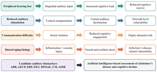
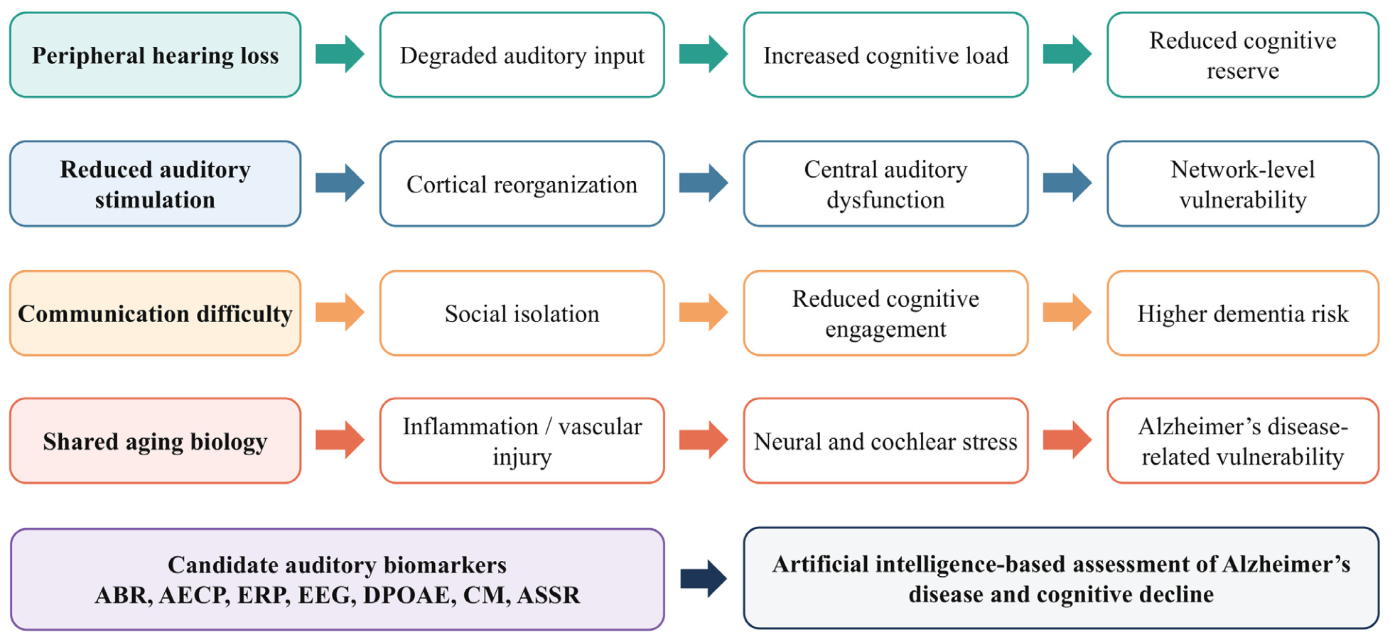
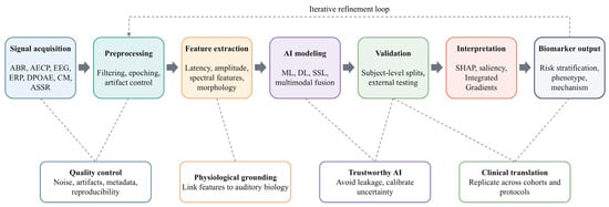
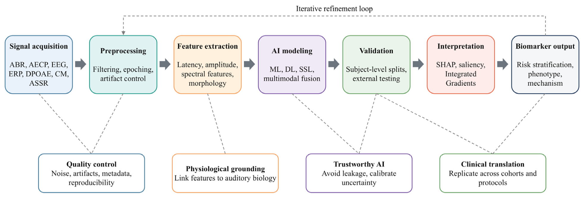
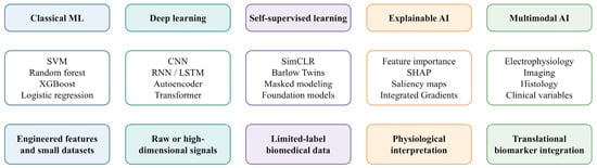
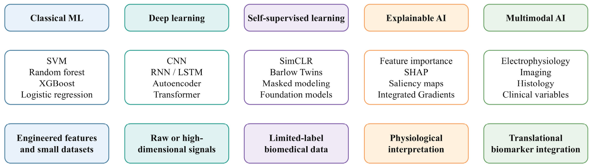
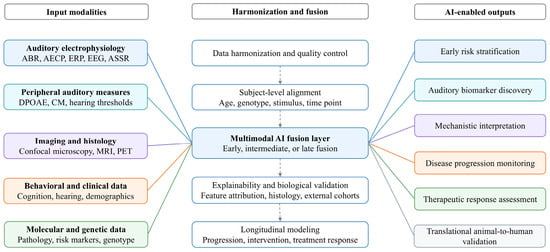
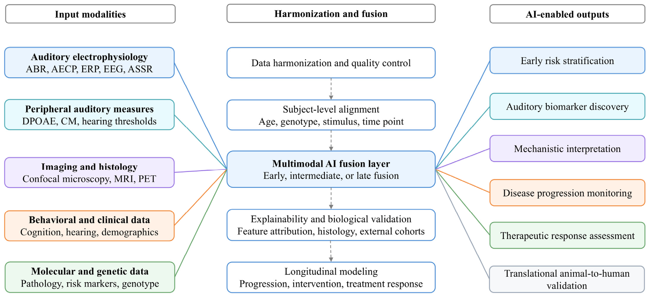

Disclaimer/Publisher’s Note: The statements, opinions and data contained in all publications are solely those of the individual author(s) and contributor(s) and not of MDPI and/or the editor(s). MDPI and/or the editor(s) disclaim responsibility for any injury to people or property resulting from any ideas, methods, instructions or products referred to in the content. |
© 2026 by the authors. Licensee MDPI, Basel, Switzerland. This article is an open access article distributed under the terms and conditions of the Creative Commons Attribution (CC BY) license.