


Highlights
What are the main findings?
- TRAEC 1.1 integrated epidemiological, in vivo, and in vitro evidence, including an original hESC experiment, on MNP-related endometrial injury and adverse pregnancy outcomes.
- The integrated evidence yielded a CES of 7.67, indicating a preliminary moderate-risk classification within the predefined TRAEC framework.
- Dual-assessor scoring across four predefined TRAEC dimensions supports transparent and traceable evidence integration.
Abstract
Micro- and nanoplastics (MNPs) are emerging environmental pollutants, but evidence concerning their effects on endometrial injury and adverse pregnancy outcomes remains limited. Therefore, we employed the Targeted Risk Assessment of Environmental Chemicals (TRAEC) strategy to integrate evidence concerning endometrial injury and adverse pregnancy outcomes associated with exposure to MNPs. Following targeted screening, 21 unique studies yielded 24 literature-derived evidence entries. Together with one additional in vitro evidence item from the present study, 25 evidence items were included in the final CES calculation. The available evidence was quantitatively integrated across four predefined TRAEC dimensions: Reliability Scores, Weight of Concentrations, Risk Intensity, and Correlation Scores. Under the tested in vitro conditions, exposure to PS-NPs increased intracellular ROS accumulation, reduced mitochondrial membrane potential, promoted apoptosis, and decreased the expression of key endometrial receptivity proteins, namely HOXA10, FOXO1, and ITGB3, in primary human endometrial stromal cells. Based on this combined evidence pool, the final Comprehensive Evidence Score was 7.67, corresponding to a preliminary moderate-risk classification within the predefined TRAEC framework. In summary, the TRAEC strategy provides a structured approach for integrating multi-level evidence and deriving a framework-specific health-risk classification for exposure to MNPs.
1. Introduction
Since the 1950s, the global plastics industry has experienced explosive growth. By 2017, global plastic production had reached 348 million tons, and it is projected to surge to 33 billion tons by 2050 [1]. Plastics are widely used in various fields such as packaging, textiles, construction, healthcare, and consumer goods due to their lightweight, durable, and low-cost properties [2]. However, this large-scale production and use, coupled with their extremely difficult natural degradation characteristics, has led to serious global plastic pollution problems. Waste plastics enter the ocean, soil, fresh water, and even the air through a variety of ways and are decomposed into smaller particles under physical, chemical, and biological effects, namely microplastics (MPs, 100 nm–5 mm in diameter) and nanoplastics (NPs, <100 nm in diameter) [3]. Human exposure to micro- and nanoplastics (MNPs) may occur through diet, inhalation, and other routes [4,5]. MNPs have been detected in several human tissues and biological samples, including blood, lung, liver, kidney, and brain samples [6,7,8]. MPs have also been reported in human female reproductive tissues, while experimental studies suggest that selected particles may cross biological barriers and affect reproductive tissues [9]. Experimental evidence further suggests that exposure to MNPs may alter endometrial function and implantation-related processes through inflammatory and endocrine pathways [10]; however, causal effects on human reproductive outcomes have not been established.
The endometrium plays a central role in women’s reproductive health and pregnancy. In each menstrual cycle, the endometrium will undergo changes such as proliferation, secretion, and shedding under the action of estrogen and progesterone, preparing for possible embryo implantation. The key to this process lies in the ‘receptivity’ of the endometrium; that is, the mature endometrium during the implantation window allows embryonic trophoblast cells to attach to the endometrial epithelium and further invade the stroma and blood vessels [10]. Once the embryo is successfully implanted, the endometrium will undergo further decidualization, providing nutrition, oxygen, and immune protection for early embryos [11]. The normal endometrial function is closely related to a variety of adverse pregnancy outcomes. When endometrial receptivity is impaired, the best-quality embryos are also difficult to implant, which is one of the main causes of in vitro fertilization-embryo transfer (IVF-ET) failure and unexplained infertility [12]. Endometrial dysfunction is also closely related to a variety of common gynecological diseases, such as endometriosis, adenomyosis, and endometrial polyps [13,14]. Studies have shown that pregnant women are highly sensitive to environmental pollutants. At this time, pollutants such as MNPs are more likely to cross the placental barrier into the fetus and amniotic fluid, may induce oxidative stress and mitochondrial dysfunction and may be associated with adverse pregnancy outcomes [9].
With advances in analytical technologies, several studies have detected and characterized MPs in human endometrial tissue using Raman spectroscopy, laser direct infrared spectroscopy (LDIR), and pyrolysis–gas chromatography/mass spectrometry (Py-GC/MS) [15,16]. One study detected MPs composed mainly of polyamide (PA), polyurethane (PU), and polyethylene terephthalate (PET), with particle sizes ranging from 2 μm to 200 μm, in the endometrial tissues from 22 women [17]. Another study performed the first quantitative analysis of microplastics in the endometrium, finding a median abundance of 21 particles per 100 mg of tissue, with 88.35% of particles measuring between 20 and 100 micrometers. This study also identified certain lifestyle habits (such as specific drinking water habits and chewing gum) as significantly associated with higher microplastic exposure levels [15]. In a three-dimensional human endometrial model, exposure to PS-MPs reduced epithelial barrier function, activated TGF-β/SMAD signaling, and increased the deposition of type I, III, and IV collagen, indicating fibrotic remodeling under the tested conditions [18]. Another study reported higher MP abundance in endometrial polyps than in normal endometrium. In vitro, PS microspheres promoted the proliferation, migration, and invasion of endometrial stromal cells via PI3K/AKT activation, suggesting a potential role in polyp development [16]. However, the study did not establish whether MPs were encapsulated, and passive accumulation in pre-existing polyps cannot be excluded; thus, the direction of causality remains uncertain.
Established human health risk-assessment frameworks, such as those developed by the World Health Organization (WHO) and the US Environmental Protection Agency (US EPA), typically address hazard identification, dose–response assessment, exposure assessment, and risk characterization [19,20]. OECD Integrated Approaches to Testing and Assessment (IATA) combine multiple sources of information to address a defined hazard, safety-assessment, or regulatory question [21]. However, for emerging contaminants such as MNPs, quantitative risk characterization remains constrained by the heterogeneity of particle size, polymer composition, morphology, and surface properties, together with limited quantitative information linking external human exposure to internal tissue doses and adverse outcomes [9]. TRAEC was introduced through initial application studies in 2024 and subsequently refined as a standardized, health outcome-oriented framework for integrating epidemiological, in vivo, and in vitro evidence [22,23]. It quantitatively integrates study reliability, concentration-related information, effect intensity, and association direction to generate a framework-specific risk classification. In an initial PFAS neurodevelopment case study, TRAEC classifications showed concordance with evaluations using ToxRTool, SciRAP, the OHAT risk-of-bias tool, and IRIS-based approaches [22]. However, this comparison provides preliminary case-based methodological support rather than comprehensive external validation. Accordingly, TRAEC is applied here as a complementary evidence-integration framework that can identify evidence gaps, rather than as a replacement for established regulatory risk-assessment approaches. In the present study, TRAEC was used to integrate the available evidence concerning exposure to MNPs, endometrial injury, and adverse pregnancy outcomes and to supplement the evidence base with original in vitro experiments using primary human endometrial stromal cells.
2. Materials and Methods
2.1. The Framework of Targeted Risk Assessment of Environmental Chemicals (TRAEC)
In this study, the TRAEC 1.1 strategy was employed to conduct a targeted health-risk assessment of evidence relating exposure to MNPs to endometrial function and pregnancy outcomes. TRAEC 1.1 is an enhanced version of TRAEC 1.0 [22].
The TRAEC framework consists of three sequential steps. First, a scientific question is formulated: “What adverse effects of exposure to MNPs on endometrial function and pregnancy outcomes are supported by the available evidence, and what targeted health-risk category is indicated within the TRAEC framework?” Next, evidence was collected through targeted literature searches and laboratory studies across three evidence types: epidemiological studies, in vivo experiments, and in vitro experiments. Finally, the collected evidence was quantitatively evaluated using four complementary dimensions defined in TRAEC 1.1: Reliability Scores, Weight of Concentrations, Risk Intensity, and Correlation Scores. These four dimensions were predefined in TRAEC 1.1 and were applied because they capture complementary aspects of evidence appraisal—methodological reliability, concentration context, effect intensity, and direction of association—and were neither selected nor reweighted specifically for the present dataset. Reliability was assessed using ten criteria specific to each evidence type, with each criterion scored as 0, 0.5, or 1, yielding a total Reliability Score of 0–10. Weight of Concentrations was calculated within each evidence type using the inverse-logarithmic weighting equation. The lowest effect concentration was normalized to 10, and the remaining concentrations were rescaled proportionally, thereby assigning relatively greater weight to evidence reporting effects at lower concentrations. Risk Intensity was scored as 1, 0.8, or 0.4 according to evidence-type-specific criteria. For epidemiological evidence, these scores represented large, moderate, or small absolute effect sizes based on Cohen’s d or Hedges’ g; for in vivo evidence, they represented phenotypic changes with or without molecular or cellular changes, molecular or cellular changes alone, or no detected change; and for in vitro evidence, they represented both cellular-phenotypic and molecular changes, either type of change alone, or no detected change. Correlation was scored as 1, 0, or −1 for a positive, nonsignificant, or negative association, respectively. The score for each evidence item and the Comprehensive Evidence Score (CES) were calculated according to the predefined TRAEC 1.1 equations shown below:
where ω, ε, φ, and r denote the Reliability Scores, Weight of Concentrations, Risk Intensity, and Correlation Scores, respectively; C represents the lowest concentration of effects; n is the number of evidence items within a single evidence domain; and i is the total number of evidence items. The epidemiological, in vivo, and in vitro evidence types were scored separately using their respective criteria and were then integrated at the item level through the CES equation; no additional fixed evidence-type coefficients were applied. In accordance with TRAEC 1.1, the original hESC experiment conducted in the present study was included as one additional in vitro evidence item in the final CES calculation. Following the TRAEC 1.1 procedure, all evidence items were independently scored by two researchers, and the two assessor-specific item-level scores were averaged to obtain the final score for each evidence item. The final CES was then calculated from these averaged item-level scores. A third researcher would be consulted if the two assessor-specific Reliability Scores differed by ≥2 points. Risk Intensity judgments were required to be consistent between assessors. For transparency, the two assessor-specific scoring datasets and corresponding CES values are provided in Supplementary Tables S2 and S3. All calculations were performed using the TRAEC web calculator (https://traec.njmu.edu.cn/; accessed on 19 July 2026).
According to the predefined TRAEC 1.1 thresholds, CES values in the ranges of (0, 4], (4, 8], and (8, 10] correspond to low, moderate, and high risk categories, respectively. Negative CES values are correspondingly interpreted as protective categories of low, moderate, or high magnitude. For the purposes of this study, hazard denotes the capacity of exposure to MNPs to produce adverse effects under specified experimental conditions, whereas risk denotes the likelihood and magnitude of such effects under defined exposure conditions. Accordingly, experimental studies were interpreted primarily as hazard evidence, while the CES was treated as a framework-specific risk classification rather than a direct estimate of real-world human risk. The concentration-weighting term represents a relative adjustment within each evidence type and was not interpreted as a formal quantitative dose–response assessment, a point of departure, or a basis for direct extrapolation to human exposure levels.
2.2. Existing Research on the Impact of Exposure to MNPs on Uterine Health
2.2.1. Search Strategy
A systematic search was conducted in the PubMed, Web of Science, Embase, and Scopus databases up to November 2025, focusing on studies examining the associations between “microplastics/nanoplastics” and “uterine health,” “endometriosis,” “uterine fibroids,” and “adverse pregnancy outcomes,” among other related topics. The detailed search strategies and keywords are provided in Supplementary Table S1. In addition, the reference lists of included studies were manually searched to supplement the relevant literature.
2.2.2. Inclusion and Exclusion Criteria
Inclusion criteria:
- (a)
- Study types include cohort studies, cross-sectional studies, case–control studies, in vivo animal experiments, or in vitro cell experiments;
- (b)
- The study examined the occurrence, distribution, association, or biological effects of MNPs in relation to female reproductive tissues, uterine or endometrial function, implantation, placental function, pregnancy outcomes, or fetal/offspring outcomes in human populations, animal models, or relevant in vitro models;
- (c)
- Provides quantitative effect indicators or specific toxicity detection data;
- (d)
- Clearly defines the characteristics of exposure to MNPs (e.g., polymer type, particle size, etc.);
- (e)
- The study included at least one outcome relevant to female reproductive tissues, uterine or endometrial health, implantation, placental function, pregnancy, or fetal/offspring development.
Exclusion criteria:
- (a)
- Duplicate publications, non-original research, or literature that does not meet the inclusion criteria;
- (b)
- Studies with incomplete information or missing data that cannot be extracted;
- (c)
- Studies not relevant to MNP exposure and female reproductive tissues, uterine or endometrial health, implantation, placental function, pregnancy outcomes, or fetal/offspring outcomes;
- (d)
- Literature for which the full text or key data cannot be obtained.
The full-text information of the literature was independently extracted by two researchers, including the title, authors, study period, study design, region, population characteristics (or experimental subject information), details of exposure to MNPs (polymer type, particle size, concentration, and exposure route), outcome indicators, and detection methods. Discrepancies were resolved through group discussion, and original authors were contacted to supplement any missing key data.
2.3. PS-NPs and Exposure Design
Polystyrene nanoplastics (PS-NPs; manufacturer-specified nominal diameter, 50 nm; Cat. No. DS50) were obtained from Huge Biotechnology (Shanghai, China). The stock suspension was stored in a sealed container at 4 °C and sonicated for 5–10 min before each use. Working suspensions were freshly prepared in complete culture medium immediately before exposure. Concentrations of 50, 100, and 200 μg/mL were selected on the basis of preliminary range-finding experiments conducted by our group and concentration ranges reported in relevant in vitro studies. The preliminary data were used solely to guide concentration selection and are not presented in the current manuscript. hESCs were exposed to 0, 50, 100, or 200 μg/mL PS-NPs for 24 h.
2.4. Isolation and Culture of Human Primary Endometrial Stromal Cells
Endometrial tissue was treated with collagenase I (Solarbio Science & Technology, Beijing, China) to obtain human primary endometrial stromal cells (hESCs). The isolated hESCs were cultured in a DMEM/F12 medium, supplemented with 5% fetal bovine serum and 1% penicillin–streptomycin (all from Sigma-Aldrich, Inc., St. Louis, MO, USA), and cultured in a cell incubator at 37 °C and 5% carbon dioxide.
2.5. Detection of Reactive Oxygen Species
The hESCs treated with different concentrations of PS-NPs were observed and photographed using a Ti2-U inverted microscope equipped with a Digital Sight 10 camera and NIS-Elements D software (version 6.10.00, Build 2017; Nikon Corporation, Tokyo, Japan). The reactive oxygen species (ROS) kit (Solarbio Science & Technology, Beijing, China) was used to detect intracellular reactive oxygen species levels. The hESCs treated with different concentrations of PS-NPs were washed three times with PBS, then DCFH-DA solution (1 mL of 10 μM) was added and incubated at 37 °C for 30 min in the dark. After washing three times with PBS, the cells were observed and photographed using the same microscope in fluorescence mode.
2.6. Mitochondrial Membrane Potential Detection (JC-1)
The mitochondrial membrane potential detection kit (JC-1) (Solarbio Science & Technology, Beijing, China) was used to detect the level of mitochondrial membrane potential of hESCs treated with PS-NPs. The hESCs treated with PS-NPs were washed three times with PBS, incubated with JC-1 staining working solution at 37 °C for 20 min in the dark, and then washed twice with JC-1 staining buffer to remove unbound dyes. The red and green fluorescence brightness was detected by fluorescence microscopy.
2.7. Cell Apoptosis Assay
Annexin V-FITC/PI Apoptosis Detection Kit (Multi Sciences, Hangzhou, China) was used to detect the level of apoptosis according to the standard process provided with the kit. The hESCs were treated with different concentrations of PS-NPs, washed with PBS, digested with trypsin, and centrifuged. The precipitate was suspended in the binding buffer and incubated with 5 μL of Annexin V-FITC and 10 μL of propidium iodide (PI) at room temperature and in the dark for 5 min, followed by quantitative analysis of apoptosis by flow cytometry.
2.8. Western Blot
Protein samples were electrophoresed on 8% or 10% polyacrylamide gels and transferred to a 0.22 μm PVDF membranes (Immobilon Transfer Membrane; MilliporeSigma, Burlington, MA, USA). The membrane was blocked in TBST buffer containing 5% skim milk powder for 2 h at room temperature and then incubated with different antibodies, GAPDH, BCL2, BAX, ITGB3, FOXO1, and HOXA10 (Proteintech Group, Inc., Wuhan, China), at 4 °C overnight. Then it was incubated with HRP-labeled secondary antibody (Proteintech Group, Inc., Wuhan, China) at room temperature for 2 h and then immersed in ECL luminescent solution (Coolaber Technology, Beijing, China) and exposed. Finally, ImageJ software (version 1.53a; National Institutes of Health, Bethesda, MD, USA) was used to analyze the gray level of the band.
2.9. Statistical Analysis
All quantitative experiments were performed with at least three independent biological replicates, and the data are presented as the mean ± standard deviation (SD). Normality was assessed using the Shapiro–Wilk test, and homogeneity of variance was evaluated using Levene’s test. Comparisons among multiple groups were performed using one-way analysis of variance (ANOVA). Fisher’s least significant difference (LSD) post hoc test was used when the assumption of homogeneity of variance was satisfied, whereas Tamhane’s T2 test was used when variances were unequal. Statistical analyses were performed using SPSS version 25.0 (IBM Corp., Armonk, NY, USA). All tests were two-sided, and a p value < 0.05 was considered statistically significant.
3. Results
3.1. Problem Statement
The potential effects of exposure to MNPs on female reproductive health have raised increasing concern. Based on the TRAEC risk assessment framework, we formulated the following scientific question: ‘What adverse effects of exposure to MNPs on endometrial function and pregnancy outcomes are supported by the available evidence, and what risk category is indicated by the integrated evidence within the TRAEC framework?’ Problem formulation constitutes the foundational step in TRAEC-based risk assessment.
3.2. Evidence Collection and TRAEC Results
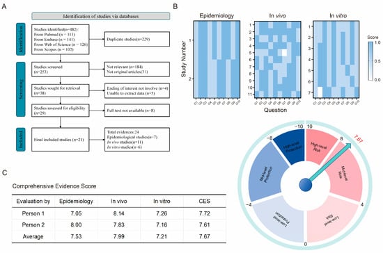
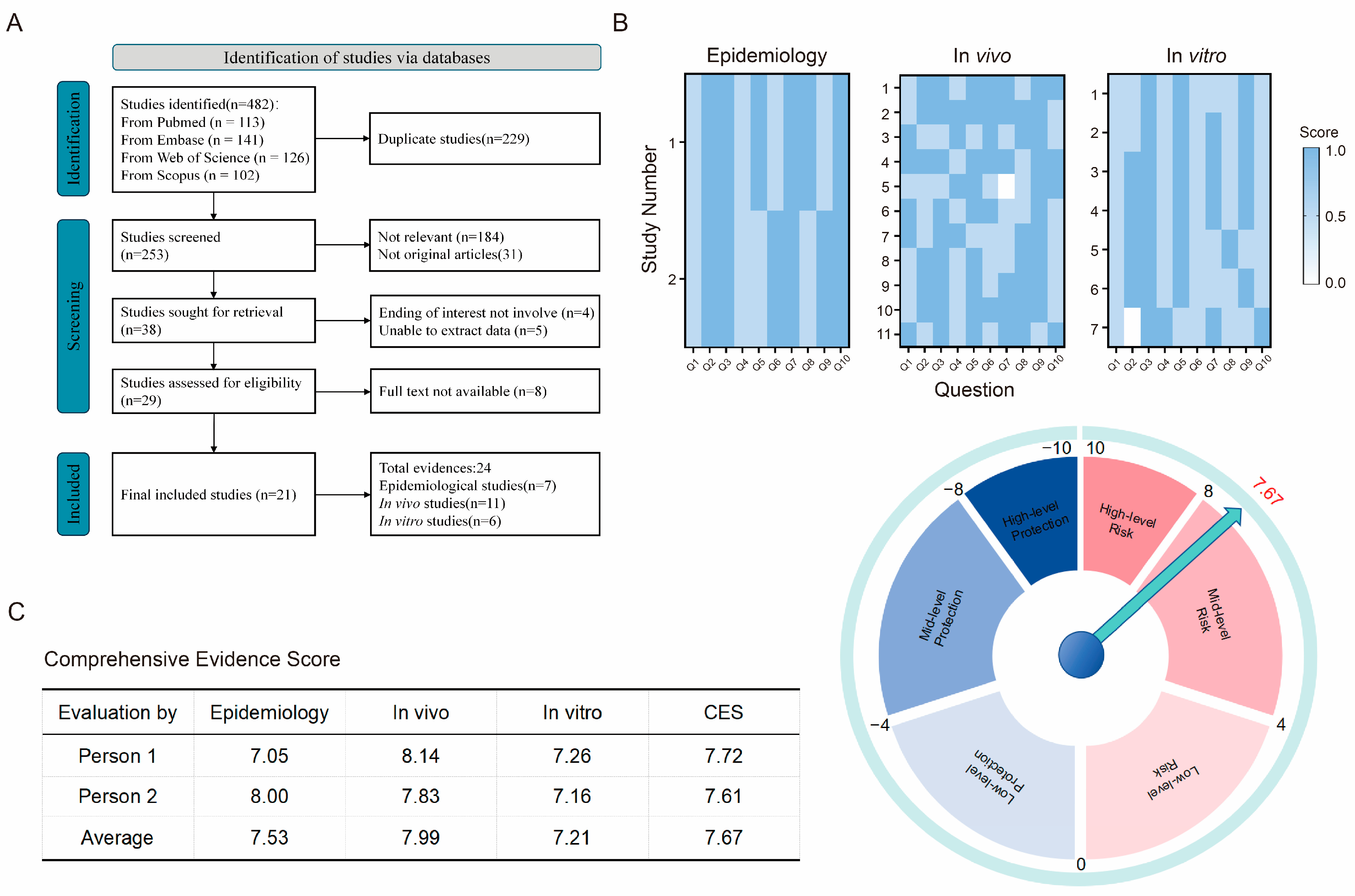
A total of 482 records were identified. After removal of 229 duplicates, 253 records were screened; 184 irrelevant records and 31 non-original articles were excluded, leaving 38 records for further assessment. Of these, 4 did not report outcomes of interest and 5 lacked extractable data, leaving 29 records for eligibility assessment. Eight of these records were subsequently excluded because their full texts were unavailable. Consequently, 21 unique studies were included in the TRAEC assessment (Figure 1A). Because some studies contributed to more than one evidence domain, these 21 studies generated 24 domain-specific evidence entries: 7 epidemiological, 11 in vivo, and 6 in vitro. The original hESC experiment conducted in the present study was included as one additional in vitro evidence item, resulting in 25 evidence items for the final CES calculation: 7 epidemiological, 11 in vivo, and 7 in vitro. Table 1, Table 2 and Table 3 summarize the literature-derived evidence, whereas the original hESC experiment is presented in Section 3.3. All 21 studies were published between 2020 and 2025, of which 12 were published in 2025 and 6 in 2024.
Figure 1.
Study selection and evidence scoring under the TRAEC framework. (A) Study selection according to the TRAEC methodology. (B) Reliability scores for epidemiological, in vivo, and in vitro studies evidence items. (C) Comprehensive Evidence Score (CES). In the dial chart, the arrow indicates the final average CES (7.67); the pink and blue sectors represent risk and protection, respectively, with darker shades indicating higher levels. The 24 literature-derived evidence entries were supplemented with one original in vitro evidence item for the CES calculation.
Table 1.
The contextual details of included epidemiological studies.
Table 2.
The contextual details of included in vivo studies.
Table 3.
The contextual details of included in vitro studies.
Among the seven epidemiological studies included, the research primarily focused on the occurrence of MNPs in endometrial, placental, and other female reproductive samples, as well as their associations with uterine conditions and pregnancy outcomes. There were four cross-sectional studies and three case–control studies. Four of these studies were conducted in China, one in the United Kingdom, one in the United States, and one in Iran. Among these seven studies, two studies reported associations between placental MNP burden and adverse pregnancy outcomes. One study found higher placental MNP levels in preterm pregnancies, whereas another reported inverse associations between placental MP burden and neonatal anthropometric measurements. Another study reported higher MP abundance and significantly higher PS concentrations in endometrial polyps than in normal endometrium. The detection methods for MPs mainly included micro-Raman spectroscopy, micro-Fourier transform infrared spectroscopy, pyrolysis-gas chromatography/mass spectrometry (Py-GC/MS), and laser direct infrared spectroscopy (LDIR). The concentration units of MPs in the tested samples were not standardized, with units such as particles/100 mg, μg/g, particles/L, and particles/g, making direct comparisons between studies impossible. Therefore, future studies should harmonize reporting metrics, including tissue-mass normalization, particle abundance, polymer-specific mass, particle-size distributions, and procedural blank correction, to improve comparability and validation across studies.
A total of 11 in vivo studies evaluated the effects of exposure to MNPs on endometrial function, adverse pregnancy outcomes, and offspring health. In the included studies, ten studies reported alterations in uterine, endometrial, decidual, endocrine, or uterine-vascular endpoints after experimental exposure, including endometrial thinning, adhesions, inflammatory infiltration, reduced uterine gland numbers, altered epithelial or mitochondrial ultrastructure, impaired decidualization, decreased uterine arteriole number or diameter, uterine endothelial dysfunction, altered reproductive hormone signaling, and reduced receptivity-related markers. Six studies reported impaired implantation, reduced litter size, or increased embryo resorption following experimental exposure to MNPs. Five studies assessed fetal or offspring outcomes: two reported reduced fetal weight; one identified altered metabolomic profiles in female offspring; one reported an altered offspring sex ratio together with a reduced litter size, and one found no significant changes in the measured litter or fetal outcomes.
A total of 6 in vitro studies were included. One of the studies was related to placental trophoblast cells, using human primary villous trophoblast cells and chorionic-villus explants. The other 5 studies were related to the endometrium, using human primary endometrial stromal cells, human primary endometrial epithelial cells, mouse endometrial epithelial cell lines, a scaffold-based three-dimensional human endometrial co-culture model, and human endometrial organoids. Across these models, exposure to PS particles produced concentration- and size-dependent changes in cellular uptake, viability, epithelial-barrier integrity, apoptosis, migration, oxidative stress, inflammatory signaling, endocrine function, and pro-fibrotic responses.
The TRAEC Comprehensive Evidence Score (CES) integrated evidence across the epidemiological, in vivo, and in vitro domains. Application of the TRAEC 1.1 scoring procedure yielded assessor-specific CES values of 7.72 and 7.61. Following the predefined dual-assessor procedure, the final CES calculated from the averaged item-level scores was 7.67. This value falls within the predefined TRAEC moderate-risk interval of (4, 8] (Supplementary Tables S2 and S3; Figure 1B,C).
3.3. Effects of Exposure to PS-NPs on Cellular Injury and Endometrial Receptivity Markers in hESCs
Based on the TRAEC framework, we conducted additional experiments to evaluate the effects of exposure to PS-NPs on primary human endometrial stromal cells (hESCs). Multiple polymer types were identified in the included epidemiological studies, whereas polystyrene was the predominant polymer used in the included in vivo and in vitro experimental studies (Table 2 and Table 3). Therefore, PS-NPs were selected for the present experiments.
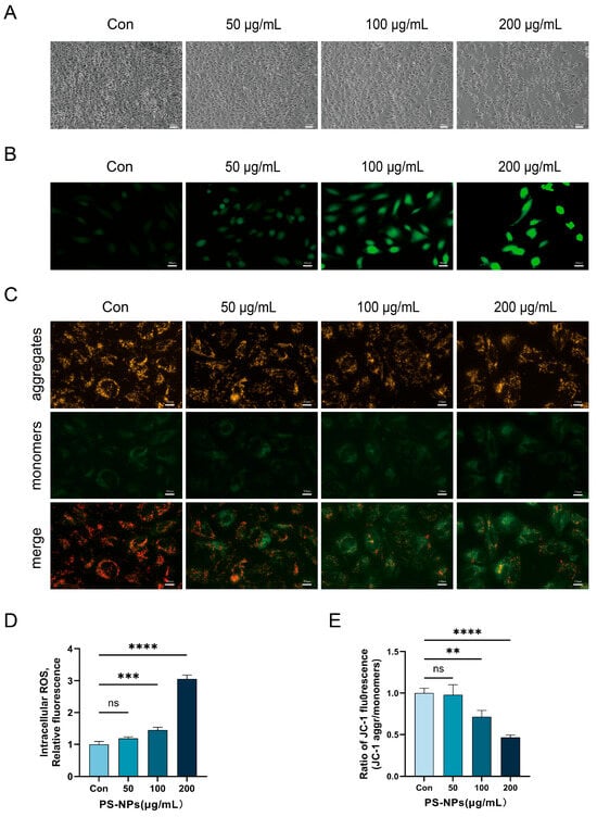
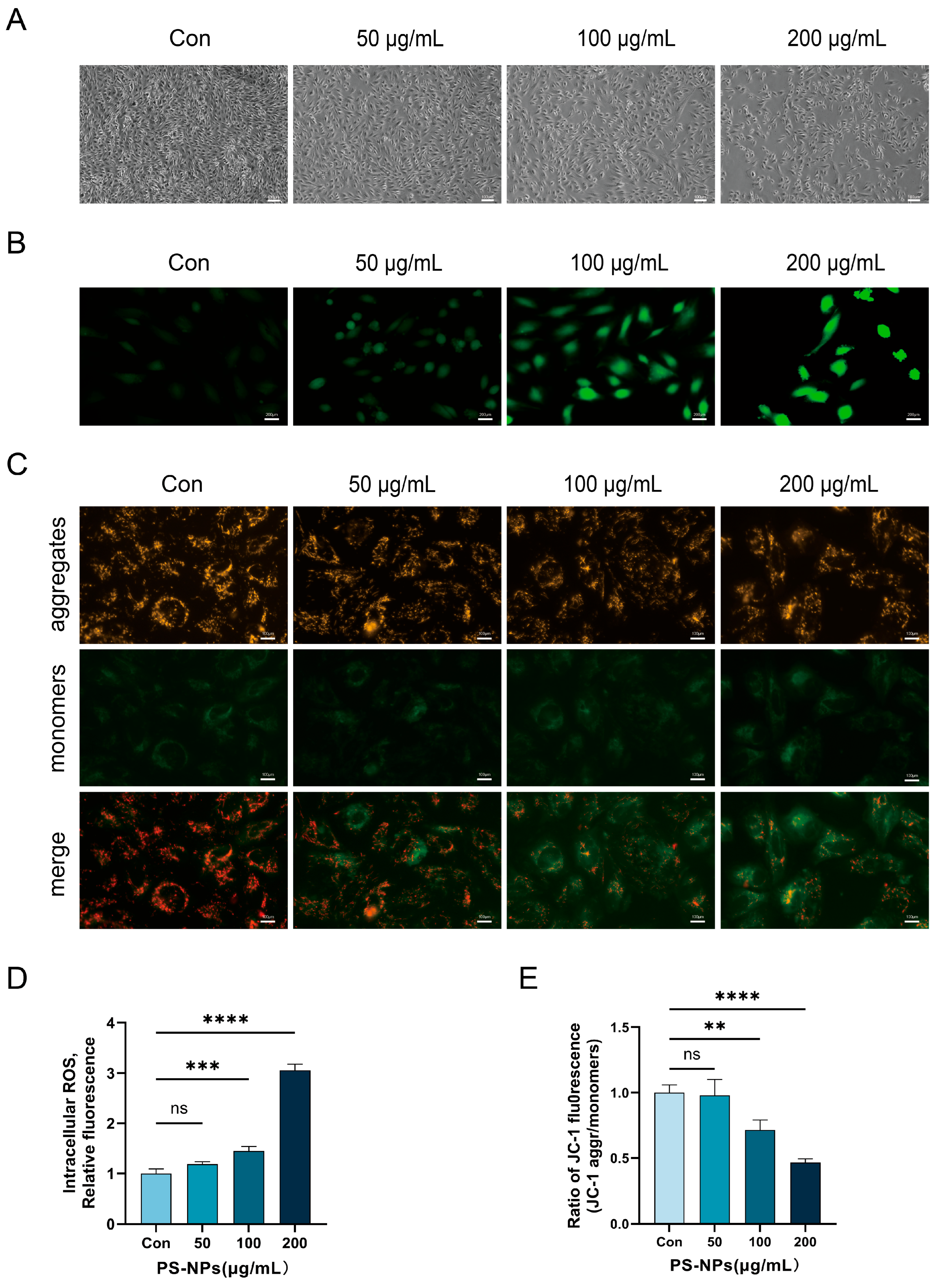
First, we examined the morphological changes in hESCs after exposure to PS-NPs. As shown in Figure 2A, with increasing concentrations of PS-NPs, the cell number gradually decreased, and the spindle-shaped morphology became increasingly irregular. Subsequently, intracellular ROS levels were assessed. The results showed that exposure to 50 μg/mL PS-NPs for 24 h did not significantly alter intracellular ROS levels. However, when the concentration of PS-NPs reached 100 μg/mL, intracellular ROS levels were significantly higher than those in the control group (p < 0.01) and increased with increasing concentrations of PS-NPs (Figure 2B,D). Further assessment of mitochondrial membrane potential was performed using JC-1 staining (Figure 2C,E). The intensity of intracellular green fluorescence gradually increased with increasing PS-NP concentrations, consistent with progressive mitochondrial membrane depolarization.
Figure 2.
Morphological changes, intracellular ROS accumulation, and reduced mitochondrial membrane potential in hESCs after exposure to PS-NPs. hESCs were treated with 0, 50, 100, and 200 μg/mL PS-NPs for 24 h. (A) Morphological changes in hESCs; scale bar = 100 μm. (B,D) Intracellular ROS production detected using the DCFH-DA probe; scale bar = 200 μm. (C,E) Changes in mitochondrial membrane potential (MMP) detected via the JC-1 probe. JC-1 monomers are represented by green fluorescence, while JC-1 aggregates are represented by red fluorescence; scale bar = 100 μm. ns, not significant; ** p < 0.01, *** p < 0.001, and **** p < 0.0001 vs. the control group.
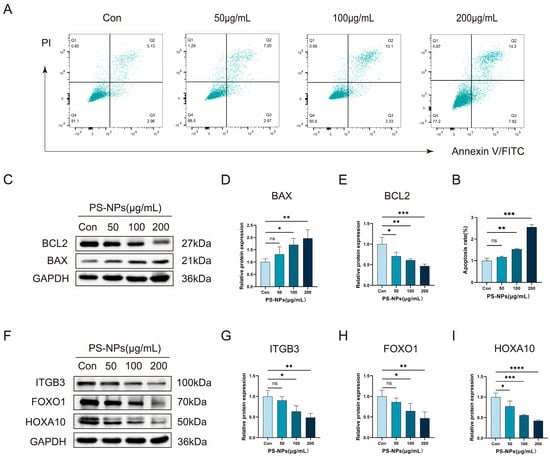
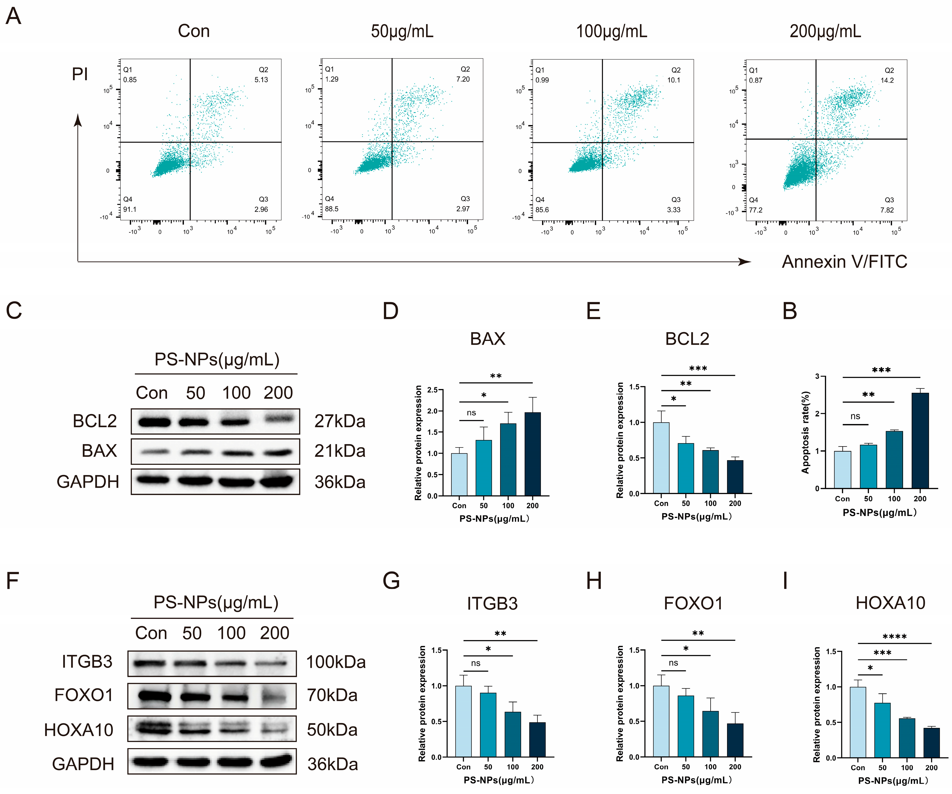
Because loss of mitochondrial membrane potential is associated with early apoptotic changes, apoptosis was further assessed by flow cytometry and Western blotting. The results of flow cytometry (Figure 3A,B) showed that the percentages of early apoptotic cells were 2.96%, 2.97%, 3.33%, and 7.82%, whereas the percentages of late apoptotic cells were 5.13%, 7.20%, 10.10%, and 14.20% after exposure to 0, 50, 100, and 200 μg/mL PS-NPs, respectively. Both early and late apoptosis increased with increasing concentrations of PS-NPs, and total apoptosis was significantly higher after exposure to 100 μg/mL PS-NPs than in the control group (p < 0.01). The pro-apoptotic effect of PS-NPs was further verified by detecting the expression of key apoptosis regulatory proteins. The results showed that exposure to PS-NPs significantly increased the expression of the pro-apoptotic protein BAX and decreased the expression of the anti-apoptotic protein BCL2 (Figure 3C–E). To evaluate endometrial receptivity-related molecular changes, we measured the protein expression of HOXA10, FOXO1, and ITGB3. The expression of all three proteins decreased significantly with increasing concentrations of PS-NPs (Figure 3F–I), demonstrating concentration-dependent downregulation of endometrial receptivity-related proteins.
Figure 3.
Apoptosis and endometrial receptivity-related protein expression in hESCs after exposure to PS-NPs. hESCs were treated with 0, 50, 100, and 200 μg/mL PS-NPs for 24 h. (A,B) Apoptosis levels detected by flow cytometry. (C–E) Protein expression levels of BAX and BCL2 determined by Western blot. (F–I) Protein expression levels of FOXO1, HOXA10, and ITGB3 determined by Western blot. ns: not significant; * p < 0.05, ** p < 0.01, *** p < 0.001, and **** p < 0.0001 vs. the control group.
4. Discussion
MNPs are widespread pollutants whose biological effects vary with polymer type, size, shape, and surface properties [40,41]. In this study, TRAEC was used to integrate epidemiological, in vivo, and in vitro evidence related to endometrial function and pregnancy outcomes.
Real-world exposure to MNPs may occur through ingestion of contaminated food and drinking water and inhalation of airborne particles [40,41]. MPs have also been detected in the human endometrium. Sun et al. identified 13 polymer types (20–500 μm) in human endometrial samples and reported potential associations with lifestyle factors [15]. Occupational exposure was not specifically evaluated in the included reproductive studies and remains an important evidence gap. Recent studies have detected MNPs in human follicular fluid and MPs in semen. Higher follicular-fluid PA66 and PVC concentrations were associated with greater odds of diminished ovarian reserve in a recent case–control study [42]. A semen study detected multiple MP polymers and observed a nonsignificant trend toward lower progressive sperm motility in PET-positive samples [43]. These findings document reproductive exposure but do not yet establish clinically meaningful effects on fertility.
Experimental evidence suggests that MNPs may affect female reproductive function through several interacting, but not yet unified, pathways [44]. Complementary cross-species studies in male mussels reported oxidative and metabolic stress, sperm-chromatin disorganization, and altered protamine-like protein-DNA interactions [45,46]. In our hESC model, PS-NPs increased intracellular ROS, reduced mitochondrial membrane potential, promoted apoptosis, and decreased HOXA10, FOXO1, and ITGB3 expression under the tested conditions. These concurrent changes are consistent with, but do not prove, a mechanistic sequence linking oxidative and mitochondrial injury to apoptosis and downregulation of receptivity-related proteins. Complementary experimental studies provided pathway-level support for several of these responses. In mouse endometrial epithelial cells, pharmacological inhibition of TLR4 or NOX2 attenuated PS-MP-induced oxidative stress, downstream Notch/TGF-β signaling, and pro-fibrotic changes [28]. In mice, JNK inhibition partially restored implantation, decidualization, and stromal-cell proliferation after PS-NP exposure, whereas another study reported activation of uterine TLR4/MyD88/NF-κB and NLRP3 signaling together with reduced implantation and receptivity-related markers [29,35]. Peri-implantation exposure to PS-MPs also altered decidual natural killer cells, macrophage polarization, placental CD4+ T cells, and cytokine secretion, together with reduced uterine arterioles and increased embryo resorption [34]. Because successful implantation requires a temporally balanced immune environment at the maternal–fetal interface [11], these findings support biologically plausible inflammatory, immune, and vascular links to impaired implantation. However, the relevant immune-cell and cytokine endpoints were not measured in our hESC experiments, and their relevance to humans remains unconfirmed. Endocrine disruption may provide a parallel pathway. One rat study reported reduced StAR expression in luteal cells and lower serum progesterone [30], whereas exposure of primary human villous cytotrophoblasts to PS-NPs reduced hCG secretion and elicited particle-size- and concentration-dependent pro-inflammatory responses without significantly altering VCT-to-ST fusion [39].
Beyond early reproductive events, evidence linking exposure to MNPs to adverse pregnancy outcomes remains limited and heterogeneous. Experimental work showed that gestational exposure to MNPs impaired placental trophoblast syncytialization and activated the PERK/eIF2α/ATF4 signaling pathway; pharmacological PERK inhibition partially restored syncytialization in BeWo cells [47], whereas observational data reported higher placental MP burdens in pregnancies complicated by fetal growth restriction and inverse associations with selected neonatal anthropometric measurements [27]. MPs have also been detected on the maternal and fetal sides of human placentas and in the chorioamniotic membranes [48], and an ex vivo human placental perfusion model demonstrated size-dependent passage of fluorescent PS beads up to 240 nm without altering the assessed viability or function of the perfused placental tissue [49]. However, placental detection alone does not establish transplacental transfer or quantify fetal exposure, and the perfusion findings were obtained under controlled experimental conditions. Overall, the available evidence supports biologically plausible links to impaired decidualization, implantation, placental function, embryo survival, and fetal growth, but it does not establish a unified causal mechanism or quantify real-world human reproductive risk.
In vitro models provide controlled mechanistic evidence but cannot reproduce whole-body absorption, biodistribution, clearance, or interactions among reproductive, immune, vascular, endocrine, placental, and embryonic compartments, nor can they directly assess implantation or fetal development. More broadly, the experimental evidence is dominated by rodent and in vitro models, often using relatively high doses and short exposure durations. Heterogeneous dose metrics prevent direct comparison with human tissue burdens; most experiments examined polystyrene particles under controlled laboratory conditions rather than real-world mixtures; and the available human studies were observational and generally involved limited sample sizes. Accordingly, experimental studies mainly inform hazard identification, biological plausibility, and model-specific adverse effects, whereas human studies establish tissue occurrence and preliminary associations. Together, these limitations constrain causal interpretation and quantitative risk estimation.
Although experimental evidence was dominated by polystyrene, epidemiological studies identified polyethylene, polypropylene, polyvinyl chloride, polyethylene terephthalate, and mixed polymer profiles in human reproductive tissues [15,24,25]. TRAEC is not intrinsically polymer-specific and could, in principle, be applied to individual polymers or mixtures when sufficiently detailed evidence is available [9,22]. However, the present CES should not be interpreted as polymer- or mixture-specific because particle properties, additives, weathering, and co-contaminants may modify biological effects [40]. Future assessments should therefore stratify evidence by polymer and particle characteristics.
The environmental relevance of the experimental exposure levels remains uncertain. One included mouse study selected 50 mg/kg/day with reference to published estimates of human MP ingestion and a mouse-equivalent dose range calculated using body-surface-area scaling [29,50,51,52]. However, the ingestion estimates were derived from heterogeneous data and conservative assumptions, and the allometric conversion does not directly account for particle-specific absorption, biodistribution, clearance, or uterine deposition [51,52]. Human tissue burdens cannot be directly compared with administered animal doses or nominal in vitro concentrations. Accordingly, the experimental concentrations, including 50–200 μg/mL in our hESC experiments, should be interpreted as hazard-identification conditions rather than as quantitatively equivalent to realistic human uterine exposure.
TRAEC provides a transparent framework for integrating epidemiological, in vivo, and in vitro evidence using predefined criteria [22,23]. The final CES of 7.67 falls within the framework’s moderate-risk category, but the evidence streams contribute differently: experimental studies mainly support hazard identification, biological plausibility, and model-specific adverse effects, whereas epidemiological studies report MNP occurrence and preliminary associations without standardized exposure estimates or quantitative links between external exposure, tissue dose, and adverse outcomes. Initial concordance with ToxRTool, SciRAP, OHAT RoB, and IRIS-based evaluations, together with subsequent applications, supports the feasibility of TRAEC [9,22,23], but its external validation remains preliminary. The CES therefore summarizes the current evidence but does not replace formal dose–response or exposure assessment or quantify real-world human risk. TRAEC should support evidence synthesis and gap identification rather than serve as a stand-alone basis for regulatory decision-making.
5. Conclusions
In this study, TRAEC 1.1 was used to integrate epidemiological, in vivo, and in vitro evidence concerning exposure to MNPs, endometrial injury, and adverse pregnancy outcomes. The final CES of 7.67 falls within the moderate-risk category of the predefined TRAEC framework and should be considered a preliminary classification based on the currently available evidence. Our hESC experiments provide mechanistic hazard evidence that exposure to PS-NPs increased intracellular ROS, reduced mitochondrial membrane potential, promoted apoptosis, and decreased the expression of key endometrial receptivity proteins such as HOXA10, FOXO1, and ITGB3 under the tested conditions. However, the experimental evidence is dominated by in vitro systems and rodent models; many studies used relatively high exposure levels, and most examined polystyrene particles rather than the heterogeneous mixtures encountered under real-world conditions. These limitations reduce confidence in direct extrapolation to environmentally relevant human exposure conditions. Well-designed prospective human studies and experimental models incorporating environmentally informed exposure ranges, diverse polymers, and more realistic particle characteristics are required to refine this classification.
Supplementary Materials
The following supporting information can be downloaded at: https://www.mdpi.com/article/10.3390/toxics14080677/s1, Table S1: Detailed search strategies; Table S2: TRAEC scoring results from Assessor 1; Table S3: TRAEC scoring results from Assessor 2.
Author Contributions
Conceptualization, H.S. and X.S.; methodology, A.L. and X.L.; validation, N.J. and X.L.; formal analysis, N.J., N.W., J.G. and X.L.; investigation, X.L.; resources, H.S. and X.M.; data curation, N.J., N.W., J.G. and X.L.; writing—original draft preparation, N.J., A.L., N.W., J.G. and X.L.; writing—review and editing, H.S., N.J. and X.L.; visualization, A.L. and X.L.; supervision, H.S., X.S. and X.M.; project administration, H.S. and X.S.; funding acquisition, H.S. All authors have read and agreed to the published version of the manuscript.
Funding
This research was supported by the Gansu Youth Science and Technology Fund (grant numbers 25JRRA1015 and 23JRRA1614); the Lanzhou Science and Technology Development Guiding Plan Project (grant number 2024-9-111); the Gansu Province University Young Doctor “Entering Enterprises and Parks” Program (grant number 2026QB-004); the First Hospital of Lanzhou University (grant number ldyyyn2025-125); the Gansu Province Traditional Chinese Medicine Research Project (grant number GZKG-2025-85); and the Lanzhou University Student Innovation and Entrepreneurship Action Plan (grant number 20260060327).
Institutional Review Board Statement
The study was conducted in accordance with the Declaration of Helsinki and approved by the Ethics Committee of the First Hospital of Lanzhou University (protocol code LDYYLL2026-692; approval date: 12 June 2026).
Informed Consent Statement
The endometrial tissues used were residual clinical specimens that would otherwise have been discarded. As the study involved no more than minimal risk to participants, the requirement for written informed consent was waived.
Data Availability Statement
The raw data supporting the conclusions of this article will be made available by the authors on request.
Conflicts of Interest
The authors declare no conflicts of interest.
Abbreviations
The following abbreviations are used in this manuscript:
| MNPs | micro- and nanoplastics |
| TRAEC | Targeted Risk Assessment of Environmental Chemicals |
| PS-NPs | polystyrene nanoplastics |
| ROS | reactive oxygen species |
| MPs | microplastics |
| NPs | nanoplastics |
| IVF-ET | in vitro fertilization-embryo transfer |
| LDIR | laser direct infrared spectroscopy |
| Py-GC/MS | pyrolysis–gas chromatography/mass spectrometry |
| PA | polyamide |
| PU | polyurethane |
| PET | polyethylene terephthalate |
| PS-MPs | polystyrene microplastics |
| hESCs | human primary endometrial stromal cells |
| CES | Comprehensive Evidence Score |
| PE | polyethylene |
| PS | polystyrene |
| PVC | polyvinyl chloride |
| PP | polypropylene |
| uNK cells | uterine natural killer cells |
References
- Wu, M.; Yang, C.; Du, C.; Liu, H. Microplastics in waters and soils: Occurrence, analytical methods and ecotoxicological effects. Ecotoxicol. Environ. Saf. 2020, 202, 110910. [Google Scholar] [CrossRef] [Scilit]
- Andrady, A.L.; Neal, M.A. Applications and societal benefits of plastics. Philos. Trans. R. Soc. B Biol. Sci. 2009, 364, 1977–1984. [Google Scholar] [CrossRef] [Scilit] [PubMed]
- Yang, Z.; DeLoid, G.M.; Zarbl, H.; Baw, J.; Demokritou, P. Micro- and nanoplastics (MNPs) and their potential toxicological outcomes: State of science, knowledge gaps and research needs. NanoImpact 2023, 32, 100481. [Google Scholar] [CrossRef] [Scilit] [PubMed]
- Kannan, K.; Vimalkumar, K. A Review of Human Exposure to Microplastics and Insights Into Microplastics as Obesogens. Front. Endocrinol. 2021, 12, 724989. [Google Scholar] [CrossRef] [Scilit] [PubMed]
- De-La-Torre, G.E. Microplastics: An emerging threat to food security and human health. J. Food Sci. Technol. 2020, 57, 1601–1608. [Google Scholar] [CrossRef] [Scilit] [PubMed]
- Leslie, H.A.; van Velzen, M.J.; Brandsma, S.H.; Vethaak, A.D.; Garcia-Vallejo, J.J.; Lamoree, M.H. Discovery and quantification of plastic particle pollution in human blood. Environ. Int. 2022, 163, 107199. [Google Scholar] [CrossRef] [Scilit] [PubMed]
- Jenner, L.C.; Rotchell, J.M.; Bennett, R.T.; Cowen, M.; Tentzeris, V.; Sadofsky, L.R. Detection of microplastics in human lung tissue using μFTIR spectroscopy. Sci. Total. Environ. 2022, 831, 154907. [Google Scholar] [CrossRef] [Scilit] [PubMed]
- Nihart, A.J.; Garcia, M.A.; El Hayek, E.; Liu, R.; Olewine, M.; Kingston, J.D.; Castillo, E.F.; Gullapalli, R.R.; Howard, T.; Bleske, B.; et al. Bioaccumulation of microplastics in decedent human brains. Nat. Med. 2025, 31, 1114–1119. [Google Scholar] [CrossRef] [Scilit] [PubMed]
- Wan, D.; Liu, Y.; Chang, Q.; Liu, Z.; Wang, Q.; Niu, R.; Gao, B.; Guan, Q.; Xia, Y. Micro/Nanoplastic Exposure on Placental Health and Adverse Pregnancy Risks: Novel Assessment System Based upon Targeted Risk Assessment Environmental Chemicals Strategy. Toxics 2024, 12, 553. [Google Scholar] [CrossRef] [Scilit] [PubMed]
- Pirtea, P.; de Ziegler, D.; Ayoubi, J.M. Endometrial receptivity in adenomyosis and/or endometriosis. Fertil. Steril. 2023, 119, 741–745. [Google Scholar] [CrossRef] [Scilit] [PubMed]
- Mori, M.; Bogdan, A.; Balassa, T.; Csabai, T.; Szekeres-Bartho, J. The decidua—The maternal bed embracing the embryo—Maintains the pregnancy. Semin. Immunopathol. 2016, 38, 635–649. [Google Scholar] [CrossRef] [Scilit] [PubMed]
- Ma, J.; Gao, W.; Li, D. Recurrent implantation failure: A comprehensive summary from etiology to treatment. Front. Endocrinol. 2023, 13, 1061766. [Google Scholar] [CrossRef] [Scilit] [PubMed]
- Liu, D.; Yin, X.; Guan, X.; Li, K. Bioinformatic analysis and machine learning to identify the diagnostic biomarkers and immune infiltration in adenomyosis. Front. Genet. 2023, 13, 1082709. [Google Scholar] [CrossRef] [Scilit] [PubMed]
- Nijkang, N.P.; Anderson, L.; Markham, R.; Manconi, F. Endometrial polyps: Pathogenesis, sequelae and treatment. SAGE Open Med. 2019, 7, 2050312119848247. [Google Scholar] [CrossRef] [Scilit] [PubMed]
- Sun, J.; Sui, M.; Wang, T.; Teng, X.; Sun, J.; Chen, M. Detection and quantification of various microplastics in human endometrium based on laser direct infrared spectroscopy. Sci. Total Environ. 2024, 906, 167760. [Google Scholar] [CrossRef] [Scilit] [PubMed]
- He, S.; Zhang, Y. Detection and quantification of microplastics in endometrial polyps and their role in polyp formation. Reprod. Toxicol. 2025, 132, 108757. [Google Scholar] [CrossRef] [Scilit] [PubMed]
- Qin, X.; Cao, M.; Peng, T.; Shan, H.; Lian, W.; Yu, Y.; Shui, G.; Li, R. Features, Potential Invasion Pathways, and Reproductive Health Risks of Microplastics Detected in Human Uterus. Environ. Sci. Technol. 2024, 58, 10482–10493. [Google Scholar] [CrossRef] [Scilit] [PubMed]
- Arcuri, S.; Pennarossa, G.; Bebbere, D.; Gandolfi, F.; Ledda, S.; Brevini, T.A.L. Microplastic exposure induces epithelial barrier alterations and increases collagen deposition in a 3D human endometrial model in vitro. J. Assist. Reprod. Genet. 2025, 42, 3065–3077. [Google Scholar] [CrossRef] [Scilit] [PubMed]
- World Health Organization. WHO Human Health Risk Assessment Toolkit: Chemical Hazards, 2nd ed.; World Health Organization: Geneva, Switzerland, 2021. [Google Scholar]
- U.S. Environmental Protection Agency. Framework for Human Health Risk Assessment to Inform Decision Making; U.S. Environmental Protection Agency: Washington, DC, USA, 2014.
- OECD. Overview of Concepts and Available Guidance Related to Integrated Approaches to Testing and Assessment (IATA), in OECD Series on Testing and Assessment; OECD Publishing: Paris, France, 2020; p. 55. [Google Scholar]
- Ning, J.; Ding, C.; Xu, H.; Liu, Z.; Guan, Q.; Xia, Y.; Xu, Q. Effect of per- and polyfluoroalkyl substances on neurodevelopment: Evidence-based risk assessment in the TRAEC strategy context. Environ. Int. 2024, 191, 109003. [Google Scholar] [CrossRef] [Scilit]
- Zhu, Y.; Tan, Z.; An, Z.; Li, L.; Guo, M.; Duan, W.; Yu, Z.; Chen, Z.; Liu, Y.; Li, A.; et al. Effects of Per- and polyfluoroalkyl substances on atherosclerosis: An evidence-based risk assessment in the context of the TRAEC strategy. Environ. Res. 2025, 285, 122698. [Google Scholar] [CrossRef] [Scilit] [PubMed]
- Jochum, M.; Garcia, M.; Hammerquist, A.; Howell, J.; Stanford, M.; Liu, R.; Olewine, M.; El Hayek, E.; Phan, E.; Showalter, L.; et al. Elevated Micro- and Nanoplastics Detected in Preterm Human Placentae. Res. Sq. 2025, rs.3.rs-5903715. [Google Scholar] [CrossRef] [Scilit] [PubMed]
- Dong, C.; Xu, H.; Lin, Y.; Zhang, B.; Yu, Z.; Xie, Y.; Yu, J.; Ma, D. Microplastics detected in three types of female reproductive organs using micro-Raman spectroscopy. Ecotoxicol. Environ. Saf. 2024, 285, 117099. [Google Scholar] [CrossRef] [Scilit] [PubMed]
- Rotchell, J.M.; Austin, C.; Chapman, E.; Atherall, C.A.; Liddle, C.R.; Dunstan, T.S.; Blackburn, B.; Mead, A.; Filart, K.; Beeby, E.; et al. Microplastics in human urine: Characterisation using μFTIR and sampling challenges using healthy donors and endometriosis participants. Ecotoxicol. Environ. Saf. 2024, 274, 116208. [Google Scholar] [CrossRef] [Scilit] [PubMed]
- Amereh, F.; Amjadi, N.; Mohseni-Bandpei, A.; Isazadeh, S.; Mehrabi, Y.; Eslami, A.; Naeiji, Z.; Rafiee, M. Placental plastics in young women from general population correlate with reduced foetal growth in IUGR pregnancies. Environ. Pollut. 2022, 314, 120174. [Google Scholar] [CrossRef] [Scilit] [PubMed]
- Wu, H.; Xu, T.; Chen, T.; Liu, J.; Xu, S. Oxidative stress mediated by the TLR4/NOX2 signalling axis is involved in polystyrene microplastic-induced uterine fibrosis in mice. Sci. Total. Environ. 2022, 838, 155825. [Google Scholar] [CrossRef] [Scilit] [PubMed]
- Sun, J.; Zhou, D.; Ji, M.; Zhao, Y.; Li, L.; Gan, L.; Chen, M. Dual impact of microplastic exposure in a mouse model: Impaired uterine receptivity and altered maternal-offspring metabolism. J. Hazard. Mater. 2025, 498, 139819. [Google Scholar] [CrossRef] [Scilit] [PubMed]
- Valencise, L.; de Barros, J.W.F.; Lozano, A.F.Q.; Calixto, L.R.; Cyr, D.G.; Kempinas, W.D.G. Exposure to low-dose polystyrene nanoplastics impairs the estrous cycle by decreasing ovarian levels of steroidogenic acute regulatory protein and serum progesterone levels in rats. Reprod. Toxicol. 2025, 138, 109069. [Google Scholar] [CrossRef] [Scilit] [PubMed]
- Sun, J.; Geng, L.; Zhou, D.; Teng, X.; Chen, M. Gut microbiota participates in polystyrene microplastics-induced defective implantation through impairing uterine receptivity. J. Environ. Manag. 2025, 380, 124997. [Google Scholar] [CrossRef] [Scilit] [PubMed]
- Cary, C.M.; Moore, T.L.; Gow, A.J.; Stapleton, P.A. Micro and nanoplastic inhalation during pregnancy elicits uterine endothelial dysfunction in Sprague Dawley rats by impeding nitric oxide signaling. Am. J. Physiol. Heart Circ. Physiol. 2026, 330, H230–H242. [Google Scholar] [CrossRef] [Scilit] [PubMed]
- Fournier, S.B.; D’Errico, J.N.; Adler, D.S.; Kollontzi, S.; Goedken, M.J.; Fabris, L.; Yurkow, E.J.; Stapleton, P.A. Nanopolystyrene translocation and fetal deposition after acute lung exposure during late-stage pregnancy. Part. Fibre Toxicol. 2020, 17, 55. [Google Scholar] [CrossRef] [Scilit] [PubMed]
- Hu, J.; Qin, X.; Zhang, J.; Zhu, Y.; Zeng, W.; Lin, Y.; Liu, X. Polystyrene microplastics disturb maternal-fetal immune balance and cause reproductive toxicity in pregnant mice. Reprod. Toxicol. 2021, 106, 42–50. [Google Scholar] [CrossRef] [Scilit] [PubMed]
- Li, J.-A.; Wang, L.-Q.; Xu, Q.-J.; Wu, X.-M.; Pang, X.-Q.; He, J.-X.; Qiao, Q.-F.; You, H.-N.; Wu, Y.-C.; Zhou, Y.-J. Polystyrene nanoplastics impair endometrial decidualization via cell cycle arrest and JNK-MAPK pathway-mediated oxidative stress in early pregnant mice. Food Chem. Toxicol. 2025, 205, 115707. [Google Scholar] [CrossRef] [Scilit] [PubMed]
- Amran, N.H.; Zaid, S.S.M.; Meng, G.Y.; Salleh, A.; Mokhtar, M.H. Protective Role of Kelulut Honey against Toxicity Effects of Polystyrene Microplastics on Morphology, Hormones, and Sex Steroid Receptor Expression in the Uterus of Rats. Toxics 2023, 11, 324. [Google Scholar] [CrossRef] [Scilit] [PubMed]
- Wang, Z.; Zhang, R.; Zhang, Y.; Xiong, Y.; Zhang, M. The risk of short-term microplastic exposure on female reproductive function: A rat model study. NanoImpact 2025, 37, 100545. [Google Scholar] [CrossRef] [Scilit] [PubMed]
- Kim, N.; Lee, J.H.; Lee, I.; Park, J.H.; Jung, G.S.; Lee, M.J.; Im, W.; Cho, S.; Choi, Y.S. Investigation of potential toxic effects of nano- and microplastics on human endometrial stromal cells. Reprod. Toxicol. 2025, 132, 108848. [Google Scholar] [CrossRef] [Scilit] [PubMed]
- Poinsignon, L.; Lefrère, B.; Ben Azzouz, A.; Chissey, A.; Colombel, J.; Djelidi, R.; Ferecatu, I.; Fournier, T.; Beaudeux, J.-L.; Lespes, G.; et al. Exposure of the human placental primary cells to nanoplastics induces cytotoxic effects, an inflammatory response and endocrine disruption. J. Hazard. Mater. 2025, 490, 137713. [Google Scholar] [CrossRef] [Scilit] [PubMed]
- Lamoree, M.H.; van Boxel, J.; Nardella, F.; Houthuijs, K.J.; Brandsma, S.H.; Béen, F.; van Duursen, M.B.M. Health impacts of microplastic and nanoplastic exposure. Nat. Med. 2025, 31, 2873–2887. [Google Scholar] [CrossRef] [Scilit] [PubMed]
- Talaie, A.; Alaee, S.; Hosseini, E.; Rezania, S.; Tamadon, A. Toxicological effects of micro/nano-plastics on human reproductive health: A review. Toxicol. Lett. 2025, 412, 1–20. [Google Scholar] [CrossRef] [Scilit] [PubMed]
- Si, M.; Xiong, X.; Yun, C.; Chen, Y.; Niu, H.; Qu, Y.; Liu, M.; Wang, Y.; Huang, L.; Long, X.; et al. Microplastics and nanoplastics in follicular fluid are associated with diminished ovarian reserve: Clinical and molecular insights. J. Adv. Res. 2026; in press. [CrossRef] [Scilit] [PubMed]
- Guo, Y.; Rong, M.; Fan, Y.; Teng, X.; Jin, L.; Zhao, Y. The Presence of Microplastics in Human Semen and Their Associations with Semen Quality. Toxics 2025, 13, 566. [Google Scholar] [CrossRef] [Scilit] [PubMed]
- Peng, Y.; He, Q. Reproductive toxicity and related mechanisms of micro(nano)plastics in terrestrial mammals: Review of current evidence. Ecotoxicol. Environ. Saf. 2024, 279, 116505. [Google Scholar] [CrossRef] [Scilit] [PubMed]
- Marinaro, C.; Scarciello, G.; Bianchi, A.R.; Berman, B.; Chianese, T.; Scudiero, R.; Rosati, L.; De Maio, A.; Lettieri, G.; Piscopo, M. Toxicological effects and potential reproductive risk of microplastic-induced molecular changes in protamine-like proteins and their DNA binding. Chem. Biol. Interact. 2025, 405, 111309. [Google Scholar] [CrossRef] [Scilit] [PubMed]
- Chianese, T.; Galati, M.; Cappello, T.; Maisano, M.; Lettieri, G.; Marinaro, C.; Piscopo, M.; Fasciolo, G.; Gravato, C.; Napolitano, G.; et al. Exploring the Impact of Polystyrene Microplastic Beads on Male Gonads of the Marine Mussel, Mytilus galloprovincialis. Environ. Toxicol. 2026, 41, 379–394. [Google Scholar] [CrossRef] [Scilit] [PubMed]
- Cheng, Y.; Li, Y.; Zhang, Y.; Liu, H.; Yang, B.; Zhu, J.; Kuang, H. Gestational exposure to micro- and nanoplastics leads to poor pregnancy outcomes by impairing placental trophoblast syncytialization. Environ. Pollut. 2025, 381, 126520. [Google Scholar] [CrossRef] [Scilit] [PubMed]
- Ragusa, A.; Svelato, A.; Santacroce, C.; Catalano, P.; Notarstefano, V.; Carnevali, O.; Papa, F.; Rongioletti, M.C.A.; Baiocco, F.; Draghi, S.; et al. Plasticenta: First evidence of microplastics in human placenta. Environ. Int. 2021, 146, 106274. [Google Scholar] [CrossRef] [Scilit] [PubMed]
- Wick, P.; Malek, A.; Manser, P.; Meili, D.; Maeder-Althaus, X.; Diener, L.; Diener, P.-A.; Zisch, A.; Krug, H.F.; Von Mandach, U. Barrier Capacity of Human Placenta for Nanosized Materials. Environ. Health Perspect. 2010, 118, 432–436. [Google Scholar] [CrossRef] [Scilit] [PubMed]
- Xu, W.; Yuan, Y.; Tian, Y.; Cheng, C.; Chen, Y.; Zeng, L.; Yuan, Y.; Li, D.; Zheng, L.; Luo, T. Oral exposure to polystyrene nanoplastics reduced male fertility and even caused male infertility by inducing testicular and sperm toxicities in mice. J. Hazard. Mater. 2023, 454, 131470. [Google Scholar] [CrossRef] [Scilit] [PubMed]
- Nair, A.B.; Jacob, S. A simple practice guide for dose conversion between animals and human. J. Basic Clin. Pharm. 2016, 7, 27–31. [Google Scholar] [CrossRef] [Scilit] [PubMed]
- Senathirajah, K.; Attwood, S.; Bhagwat, G.; Carbery, M.; Wilson, S.; Palanisami, T. Estimation of the mass of microplastics ingested—A pivotal first step towards human health risk assessment. J. Hazard. Mater. 2021, 404, 124004. [Google Scholar] [CrossRef] [Scilit] [PubMed]

Disclaimer/Publisher’s Note: The statements, opinions and data contained in all publications are solely those of the individual author(s) and contributor(s) and not of MDPI and/or the editor(s). MDPI and/or the editor(s) disclaim responsibility for any injury to people or property resulting from any ideas, methods, instructions or products referred to in the content. |
© 2026 by the authors. Licensee MDPI, Basel, Switzerland. This article is an open access article distributed under the terms and conditions of the Creative Commons Attribution (CC BY) license.