


Abstract
Immune dysregulation is a critical driver of various pathological processes. Fish maw (FM) serves as a traditional immunomodulatory food. However, the immunomodulatory properties and mechanisms of fish maw hydrolysate (FMH) remain unclear. Here, low-molecular-weight FMH was prepared from Perca fluviatilis, exhibiting a major molecular weight distribution of 73–580 Da (83.89%), enriched in charged and hydrophobic amino acids (28.61% and 67.33%, respectively). Moreover, high-resolution mass spectrometry (HRMS) analysis identified 5 small peptides, including Asp-Leu and Gly-Pro-Ala, alongside 7 collagen-derived polypeptides with characteristic Gly-X-Y repetitive motifs. In cyclophosphamide (CTX)-induced immunosuppressed C57BL/6J mice, FMH significantly ameliorated alterations in peripheral blood cell parameters, regulated cytokine homeostasis, attenuated splenic histopathological lesions, and enhanced splenic lymphocyte proliferation. Mechanistically, thymic transcriptomic profiling identified 2237 DEGs in the CTX vs. CON comparison and 212 DEGs in the CTX+FMH vs. CTX comparison, with the NF-κB signaling pathway significantly enriched. Furthermore, qRT-PCR validated the expression of key NF-κB-related genes, including IκBα, P50, P65, CHUK, IL-1β, and IL-6, while immunohistochemical analysis confirmed reduced PI3K and P65 expression, thereby partly restoring immune homeostasis. These findings support FMH as a potential dietary immunomodulator.
1. Introduction
Immunocompromised status may impair host defense and increase susceptibility to disease [1]. Notably, accumulating evidence indicates that immune dysregulation contributes to the pathogenesis of a wide spectrum of diseases, including autoimmune disorders, chronic inflammatory conditions, malignancies, and neurodegenerative diseases [2,3,4,5]. Therapeutic enhancement of host defense responses through immunomodulators has been widely explored as a strategy for improving disease resistance [6]. However, conventional immunomodulators, such as corticosteroids and cyclosporine, may have limitations during long-term use, and prolonged use of some agents may impair immune function or cause other adverse effects [7,8]. Consequently, the development of novel dietary immunomodulatory ingredients with good biocompatibility and bioactivity is of considerable significance.
Fish maw, the dried product derived from swim bladders, contains abundant type I collagen and functional amino acids while remaining low in fat, making it a high-protein and low-fat nutritional resource [9]. Current processing of fish maw mainly relies on conventional cooking methods, which are time-consuming and may cause nutrient loss. Moreover, the bioefficacy of native collagen may be limited by its relatively low absorption efficiency [10]. Enzymatic hydrolysis has recently enabled the production of low-molecular-weight hydrolysates and derived peptides [11]. These bioactive products exhibit enhanced absorption and utilization efficiency [12]. Dai et al. revealed the potential of swim bladder-derived peptides in preventing ulcerative colitis [13]. Li et al. demonstrated that enzymatic hydrolysates from Gadus morhua swim bladder possess notable antioxidant and anti-aging properties [14]. Cai et al. identified antioxidant peptides from Miichthys miiuy swim bladder, and FPYLRH significantly alleviated H2O2-induced oxidative damage in human umbilical vein endothelial cells (HUVECs) [15]. Nevertheless, existing research predominantly focuses on anti-inflammatory, antioxidant, and anti-aging activities of fish maw hydrolysates and derived peptides, while their immunomodulatory mechanisms remain insufficiently explored.
Furthermore, current commercial fish maw resources predominantly originate from marine species such as yellow croaker, brown croaker, and codfish, whereas the utilization of freshwater fish maw remains relatively limited [16]. Perca fluviatilis (Eurasian perch), a freshwater species widely distributed across Eurasia, has attracted attention because of its rapid growth, disease resistance, and favorable edible quality [17]. The feasibility and mechanism of fish maw hydrolysate derived from Perca fluviatilis as an immunomodulatory ingredient deserve further investigation. Therefore, to diversify the raw material sources and processing strategies for fish maw while exploring its potential in immune restoration, FMH was prepared from Perca fluviatilis maw and systematically evaluated in a CTX-induced immunosuppressed mouse model. CTX is widely used to establish a robust and reproducible immunosuppressed mouse model by inducing myelosuppression and systemic immune dysfunction, and it is therefore suitable for evaluating immunomodulatory interventions in vivo [18,19]. This study aimed to investigate the immunomodulatory efficacy and mechanism of FMH and to provide a theoretical basis for developing dietary immunomodulators.
2. Materials and Methods
2.1. Materials and Reagents
The fish maw was purchased from Sanhai Industrial Co., Ltd., Guangdong, China. CTX (PHR1404) was purchased from Sigma Aldrich, Shanghai, China. Alcalase 2.4 L (2.4 AU-A/g) was purchased from Novozymes (China) Investment Co., Ltd., Beijing, China. Interleukin-3 (IL-3), interleukin-6 (IL-6), and tumor necrosis factor alpha (TNF-α) assay kits were purchased from Xiamen Huijia Biotechnology Co., Ltd., Xiamen, China. The target gene primers were purchased from Sangon Biotech Co., Ltd., Shanghai, China.
2.2. Preparation of the FMH
The fish maw (FM) was initially purified through a series of steps to remove impurities and enhance its quality. At the beginning, degreasing was performed by soaking the FM in petroleum ether for 24 h to eliminate lipid content, using a solid-to-solvent ratio of 1:6 (w/v). Subsequently, decalcification was carried out using a 0.8 M HCl solution for 12 h to remove calcium deposits, using a solid-to-solvent ratio of 1:10 (w/v), followed by salting out with a Na2SO4 solution for an additional 12 h. The purified FM was then dried in a forced air drying oven at 50 °C until constant weight. The dried FM was placed in sealed plastic bags and stored at −20 °C until use. For enzymatic hydrolysis, the dried FM was rehydrated by soaking in purified water (1:20, w/v) for 12 h. The hydrated FM was boiled for 20 min to soften its structure and then homogenized using a blender at low speed for 30 s followed by high speed for 30 s to obtain a slurry. The pH of the homogenate was adjusted to 9.0 using 0.5 M NaOH or 0.5 M HCl, and alcalase 2.4 L was added at 200 μL/g FM to initiate hydrolysis. The mixture was incubated at 50 °C for 6 h to optimize peptide release, as previously reported for similar collagen hydrolysis processes [20]. After hydrolysis, the reaction mixture was boiled at 100 °C for 10–15 min to inactivate the enzyme. The mixture was then cooled to room temperature and centrifuged at 1800× g for 20 min at 4 °C to separate the insoluble residues. The supernatant was filtered through a 0.45 µm microporous membrane to obtain the FMH, which was subsequently freeze-dried for further analysis and application.
2.3. Structural Characterization of FMH
2.3.1. Molecular Weight Distribution Determination
FMH was dissolved in ultrapure water to prepare a 10 mg/mL clear solution. The relative molecular weight distribution was determined by high-performance liquid chromatography (HPLC) using a Waters 1525EF system (Waters Corporation, Milford, MA, USA) equipped with a TSK gel G2000 SWXL column (7.8 mm × 300 mm) with a mobile phase of acetonitrile/water/trifluoroacetic acid (40:60:0.1, v/v/v). The sample was filtered through a 0.45 μm membrane, and 10 μL aliquots were injected at a flow rate of 0.5 mL/min [21].
2.3.2. Analysis of Amino Acid Composition
A total of 100.0 mg of FMH was accurately weighed and transferred into a hydrolysis tube containing 8 mL of 6 mol/L HCl. After nitrogen purging for 3 min to remove oxygen, the tube was sealed and hydrolyzed at 120 °C for 22 h. The hydrolysate was cooled, transferred to a 25 mL volumetric flask, and neutralized with 4.80 mL of 10 mol/L NaOH, followed by ultrapure water dilution to volume. The solution was filtered through a 0.45 μm membrane, and 1 mL of the filtrate was centrifuged at 6750× g for 10 min. Subsequently, 400 μL of the supernatant was analyzed using an Agilent 1260 liquid chromatography system (Agilent Technologies, Inc., Santa Clara, CA, USA).
2.3.3. FTIR Spectral Analysis
FTIR spectroscopic analysis of FMH was conducted on an iS10 Fourier-transform infrared spectrometer (Thermo Fisher Scientific Inc., Waltham, MA, USA). Lyophilized samples were directly deposited onto the ATR crystal, and spectral data were collected over the 4000–600 cm−1 range with 32 cumulative scans at 4 cm−1 resolution.
2.3.4. Mass Spectrometry Identification of FMH
Nano liquid chromatography-tandem mass spectrometry (nanoLC-MS/MS) analysis was performed on an Orbitrap Exploris 480 mass spectrometer (Thermo Fisher Scientific, Bremen, Germany) coupled with a Dionex UltiMate 3000 nano HPLC system (Thermo Fisher Scientific, Waltham, MA, USA). Mass spectra were acquired in positive ion mode under data-dependent acquisition (DDA) settings: a full-scan Orbitrap MS survey followed by high-energy collisional dissociation (HCD) fragmentation of the most intense ions detected in the MS scan, with Orbitrap detection at a 2-s duty cycle.
Mass spectrometry data files were processed using Proteome Discoverer 2.4.0.305 software with the MSPepSearch and SequestHT search engines. Database searches against the UniProt/Swiss-Prot database for Perca fluviatilis species were conducted with the following parameters: trypsin digestion (maximum two missed cleavage sites); precursor and fragment mass tolerances set to 10 ppm and 0.05 Da, respectively; dynamic modifications including methionine oxidation and N-terminal acetylation; static modification of cysteine carbamidomethylation. Peptide identifications were filtered at a high-confidence level (false discovery rate, FDR < 1%).
For unassigned low-molecular-weight peptides, the raw MS/MS data were converted to mzXML format using MSConvert and subjected to molecular networking analysis via the Classical Molecular Networking workflow on the Global Natural Products Social Molecular Networking (GNPS, http://gnps.ucsd.edu) platform. Spectral similarity clustering was conducted with the following parameters: Precursor Ion Mass Tolerance: 0.2 Da, Fragment Ion Mass Tolerance: 0.02 Da, Cosine Score: 0.6, Library Search Min Matched Peaks: 6, and maximum analog search mass difference = 100.0 Da. The results were visualized using Cytoscape v3.8.2 (The Cytoscape Consortium, San Diego, CA, USA). The molecular network filtering parameter was set at a precursor mass < 400 Da.
2.4. Animals and Treatments
32 male C57BL/6J mice, 6 weeks old, were purchased from Beijing Weitong Lihua Experimental Animal Technology Co., Ltd., Beijing, China, with the license number SYXK (SU) 2021-0056. All animal experiments were approved by the Experimental Animal Welfare and Ethics Review Committee of Jiangnan University on 15 November 2023, with the approval number JN.No20231115c0900104[537]. The mice were raised in an SPF-level barrier environment, with a controlled temperature of 23–27 °C and a relative humidity of 40–60%, under an artificial light/dark cycle for 12 h.
After a one-week adaptation period, mice were randomly assigned to four groups: the normal control group (CON, n = 8), the CTX-induced model group (CTX, n = 8), the FMH-treated control group (CON+FMH, n = 8), and the CTX combined with FMH-treatment group (CTX+FMH, n = 8).
The intervention began on day 8. Mice in the CON and CTX groups were gavaged with 200 µL of normal saline daily, while those in the CON+FMH and CTX+FMH groups received 200 µL of FMH (1.67 g/kg·bw) via gavage daily for 12 days. Body weight (BW) and food intake were recorded every 3 days. On day 17, modeling was initiated by intraperitoneal injection of 200 µL cyclophosphamide (80 mg/kg, diluted in normal saline) daily for 3 days in the CTX and CTX+FMH groups. The dose of cyclophosphamide was determined based on the method described by Cheng [22]. FMH was dissolved in normal saline and administered by oral gavage. The dose of FMH (1.67 g/kg bw) was selected based on a previous study and our preliminary experiments [22]. On day 20, mice were fasted overnight with free access to water, weighed, and then anesthetized with isoflurane inhalation. Euthanasia was performed by cervical dislocation, followed by immediate dissection. Organ samples were collected on ice, weighed, and plasma, serum, and tissues were stored at −80 °C for further analysis.
2.5. Determination of Whole Blood Hemogram Indexes
50 µL of blood was collected from the orbital sinuses of mice into heparinized tubes for the analysis of basic hematological parameters. An automated hematology analyzer (BC-5000Vet, Shenzhen Mindray Bio-Medical Electronics Co., Ltd., Shenzhen, China) was used to measure the counts of white blood cells (WBC), red blood cells (RBC), as well as the proportions of neutrophils (Neu), eosinophils (Eos), basophils (Bas), monocytes (Mon), and lymphocytes (Lym).
2.6. Histopathological Examination
After euthanasia, the spleen was removed and immediately fixed in 10% neutral buffered formalin. The tissue samples were then processed through graded ethanol dehydration, cleared with xylene, and embedded in paraffin wax. The paraffin-embedded tissue blocks were processed at 65–70 °C. Sections of 4–5 µm thickness were cut and baked at 60 °C for 30 min to remove residual moisture. Following sectioning, the slides were stained with hematoxylin and eosin (H&E) for histological evaluation. The stained sections were examined and analyzed under a light microscope (NIKON ECLIPSE E100, Nikon Corporation, Tokyo, Japan).
2.7. Determination of Splenic Lymphocyte Proliferation Ability
Splenic lymphocyte suspensions were prepared in a procedure consistent with Li et al. [23]. In brief, a portion of the mouse spleen was collected in a sterile environment to prepare a splenic lymphocyte suspension. The proliferative capacity of mouse spleen lymphocytes induced by concanavalin A (ConA) was evaluated using the Cell Counting Kit-8 (CCK-8) assay. Lymphocytes were inoculated into 96-well plates with a total volume of 210 µL per well. The blank control group received 210 µL of RPMI-1640 complete medium, the control group received splenic lymphocyte suspension without ConA, while the ConA group received splenic lymphocyte suspension with ConA. After incubation for 68 h at 37 °C in a 5% CO2 incubator, 10 µL of CCK-8 solution was added to each well, followed by an additional 4 h of incubation. The optical density (OD) value of each well was measured with an enzyme-labeled instrument (BioTek Instruments, Winooski, VT, USA) at 450 nm, and the stimulation index (SI) was used as an evaluation index to reflect the proliferation ability of lymphocytes [24]. The stimulation index was calculated according to Formula (1), as follows:
2.8. Determination of Cytokines in Serum
The levels of IL-3, IL-6, and TNF-α in serum were determined using an enzyme-linked immunosorbent assay (ELISA) kit (Huijia Biotechnology Co., Ltd., Xiamen, China). The enzyme-labeled reagents were added to 96-well plates according to the manufacturer’s instructions, followed by incubation and washing steps. After the color development reaction, the reaction was terminated by adding 1.0 M H2SO4 solution. The absorbance of each well was then measured at a wavelength of 450 nm, and the concentrations of the cytokines were calculated based on the standard curve equation.
2.9. Thymus Transcriptome Sequencing and Analysis
2.9.1. RNA Extraction, Library Construction, and Quality Control
Total RNA was extracted from mouse thymus tissues, followed by removal of rRNA to enrich for mRNA. The enriched mRNA was reverse-transcribed into double-stranded cDNA. After end repair and A-tailing of the cDNA, adapters were ligated, and the library was amplified by PCR to construct the sequencing library.
Specifically, mRNA was captured using mRNA Capture Beads. After purification, the mRNA was fragmented at elevated temperatures. Using the fragmented mRNA as a template, the first strand of cDNA was synthesized in a reverse transcription mixture. The second strand of cDNA was synthesized concurrently with end repair and A-tailing. Adapters were then ligated to the cDNA fragments. The ligation products were purified using Hieff NGS® DNA Selection Beads to select target fragments, followed by PCR amplification to complete the library construction. The quality of the constructed libraries was assessed using agarose gel electrophoresis, NanoPhotometer spectrophotometry (Implen, Munich, Germany), Qubit 2.0 Fluorometer (Thermo Fisher Scientific, Waltham, MA, USA), and Agilent 2100 Bioanalyzer (Agilent Technologies, Santa Clara, CA, USA). Sequencing was subsequently performed on the Illumina Novaseq X Plus platform (Illumina, San Diego, CA, USA).
2.9.2. Differentially Expressed Genes (DEGs) Analysis
Thymus tissues were collected from three experimental groups: CON, CTX, and CTX+FMH, with three biological replicates per group. Raw reads were filtered using fastp (v0.18.0) to obtain clean reads, and rRNA reads were removed by alignment to the rRNA database using Bowtie2 (v2.2.8). The resulting clean reads were then aligned to the reference genome using HISAT2 (v2.1.0). Transcript assembly was performed using StringTie (v1.3.1), and gene expression levels were quantified by RSEM (v1.3.3). DESeq2 (v1.24.0) was utilized to identify DEGs with |log2FoldChange| > 1 and p < 0.05. Furthermore, KEGG pathway enrichment analysis was performed using the KEGG Pathway database and a hypergeometric test, as implemented on the Gene Denovo analysis platform. Pathways with Q value ≤ 0.05 were considered significantly enriched.
2.10. Real-Time Quantitative PCR
Total RNA was extracted from thymus tissues using the TRIzol method and reverse-transcribed into cDNA with the HiScript® III RT SuperMix kit (Vazyme Biotechnology Co., Ltd., Nanjing, China) according to the manufacturer’s instructions. Quantitative real-time PCR (qRT-PCR) was performed in triplicate using the 2× Taq Pro Universal SYBR qPCR Master Mix (Vazyme Biotechnology Co., Ltd., Nanjing, China) on a real-time quantitative PCR instrument (Monad, Suzhou, China). The amplification program was as follows: initial denaturation at 95 °C for 30 s, followed by 40 cycles of 95 °C for 5 s, 60 °C for 30 s, and 72 °C for 30 s. The β-actin gene served as the endogenous reference for normalization, and relative gene expression levels were calculated using the 2−ΔΔCt method. The primer sequences were obtained from previously published studies and are listed in Table 1 [25,26,27,28,29].
Table 1.
Primer sequences used for qRT-PCR.
2.11. Immunohistochemical Analysis
Immunohistochemical staining was performed on paraffin-embedded tissue sections. Sections were deparaffinized in a series of dewaxing solutions (10 min each) and rehydrated through graded ethanol solutions (5 min each) to distilled water. Antigen retrieval was conducted using citrate buffer (pH 6.0) in a microwave for 15 min, followed by cooling and washing in PBS (pH 7.4). Endogenous peroxidase activity was blocked with 3% hydrogen peroxide for 25 min at room temperature, followed by washing in PBS. Sections were blocked with 3% BSA for 30 min at room temperature. Primary antibodies against PI3K (1:200 dilution) and P65 (1:200 dilution) were applied overnight at 4 °C. Sections were then incubated with HRP-conjugated secondary antibodies for 50 min at room temperature. DAB substrate was applied until a brownish-yellow color appeared, indicating positive staining. Nuclei were counterstained with hematoxylin, and sections were dehydrated through graded ethanol solutions and cleared in xylene before mounting [30,31,32]. Slides were examined under an optical microscope, and the average optical density of positive signals was analyzed using ImageJ 1.54p software.
2.12. Statistical Analysis
Statistical analyses were performed using GraphPad Prism 8.0.2 software. All results are expressed as mean ± standard deviation (SD). Significant differences among groups were assessed by one-way analysis of variance (ANOVA), with a significance level set at p < 0.05.
3. Results
3.1. Structural Characterization Results of FMH
The molecular weight (Mw) distribution of FMH was determined by HPLC (Table 2). FMH is composed of peptides with molecular weights below 1755 Da, with the majority (83.89%) distributed within the 73–580 Da range, while peptides above 1111 Da accounted for only 3.43%.
Table 2.
Molecular weight distribution of FMH.
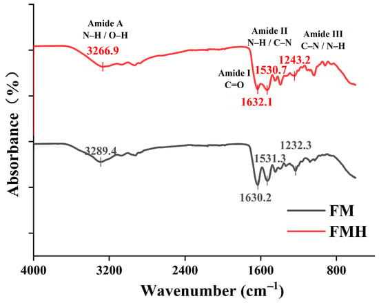
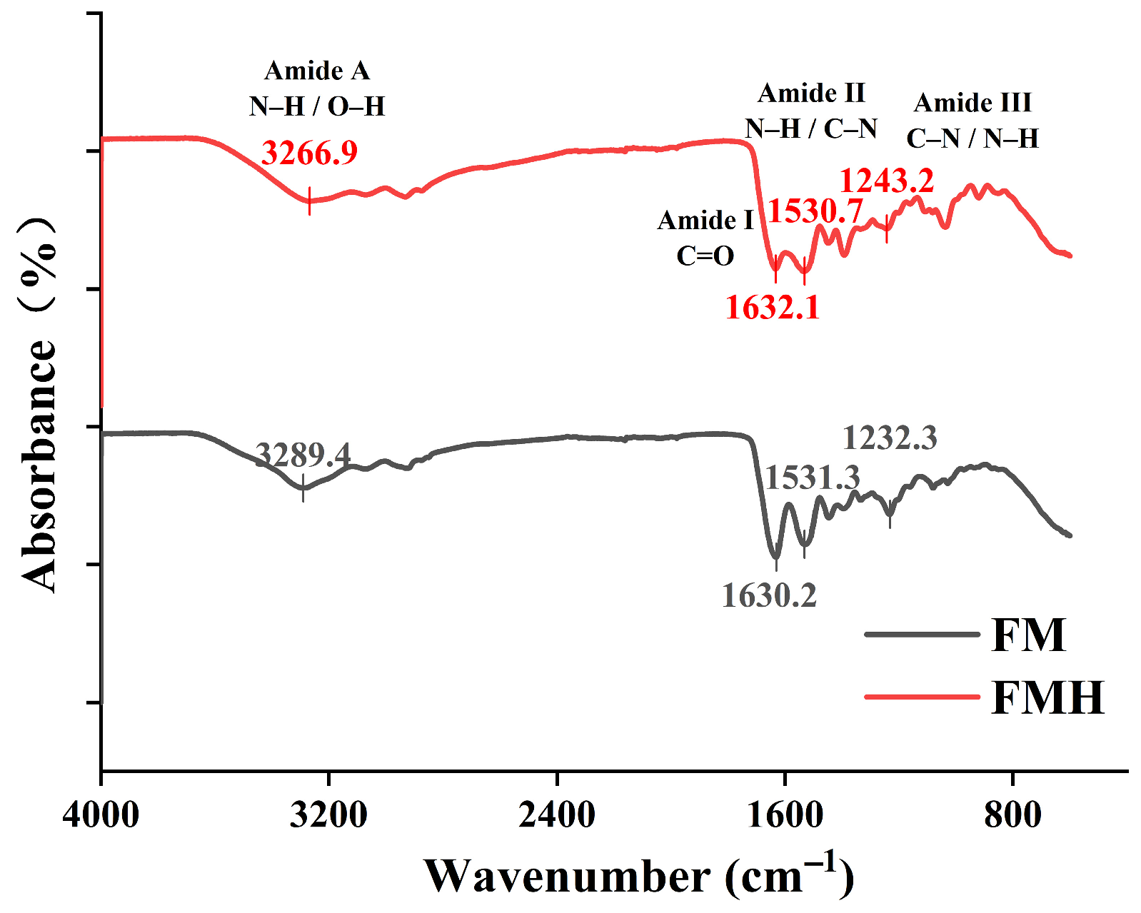
FTIR spectroscopy analysis was performed on components during FMH preparation (Figure 1). Both spectra displayed characteristic peaks of type I collagen, particularly in the amide A, I, II and III regions. The amide A band appeared at 3289.4 cm−1 for FM and 3266.9 cm−1 for FMH, mainly associated with N-H and O-H stretching vibrations. Compared with FM, FMH showed a redshift of 22.5 cm−1, indicating a change in the hydrogen-bonding interactions [33]. The amide I band (1700–1600 cm−1), dominated by C=O stretching vibrations, showed peaks at 1630.2 cm−1 (FM) and 1632.1 cm−1 (FMH) [34]. The amide II band showed peaks at 1531.3 cm−1 (FM) and 1530.7 cm−1 (FMH), mainly arising from N-H bending coupled with C-N stretching vibrations. In the amide III region (1400–1200 cm−1), FM and FMH exhibited bands at 1232.3 and 1243.2 cm−1, which are commonly used as indicators of collagen-related structural features [35]. Overall, the presence of these characteristic amide bands in FMH is consistent with the retention of collagen-derived chemical features after hydrolysis, although slight shifts in peak positions suggest changes in the local molecular environment and intermolecular interactions.
Figure 1.
Fourier transform infrared (FTIR) spectra of FMH.
As shown in Table 3, FMH is enriched in charged amino acids (e.g., Asp, Glu, Arg, Lys, and His) and hydrophobic amino acids (e.g., Gly, Ala, Tyr, Val, Met, Phe, Ile, Leu, and Pro), which account for 28.61% and 67.33% of the total amino acid content, respectively.
Table 3.
Amino Acid Composition Analysis of FMH.
High-resolution mass spectrometry (HRMS) was employed to characterize the peptide composition of FMH. Low-molecular-weight peptides (precursor mass < 400 Da) were identified through spectral matching using the GNPS online database, while larger polypeptides were further characterized by database searching against the UniProt/Swiss-Prot database of Perca fluviatilis. The molecular network generated 1032 nodes, with each node representing a precursor ion feature (Figure S1). Cross-referencing with the GNPS spectral library enabled the identification of five small peptides, including Asp-Leu, Gly-Pro-Ala, Glu-Val-Phe, pyroGlu-Tyr, and pyroGlu-Phe. In addition, seven polypeptides with high signal intensity were identified (relative intensity > 107, Table 4), which were mainly assigned to collagen alpha-1(I), alpha-1(XII), and alpha-1(XXVIII) chains. These peptides exhibited molecular weights below 1500 Da, contained characteristic collagen-like Gly-X-Y repetitive motifs, and were enriched in amino acid residues including Gly (G), Pro (P), Ala (A), Arg (R), and Glu (E) [36].
Table 4.
Polypeptide Sequence Analysis of FMH.
3.2. FMH Ameliorates Systemic Immune Function
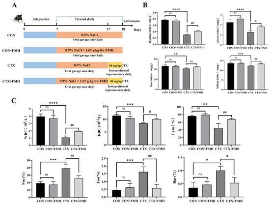
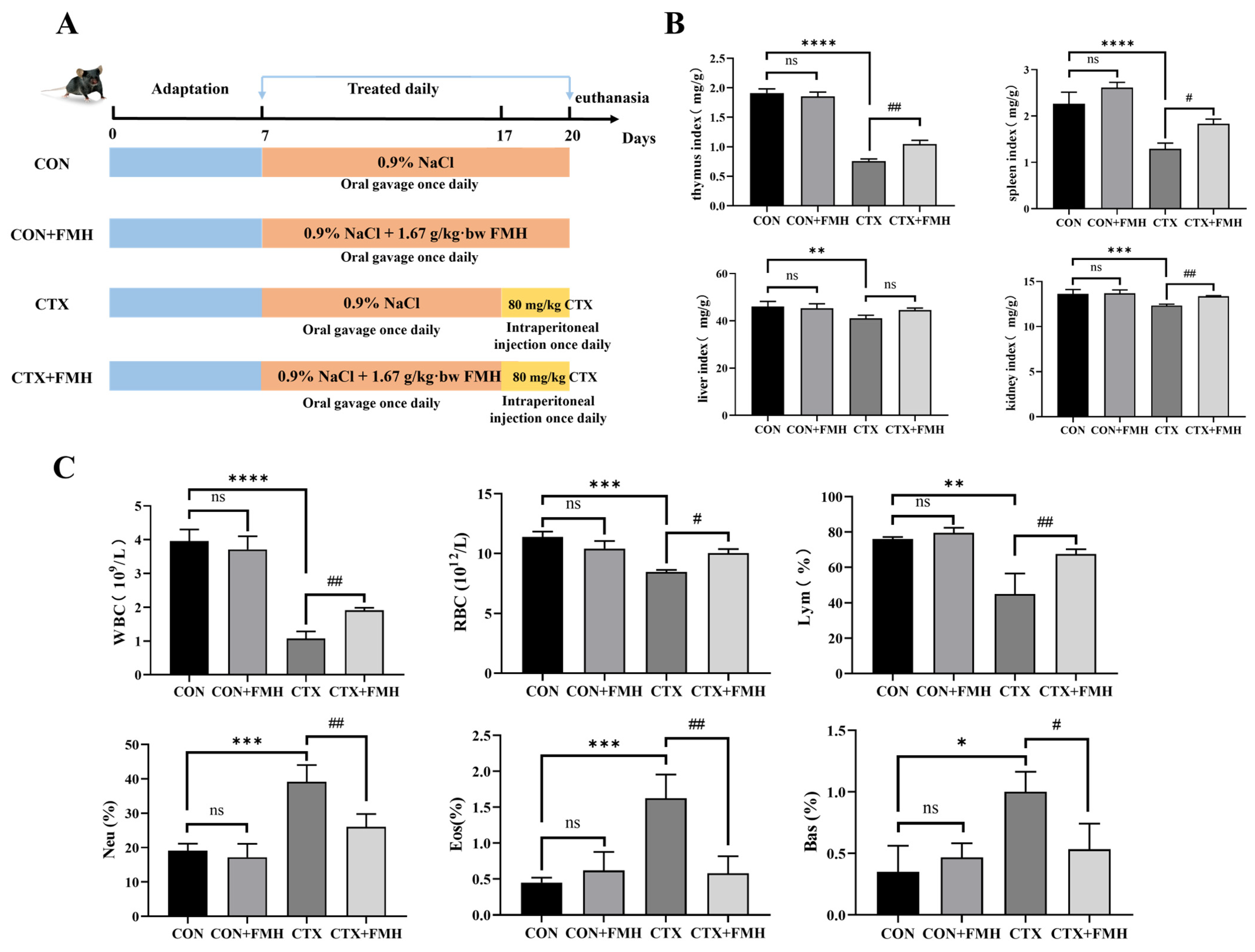
A total of 32 male C57BL/6J mice were used to investigate whether FMH could alleviate immunosuppression (Figure 2A). Body weight and food intake of mice in each group were monitored during the feeding period (Figure S2). After CTX injection, both body weight gain and food intake decreased in the CTX and CTX+FMH groups, with larger decreases in the CTX group. These findings suggest that FMH may partially alleviate the CTX-induced reductions in body weight and food intake in immunosuppressed mice.
Figure 2.
Effect of FMH on CTX-induced immunosuppressed model. (A) Schematic of the experimental design. (B) Organ indices. (C) peripheral blood count. Data are expressed as the mean ± SD. Compared with the CON group: ns, p ≥ 0.05; * p < 0.05, ** p < 0.01, *** p < 0.001, **** p < 0.0001; Compared with the CTX group: ns, p ≥ 0.05; # p < 0.05, ## p < 0.01.
At the end of the intervention, organ indices (thymus, spleen, liver, and kidney) were evaluated across all groups (Figure 2B). Mice treated with CTX exhibited significant decreases in thymus, spleen, liver, and kidney indices compared with the CON group (p < 0.01). Notably, FMH intervention significantly improved the thymus, spleen, and kidney indices in CTX-treated mice (p < 0.05). No significant differences in organ indices were observed between the CON group and the CON+FMH group (p ≥ 0.05), indicating that FMH administration under physiological conditions induced no adverse effects.
Blood cell counts for each group of mice were measured (Figure 2C). A 3-day continuous intraperitoneal injection of CTX significantly reduced the levels of WBC, RBC, and Lym in the peripheral blood of mice (p < 0.01). Neu, Eos, and Bas increased significantly (p < 0.05). In contrast, no significant differences in blood cell counts were observed between the CON and CON+FMH groups (p ≥ 0.05), suggesting that FMH did not markedly affect peripheral blood cell profiles in normal mice. Notably, when compared with the CTX group, the CTX+FMH group exhibited significant improvement in WBC, RBC, Lym, Neu, Eos and Bas counts (p < 0.05).
The experimental findings suggested that FMH was well tolerated under physiological conditions and improved the thymus, spleen, and kidney indices, partially restored several peripheral blood cell parameters, and alleviated CTX-induced systemic immunosuppression.
3.3. FMH Enhances Splenic Immune Activity
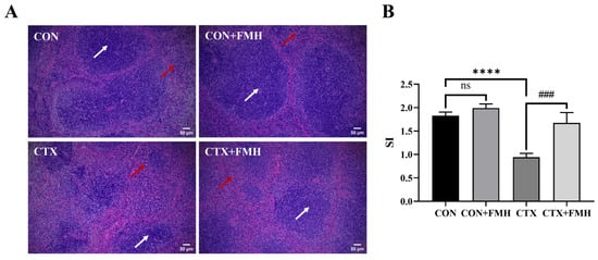
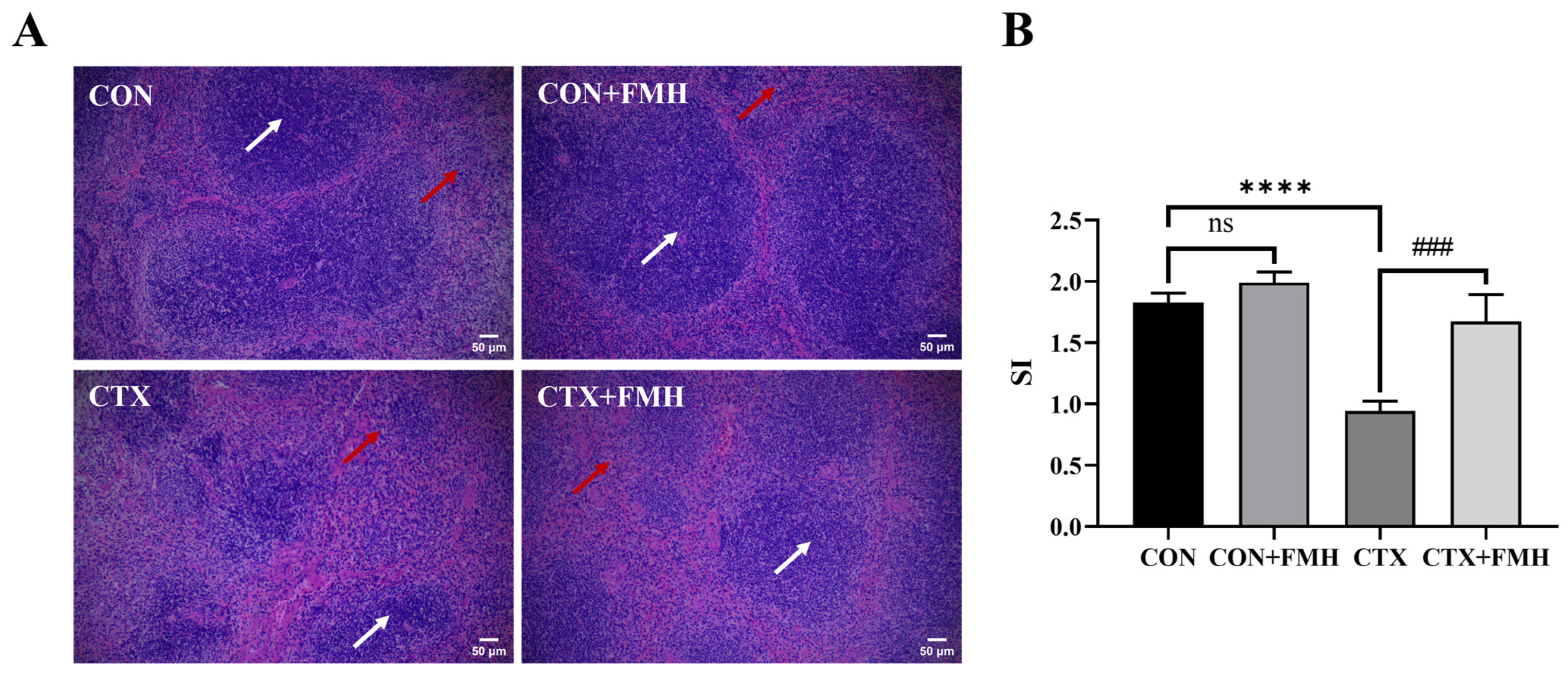
Histopathological images of H&E-stained splenic tissues are illustrated in Figure 3A. In the CON and CON+FMH groups, the splenic tissues exhibited an intact capsule, clear demarcation between red pulp and white pulp, and lymphocytes arranged in orderly patterns. In CTX-treated mice, the boundary between red pulp and white pulp was blurred, the white pulp structure was reduced, lymphocyte count decreased, cells were loosely arranged, and staining intensity was diminished, indicating that CTX caused significant structural damage to the spleen and induced immunosuppression. Compared with the CTX group, the CTX+FMH group showed distinct demarcation between red and white pulp, increased white pulp structure, and a mitigated reduction in lymphocyte count, demonstrating that FMH significantly alleviated splenic lesions in CTX-induced immunosuppressed mice. Compared with the CON group, the CTX group exhibited a significantly decreased stimulation index (SI). Notably, the SI value in the CTX+FMH group was significantly increased compared to the CTX group (p < 0.001), demonstrating that FMH enhances the proliferative capacity of splenic lymphocytes in CTX-induced immunosuppressed mice (Figure 3B).
Figure 3.
Effect of FMH on splenic immune activity. (A) Histological architecture of the spleen in each group (H&E staining, ×100). White arrows demarcate the white pulp regions, while red arrows highlight the red pulp areas. (B) Splenic lymphocyte proliferation in mice. Stimulation index (SI) was quantified to assess mitogen-induced proliferative activity. Data are expressed as the mean ± SD. Compared with the CON group: ns, p ≥ 0.05; **** p < 0.0001; Compared with the CTX group: ### p < 0.001.
3.4. FMH Regulates Cytokine Homeostasis
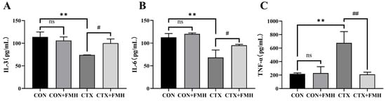
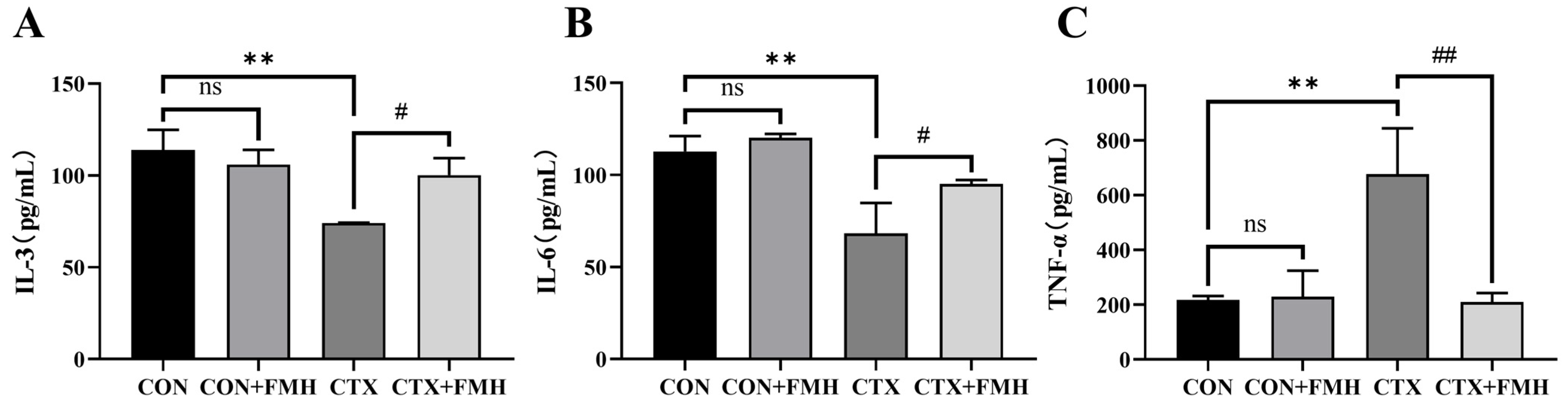
ELISA revealed no significant differences (p ≥ 0.05) in serum cytokine levels between the CON and CON+FMH groups (Figure 4). Compared to the CON group, the CTX group exhibited significantly decreased IL-3 and IL-6 levels (p < 0.01) and increased TNF-α levels (p < 0.01). Relative to the CTX group, the CTX+FMH group showed significantly increased IL-3 and IL-6 levels (p < 0.05) and decreased TNF-α levels (p < 0.01), indicating that FMH effectively regulates cytokine levels.
Figure 4.
Effect of FMH on serum cytokine levels. (A) IL-3. (B) IL-6. (C) TNF-α. Data are expressed as the mean ± SD. Compared with the CON group: ns, p ≥ 0.05; ** p < 0.01; Compared with the CTX group: # p < 0.05, ## p < 0.01.
3.5. Transcriptome Reveals FMH-Mediated Immunomodulation via NF-κB Pathway
3.5.1. Inter-Sample Relationship and Differentially Expressed Genes (DEGs) Analysis
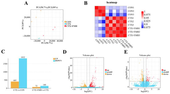
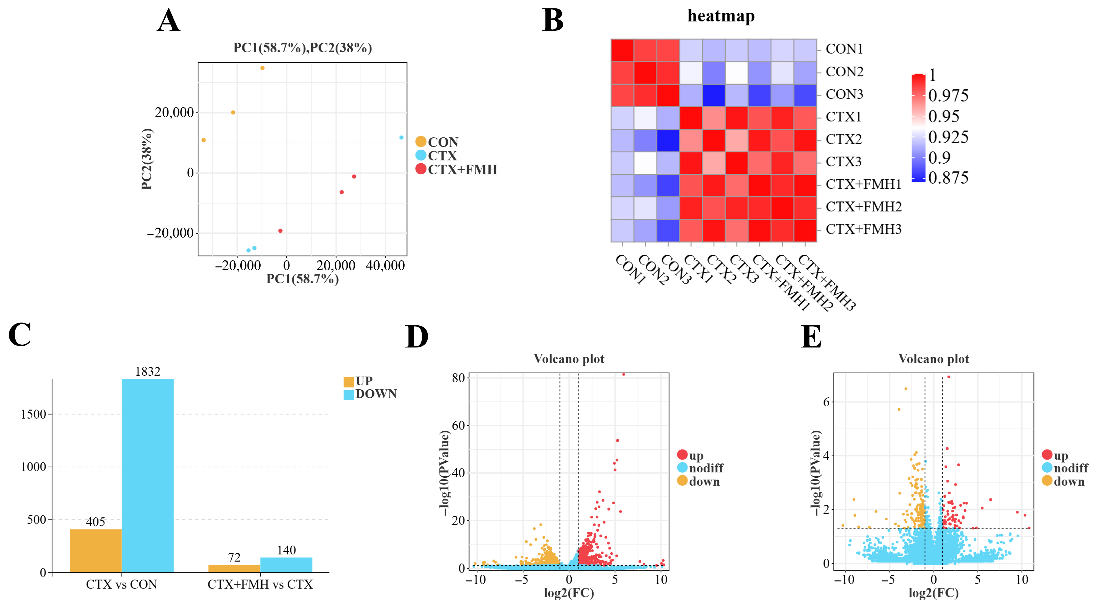
The PCA scatter plot revealed a clear grouping trend among the different groups (Figure 5A). Specifically, the data points for the CTX and CTX+FMH groups showed some overlap along the first principal component (PC1) but were more dispersed along the second principal component (PC2). In contrast, the CON group data points were distinctly separated from those of the CTX and CTX+FMH groups along PC1, and they also exhibited significant differences along PC2. This distribution pattern suggests that the CTX and CTX+FMH groups showed partial similarity in overall transcriptomic profiles, while still exhibiting separation along the second principal component. Although one sample showed a relatively distinct distribution in the PCA plot, this may reflect biological variability among individual mice. Nevertheless, the overall grouping trend remained clear, indicating that CTX markedly altered thymus transcriptomic profiles.
Figure 5.
Thymus transcriptome analysis. (A) Principal component analysis (PCA). (B) Sample correlation heatmap based on Pearson correlation coefficients. (C) Counts of up-and downregulated differentially expressed genes (DEGs). (D) Volcano plot: CTX vs. CON. (E) Volcano plot: CTX+FMH vs. CTX.
To further validate the correlations between samples, we generated a heatmap based on Pearson correlation coefficients to visualize sample-to-sample correlations (Figure 5B). The correlation coefficients between all samples were above 0.875, indicating good inter-sample consistency. This provides assurance of data quality for subsequent differential gene expression analysis. In the CTX vs. CON group, a total of 2237 DEGs were identified, with 405 upregulated genes and 1832 downregulated genes. In the CTX+FMH vs. CTX group, a total of 212 DEGs were identified, with 72 upregulated genes and 140 downregulated genes (Figure 5C–E). The full lists of identified genes and DEGs for these two comparisons are provided in Supplementary Tables S1–S4.
3.5.2. KEGG Enrichment Analysis
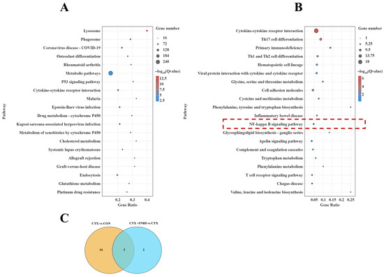
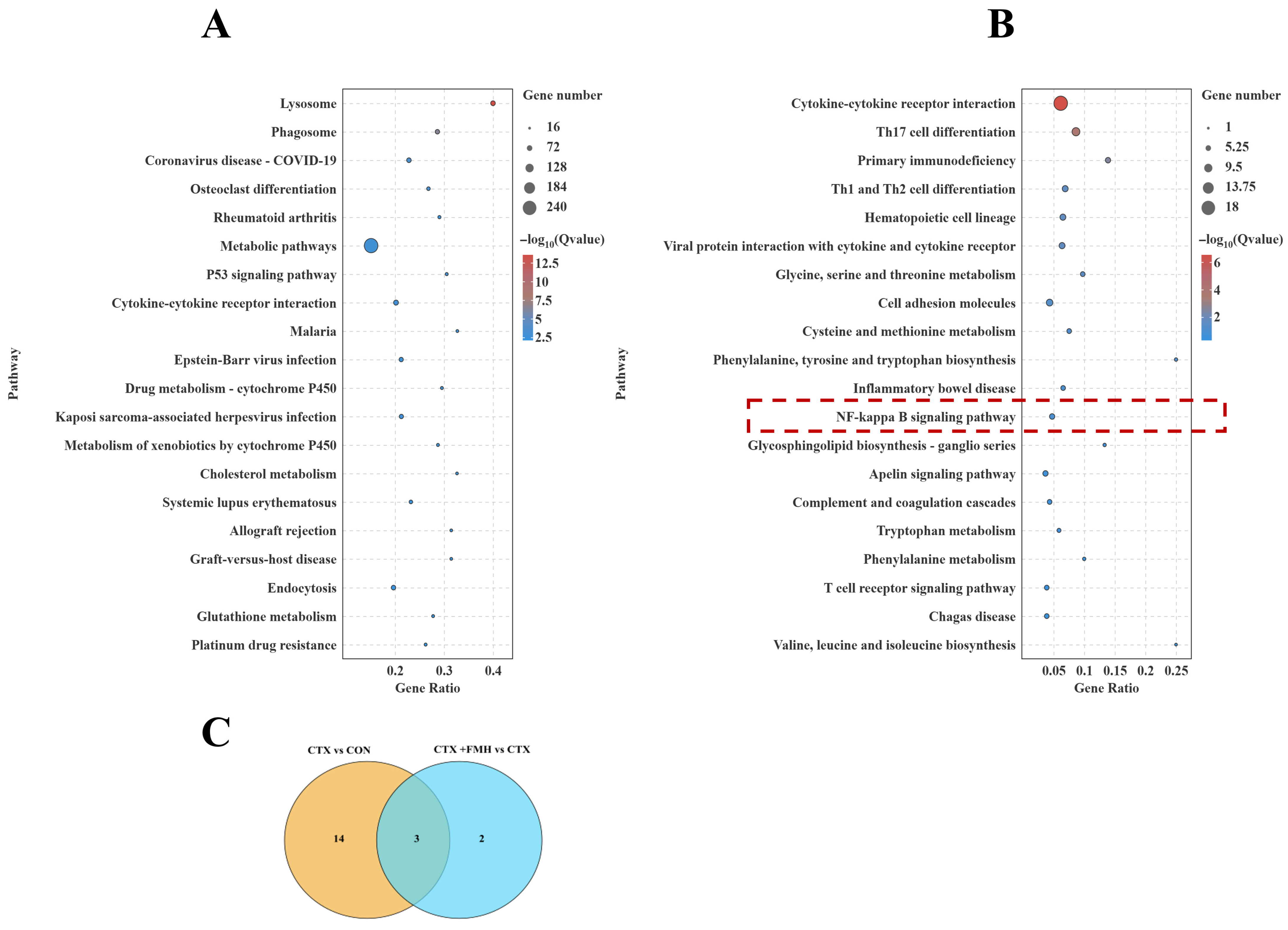
KEGG enrichment analysis was performed on the DEGs identified in each comparison (Figure 6). In the CTX vs. CON comparison, the top enriched pathways included lysosome, phagosome, coronavirus disease-COVID-19, osteoclast differentiation, rheumatoid arthritis, metabolic pathways, p53 signaling pathway, and cytokine-cytokine receptor interaction, suggesting that CTX induced broad transcriptomic alterations associated with immune regulation, stress responses, and metabolic disturbance. In the CTX+FMH vs. CTX comparison, the enriched pathways were mainly associated with cytokine-cytokine receptor interaction, Th17 cell differentiation, primary immunodeficiency, Th1 and Th2 cell differentiation, hematopoietic cell lineage, viral protein interaction with cytokine and cytokine receptor, inflammatory bowel disease, T cell receptor signaling pathway, and NF-κB signaling pathway. These results suggest that FMH exerts its protective effect against CTX-induced immunosuppression mainly through modulation of immune-related signaling networks.
Figure 6.
KEGG pathway enrichment bubble plots of DEGs (bubble size: gene count; color: −log10(Q value)). (A) CTX vs. CON. (B) CTX+FMH vs. CTX; the dotted red box highlights the NF-κB signaling pathway selected for further analysis. (C) Venn diagram of DEGs enriched in the NF-κB signaling pathway.
Notably, the NF-κB signaling pathway was significantly enriched in the CTX+FMH vs. CTX comparison. As NF-κB is a central regulator of innate and adaptive immune responses and controls the transcription of multiple inflammatory mediators, this pathway was considered particularly relevant to the immunological focus of the present study and was therefore selected for further mechanistic validation [37]. The Venn diagram showed that three NF-κB pathway-related DEGs overlapped between the two comparisons, namely IL-1β, Ltb, and Zap70. Among them, IL-1β was upregulated in the CTX vs. CON comparison but downregulated in the CTX+FMH vs. CTX comparison, whereas Ltb and Zap70 showed the opposite trend, being downregulated in the CTX vs. CON comparison but upregulated in the CTX+FMH vs. CTX comparison (Figure 6C).
3.6. Mechanistic Validation of FMH-Mediated NF-κB Pathway Modulation
3.6.1. Transcriptional Profiling via Quantitative Real-Time PCR (qRT-PCR)
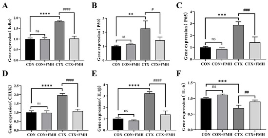
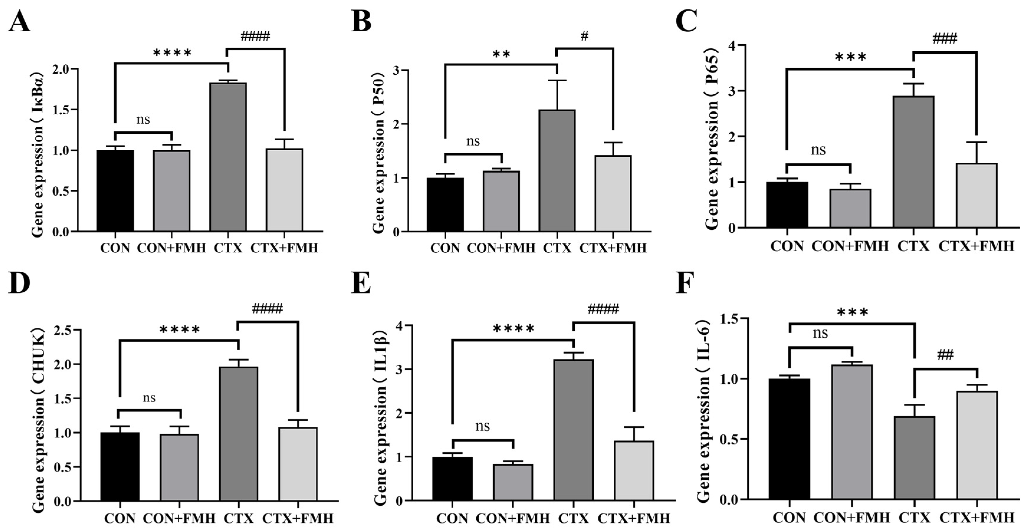
To further investigate the mechanisms by which FMH mitigates CTX-induced immunosuppression, the thymic mRNA expression levels of key NF-κB signaling pathway-related genes, including IκBα, P50, P65, CHUK, IL-1β and IL-6, were determined by qRT-PCR (Figure 7). Although Ltb and Zap70 were also identified among the overlapping NF-κB pathway-related DEGs in the transcriptome analysis, they were not prioritized for RT-qPCR validation because they mainly act upstream of NF-κB signaling, whereas the selected genes more directly reflect the core NF-κB cascade and its downstream inflammatory outputs [38,39]. No significant differences in NF-κB-related gene expression were observed between the CON and CON+FMH groups (p ≥ 0.05). In contrast, compared with the CON group, CTX treatment significantly increased the expression levels of IκBα, P50, P65, CHUK, and IL-1β (p < 0.01), alongside marked downregulation of IL-6 (p < 0.001). Notably, compared with the CTX group, FMH treatment significantly reversed the CTX-induced changes in the expression of IκBα, P50, P65, CHUK, IL-1β, and IL-6 (p < 0.05), suggesting that FMH may alleviate CTX-induced immunosuppression by modulating NF-κB signaling and its downstream inflammatory responses, which is consistent with the transcriptomic findings.
Figure 7.
Effect of FMH on the expression of NF-κB pathway-related genes in CTX-induced immunosuppressed mice. (A) IκBα, (B) P50, (C) P65, (D) CHUK, (E) IL-1β, (F) IL-6. Data are expressed as mean ± SD. Compared with the CON group: ns, p ≥ 0.05; ** p < 0.01, *** p < 0.001, **** p < 0.0001; compared with the CTX group: # p < 0.05, ## p < 0.01, ### p < 0.001, #### p < 0.0001.
3.6.2. Validation Through Immunohistochemical (IHC) Staining
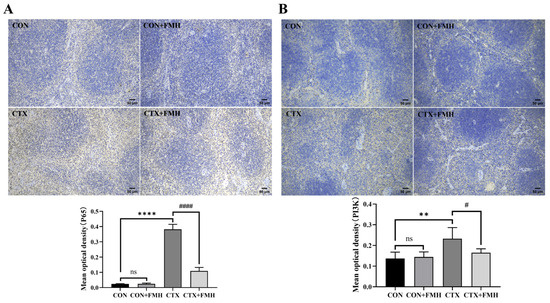
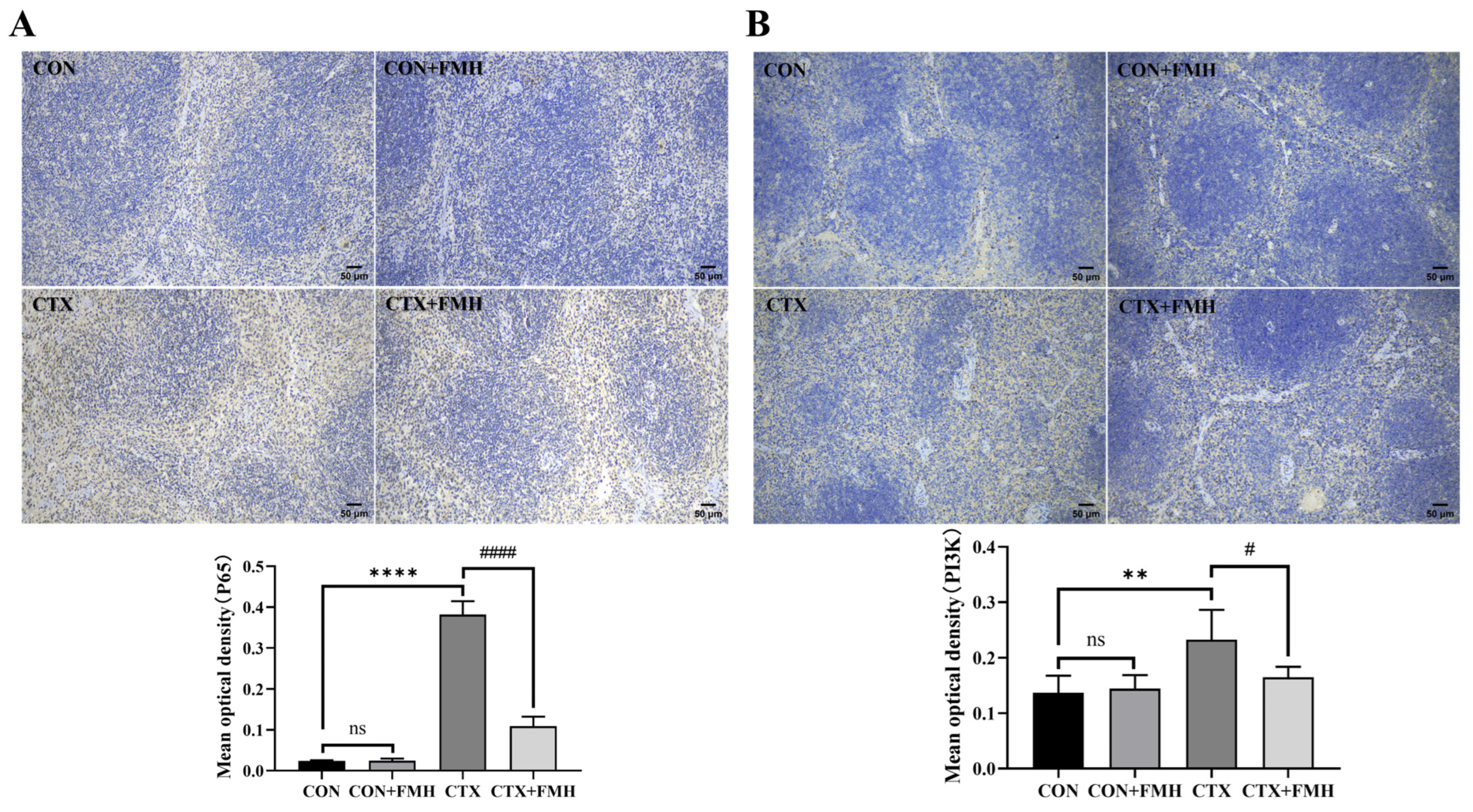
Immunohistochemical (IHC) staining was performed to evaluate the protein expression of P65 and PI3K in splenic tissues (Figure 8). No significant differences in P65 or PI3K expression were observed between the CON and CON+FMH groups (p ≥ 0.05). Compared with the CON group, the CTX group showed significantly increased expression of both P65 and PI3K (p < 0.01). In contrast, FMH significantly reduced the expression levels of these proteins compared with the CTX group (p < 0.05).
Figure 8.
Immunohistochemical staining (IHC, ×100) of P65 and PI3K in splenic tissues from each group. (A) NF-κB P65, (B) PI3K. Data are expressed as mean ± SD. Compared with the CON group: ns, p ≥ 0.05; ** p < 0.01, **** p < 0.0001; compared with the CTX group: # p < 0.05, #### p < 0.0001.
4. Discussion
The immune system protects the host against pathogenic invasions and maintains physiological homeostasis [40]. Recently, diet-derived bioactive immunomodulators have attracted significant interest due to their natural origin and functional efficacy [41]. Studies indicate that bioactive peptides from natural products, such as soybean [42] and Mytilus coruscus [43] exhibit immunomodulatory activity through regulation of immune cell function and cytokine secretion. While extensive investigations have been conducted on the immunomodulatory functions of bioactive peptides from diverse biological sources, the immunomodulatory properties and mechanisms of peptides in fish maw hydrolysate (FMH) remain unclear. In the present study, FMH derived from freshwater perch (Perca fluviatilis) maw exhibited immunomodulatory activity in CTX-immunosuppressed mice. FMH consisted mainly of low-molecular-weight peptides and was enriched in charged and hydrophobic amino acids. Moreover, FMH ameliorated CTX-induced alterations in immune organ indices, peripheral blood cell parameters, and cytokine profiles, possibly through modulation of the NF-κB signaling pathway. These findings expand the potential raw material sources for fish maw product development and support the development of dietary immunomodulators.
The immunomodulatory activity of bioactive peptides is linked to their structural characteristics, including relative molecular mass, amino acid composition, sequence arrangement, chain length, and hydrophobicity [44]. The FTIR spectrum of FMH exhibited characteristic peaks of type I collagen, indicating the presence of collagen-derived chemical features after hydrolysis. Low-molecular-weight peptides are often associated with enhanced immunomodulatory activity [45]. FMH contains peptides with molecular weights below 1755 Da, with the majority (83.89%) distributed within 73–580 Da. Consequently, the potential bioactivity of FMH may be partly attributed to the short-chain peptides generated during hydrolysis. In addition, HRMS analysis identified multiple small peptides and collagen-derived peptide fragments in FMH, further supporting the compositional complexity of the low-molecular-weight peptide fraction that may contribute to its bioactivity. Previous studies have reported that hydrophobic amino acids enhance immunomodulatory activity by strengthening peptide-membrane interactions [46]. Charged amino acids may also contribute to immunomodulatory effects through interactions with immune cells or related receptors [47,48]. FMH is enriched in charged amino acids and hydrophobic amino acids, which account for 28.61% and 67.33% of the total amino acid content, respectively. This compositional profile may contribute to its potential immunomodulatory activity.
CTX, an alkylating cytotoxic agent, is widely used to induce immunosuppression in experimental models [19]. Mechanistically, CTX can cause oxidative DNA damage and impair the proliferation and function of immune cells, thereby inducing immune dysfunction [49,50]. Compared with the CON group, CTX significantly reduced the organ indices of mice (spleen, thymus, liver, and kidney indices; p < 0.01). The decreases in WBCs, RBCs, and lymphocytes further indicated that immune function was significantly suppressed in CTX-treated mice [51,52]. H&E staining revealed structural lesions in splenic tissues characterized by white pulp atrophy and disordered lymphocyte arrangement. Furthermore, CTX significantly suppressed the SI of splenic lymphocytes (p < 0.0001) and concurrently decreased serum levels of IL-3 and IL-6, while elevating TNF-α expression (p < 0.01). These findings confirm the successful establishment of an immunosuppressive murine model induced by CTX.
As pivotal immune organs, the spleen and thymus are essential for immune cell differentiation, maturation, and antigen-specific responses [53]. FMH intervention significantly reversed CTX-induced atrophy of immune organs (p < 0.05). Histopathological evaluation showed that the CTX+FMH group exhibited marked restoration of splenic white pulp architecture, suggesting that FMH may alleviate CTX-induced splenic injury. Additionally, the proliferative capacity of splenic lymphocytes is widely used as an indicator of immune function [54]. FMH significantly enhanced the SI in CTX-treated mice (p < 0.001), suggesting its potential to ameliorate CTX-induced immune impairment by restoring cellular immune function. Importantly, no significant differences in immune organ indices or SI were observed between the CON and CON+FMH groups (p ≥ 0.05). Therefore, FMH appears to be well tolerated under physiological conditions and may mitigate CTX-induced immunosuppression by preserving immune organ integrity and improving cellular immune function.
Peripheral blood cells are important indicators of immune status. No significant differences in peripheral blood cell profiles were observed between the CON and CON+FMH groups (p ≥ 0.05), suggesting that FMH was well tolerated under physiological conditions. FMH intervention ameliorated CTX-induced alterations in several peripheral blood cell parameters, indicating its potential to alleviate CTX-driven immune cell dysregulation.
Cytokines play important roles in immune regulation by modulating innate and adaptive immune responses [55,56]. Octopus-derived peptides ameliorate CTX-mediated immunosuppression by activating IL-1β, IL-6, and TNF-α secretion pathways [57]. Shark peptides achieve immune homeostasis by modulating the secretion balance of IL-4, IL-6, IL-10, and TNF-α [58]. Cyclina sinensis pentadecapeptide effectively counteracts CTX-induced immunotoxicity through upregulation of IL-6, IL-1β, and TNF-α expression levels [59]. In the present study, FMH significantly modulated the serum levels of IL-3, IL-6, and TNF-α in immunocompromised mice (p < 0.05), suggesting its involvement in the regulation of systemic immune responses.
The thymus provides a unique microenvironment essential for the differentiation and maturation of T lymphocytes [60]. To further investigate the regulatory mechanisms of FMH on CTX-induced immunosuppression, thymic tissues from mice were collected for transcriptome profiling. KEGG analysis showed CTX-induced perturbations in pathways related to transport and catabolism, cell growth and death, immune diseases, and signaling interactions, including lysosome, phagosome, p53 signaling pathway, and cytokine-cytokine receptor interaction. FMH modulated CTX-induced immunosuppression through pathways associated with cytokine signaling, T-cell differentiation, immune dysfunction, and inflammatory signal transduction, including cytokine-cytokine receptor interaction, Th17 cell differentiation, Th1 and Th2 cell differentiation, primary immunodeficiency, inflammatory bowel disease, and the NF-κB signaling pathway.
The NF-κB pathway, governing innate immunity, adaptive immunity, inflammatory stress responses, B cell development, and lymphocyte formation, can be activated through canonical and non-canonical mechanisms [61,62]. Recent studies have demonstrated that collagen peptides from cod skin alleviate colitis by suppressing the phosphorylation of NF-κB P65, IκBα, and P38 MAPK in the NF-κB/MAPK pathway [34], while Lee et al. reported that fish collagen peptides promote thymic regeneration in CTX-damaged mice via the NF-κB signaling pathway [63]. In the current study, transcriptomic analysis revealed significant enrichment of the NF-κB signaling pathway in the CTX+FMH vs. CTX comparison, suggesting that NF-κB signaling may be involved in the immunomodulatory effect of FMH.
qRT-PCR validation of key NF-κB pathway genes demonstrated that FMH modulated the expression of both core regulatory components and downstream targets in thymic tissues of CTX-induced immunocompromised mice. Specifically, FMH modulated the expression of IκBα (a member of the IκB family), P50/P65 (NF-κB family transcription factors), and IKKα (CHUK, a catalytic subunit of the IKK complex), while also modulating downstream inflammatory mediators IL-6 and IL-1β. These results align with prior findings, suggesting that FMH mitigates thymic inflammatory injury and ameliorates CTX-induced immune dysfunction through modulation of the NF-κB-related inflammatory pathway and its downstream cascades.
P65, a key component of the NF-κB pathway, undergoes nuclear translocation following dissociation from IκB, thereby activating the transcription of cytokines and chemokines [37]. PI3K activation triggers AKT-mediated phosphorylation of the IKK complex, subsequently inducing cascading regulation of the NF-κB pathway [64]. In this study, no significant differences in P65 and PI3K protein expression were observed between the CON and CON+FMH groups in splenic tissues (p ≥ 0.05). CTX treatment significantly upregulated the expression levels of both P65 and PI3K proteins (p < 0.01), whereas FMH intervention markedly attenuated their overexpression in immunosuppressed mice (p < 0.05). Together with the transcriptomic and qRT-PCR results obtained from thymic tissues, these findings suggest that FMH may ameliorate CTX-induced immune dysregulation through coordinated modulation of the NF-κB pathway.
5. Conclusions
This study demonstrates the immunomodulatory activity and a possible mechanism of FMH derived from Perca fluviatilis maw. Structural characterization showed that FMH, predominantly distributed within 73–580 Da (83.89%), is enriched in hydrophobic and charged amino acids and contains small peptides and collagen-derived polypeptides. Moreover, in a CTX-induced immunosuppressed mouse model, FMH alleviated CTX-induced immune suppression by partially improving immune organ indices, ameliorating alterations in peripheral blood cell parameters, and regulating cytokine balance. Mechanistically, FMH may enhance immune responses through the modulation of the NF-κB pathway and its downstream pro-inflammatory cytokine cascades. These findings provide a new strategy for the high-value utilization of freshwater fish maw and offer a basis for expanding dietary sources of immunomodulatory hydrolysate. Further studies on dose dependence, safety, and validation in other relevant models would help support its future application.
Supplementary Materials
The following supporting information can be downloaded at: https://www.mdpi.com/article/10.3390/foods15071227/s1, Figure S1: Molecular network of FMH generated by GNPS; Figure S2: Effects of FMH on body weight and food intake; Table S1: Full list of identified genes in the CTX vs. CON comparison; Table S2: Full list of identified genes in the CTX+FMH vs. CTX comparison; Table S3: Differentially expressed genes (DEGs) in the CTX vs. CON comparison; Table S4: Differentially expressed genes (DEGs) in the CTX+FMH vs. CTX comparison.
Author Contributions
Conceptualization, M.-Z.P., H.-P.L. and X.-R.C.; Investigation, Z.-W.Z.; Resources, W.-S.L., X.T. and H.-P.L.; Validation, X.-M.G.; Data curation, J.S., Z.-W.Z. and Q.-T.Z.; Formal analysis, J.S., Z.-W.Z., Q.-T.Z. and X.T.; Visualization, J.S., Q.-T.Z. and X.-R.C.; Writing—original draft preparation, J.S.; Writing—review and editing, J.S. and X.-R.C.; Supervision, X.-M.G., W.-S.L. and M.-Z.P. All authors have read and agreed to the published version of the manuscript.
Funding
This research received no external funding.
Institutional Review Board Statement
The study was conducted according to the guidelines of the Declaration of Helsinki and approved by the Experimental Animal Welfare and Ethics Review Committee of Jiangnan University(Approval number JN.No20231115c0900104[537] was granted on 15 November 2023).
Informed Consent Statement
Not applicable.
Data Availability Statement
The raw transcriptomic sequencing data generated in this study have been deposited in the NCBI Sequence Read Archive (SRA) under BioProject accession number PRJNA1440437. The original contributions presented in the study are included in the article and its Supplementary Materials. Further inquiries can be directed to the corresponding authors.
Acknowledgments
We thank Figdraw for providing the drawing tools.
Conflicts of Interest
Author Wen-Sen Liu, Mei-Zhen Peng, and Hui-Ping Liu are affiliated with Guangdong Food Nutrition (Era Biotech) Engineering Technology Research Center (Shenzhen, China) asl researchers. The remaining authors (Jie Song, Zi-Wei Zhao, Qing-Tao Zhan, Xue-Mei Ge. Xue Tang, and Xiang-Rong Cheng) declare that the research was conducted in the absence of any commercial or financial relationships that could be construed as a potential conflict of interest. Guangdong Food Nutrition (Era Biotech) Engineering Technology Research Center focuses on technological research and development in the field of food nutrition and functional food-related technologies, and supports the transformation and application of related technologies. This study was advanced based on independent academic research plans and complementary research resources. The authors solemnly declare that there are no conflicts of interest involved, Thel research work described in this manuscript was carried out in strict accordance with scientific principles and ethical standards, and all authors have fulfilled their responsibilities in an unbiased manner, ensuring that the presented research findings are reliable and free from any undue influence. The institution did not influence the study design, methodologv. data analysis, interpretation, or conclusions. This manuscript is submitted solely for academic purposes without any commercial purposes.
References
- Ren, Z.; Yang, F.; Yao, S.; Bi, L.; Jiang, G.; Huang, J.; Tang, Y. Effects of low molecular weight peptides from monkfish (Lophius litulon) roe on immune response in immunosuppressed mice. Front. Nutr. 2022, 9, 929105. [Google Scholar] [CrossRef] [Scilit]
- Pisetsky, D.S. Pathogenesis of autoimmune disease. Nat. Rev. Nephrol. 2023, 19, 509–524. [Google Scholar] [CrossRef] [Scilit] [PubMed]
- Wellen, K.E.; Hotamisligil, G.S. Inflammation, stress, and diabetes. J. Clin. Investig. 2005, 115, 1111–1119. [Google Scholar] [CrossRef] [Scilit] [PubMed]
- Chen, D.S.; Mellman, I. Oncology Meets Immunology: The Cancer-Immunity Cycle. Immunity 2013, 39, 1–10. [Google Scholar] [CrossRef] [Scilit]
- Heneka, M.T.; van der Flier, W.M.; Jessen, F.; Hoozemanns, J.; Thal, D.R.; Boche, D.; Brosseron, F.; Teunissen, C.; Zetterberg, H.; Jacobs, A.H.; et al. Neuroinflammation in Alzheimer disease. Nat. Rev. Immunol. 2025, 25, 321–352. [Google Scholar] [CrossRef] [Scilit]
- Si, S.; Guo, Y.; Xu, B.; Qin, Y.; Song, S. Protective Effects of Collagen Peptides on the Dexamethasone-Induced Immunosuppression in Mice. Int. J. Pept. Res. Ther. 2021, 27, 1493–1499. [Google Scholar] [CrossRef] [Scilit]
- Hojo, M.; Morimoto, T.; Maluccio, M.; Asano, T.; Morimoto, K.; Lagman, M.; Shimbo, T.; Suthanthiran, M. Cyclosporine induces cancer progression by a cell-autonomous mechanism. Nature 1999, 397, 530–534. [Google Scholar] [CrossRef] [Scilit]
- Davis, T.E.; Kis-Toth, K.; Szanto, A.; Tsokos, G.C. Glucocorticoids Suppress T Cell Function by Up-Regulating MicroRNA-98. Arthritis Rheum. 2013, 65, 1882–1890. [Google Scholar] [CrossRef] [Scilit]
- Wen, J.; Zeng, L.; Chen, Z.; Xu, Y. Comparison of nutritional quality in fish maw product of croaker Protonibea diacanthus and perch Lates niloticus. J. Ocean Univ. China 2016, 15, 726–730. [Google Scholar] [CrossRef] [Scilit]
- Lan, X.; Luo, M.; Li, M.; Mu, L.; Li, G.; Chen, G.; He, Z.; Xiao, J. Swim bladder-derived biomaterials: Structures, compositions, properties, modifications, and biomedical applications. J. Nanobiotechnol. 2024, 22, 186. [Google Scholar] [CrossRef] [Scilit]
- Sapatinha, M.; Camacho, C.; Pais-Costa, A.J.; Fernando, A.L.; Marques, A.; Pires, C. Enzymatic Hydrolysis Systems Enhance the Efficiency and Biological Properties of Hydrolysates from Frozen Fish Processing Co-Products. Mar. Drugs 2025, 23, 14. [Google Scholar] [CrossRef] [Scilit]
- Xu, C.; Liu, Y.; Li, K.; Zhang, J.; Wei, B.; Wang, H. Absorption of food-derived peptides: Mechanisms, influencing factors, and enhancement strategies. Food Res. Int. 2024, 197, 115190. [Google Scholar] [CrossRef] [Scilit] [PubMed]
- Dai, C.; Dai, L.; Yu, F.-J.; Li, X.-N.; Wang, G.-X.; Chen, J.; Wang, C.; Lu, Y.-P. Chemical and biological characteristics of hydrolysate of crucian carp swim bladder: Focus on preventing ulcerative colitis. J. Funct. Foods 2020, 75, 104256. [Google Scholar] [CrossRef] [Scilit]
- Li, N.; Shiwei, L.; Yujie, M.; Nan, L.; Shanshan, W.; Zhou, D. In vitro antioxidant and anti-aging properties of swim bladder peptides from Atlantic cod (Gadus morhua). Int. J. Food Prop. 2020, 23, 1416–1429. [Google Scholar] [CrossRef] [Scilit]
- Cai, S.Y.; Wang, Y.M.; Zhao, Y.Q.; Chi, C.F.; Wang, B. Cytoprotective Effect of Antioxidant Pentapeptides from the Protein Hydrolysate of Swim Bladders of Miiuy Croaker (Miichthys miiuy) against H(2)O(2)-Mediated Human Umbilical Vein Endothelial Cell (HUVEC) Injury. Int. J. Mol. Sci. 2019, 20, 5425. [Google Scholar] [CrossRef] [Scilit]
- Wei, S.; Xu, L.; Zhu, Y.; Sun, C.; Lu, W.; Cao, Y.; Zhang, Y.; Zhao, Y.; Fang, Y. Characteristics analysis of microstructure and physicochemical properties of fresh and water-soaked fish maws derived from various swim bladder species. Food Res. Int. 2025, 202, 115761. [Google Scholar] [CrossRef] [Scilit]
- Xu, P.; Lu, C.; Sun, Z.; Kuang, Y.; Cao, D.; Huo, T.; Li, C.; Jin, H.; Zheng, X. In Silico Screening and Development of Microsatellite Markers for Genetic Analysis in Perca fluviatilis. Animals 2022, 12, 1809. [Google Scholar] [CrossRef] [Scilit]
- Yu, F.; Zhang, Z.; Ye, S.; Hong, X.; Jin, H.; Huang, F.; Yang, Z.; Tang, Y.; Chen, Y.; Ding, G. Immunoenhancement effects of pentadecapeptide derived from Cyclina sinensis on immune-deficient mice induced by Cyclophosphamide. J. Funct. Foods 2019, 60, 103408. [Google Scholar] [CrossRef] [Scilit]
- Meng, M.; Wang, H.; Li, Z.; Guo, M.; Hou, L. Protective effects of polysaccharides from Cordyceps gunnii mycelia against cyclophosphamide-induced immunosuppression to TLR4/TRAF6/NF-κB signalling in BALB/c mice. Food Funct. 2019, 10, 3262–3271. [Google Scholar]
- Liang, R.; Xu, L.; Fan, C.; Cao, L.; Guo, X. Optimization of Enzymatic Hydrolysis Process for Preparing Peptides from Asini Corii Colla and Its Antioxidant Activity. Sci. Technol. Food Ind. 2024, 45, 217–224. [Google Scholar]
- Ling, C.; Yuan, L.; Feifei, X.; Fang, Z. The effect of oral supplements containing collagen peptides rich in X-Hyp or X-Hyp-Gly compared with normal collagen hydrolysates on skin elasticity and collagen holes: A randomised double-blind clinical study. Food Funct. 2023, 14, 10628–10638. [Google Scholar]
- Cheng, X.-R.; Zhao, Z.-W.; Chen, Y.-Y.; Song, J.; Ma, J.-H.; Zhang, C.-X.; Amadou, I.; Lu, N.-Y.; Tang, X.; Guan, B. Interventional Effect of Donkey Bone Collagen Peptide Iron Chelate on Cyclophosphamide Induced Immunosuppressive Mice. Nutrients 2024, 16, 2413. [Google Scholar] [CrossRef] [Scilit] [PubMed]
- Li, Q.; Chen, G.; Chen, H.; Zhang, W.; Ding, Y.; Yu, P.; Zhao, T.; Mao, G.; Feng, W.; Yang, L.; et al. Se-enriched G-frondosa polysaccharide protects against immunosuppression in cyclophosphamide-induced mice via MAPKs signal transduction pathway. Carbohydr. Polym. 2018, 196, 445–456. [Google Scholar] [CrossRef] [Scilit]
- Xiang, L.; Shen, Y.; Liu, S.; Fan, B.; Zhan, J.; Zhou, Y.; Jiang, B.; Wang, M.; Liu, Q.; Liu, X.; et al. Guggulsterone ameliorates psoriasis by inhibiting keratinocyte proliferation and inflammation through induction of miR-17 directly targeting JAK1 and STAT3. Biochem. Pharmacol. 2025, 233, 116745. [Google Scholar] [CrossRef] [Scilit] [PubMed]
- Shi, L.; Fei, X.; Huang, J.; He, B.; Sun, Z.; Sun, G. DMC-BH derivative DMC-GF inhibits the growth of glioma stem cells by targeting the TRIM33/SLC25A1/mitochondrial oxidative phosphorylation pathway. J. Transl. Med. 2025, 23, 363. [Google Scholar] [CrossRef] [Scilit]
- Yao, J.; Liu, J.; He, Y.; Liu, L.; Xu, Z.; Lin, X.; Liu, N.; Kai, G. Systems pharmacology reveals the mechanism of Astragaloside IV in improving immune activity on cyclophosphamide-induced immunosuppressed mice. J. Ethnopharmacol. 2023, 313, 116533. [Google Scholar] [CrossRef] [Scilit]
- Pan, B.; Gao, J.; Chen, W.; Liu, C.; Shang, L.; Xu, M.; Fu, C.; Zhu, S.; Niu, M.; Xu, K. Selective inhibition of interleukin-1 receptor-associated kinase 1 ameliorates lipopolysaccharide-induced sepsis in mice. Int. Immunopharmacol. 2020, 85, 106597. [Google Scholar] [CrossRef] [Scilit]
- Kong, W.; Liu, Y.; Ai, P.; Bi, Y.; Wei, C.; Guo, X.; Cai, Z.; Gao, G.; Hu, P.; Zheng, J.; et al. Genetically modified E. Coli secreting melanin (E.melanin) activates the astrocytic PSAP-GPR37L1 pathway and mitigates the pathogenesis of Parkinson’s disease. J. Nanobiotechnol. 2024, 22, 690. [Google Scholar] [CrossRef] [Scilit]
- García-García, V.A.; Alameda, J.P.; Page, A.; Mérida-García, A.; Navarro, M.; Tejero, A.; Paramio, J.M.; García-Fernández, R.A.; Casanova, M.L. IKKα Induces Epithelial–Mesenchymal Changes in Mouse Skin Carcinoma Cells That Can Be Partially Reversed by Apigenin. Int. J. Mol. Sci. 2022, 23, 1375. [Google Scholar] [CrossRef] [Scilit]
- Ibrahim, M.F.G.; Ali, F.F.; Ali, S.F.E.-S.; Shaker, E.S.; Mahmoud, H.I.; Abdellatif, F.E.M.; Mokhemer, S.A. Neuroprotective effect of red dragon fruit extract ameliorates oxidative stress and inflammation in D-galactose-induced aging rat model: Biochemical, histological and immunohistochemical study. J. Mol. Histol. 2025, 56, 51. [Google Scholar] [CrossRef] [Scilit]
- Kinoshita, H.; Takenouchi, K.; Tsukamoto, N.; Ohnuki, K.; Suzuki, T.; Nakatsura, T. Identification of 68 HLA-A24 and -A2-restricted cytotoxic T lymphocyte-inducing peptides derived from 10 common cancer-specific antigens frequently expressed in various solid cancers. Neoplasia 2025, 61, 101135. [Google Scholar] [CrossRef] [Scilit] [PubMed]
- Salam, S.A.; George, C.S.; Al Abdulsalam, N.K.; Abdel-Moneim, A.M.; Essawy, A.E. Ginkgo biloba attenuates complete Freund’s adjuvant-induced inflammatory pain by suppressing the NF-κB-CXCL1/CXCR2 signaling cascade in the rat spinal cord. Redox Rep. 2025, 30, 2447778. [Google Scholar] [CrossRef] [Scilit]
- Petcharat, T.; Benjakul, S.; Karnjanapratum, S.; Nalinanon, S. Ultrasound-assisted extraction of collagen from clown featherback (Chitala ornata) skin: Yield and molecular characteristics. J. Sci. Food Agric. 2021, 101, 648–658. [Google Scholar] [CrossRef] [Scilit] [PubMed]
- Xin, X.; Zhou, J.; Liu, G.; Zhang, M.; Li, X.; Wang, Y. Anti-inflammatory activity of collagen peptide in vitro and its effect on improving ulcerative colitis. npj Sci. Food 2025, 9, 1. [Google Scholar] [CrossRef] [Scilit]
- Rozi, P.; Maimaiti, P.; Abuduwaili, A.; Wali, A.; Yili, A.; Akber Aisa, H. Isolation and Evaluation of Bioactive Protein and Peptide from Domestic Animals’ Bone Marrow. Molecules 2018, 23, 1673. [Google Scholar] [CrossRef] [Scilit]
- Silva, T.H.; Moreira-Silva, J.; Marques, A.L.P.; Domingues, A.; Bayon, Y.; Reis, R.L. Marine Origin Collagens and Its Potential Applications. Mar. Drugs 2014, 12, 5881–5901. [Google Scholar] [CrossRef] [Scilit]
- Liu, T.; Zhang, L.; Joo, D.; Sun, S.-C. NF-κB signaling in inflammation. Signal Transduct. Target. Ther. 2017, 2, 17023. [Google Scholar] [CrossRef] [Scilit]
- Courtney, A.H.; Lo, W.-L.; Weiss, A. TCR Signaling: Mechanisms of Initiation and Propagation. Trends Biochem. Sci. 2018, 43, 108–123. [Google Scholar] [CrossRef] [Scilit]
- Wolf, M.J.; Seleznik, G.M.; Zeller, N.; Heikenwalder, M. The unexpected role of lymphotoxin β receptor signaling in carcinogenesis: From lymphoid tissue formation to liver and prostate cancer development. Oncogene 2010, 29, 5006–5018. [Google Scholar] [CrossRef] [Scilit]
- Medzhitov, R.; Iwasaki, A. Exploring new perspectives in immunology. Cell 2024, 187, 2079–2094. [Google Scholar] [CrossRef] [Scilit] [PubMed]
- Gao, S.; Hong, H.; Zhang, C.; Wang, K.; Zhang, B.; Han, Q.-a.; Liu, H.; Luo, Y. Immunomodulatory effects of collagen hydrolysates from yak (Bos grunniens) bone on cyclophosphamide-induced immunosuppression in BALB/c mice. J. Funct. Foods 2019, 60, 103420. [Google Scholar] [CrossRef] [Scilit]
- Fan, X.; Zhang, Z.; Hu, Y.; Richel, A.; Wang, F.; Zhang, L.; Ren, G.; Zou, L. Current research status on the structure, physicochemical properties, bioactivities, and mechanism of soybean-derived bioactive peptide lunasin. Food Chem. 2025, 479, 143836. [Google Scholar] [CrossRef] [Scilit]
- Wang, Z.; Fang, Y.; Zeng, Y.; Yang, X.; Yu, F.-M.; Wang, B. Immunomodulatory peptides from thick-shelled mussel (Mytilus coruscus): Isolation, identification, molecular docking and immunomodulatory effects on RAW264.7 cells. Food Biosci. 2024, 59, 103874. [Google Scholar] [CrossRef] [Scilit]
- Chalamaiah, M.; Yu, W.; Wu, J. Immunomodulatory and anticancer protein hydrolysates (peptides) from food proteins: A review. Food Chem. 2018, 245, 205–222. [Google Scholar] [CrossRef] [Scilit]
- Yu, F.; He, K.; Dong, X.; Zhang, Z.; Wang, F.; Tang, Y.; Chen, Y.; Ding, G. Immunomodulatory activity of low molecular-weight peptides from Nibea japonica skin in cyclophosphamide-induced immunosuppressed mice. J. Funct. Foods 2020, 68, 103888. [Google Scholar] [CrossRef] [Scilit]
- Prommasith, P.; Surayot, U.; Autsavapromporn, N.; Rod-In, W.; Rachtanapun, P.; Wangtueai, S. Immunomodulatory, Anticancer, and Antioxidative Activities of Bioactive Peptide Fractions from Enzymatically Hydrolyzed White Jellyfish (Lobonema smithii). Foods 2024, 13, 3350. [Google Scholar] [CrossRef] [Scilit]
- Yang, Q.; Cai, X.; Huang, M.; Jia, L.; Wang, S. Immunomodulatory effects of Pseudostellaria heterophylla peptide on spleen lymphocytes via a Ca(2+)/CaN/NFATc1/IFN-γ pathway. Food Funct. 2019, 10, 3466–3476. [Google Scholar] [CrossRef] [Scilit]
- Jiang, Y.; Li, S.; Jiang, L.; Mu, G.; Jiang, S. Immunomodulatory activity and molecular mechanisms of action of peptides derived from casein hydrolysate by alcalase and flavourzyme based on virtual screening. J. Dairy Sci. 2025, 108, 2152–2168. [Google Scholar] [CrossRef] [Scilit] [PubMed]
- Zhang, J.; Zhou, H.-C.; He, S.-B.; Zhang, X.-F.; Ling, Y.-H.; Li, X.-Y.; Zhang, H.; Hou, D.-D. The immunoenhancement effects of sea buckthorn pulp oil in cyclophosphamide-induced immunosuppressed mice. Food Funct. 2021, 12, 7954–7963. [Google Scholar] [CrossRef] [Scilit] [PubMed]
- Deng, J.; Zhong, Y.-F.; Wu, Y.-P.; Luo, Z.; Sun, Y.-M.; Wang, G.-E.; Kurihara, H.; Li, Y.-F.; He, R.-R. Carnosine attenuates cyclophosphamide-induced bone marrow suppression by reducing oxidative DNA damage. Redox Biol. 2017, 14, 1–6. [Google Scholar] [CrossRef] [Scilit] [PubMed]
- O’Donnell, T.; Vabret, N. Red blood cells join the ranks as immune sentinels. Nat. Rev. Immunol. 2021, 21, 760–761. [Google Scholar] [CrossRef] [Scilit]
- Chapman, N.M.; Chi, H. Metabolic adaptation of lymphocytes in immunity and disease. Immunity 2022, 55, 14–30. [Google Scholar] [CrossRef] [Scilit]
- Cui, H.; Li, T.; Wang, L.; Su, Y.; Xian, C.J. Dioscorea bulbifera polysaccharide and cyclophosphamide combination enhances anti-cervical cancer effect and attenuates immunosuppression and oxidative stress in mice. Sci. Rep. 2016, 6, 19185. [Google Scholar] [CrossRef] [Scilit]
- Liu, N.; Dong, Z.; Zhu, X.; Xu, H.; Zhao, Z. Characterization and protective effect of Polygonatum sibiricum polysaccharide against cyclophosphamide-induced immunosuppression in Balb/c mice. Int. J. Biol. Macromol. 2018, 107, 796–802. [Google Scholar] [CrossRef] [Scilit]
- Saxton, R.A.; Glassman, C.R.; Garcia, K.C. Emerging principles of cytokine pharmacology and therapeutics. Nat. Rev. Drug Discov. 2023, 22, 21–37. [Google Scholar] [CrossRef] [Scilit] [PubMed]
- Luo, W.; Li, X.; Zhang, C.; Shen, K.; Li, M.; Zhuang, Y.; Huo, J.; Wang, W. Physicochemical characterization and protective effects of raw and nine-steamed Polygonatum cyrtonema polysaccharides on cyclophosphamide-induced immunosuppression in mice. Int. J. Biol. Macromol. 2025, 307, 141911. [Google Scholar] [CrossRef] [Scilit]
- Ali, M.; Ullah, H.; Farooqui, N.A.; Deng, T.; Siddiqui, N.Z.; Ilyas, M.; Ali, S.; Rahman, M.U.; Rehman, A.U.; Alioui, Y.; et al. NF-κB pathway activation by Octopus peptide hydrolysate ameliorates gut dysbiosis and enhances immune response in cyclophosphamide-induced mice. Heliyon 2024, 10, e38370. [Google Scholar] [CrossRef] [Scilit]
- Mallet, J.; Duarte, J.; Vinderola, G.; Anguenot, R.; Beaulieu, M.; Matar, C. The immunopotentiating effects of shark-derived protein hydrolysate. Nutrition 2014, 30, 706–712. [Google Scholar] [CrossRef] [Scilit]
- Zhao, R.; Jiang, X.; Zhao, Q.; Ye, H.; Lin, Y.; Huang, J.; Tang, Y. Immunoenhancing Effects of Cyclina sinensis Pentadecapeptide through Modulation of Signaling Pathways in Mice with Cyclophosphamide-Induced Immunosuppression. Mar. Drugs 2022, 20, 560. [Google Scholar] [CrossRef] [Scilit] [PubMed]
- Alawam, A.S.; Cosway, E.J.; James, K.D.; Lucas, B.; Bacon, A.; Parnell, S.M.; White, A.J.; Jenkinson, W.E.; Anderson, G. Failures in thymus medulla regeneration during immune recovery cause tolerance loss and prime recipients for auto-GVHD. J. Exp. Med. 2021, 219, e20211239. [Google Scholar] [CrossRef] [Scilit] [PubMed]
- Zhang, W.; Mao, G.; Feng, W.; Chen, Y.; Zhang, M.; Li, Q.; Wu, X.; Cheng, G.; Yang, L.; Zhao, T. Polysaccharide from Abelmoschus esculentus L. flower ameliorates cyclophosphamide-induced immunosuppression via the TLR4/endocytosis and MAPK-NF-κB signaling pathways in mice. Process Biochem. 2023, 130, 659–673. [Google Scholar] [CrossRef] [Scilit]
- Yu, H.; Lin, L.; Zhang, Z.; Zhang, H.; Hu, H. Targeting NF-κB pathway for the therapy of diseases: Mechanism and clinical study. Signal Transduct. Target. Ther. 2020, 5, 209. [Google Scholar] [CrossRef] [Scilit] [PubMed]
- Lee, D.Y.; Song, W.H.; Lim, Y.S.; Lee, C.; Rajbongshi, L.; Hwang, S.Y.; Kim, B.S.; Lee, D.; Song, Y.J.; Kim, H.-G.; et al. Fish Collagen Peptides Enhance Thymopoietic Gene Expression, Cell Proliferation, Thymocyte Adherence, and Cytoprotection in Thymic Epithelial Cells via Activation of the Nuclear Factor-κB Pathway, Leading to Thymus Regeneration after Cyclophosphamide-Induced Injury. Mar. Drugs 2023, 21, 531. [Google Scholar]
- Yang, D.; Yang, L.; Cai, J.; Li, H.; Xing, Z.; Hou, Y. Phosphoinositide 3-kinase/Akt and its related signaling pathways in the regulation of tumor-associated macrophages polarization. Mol. Cell. Biochem. 2022, 477, 2469–2480. [Google Scholar] [CrossRef] [Scilit]

Disclaimer/Publisher’s Note: The statements, opinions and data contained in all publications are solely those of the individual author(s) and contributor(s) and not of MDPI and/or the editor(s). MDPI and/or the editor(s) disclaim responsibility for any injury to people or property resulting from any ideas, methods, instructions or products referred to in the content. |
© 2026 by the authors. Licensee MDPI, Basel, Switzerland. This article is an open access article distributed under the terms and conditions of the Creative Commons Attribution (CC BY) license.