


Abstract
Aflatoxin B1 (AFB1), classified as a Group 1 carcinogen by the International Agency for Research on Cancer (IARC), is regulated at extremely low levels in food. Surface-enhanced Raman scattering (SERS), owing to its molecular fingerprinting capability and trace-level sensitivity, has emerged as a promising strategy for rapid and on-site AFB1 analysis. This review focuses on AFB1 as a single analytical target and provides a critical narrative review of recent advances across three sequential dimensions: detectability, specific recognition, and quantitative robustness. Representative direct AFB1-SERS developments include hotspot engineering, magnetic separation-assisted enrichment, nucleic acid cascade and CRISPR-based amplification, aptamer- and antibody-mediated recognition, ratiometric correction strategies, multipoint sampling, and portable platforms. Current literature increasingly emphasizes cross-batch reproducibility, matrix tolerance, validation using naturally contaminated samples, and agreement with HPLC/LC-MS/MS in addition to sensitivity improvement. At the same time, an ultra-low nominal limit of detection (LOD) alone is not indicative of analytical maturity, and many portable implementations simplify readout rather than deliver full workflow validation. This review provides an updated perspective on the opportunities and current limitations of AFB1-SERS for regulatory-relevant and field-deployable analysis.
1. Introduction
Aflatoxin B1 (AFB1) is a high-risk and representative analytical target among mycotoxin contaminants. The International Agency for Research on Cancer (IARC) has explicitly classified aflatoxins as Group 1 carcinogens, with AFB1 making the greatest contribution to their overall carcinogenicity [1]. The European Food Safety Authority (EFSA) has further confirmed the pronounced genotoxicity and carcinogenicity of AFB1, concluding that no acceptable daily intake (ADI) should be established and that dietary exposure should be reduced to as low as reasonably achievable [2]. Accordingly, research on AFB1 detection technologies is no longer concerned solely with sensitivity and is now closely linked to regulatory compliance, exposure assessment, and early warning system development.
The current regulatory framework defines AFB1 control through clearly specified quantitative limits. Schneidemann-Bostelmann et al. [3] reported that Commission Regulation (EU) 2023/915 sets a maximum AFB1 level of 0.10 μg/kg for cereal-based infant foods. AFB1 has consistently remained a key individual indicator in the regulation of nuts and foods for special dietary uses [4]. Global differences in regulatory limits reflect the underlying tension among risk assessment, regulatory effectiveness, and trade capacity [5]. In addition, the Codex Alimentarius Commission has established maximum limits for total aflatoxins in multiple food categories, and these standards are frequently used as important reference points in international trade rules [6].
AFB1 remains a widespread concern across global supply chains. Studies published from 2016 to 2022 indicate that cross-category contamination has caused the Margin of Exposure (MOE) in most samples to fall to levels indicative of health concern [7]. Long-term retrospective analyses of EU RASFF data (e.g., statistics from 1997 to 2022 or 2011–2021) further show consistently high numbers of aflatoxin notifications in nuts and nut products, especially in categories such as peanuts and pistachios, indicating that these remain high-risk commodities [8,9]. These observations identify AFB1 as a continuing analytical priority across multiple high-risk food commodities.
Recent studies have further confirmed the particular vulnerability of exposed populations to AFB1 [10]. The associated risks extend beyond raw grains to processed foods [8] and to products at the food–medicine interface, including herbal medicines, dietary supplements, and traditional Chinese medicinal materials [11,12]. AFB1 is therefore relevant to risk management across the food supply chain [13]. Conventional AFB1 analysis relies mainly on chromatography, mass spectrometry, and immunoassays, which have been summarized in earlier reviews [14,15]. High-performance liquid chromatography (HPLC) [16], liquid chromatography–tandem mass spectrometric (LC-MS/MS) [17] and enzyme-linked immunosorbent assay (ELISA) [18] methods provide high sensitivity and reliable quantification as confirmatory techniques, but they require specialized instrumentation, time-consuming sample preparation, and controlled laboratory conditions. Traditional immunoassays support rapid screening but may show limited matrix tolerance and variable quantitative performance in cereals, edible oils, and nuts. These constraints restrict the ability of conventional approaches to meet the growing demand for on-site screening, early warning, and high-throughput analysis across the food supply chain.
Spectroscopy-based techniques, including visible/near-infrared (Vis/NIR) spectroscopy [19], near-infrared spectroscopy [20], terahertz spectroscopy [21], colorimetry [22], fluorescence [23], hyperspectral imaging [24], and surface-enhanced Raman scattering (SERS) [25] have shown their potential and applicability for the rapid, routine detection of AFB1 in food crops. SERS technology is designed to improve Raman scattering signals and offers considerable analytical potential by exploiting plasmonic electromagnetic enhancement, chemical enhancement and molecular vibrational fingerprinting [26,27,28,29,30]. SERS can, in principle, achieve ultra-trace sensitivity, structural specificity, and rapid readout within a single measurement [31,32,33]. These attributes position SERS as a promising technology for the rapid and sensitive determination of pesticides [34,35,36,37], heavy metals [38,39], biotoxins [40], and fungi and bacteria [41,42] in food matrices. Particularly, previous studies have also extended SERS-based analysis to multiple mycotoxins, including the detection of AFB1, ochratoxin A (OTA) [43], ochratoxin B (OTB) [44], deoxynivalenol (DON) [45], zearalenone (ZEN) [46,47,48,49], and patulin [50,51].
Existing SERS reviews have mainly addressed substrate engineering, recognition elements, and applications across multiple food safety targets. They identify noble-metal nanostructures, semiconductors, and hybrid systems as the principal material platforms for improving detection performance [52]. A focused review of Au–Ag bimetallic nanoparticles further summarizes their classification, synthesis methods, and applications as SERS substrates for food contaminant detection, complementing broader discussions of noble-metal and hybrid nanostructures [53]. Broad surveys demonstrate the platform-level applicability of SERS across target classes [31], whereas thematic reviews of aptamer-based sensing and multi-target detection emphasize recognition design and rapid on-site testing [54,55]. More recent articles have reviewed SERS-based mycotoxin detection from substrate construction to application and have separately summarized aptamer-based strategies for aflatoxins [56,57]. Reviews of fruit and vegetable applications likewise identify substrate reproducibility and portable instrumentation as persistent barriers to implementation [58]. Recent reviews have further examined SERS-assisted multimodal biosensing for food hazards, simultaneous SERS detection of multiple food contaminants, and multimodal fusion detection strategies [59,60]. Reviews of Raman-plus-X dual-modal spectroscopy and emerging Raman technologies further discuss combining Raman spectroscopy with complementary analytical modalities to broaden food-analysis applications and facilitate the translation of laboratory methods toward practical use [61,62]. Together with broader reviews of mycotoxin occurrence, exposure control, and conventional analysis comparison [63,64], this literature provides the context for the present review. The distinct contribution here is a target-focused analysis of AFB1-SERS that separates direct AFB1-SERS evidence from the broader SERS literature. Table S1 in the Supporting Information summarizes representative prior reviews and their principal emphases. However, whether SERS can consistently deliver reliable quantitative results under the complex food matrix conditions and stringent regulatory limits characteristic of AFB1 remains an open question. This discrepancy between analytical potential and validated performance is the rationale for treating AFB1 as the sole target of this review.
In recent years, research on AFB1 in grains has moved beyond simple comparisons of technical performance toward an integrated paradigm of non-destructive handheld devices, which combines feature extraction, algorithmic discrimination, and performance validation. This underscores the growing trend toward real-time, non-destructive, and portable analysis [65]. For the portable devices, microfluidic chips are combined with micro-Raman spectroscopy to capture, enrich, and identify target samples. This reflects the potential of integrating sample pre-treatment with spectroscopic detection in miniaturized agricultural monitoring processes, which could facilitate the transfer of this approach to the detection of AFB1 [66]. To improve quantitative robustness, core–internal standard–shell architectures and ratiometric SERS strategies have also been investigated for food contaminants [40,67,68]. At the same time, reviews of machine learning applications in AFB1 analysis show that the major technical bottlenecks no longer lie primarily in model accuracy, but in reproducibility gaps arising from limited data scale, insufficient parameter transparency, and a lack of code openness [69]. This shift indicates that the evaluation of AFB1 detection methods has moved from raw discriminative capability toward the analytical robustness needed to support real-world decision-making.
This review outlines the primary challenges in the SERS detection of AFB1. By integrating recent advancements in the field of SERS, this paper focuses on discussing three critical issues: acquiring the characteristic signals of the target at ultra-trace levels and under complex matrix conditions; maintaining the specificity and stability of recognition amidst interference from structural analogs and matrices; and achieving reliable, universal quantitative results across diverse substrates, batches, and application scenarios [54,55,70].
The review first examines the molecular structure, interfacial adsorption behavior, and representative SERS features of AFB1 to establish a common basis for comparing direct readout, label-based amplification, and displacement-driven detection. It then considers how hotspot engineering, functional carriers, and cascade reactions enhance detectability, tracing the progression from nanostructure-level electromagnetic enhancement to carrier-assisted enrichment and nucleic acid-mediated signal amplification. The discussion of specific recognition compares label-free spectral differentiation, aptamer- and antibody-mediated recognition, and synthetic-material-assisted strategies, with attention to congener discrimination and matrix interference. Quantitative robustness is subsequently assessed through internal standard calibration, multipoint sampling, portable integration, and comparison with the gold standard approach (such as LC-MS/MS), including the reported limitations in reproducibility, matrix-matched calibration, and cross-batch stability. Field deployment is treated as a related translational dimension through portable Raman devices, disposable test strips, and integrated microfluidic platforms [31,32,33,65]. The review thereby assesses how close AFB1-SERS is to becoming a reliable analytical approach and identifies the obstacles that still separate laboratory performance from practical use.
Figure 1 summarizes the underlying rationale for AFB1 as a critical analytical target, and provides an overview of the main thematic pillars of SERS-based AFB1 analysis addressed in this review.
Figure 1.
Rationale for targeting aflatoxin B1 and core dimensions of SERS-based detection. Abbreviations: AFB1, aflatoxin B1; AFB2, aflatoxin B2; AFG1, aflatoxin G1; AFG2, aflatoxin G2; MIP, molecularly imprinted polymer; LOD, limit of detection; SERS, surface-enhanced Raman scattering; AI, artificial intelligence.
Scope and Evidence Classification of This Review
This article is a focused narrative review of AFB1-SERS, with particular emphasis on analytical performance, validation-related considerations, and on-site deployment. Direct AFB1-SERS experimental studies constitute the primary evidence base of the review, with emphasis on peer-reviewed research published from 2018 to 2026. Selected foundational studies published before this period are retained only when necessary to clarify vibrational assignments, interfacial chemisorption mechanisms, recognition chemistry, or the early technical development of AFB1-SERS sensing platforms. The literature was identified through targeted searches of PubMed and major publisher article databases, supplemented by backward reference checking of relevant primary studies and reviews. The literature searches were last updated in March 2026. Search terms combined “aflatoxin B1” or “AFB1” with “surface-enhanced Raman” or “SERS”, “aptamer”, “matrix”, “quantification”, “validation”, and “portable”. Direct experimental studies in which SERS was applied for AFB1 detection, discrimination, or quantification were prioritized during screening. Non-AFB1 SERS studies were retained only when they provided experimentally demonstrated strategies addressing a clearly defined methodological gap relevant to AFB1 analysis, whereas regulatory documents and reference-method literature were used solely to establish standard validation benchmarks for food mycotoxin testing.
2. Analytical Foundations and Evaluation Framework of AFB1-SERS
2.1. Molecular Structure and SERS Features of AFB1
AFB1 belongs to the class of difuranocoumarin derivatives. Its molecular structure consists of a rigid aromatic conjugated skeleton, a lactone ring, and an embedded bifuran ring. This distinctive chemical architecture gives rise to a rich molecular vibrational fingerprint and governs its orientation-selective adsorption on metal surfaces, thereby making the SERS signal of AFB1 highly dependent on the interfacial environment [33].
The information content of different structural units within the AFB1 molecule is not equivalent. The lactone ring and bifuran moiety are major structural determinants of AFB1 toxicity, whereas the carbonyl group and other oxygen-containing sites are more directly implicated in molecule–metal adsorption and interfacial charge-transfer processes [33,71,72]. Perturbations at these sites are often reflected in SERS spectra as characteristic peak shifts and relative intensity rearrangements [71,72,73]. Therefore, structural analysis in AFB1-SERS must distinguish the vibrational modes of the toxic core skeleton from spectral changes induced by interfacial interactions.
Theoretical studies provide important support for understanding this interfacial adsorption behavior. Density functional theory (DFT) calculations on silver-based interfaces show that the SERS response of AFB1 is strongly influenced by both the adsorption site and the excitation wavelength, with the chemical enhancement contribution reaching 102–104. These findings highlight the important role of the carbonyl group and other electron-rich oxygen atoms in mediating interfacial charge transfer [71]. The approximately 1592 cm−1 skeletal band and the carbonyl-sensitive regions around 1692–1700 and 1740–1758 cm−1 are therefore useful points of comparison, whereas 1166–1167 cm−1 is an operational quantification band for a particular Ag-IANP platform [25,73]. Studies on gold-based interfaces further confirm that AFB1 preferentially binds to the gold surface through its highly electronegative carbonyl oxygen [72]. Chemisorption of AFB1 onto SERS substrates modifies carbonyl-oxygen coordination, charge transfer, and adsorption geometry, which in turn alters both the relative intensities and peak positions of AFB1 SERS bands [71,72].
Reported acquisition settings were also different for diverse SERS substrates. Guo et al. used 638 nm excitation, a 10× objective, and a 1 s acquisition time in triplicate on the self-assembled Si-based aptasensor [74]. Wang et al. used 785 nm excitation, a 50× objective, five spatial points per sample, and a 5 s integration for peanut-oil SERS [75], whereas Singh et al. used a portable 785 nm system with three accumulations of 3 s on Ag-coated porous silicon [76]. These examples illustrate a trade-off among collection efficiency, fluorescence/background suppression, spatial sampling, and thermal or photochemical effects. Objective magnification changes the illuminated area and sampled hotspot population, while power, integration time, and the number of spots determine the spectral signal-to-noise and measurement duration. Therefore, the cited studies show that SERS spectral acquisition should be determined under their own optimized conditions rather than a universal specification.
Taken together, these results indicate that band reliability in AFB1-SERS is conditional, because substrate chemistry, excitation wavelength, and adsorption geometry can all reshape the analytical signal.
In terms of spectral resolution, the identification of AFB1 relies on a set of associated vibrational features that collectively define its skeletal structure. Comparative analyses of the SERS spectra of AFB1 and its congeners (AFB2, AFG1, AFG2), supported by theoretical assignments, have demonstrated distinguishable peak-shape differences within the fingerprint region [73]. Subsequent computational reviews further integrated these differences into a unified theoretical framework involving charge distribution, solvent effects, and molecule–metal interactions, thereby advancing the interpretation of AFB1 spectral responses from empirical discrimination to analysis based on structure–interface coupling [33]. This means that concentration-dependent analysis often relies on band patterns or multiband feature sets.
As conventional Raman evidence, studies in peanut matrices showed that models based on the synergistic optimization of multiple feature variables exhibited greater robustness than single-point responses [77]. In wheat sample analysis, the introduction of variable selection strategies such as competitive adaptive reweighted sampling (CARS), iteratively variable subset optimization (IVSO), and bootstrapping soft shrinkage (BOSS) significantly improved predictive robustness, indicating that effective information transfer depends more on feature combinations than on individual peaks [78]. Likewise, compact automated Raman systems have achieved accuracy comparable to that of laboratory platforms for detecting naturally contaminated corn flour, demonstrating the value of multivariate modeling in improving the performance of portable measurements [79]. These conventional Raman studies illustrate transferable principles of feature selection, but they do not establish AFB1-SERS performance. Using silver-coated porous silicon microarrays, researchers have successfully integrated specific recognition, fixed interfaces, and portable readout into a single sensor, thereby applying molecule–substrate interaction principles in a sensor design suitable for practical measurement [76]. By contrast, although near-infrared technology offers advantages for high-throughput, real-time screening, it lacks molecular vibrational fingerprint information directly corresponding to the vibrational modes of the AFB1 backbone and therefore cannot replace SERS in enabling detailed interpretation of the intrinsic significance of these spectral features [80].
The characteristic spectral signals of AFB1 are therefore closely related to both molecular structure and the interface state. Table 1 summarizes representative literature-supported SERS bands of AFB1 and related aflatoxin congeners, together with their principal assignments and reported analytical relevance, while explicitly distinguishing structurally assigned reference bands from platform-specific analytical bands [25,33,71,72,73,81,82]. Topics such as portable readout, chemometric modeling, and non-SERS spectroscopic comparison are discussed later in Section 3, Section 4 and Section 5 according to their analytical role.
Table 1.
Representative SERS bands of AFB1 and related aflatoxin congeners, with literature-supported assignments and analytical relevance.
In this review, the bands in the first section are retained as structure-anchored reference signals, whereas the bands in the last section are presented as platform-dependent analytical peaks reported for specific quantitative workflows. In particular, the ~1592 cm−1 region is one of the more consistently reported analytical bands in direct AFB1-SERS studies, whereas the ~1620–1625 cm−1 band is among the better-supported experimental discriminators between AF1s and AF2s. By contrast, peaks such as 1167 cm−1 or 1300 cm−1 should be understood as study-specific operational peaks rather than universally transferable AFB1 markers. The 1740–1758 cm−1 carbonyl-related band is shared across multiple aflatoxin congeners and should therefore not be described as an AFG-specific discrimination peak [25,33,71,72,73,81,82].
2.2. Major Detection Paradigms in AFB1-SERS
The evolution of AFB1-SERS detection paradigms is closely linked to advances in biorecognition chemistry. The introduction of new recognition units, such as high-affinity aptamers, specific antibodies, and, in some cases, auxiliary synthetic recognition materials, has driven detection platforms from simple spectral readout toward signal output driven by recognition events [83]. For AFB1, this means that SERS can read not only intrinsic vibrational fingerprints but also signal changes arising from probe conformational changes, dissociation, or spatial rearrangement [83].
Based on a synthesis of existing studies and differences in signal origin and quantitative logic, AFB1-SERS can be classified into three core paradigms: label-free direct detection, reporter-labeled competitive detection, and recognition element-mediated interfacial assembly and release detection [57]. Although these paradigms may overlap in practical applications, their analytical foundations differ substantially. For analytical clarity, they are best distinguished according to whether detection is based on the intrinsic spectrum of AFB1, the response of an introduced reporter molecule, or changes in interfacial assembly states [84]. These classes are also prone to different failure modes: direct readout is most sensitive to adsorption competition and matrix background, reporter systems are more exposed to probe stability and reaction kinetics, and assembly-based systems shift more of the burden toward interface reproducibility and fabrication consistency.
2.2.1. Label-Free Direct Detection
Label-free direct detection follows the most fundamental analytical logic of SERS, namely molecular fingerprint readout. It seeks to directly enhance the Raman signal of AFB1 itself using high-performance nanostructured substrates, thereby enabling qualitative identification or quantitative analysis. The main advantages of this strategy are its simple detection architecture, rapid response, and avoidance of risks associated with biosensor inactivation or non-specific interference. However, because direct detection is highly sensitive to SERS substrate surface properties, hotspot uniformity, molecular adsorption configuration, and matrix-derived background noise, its practical implementation remains challenging [85].
Systematic analyses of the SERS spectra of AFB1 congeners, together with DFT-based peak assignments, have established the scientific basis for fingerprint identification within this paradigm [73]. Early exploratory studies showed that, when combined with chemometric algorithms, direct SERS detection can effectively discriminate toxin contamination levels in corn extracts [85]. Subsequent studies further extended the practical utility of this paradigm in preliminary grain screening and rapid model development through the coupling of portable Raman systems with colloidal gold/silver substrates [86,87].
In recent years, label-free detection research has increasingly focused on improving resistance to matrix interference. Using a high-density enhancement system based on iodine-modified silver nanoparticles (Ag IANPs), researchers achieved sensitive detection of AFB1 in soybean extracts containing complex proteins and lipids [25]. In more challenging backgrounds such as vegetable oils, deep-learning models, including a convolutional neural network–long short-term memory hybrid (CNN–LSTM) have shown markedly stronger feature extraction capability than traditional linear models, improving prediction within the studied peanut-oil extracts, without yet demonstrating transfer to new oils or instruments [75]. Recent studies further indicate that, by exploiting the synergistic electromagnetic and chemical enhancement effects of aggregated silver nanocages, characteristic spectral fingerprints can be directly extracted from real samples while maintaining high selectivity, suggesting that label-free direct detection is gradually overcoming its earlier limitations in robustness [88].
Overall, the label-free paradigm is suitable for applications that prioritize rapid analysis and simplified operation. Its performance depends on the coordinated optimization of hotspot architecture, interfacial charge transfer, and spectral data analysis, rather than on substrate enhancement alone [75,88]. At the same time, many very low LOD claims in this class are obtained under simplified conditions; for regulatory-facing analysis, the more important question is whether intrinsic AFB1 bands remain quantifiable at the limit of quantification (LOQ) with acceptable recovery and precision in real extracts.
2.2.2. Reporter-Labeled Competitive Detection
Unlike label-free direct detection, labeled SERS does not depend on the inherently weak Raman scattering of AFB1 itself. Instead, it uses highly readable reporter molecules, such as 4-MBA, DTNB, and Prussian blue, to convert target binding into changes in label signals, thereby enabling indirect readout [57,89,90]. For small molecules such as AFB1, this strategy is particularly important because the small molecular size and limited sandwich-type recognition space restrict direct signal transduction; in such cases, competitive recognition, displacement response, and label release often provide more stable signals than direct readout of intrinsic peaks [83,84].
Building on this principle, reporter-labeled competitive SERS immunoassays convert the weak intrinsic Raman response of AFB1 into amplifiable, tunable, and quantifiable reporter signals [57]. This design reduces the likelihood that AFB1 signals will be masked by lipids, proteins, and pigments in grain and oil matrices and is therefore useful when low detection limits are required [84,89]. Nitrile-terminated reporters operating in the Raman-silent window (1800–2800 cm−1) provide a further means of reducing spectral overlap and autofluorescence in lipid-rich matrices [91].
A clear technical pathway for this category of AFB1 research has long been established. Coupling magnetic separation with core–shell SERS nanotags, where reporter molecules are deeply embedded between the metal core and shell, drastically shields the Raman signal from external matrix degradation and laser-induced fluctuations [92,93,94]. Li et al. [95] further introduced Exo III-assisted cyclic amplification to amplify the competitive readout, whereas He et al. [96] extended the dual-probe system from standard solutions to peanut oil, marking the initial application of this paradigm in real sample matrices. Collectively, these studies show that a modular design based on magnetic enrichment, labeled probes, and competitive recognition has become a common configuration for AFB1 reporter-labeled SERS detection.
Recent developments indicate that labeled systems are developing from single-target high-sensitivity formats toward analytical outputs with lower background, compatibility with multiplex detection, and greater robustness. Sun J. et al. introduced functionalized gold nanotags into a silicon-based photonic crystal microsphere biochip and achieved competitive SERS immunoassays for AFB1, ochratoxin A (OTA), and zearalenone (ZEN), demonstrating that the tagging strategy is inherently amenable to multiplex toxin detection [97]. Song et al. used aptamer-functionalized photonic crystal microsphere arrays for AFB1/OTA detection, further showing that three-dimensional array structures not only increase hotspot density but also improve focal localization and throughput [98]. These studies suggest that labeled detection can combine signal amplification with more standardized signal output formats, which may be advantageous for array-based and batch analysis [89].
Another important development is the pursuit of low-background operation and higher signal-to-noise ratios. Zhang J. et al. combined CRISPR/Cas12a amplification with Raman-silent-region labeling, converting AFB1 recognition into a nucleic acid-amplified readout with reduced background [99]. By contrast, Jiao et al. used aptamer-capped mesoporous silica nanoparticles to relate 4-MPBA release to AFB1 concentration. In this design, target recognition opens the pores and releases the preloaded reporter, so the signal is governed by the recognition-triggered release event [100]. Ratiometric and dual-signal designs have also been introduced to reduce errors caused by probe assembly and assay fluctuations. The GO-Au@Ag core–shell system reported by Chen P. et al. uses the intrinsic GO signal as a calibration reference and 4-MBA as the response signal, illustrating the shift from single-peak readout to calibratable output [101]. Similarly, the aptamer-based SERS/resonance Rayleigh scattering (RRS) system constructed by Lv et al. with AgMOF-derived silver single-atom catalysts demonstrates the growing diversity of reporters and signal-transduction pathways [102].
Accordingly, the role of reporter-labeled competitive detection is to replace direct reliance on the weak intrinsic Raman response of AFB1 with an amplifiable, controllable, and calibratable output signal. This strategy is particularly well suited to trace analysis at regulatory-limit levels and is more readily compatible with multiplex detection and complex matrices [90,100]. However, such systems usually involve more reaction steps and probe preparation and depend more strongly on label stability, batch consistency, and control of competitive reaction kinetics [84]. Evaluation of labeled/competitive systems should therefore not focus solely on the LOD; recovery, repeatability, and operational complexity are equally important because added recognition and amplification steps can themselves introduce new sources of variance [96,103,104].
2.2.3. Recognition-Mediated Assembly and Release Detection
Whereas labeled/competitive strategies mainly address how to make the signal stronger and easier to read, recognition element-mediated interfacial assembly and release detection, or solid-phase assembly assays, focus more on where the signal is generated and how it can be produced in a stable manner. Their common feature is the use of aptamers, antibodies, or complementary strands as the recognition core, integrating target capture, probe assembly, displacement dissociation, and signal output onto a fixed or semi-fixed interface. This design reduces fluctuations caused by random aggregation and improves controllability in complex matrices [83,84].
As a recognition core for AFB1, aptamers undergo distinct conformational rearrangements, which alters the spatial distance between SERS hotspots and the Raman reporter tags tethered on aptamer strands mainly via competitive displacement and target-triggered release mechanisms [98]. In competitive aptasensors, AFB1 binding induces aptamer folding and detaches the aptamer–reporter complex from the SERS substrate, decreasing the local reporter concentration within hotspots and weakening the measured SERS intensity. Alternatively, target-induced folding may pull reporter molecules into plasmonic hotspots to produce signal enhancement. Compared with the label-free direct detection of AFB1 intrinsic peaks, this aptamer-conformation-dependent readout is generally more robust. Direct AFB1 SERS signals are highly susceptible to adsorption geometry, substrate surface heterogeneity, and matrix interference, which cause unpredictable peak shifts and intensity fluctuations [71]. In contrast, aptamer-mediated SERS transduction relies on reporter signals rather than AFB1 molecular vibrations, decoupling the final readout from the random adsorption orientation of AFB1 molecules on the substrate surface. Nevertheless, this approach still bears intrinsic limitations from aptamer batch-to-batch variation, non-specific probe adsorption, and potential cross-reactivity toward structurally similar mycotoxin analogs, which require strict selectivity validation in spiked food matrices [105].
While reporter molecules can still be used in these methods, their core design focuses on interface ordering. These methods utilize solid silica, porous silica, magnetic carriers, Janus-like composite particles, and core–satellite structures to confine recognition events within a reproducible spatial framework, thereby stabilizing hotspot distribution, probe spacing, and the molecular trapping process [74,106,107]. This is particularly important for small molecules such as AFB1, because even minor fluctuations in interfacial configuration under trace-level conditions can be amplified into substantial quantitative errors [108]. The molecular basis of this approach is becoming increasingly clear. Xu et al. resolved the high-resolution structure of the AFB1 complex with the DNA aptamer AF26, showing that the aptamer undergoes conformational rearrangement to form an encapsulating binding pocket and enables molecular-level discrimination between AFB1 and AFG1. This finding provides a clear foundation for the design of recognition elements in subsequent interfacial assembly-based SERS systems [109]. Together, these findings indicate that solid-phase AFB1 assemblies require coordinated design of the full signal-generation pathway, including recognition conformation, binding orientation, and displacement mechanisms [109,110].
In terms of practical implementation, the silicon-based self-assembled aptamer sensor proposed by Guo et al. is a representative example. In this system, Si@AgNPs and AuMBA@AgNPs are immobilized on a unified interface, and aptamers regulate changes in the assembly state, resulting in good recovery in corn flour and oil samples. This demonstrates the advantages of solid-phase platforms in controlling spatial arrangement and reducing random fluctuations in complex matrices [74]. Nirala et al. further performed a systematic comparison of aptasensors and immunosensors on Ag-pSi substrates. The results showed that both recognition units enabled highly sensitive detection of AFB1; however, the aptamer-based system offered greater advantages in interfacial regeneration and structural tunability, whereas the antibody-based system retained the more established immunoassay logic [107]. This comparison suggests that, within this SERS detection paradigm, the matching between recognition chemistry and interfacial structure may be more important than a simple comparison of recognition-element category [110].
In addition to planar solid-phase platforms, magnetic and three-dimensional assembly structures have further advanced this paradigm. Chen Y. et al. used UiO-66-NH2-modified Fe3O4 magnetic beads to enhance signal-probe capture capacity, allowing target recognition, magnetic separation, and hotspot enrichment to occur on a single platform and thereby markedly improving interfacial assembly capability and controllability [111]. Chen P. et al. reported a 3D plasmonic aptasensor composed of Fe3O4@MoS2 magnetic nanoblooms and Au@Ag bimetallic nanostars, which integrates magnetic enrichment, 3D hotspots, and aptamer recognition into a single structure. This work shows that interfacial assembly can be designed to support target enrichment and background reduction within the same detection architecture [112]. The core–satellite aptasensor proposed by Sun D. et al. follows a similar logic: programmable nucleic acid hybridization drives the formation of high-density hotspot structures between Au nanostars and Fe3O4@Au, enabling the recognition event to directly correspond to a change in assembly state [105].
The emergence of Janus probes and intrinsically encoded probes has further advanced this paradigm toward quantitative robustness. Fu et al. employed Si–Au–Ag Janus nanocomposite probes and used the intrinsic Raman signals of the particles as encoded readouts, thereby reducing the additional uncertainty introduced by external reporter molecules and improving structural stability and quantitative consistency under acidic conditions [106]. Zhang D. et al. integrated magneto-plasmonic probes with membrane-like nanotags to construct a more stable solid-phase detection interface, achieving low RSD and prolonged shelf life in various food samples. These findings indicate that solid-phase assembly-based detection is gradually progressing from mere feasibility to stable operation [113]. This paradigm places greater emphasis than traditional labeling systems on interfacial engineering, batch consistency, and adaptability to complex samples [84]. Its main advantage is that recognition events typically occur at predefined interfaces, which facilitates spatial confinement, ratiometric correction, and substrate reuse. Its limitation is that platform construction is more complex and therefore imposes higher requirements on the stability of interfacial modification, retention of recognition element activity, and manufacturing consistency [106,107]. In other words, random aggregation errors are partly replaced by interface-manufacturing and modification-consistency errors that must also be validated. For this reason, such platforms are potentially more transferable than simple competitive labeling systems, although they still require careful validation in real samples, across batches, and against reference methods [74,108].
Flow-based lateral flow and vertical-flow formats extend some recognition-mediated designs toward multiplex field readout [45,114,115]. In these systems, the decisive feature is the recognition-controlled formation, disruption, or positioning of a SERS-active interface, even when a reporter is also present. Overall, label-free readout is operationally short and retains the intrinsic AFB1 fingerprint, but remains vulnerable to competitive adsorption in complex extracts. Reporter-labeled competitive assays support trace analysis and multiplexing, while probe fabrication and binding kinetics add variability. Recognition-mediated assembly or release can control hotspot geometry and enable solid-phase readout, although interface fabrication and functionalization require stronger process control. The three approaches therefore need to be judged against the intended matrix and task using LOQ, total assay time, recovery, repeatability, and independent validation as well as LOD.
Table 2 compares representative AFB1-specific evidence across the three paradigms. LOQ and complete sample-to-result time are reported less consistently than LOD; a short Raman integration is not a complete analytical workflow.
Table 2.
Comparative analytical characteristics of the three major AFB1-SERS detection paradigms.
2.3. Evaluation Metrics and Validation Criteria
In this review, the term “quantitative robustness” is used in a practical, literature-based sense to refer to the reported stability of calibration, reproducibility, matrix tolerance, and agreement with reference methods. For practical comparison, the present review distinguishes four validation levels: standard solutions only, spiked extracts, spiked real matrices, and naturally contaminated samples with reference-method comparison. These evidence levels are not analytically equivalent. A low LOD reported in solvent or a simplified extract does not provide the same evidential weight as acceptable recovery, precision, and chromatographic agreement obtained in naturally contaminated grain or oil matrices. In AFB1-SERS research, the LOD and linear range are often reported first. However, these metrics reflect only detectability and are not sufficient on their own to support a judgment of practical usability [116,117]. In accordance with general principles of analytical chemistry, including Eurachem guidelines and IUPAC validation specifications, an analytical method must report selectivity, working range, accuracy (recovery), repeatability, intermediate precision, and robustness, with measurement uncertainty included where necessary. Only when these parameters are adequately documented can a method progress from initial feasibility to a transferable analytical tool [116,118,119,120]. Together, these parameters determine whether the method is suitable for the intended sample type and application scenario. This requirement is particularly important for AFB1 because its analysis is primarily designed for food contaminant detection scenarios, characterized by very low regulatory limits, strong matrix interference, and genuine compliance assessments. Evaluation must therefore move beyond asking whether a signal is present and instead determine whether results can be obtained stably and accurately near regulatory concentration limits [121,122].
Regarding low-concentration performance, the LOD and LOQ should not be treated as fixed values. Guidance documents in the field of food contaminants clearly state that LOD/LOQ estimation should be based on blank matrices, spiked blanks, or pseudo-blank samples, with evaluation performed in a concentration range close to the expected detection limit; they should not be derived solely by direct extrapolation from wide-range solvent calibration curves [121]. Similarly, the true meaning of the LOQ is the ability to quantify with acceptable precision and accuracy. The LOQ is therefore inherently linked to method error, rather than being a simple mechanical extension of the LOD [116,121]. The European Commission Joint Research Center (JRC) guidance document for LOD and LOQ estimation of contaminants in feed and food supports this interpretation. The JRC guidance explicitly addresses mycotoxins and requires standardized LOD/LOQ estimation if monitoring and exposure modeling results approach the expected LOQ. The reported LODs in AFB1-SERS studies should therefore ideally be acquired through matrix-matched experimental designs, including blank matrices, spiked blanks, pseudo-blank samples, and low-concentration calibration data. Solvent-only calibration curves alone cannot serve as the sole calculation basis [121].
For AFB1-SERS, this issue is further amplified by real grain and oil matrices. Lipids, proteins, polysaccharides, and co-extracted impurities not only affect the background signal, but also alter the adsorption behavior and localized enhancement state of the target molecule. As a result, instrumental sensitivity and actual analytical capability are not equivalent [17,123]. Therefore, in strongly matrix-dependent systems such as those for AFB1, matrix-matched calibration, the standard addition method, or other calibration strategies capable of compensating for matrix effects often reflect true quantitative performance more accurately than simple solvent-based external standards [124,125]. If the calibration data exhibit heteroscedasticity, the routine application of ordinary least-squares fitting may underestimate errors at the low-concentration end [124]. This issue is particularly critical in AFB1-SERS. Although many studies report high correlation coefficients, they provide insufficient discussion of variance amplification in the low-concentration range, peak-area extraction methods, and the applicability limits of calibration models. Reliance on R2 alone is often insufficient to demonstrate analytical reliability [125,126]. It should also be emphasized that after introducing multivariate statistical models such as PLS into AFB1-SERS analysis, the detection limit is affected by both changes in background components and the correction space, and is not a constant parameter applicable to all samples [127]. Therefore, AFB1-SERS methods based on full-spectrum modeling should report both model training boundaries and external prediction performance.
Compared with conventional chromatographic methods, SERS shows greater platform dependence. Spot-to-spot and batch-to-batch variability, differences in sampling location, and the absence of unified calibration standards for spectral readout can all directly compromise the reproducibility of quantitative results. Accordingly, spot-to-spot and batch-to-batch consistency, multipoint sampling or mapping, internal standard calibration or ratiometric calibration, transparency in spectral preprocessing, and validation using an independent test set should all be regarded as essential reporting elements for AFB1-SERS [108,117,128]. Among these factors, the use of internal standards and ratiometric designs deserves particular emphasis. Related studies have shown that introducing an internal standard can effectively improve the relative standard deviation (RSD) and enhance overall model stability, serving as a key strategy for transforming random fluctuations into correctable errors [108,129].
Conventional chromatography and AOAC INTERNATIONAL-validated methods provide useful external benchmarks because specificity, accuracy, repeatability, working range, and detection limits are evaluated in real matrices under defined validation protocols [16,122]. Some recent Ag-pSi microarray studies have begun to approach a more complete validation record by reporting selectivity, reproducibility, storage stability, recovery, and RSDs in grain samples together with comparison against HPLC [76,108]. Such reports are particularly informative because they document a broader evidence chain.
This section evaluates the completeness of the evidence reported in representative AFB1-SERS studies. Table 3 compares matrix type, quantitative range, recovery, reproducibility, validation design, and reference-method comparison. Studies based only on bulk solutions or spiked samples remain informative, but they should not be treated as evidentially equivalent to studies that include real matrices, naturally contaminated samples, or chromatographic confirmation [76,117,123].
Table 3.
(A) Direct AFB1-SERS studies validated in standard solutions only. (B) Direct AFB1-SERS studies using AFB1-fortified food extracts. (C) Direct AFB1-SERS studies using fortified whole-food matrices. (D) Direct AFB1-SERS studies with verified positive or inoculated samples and reference-method comparison.
2.4. Matrix Effects in AFB1-SERS Evaluation
In real grain and oil samples, matrix components are a principal reason why performance achieved in buffer or standard solutions may not transfer directly to practical analysis. Differences in background composition and surface adsorption among grains, vegetable oils, and other foods alter the interpretation of LOD and LOQ values [32,110]. Vegetable oils are particularly challenging because their lipid-rich background affects both target extraction and interfacial adsorption [131,132]. Combining QuEChERS pretreatment with chemometric analysis has improved quantitative performance in oil samples [130], whereas magnetic extraction-assisted workflows offer a route to integrated enrichment, separation, and portable testing [133,134]. Accordingly, an AFB1-SERS workflow intended for real samples must include an appropriate strategy for compensating for matrix effects [32,76]. The following sections therefore discuss matrix mitigation in relation to recognition control, calibration validity, and workflow translation.
3. Detectability: Signal Enhancement and Enrichment
For AFB1-SERS, the central challenge is to obtain a strong, clean, and quantifiable signal at trace concentrations in the presence of severe matrix interference. Research has accordingly progressed from isolated hotspot enhancement toward the coordinated optimization of hotspot construction, target enrichment, and signal amplification. This section examines the origins and limitations of sensitivity enhancement through three routes: direct enhancement, functional-carrier coupling, and cascade amplification.
3.1. Hotspot Engineering for Direct and Recognition-Assisted SERS Readout
This section examines why label-free detection of AFB1 remains challenging in complex matrices [32]. Direct Raman studies indicate that AFB1 information is distributed across multiple characteristic bands. Variable selection and multivariate modeling are therefore important for extracting interpretable spectral information [77,78].
As discussed in Section 2.1, variable selection studies in wheat [78] and peanuts [77], as well as compact Raman-based classification of naturally contaminated corn flour [79], all indicate that information for direct AFB1 detection is more effectively extracted through feature combinations than through single-peak intensity alone. The analytical challenge for AFB1-SERS extends beyond merely achieving high local enhancement; it demands the reproducible acquisition of multiband information across the hotspot distribution. Consequently, this section examines how hotspot engineering facilitates the stabilization of these multivariate signals. From a broader SERS perspective, the value of such nanostructural optimization lies in driving signal amplification while transforming weak and variable responses into reliable analytical outputs through highly controllable local electromagnetic field distributions [31,32].
From the perspective of nanostructure design, hotspot engineering for AFB1-SERS mainly progresses along three pathways. The first is the construction of controllable nanogaps. Studies have shown that when the distance between two metal particles is reduced to 1–2 nm, electromagnetic field enhancement can increase by several orders of magnitude [135]. However, for low-molecular-weight small molecules such as AFB1, overly narrow gaps may hinder entry into the hotspot region. Thus, the compatibility between gap size and target molecules remains a parameter that requires systematic optimization. The second pathway is the use of tip-enhancement effects. High-curvature structures, such as nanostars, nanotriangles, and nanoflowers, can generate strong localized fields at the single-particle level, thereby reducing reliance on interparticle aggregation [96,112]. The third pathway is the development of ordered array platforms. As noted above, porous silicon microarrays [76] and photonic crystal microspheres [97,98] use photolithography, etching, or self-assembly to immobilize hotspots at predefined positions, thereby fundamentally reducing spatial randomness, which is especially important for the quantitative analysis of AFB1. To further eliminate the structural randomness of colloidal aggregation, recent breakthroughs employ template-free electrochemical programming to fabricate ultra-compact, monolithic substrates. Such rigidly locked nanogaps deliver exceptional batch-to-batch reproducibility [136,137].
In addition, emerging two-dimensional material substrates are expanding the scope of hotspot engineering. For MXene-based substrates, surface terminal groups (-O, -OH, -F) engage AFB1 mainly via hydrogen-bonding and weak charge-transfer interactions rather than strong covalent chemisorption [138]. Consequently, C=O peak shifts are generally milder than those observed on noble-metal surfaces, since MXene surface sites constrain molecular orientation and suppress fully random adsorption geometries, partially mitigating erratic spectral shift compared to bare plasmonic nanoparticles. It should be noted that most published MXene-AFB1 SERS work uses MXene–plasmonic hybrid structures. Wu Z. et al. loaded Au nanoparticle dimers onto Ti3C2Tx MXene to construct an AFB1-specific aptamer SERS sensor, providing preliminary evidence for the feasibility of MXene-SERS in AFB1 detection [138]. MoS2 has also been used in Fe3O4@MoS2 composite structures [112], where it mainly serves as a support with a large specific surface area and contributes to charge-transfer enhancement. However, studies of these novel substrates in AFB1-SERS remain limited, and their enhancement factors, reproducibility, and matrix compatibility still require further comparative validation.
Taken together, hotspot engineering and spectral modeling work jointly to convert the weak intrinsic signal of AFB1 into a readable fingerprint. Although label-free detection remains sensitive to complex matrices, it provides the physical and analytical foundation for composite substrates, recognition assemblies, and signal-amplification strategies. A further limitation is the absence of a unified standard for calculating the enhancement factor (EF) when substrate performance is compared. This ambiguity reinforces the need to evaluate substrate enhancement together with reproducibility and full-spectrum analytical performance.
3.2. Composite Substrates and Carrier-Assisted Enhancement
The significance of 3D assembly and functional carriers lies in integrating target transmission, interface preservation, and local field amplification into a single detection process. In this way, signal enhancement no longer depends on the random collision of target molecules with hotspots [112,113]. In the Fe3O4@Au nanoflower–Au@Ag nanosphere bridging system constructed by He et al., the enhanced sensitivity arises, first, from the magnetic capture probe bringing the target and reporter probes into the same localized space and, second, from the combined electromagnetic enhancement generated by the dendritic nanoflower surface and the nanogaps formed after bridging. The gain in sensitivity therefore arises from the combined effects of magnetic enrichment and nanogap-associated hotspots, rather than merely from the use of a stronger individual reporter probe [96]. The three-dimensional platform proposed by Chen et al., composed of Fe3O4@MoS2 magnetic nanoblooms and Au@Ag nanostars, further separates enrichment and enhancement into two functional layers. Fe3O4 enables rapid separation and attenuation of matrix interference, whereas MoS2 provides a large specific surface area together with some degree of chemical enhancement. Meanwhile, the Au@Ag nanostars generate stronger localized fields through tip-enhancement effects and their bimetallic structure. Therefore, this sensitivity stems from higher probe loading, a more three-dimensional hotspot distribution, and a cleaner background signal [112]. From a morphological perspective, extending hotspots from planar constraints to multi-layer three-dimensional vertical matrices, or employing post-assembled particle-film sandwiches, can essentially overcome the transport barriers of small molecules [139,140]. Coupling these topological designs with bimetallic synergistic effects [141,142] or integrating semiconductor quantum dots for complementary chemical enhancement [143], creates a signal amplification loop. The membrane-like nanotag system advances this concept further. High-density deposition of AuNPs on GO nanosheets creates numerous film-like nanogaps smaller than 3 nm, which are responsible for the stable generation of strong signals, whereas Fe3O4@PDA@Ag magnetic probes enable the rapid extraction of recognized complexes from complex samples. Therefore, its advantage lies in combining thin-film hotspots and magnetic separation to reduce random fluctuations and matrix noise [113]. The Janus-particle-inspired strategy follows a different logic. The Si–Au–Ag Janus system uses ordered heterogeneous interfaces and intrinsic Raman signals within the particles as encoded readouts, thereby confining enhancement, recognition, and signal readout within a more controllable single-particle structure. This reduces dependence on external reporter molecules and random aggregation. The principal improvement is greater readout stability and structural reproducibility [106].
Therefore, the core value of such platforms lies in combining target enrichment and local enhancement through 3D assembly, thereby increasing capture probability and stabilizing hotspot readout. At the same time, these gains should be interpreted together with added fabrication complexity and the need for more careful batch-to-batch validation.
3.3. Target Enrichment and Signal Amplification
The significance of reporter-assisted nucleic acid cascade amplification lies in transforming a single AFB1 recognition event into a repeatedly triggered nucleic acid reaction or multi-molecule release process, thereby amplifying the signal from single-molecule binding to a macroscopically readable signal [55,144]. These strategies should be evaluated not only by signal gain, but also by the number of reaction steps, timing requirements, and storage stability.
Exonuclease III (Exo III) cycling converts a single aptamer–AFB1 recognition event into repeated complementary strand release and cleavage, thereby increasing the number of readable nucleic acid events [95]. In hybridization chain reaction (HCR) amplification, the trigger strand repeatedly opens hairpin structures to generate long double-stranded DNA carrying multiple signal molecules. HCR amplification has been used in electrochemical AFB1 biosensing [145] and in SERS analysis of other targets such as E. coli O157:H7 [146]; direct AFB1-SERS evidence was subsequently reported by Sun et al., who applied HCR cascade amplification to trace AFB1 detection [147]. CRISPR/Cas12a-coupled SERS separates small-molecule recognition from signal amplification: AFB1 recognition generates a nucleic acid trigger, and Cas12a trans-cleavage produces multiple cleavage or reporter-release events while Raman-silent-region labels reduce background [99,148]. Mesoporous-silica release systems use a different mechanism. Aptamers regulate pore opening, and AFB1 recognition releases multiple preloaded 4-MPBA reporter molecules, converting one recognition event into amplified reporter output [100]. Target-responsive hollow mesoporous silica nanoparticles have also been used to gate SERS–fluorescence dual-mode aptasensors [149,150]. In these systems, aptamer conformational changes release dual-signal reporters and reduce false-positive responses [151].
These detection strategies have a common feature: the achievable sensitivity of AFB1-SERS is determined not only by the substrate material but also by the design of the molecular recognition cascade reaction network. Therefore, the main value of DNA nanotechnology in this context is to convert a single recognition event into a larger number of measurable output events. However, these routes should not be judged by LOD alone. Their analytical credibility depends on whether the sensitivity gain remains stable at the LOQ, across independent batches, and under real sample-handling conditions. Figure 2 depicts Ti3C2Tx MXene-supported Au nanoparticle dimers that strengthen local electromagnetic fields through substrate-level nanogap engineering.
Figure 2.
Representative detectability enhancement route in AFB1-SERS Ti3C2Tx MXene loaded with Au nanoparticle dimers for AFB1 SERS aptasensing. Panels were adapted from Ref. [138].
4. Specific Recognition: Recognition Elements and Matrix Compatibility
The specificity of AFB1-SERS in real grain and oil matrices depends on three related capabilities: distinguishing AFB1 from AFB2, AFG1, AFG2, and coexisting toxins; resisting nonspecific adsorption and spectral interference; and transferring stable recognition from standard solutions to matrices such as cornmeal, peanuts, vegetable oils, and grains [85,152,153]. Existing studies indicate that this performance arises from the combined effects of sample preprocessing, recognition elements, interfacial structure, and data analysis [154].
Overall, the detection sensitivity of AFB1-SERS is the result of three factors working together: efficient target delivery to hotspots, stable generation of readable Raman signals, and the amplification of recognition events into measurable spectral outputs. Therefore, extremely low LODs should be interpreted together with matrix compatibility, reaction-step complexity, and reproducibility of the amplification pathway.
4.1. Interference Mitigation in Label-Free Detection
In label-free direct SERS detection, the reliability of AFB1 identification depends less on recognition elements than on whether the intrinsic spectral fingerprint of AFB1 can be effectively preserved and distinguished in complex grain and oil matrices. Co-extracted components, including lipids, proteins, polysaccharides, and natural pigments, can compete for adsorption at the enhancing interface, causing peak shifts, intensity distortion, and background elevation, thereby undermining the specificity of direct spectral readout. Therefore, this strategy generally requires integration of sample purification with downstream spectral analysis. Existing studies show that, in grain and oil samples, purification pretreatment combined with chemometric analysis or deep learning modeling is a practical route for improving the interference-resistant identification of AFB1 [25,75]. Adade et al. combined quick, easy, cheap, effective, rugged, and safe (QuEChERS) extraction with AuNP-SERS and chemometric analysis for the quantitative detection of AFB1 in crude palm oil. Their results showed that, compared with single-peak methods or simple full-spectrum regression, variable selection approaches such as bootstrapping soft shrinkage–partial least squares (BOSS-PLS) and multivariate modeling significantly improved predictive accuracy and specificity in complex oil matrices [130]. Similarly, a 2025 study on peanut oil combined Au@Ag NP-SERS with deep learning models and compared partial least squares regression (PLSR), support vector regression (SVR), random forest regression (RFR), convolutional neural networks (CNN), long short-term memory networks (LSTM), and a combined CNN–LSTM. The reported CNN–LSTM prediction performance (R2P = 0.9892; RMSEP = 0.2104) exceeded those of the compared models on the study’s prediction spectra; its HPLC–MS/MS comparison applied to the investigated oil-extract protocol [75]. In other words, the anti-interference ability of label-free surface-enhanced Raman spectroscopy is closely related to sample pretreatment, spectral discrimination capability, and model robustness. Since trace-level SERS responses frequently deviate from strict linearity, non-linear diagnostic tools are crucial to correct signal distortions [155]. Furthermore, combining bimetallic nanostructures with convolutional neural networks effectively extracts overlapped multicomponent markers while bypassing subjective manual peak tracking [156]. Recently, adaptive wavelength-selective machine learning frameworks have successfully circumvented the overfitting pitfalls associated with full-spectrum modeling [157]. At the same time, the interpretability of direct detection still depends on fundamental spectroscopic support. In an earlier study, Wu et al. systematically analyzed the SERS spectra of AFB1, AFB2, AFG1, and AFG2 using DFT and PCA, demonstrating that aflatoxin congeners exhibit distinguishable congener-related differences in the fingerprint region. This provided a theoretical anchor for subsequent spectral discrimination studies [73]. Thus, chemometric and deep-learning models may integrate subtle spectral differences, but congener classification in independent mixed-food samples still requires direct validation.
4.2. Biorecognition Molecules: Structural Basis and Stability
In SERS-based AFB1 recognition technology, the main difference between nucleic acid aptamers and antibodies concerns the molecular basis and interfacial implementation of recognition. The high-resolution NMR structure of the AF26–AFB1 complex shows that AFB1 is almost fully encapsulated within a binding pocket formed by a bulged loop and a hairpin. Its nanomolar-level affinity mainly arises from the synergistic contributions of π–π stacking, hydrophobic interactions, and donor atom–π interactions provided by G·C base pairs, G·G·C triplets, and multiple T bases [109]. More importantly, the addition of a single oxygen atom in AFG1 alters the local conformation around T20 and weakens favorable stacking interactions, whereas the T20C mutation increases the constant ratio Kd(AFG1)/Kd(AFB1) to approximately 17, compared with about 5.2 for the parent AF26 aptamer, indicating that the molecular selectivity of the aptamer can be substantially enhanced through structure-guided mutation. [109]. Majdinasab et al. further found that the adenine band at 734 cm−1 and DNA-backbone O–P–O band at 1004 cm−1 decrease on binding AFB1, suggesting that conformational rearrangement itself can serve as a label-free SERS readout [158].
Mechanistically, AFB1 binding moves the aptamer’s adenine-rich 5′ end farther from the Au nanoparticle, decreasing its 734 cm−1 signal; rearrangement of the phosphate backbone also decreases the 1004 cm−1 band [158]. This is an indirect signal-off readout of aptamer geometry rather than the intrinsic Raman fingerprint of AFB1. Its interpretation can be less dependent on the adsorption orientation of free AFB1, but it remains dependent on aptamer surface density, folding, tethering, and hotspot geometry. The available evidence does not establish that conformational readout is universally more robust than direct AFB1 detection; a head-to-head matrix- and batch-matched comparison is needed.
Broader aptamer methodology reviews describe in silico design, sequence selection, and binding characterization, but they do not provide AFB1-SERS validation data [159,160]. The AF26 structure and direct spectroscopic experiments therefore remain the relevant evidence for AFB1-specific recognition [109,158].
By contrast, although antibodies have a well-established immunological basis in AFB1 analysis, retention of their activity in organic solvents or under non-neutral conditions often requires additional screening. Uchigashima et al. specifically obtained monoclonal antibodies tolerant to 40% methanol or 40% acetonitrile, which itself indicates that antibodies do not possess an inherent stability advantage in complex extraction environments [161]. Aptamers, by contrast, offer reversible folding, synthetic consistency, and modification tolerance and are better suited for coupling with magnetic separation, solid-phase assembly, and ratiometric SERS. They also tend to recover their recognition conformation more readily under fluctuations in temperature, pH, and organic phase composition [162,163,164]. As shown in the system developed by Guo et al., silicon-based solid-phase self-assembly not only achieved high sensitivity, but also conferred strong resistance to lipid interference, maintaining recoveries of 92.77–110.13% in corn flour and oil samples; comparison on the same Ag-pSi substrate by Nirala et al. further indicated that the aptasensor was overall superior to the immunosensor in regenerability, durability, and accuracy [74,107]. In addition, the Si–Au–Ag Janus system developed by Fu et al. remained stable over a pH range of 4–11, suggesting that the advantages of aptamers can be translated into analytical robustness in acidic, alkaline, and lipid-rich matrices only when they are integrated into well-controlled interfaces [106].
Notably, nanobodies (VHHs), with a molecular weight of only about 15 kDa and thermal stability and organic-solvent tolerance superior to those of conventional IgG, have already been applied in AFB1 immunoassays [165,166]. Their smaller size may improve recognition density and signal-generation efficiency at the SERS interface. However, there are still no reported studies on VHH-based AFB1-SERS, and their actual interfacial behavior, including orientation control and hotspot-distance matching, remains to be experimentally verified. In any format, optimizing the interfacial adsorption density of aptamers on plasmonic surfaces is a prerequisite to maximize binding accessibility [167]. To filter out cross-reactivity from homologous targets, deploying heterogeneous dual-recognition configurations effectively prevents false-positive alerts [168]. Alternatively, robust biomimetic extraction shells like polydopamine have been engineered to simultaneously capture multiple co-occurring mycotoxins without fragile biological ligands [169]. Overall, these targeted strategies address core bottlenecks of SERS biosensors from recognition, interface and anti-interference dimensions, offering feasible paths to improve the practical applicability of AFB1 detection.
4.3. MIPs and MOFs as Auxiliary Recognition Materials
As auxiliary recognition materials for SERS sensing, molecularly imprinted polymers (MIPs) and metal–organic frameworks (MOFs) compensate for the inherent drawbacks of bare plasmonic SERS substrates: conventional noble-metal SERS nanostructures generate strong electromagnetic hotspots but lack target-specific molecular-capturing capacity and suffer severe matrix interference in complex food or environmental samples [111]. MIPs possess template-matched imprinted cavities that deliver artificial receptor-like selective recognition, while MOFs feature tunable high porosity, large specific surface area and diverse non-covalent interaction sites [170]. Both materials are integrated with Au/Ag plasmonic nanostructures (as core SERS-active components) either by surface-coating, core–shell assembly or pre-sample-enrichment workflows. Their major shared functions include selectively enriching target analytes (AFB1) and driving analytes close to plasmonic hotspots to boost SERS signal intensity, and discriminating target molecules from structurally similar interferents to suppress background noise and false-positive signals. MOFs can additionally serve as spacer layers to regulate nanogap distance and stabilize plasmonic nanoparticles against uncontrolled aggregation, while MIPs avoid reliance on fragile biological receptors such as antibodies or aptamers, improving the robustness of SERS platforms for real-matrix detection like mycotoxin analysis in food extracts.
Within the direct AFB1-SERS literature, this auxiliary role is currently most clearly represented by the UiO-66-NH2-modified magnetic-bead aptasensor reported by Chen et al. [111]. As discussed in Section 4.2, the high specificity of aptamers toward AFB1 is already supported by a well-defined molecular structural basis [109]; thus, biological recognition sites remain the core components responsible for molecular-level discrimination. The main advantage of MIPs is manifested at the sample pretreatment stage. Liang et al. used UiO-66-NH2@MIP for the selective extraction of AFB1, AFB2, AFG1, and AFG2 from grains, showing that imprinted materials are better suited to front-end tasks such as impurity removal, enrichment, and background reduction, whereas final identification is performed by subsequent spectroscopic analysis or biological recognition elements [170]. Similarly, Shi et al. employed aptamer-functionalized magnetic MOFs for the enrichment and separation of AFB1 in grains, indicating that the practical value of MOFs lies mainly in their high specific surface area, modifiable interfaces, and rapid magnetic separation capability, which together convert complex matrices into sample environments more suitable for downstream detection [171].
It should be noted that MIP and MOF selectivity cannot be generalized across materials: the cited imprinting and extraction studies require a separate congener cross-reactivity assessment before they can support AFB1-specific SERS claims. Therefore, the precise role of MIPs and metal–organic framework materials (MOFs) is as an auxiliary recognition enhancement layer. Their role is to compensate for the defects of single biomolecule recognition systems that are easily affected by matrix interference in complex samples, and they should not currently be presented as substitutes for the structurally defined binding between aptamers and AFB1 [109,170,171]. Recent non-SERS AFB1 sensing studies based on MIP–MOF hybrids or magnetic MIP sorbents further support this functional positioning. MIP-assisted MOF-on-MOF fluorescence sensors use a dual-selectivity mechanism in which the MIP layer provides molecular-level recognition, and anti-interference capability, while MOF structures contribute structural adaptability and signal transduction [172]. Ce-MOF@MIP colorimetric systems similarly combine MOF catalytic activity with imprinted cavities to achieve selective AFB1 detection in agricultural commodities such as peanuts, chicken feed, and corn [173]. In addition, dual-monomer magnetic imprinted sorbents emphasize rapid adsorption, enrichment, and trace-level monitoring of AFB1 in complex food matrices [174]. These findings suggest that, in integrated AFB1-SERS workflows, MIP/MOF or magnetic MIP materials are better regarded as front-end enrichment, matrix purification, and auxiliary recognition modules. This division of labor, in which synthetic materials perform purification and biological recognition provides the final discrimination, establishes a clear functional framework for the design of integrated AFB1-SERS platforms. Once MIP/MOF-based materials complete impurity removal and background reduction, subsequent spectral discrimination via aptamer-mediated surface sensing or chemometric and deep learning analysis can operate in a cleaner signal space, thereby indirectly improving overall specificity and quantitative reliability.
4.4. Aflatoxin Congener Discrimination and Validation in Real Matrices
For AFB1-SERS, specific recognition requires three capabilities to be maintained simultaneously: discrimination of AFB1 from congeners such as AFB2, AFG1, and AFG2; suppression of cross-responses when multiple mycotoxins coexist; and stable, interpretable quantitative performance in real matrices like grain and oil extracts [73,74,109].
For congener discrimination, Wu et al. obtained distinguishable SERS fingerprints of AFB1, AFB2, AFG1, and AFG2 using a Ag nanorod array and further assigned these peaks by DFT, successfully differentiating the four different kinds of AFs using PCA [73]. The differentiation between AFBs and AFGs is that the AFBs have an extra peak at 1086 cm−1 of AFB1 and 1085 cm−1 of AFB2 for the v(C–C–C) ring deformation mode, and the AFGs also have an extra peak at 1115 cm−1 for the C–O–C(H2) stretching mode. While the distinction between B1/G1 and B2/G2 is partly associated with the C17=C18 unsaturation in a peak at 1620 cm−1. However, this research does not establish the classification of the four congeners in mixed, naturally contaminated foods. In addition, aptamer-level discrimination provides another dimension for congener differentiation. As discussed in Section 4.2, the high-resolution structural study of AF26–AFB1 showed that a single oxygen atom difference can markedly alter binding-pocket conformation [109]. AF26 structural data show how a binding pocket can prefer AFB1 over AFG1. Thus, congener selectivity does not depend solely on intrinsic spectral differences. In real matrices, the recoveries reported for solid-phase self-assembled aptasensors in corn flour and oil samples (92.77–110.13%) [74] and for QuEChERS-SERS analysis of crude palm oil [130] show that specific recognition ultimately depends on resistance to interference in complex extracts.
Under conditions of multi-toxin coexistence, Zhao et al. achieved dual SERS detection of AFB1 and OTA and validated the feasibility of this strategy in corn flour [175]. Li et al. subsequently expanded the target set to AFB1, ZEN, and OTA by constructing a microarray-based multiplex SERS immunoassay system [176]. Sun et al. further advanced multiplex SERS immunoassays for mycotoxins using functionalized gold nanoparticles and silica photonic crystal microsphere biochips, thereby improving multicomponent detection integration [97]. Jiao et al. developed a multiplex mycotoxin SERS array based on AuNP-loaded opal-like photonic crystal microspheres and achieved simultaneous detection of DON, FB1, and OTA, indicating that selectivity under coexisting-toxin conditions has begun to evolve from dual-target to multi-target competitive systems [177]. Yang et al. developed a multiplex mycotoxin microarray immunoassay on an engineered three-dimensional porous silicon microarray SERS substrate, showing that high-throughput and multiplex detection are gradually being integrated within a single platform [178]. Closer to real co-exposure scenarios, the Ag-pSi microarray platform developed by Singh et al. enabled the simultaneous detection of AFB1, FB1, and OTA, while specifically evaluating interference from competing toxins such as AFG1, AFG2, DON, and ZEN; the results showed that good selectivity for the target toxins was still maintained [76].
A decisive validation set would combine individually spiked and mixed B1/B2/G1/G2 standards, blank and positive grain/oil extracts, concentration ratios near regulatory decision levels, matrix-specific confusion matrices, and chromatographic reference results. None of the cited studies establishes all of these conditions simultaneously.
At the data-analysis level, chemometric methods [78] and deep learning models [75,79], both discussed earlier, provide effective support for spectral discrimination in complex samples. However, the limitations of these algorithms cannot be ignored. Reviews of machine learning-assisted Raman spectroscopy have clearly noted that the limited availability of Raman data remains a major constraint [179]; cross-device SERS studies further suggest that the generalization ability of current models is restricted by insufficient cross-platform reproducibility [180]. Overall, although deep learning can substantially improve the extraction of weak spectral differences in complex samples [75], its dependence on specific training sets and its risk of overfitting must be carefully considered [181,182]. Such gains are more persuasive when models are tested on independent sample sets, separate substrate batches, or naturally contaminated matrices, and when preprocessing and train/test partitioning are transparently reported. A more appropriate application of deep learning is therefore as a post-recognition discriminative tool applied after front-end identification and sample purification.
The modeling evidence also differs in different types of sample matrices. In the contextual conventional Raman study of ground maize, the best LDA classification using Savitzky–Golay second-derivative (SG2) preprocessing with 95.7% accuracy was achieved, and 122 physical samples were analyzed, in which a 97/25 calibration/validation partition was reported for quantification using a PLSR model [79]. For direct AFB1-SERS in peanut oil, Wang et al. expanded measured spectra by intensity changes, wavenumber shifts, and added noise to 1050 augmented spectra, then allocated 800/250 to calibration/prediction and compared raw spectra with AsLS/MSC/Savitzky–Golay-preprocessed spectra [75]. The reported of 0.9892 cannot establish external generalization by itself. In crude palm oil, standard normal variate (SNV) preprocessing preceded BOSS–PLS feature selection (Rc = 0.9929; RMSECV = 0.204 ng/g), a calibration-focused result [130].
From a regulatory perspective, real grain and nut batches often contain multiple co-occurring mycotoxins. EU Regulation 2023/915 sets separate maximum limits for several toxins, including AFB1, total aflatoxins, OTA, and DON; compliance assessment, therefore, usually requires the simultaneous acquisition of multi-target data. Accordingly, multiplex SERS detection is not only scientifically relevant but also practically important for regulatory scenarios involving co-occurring mycotoxins. Taken together, congener discrimination, multi-toxin detection, and validation in real matrices are key conditions for evaluating whether specific recognition can support quantitatively reliable AFB1-SERS analysis.
5. Quantitative Robustness: Calibration, Sampling, and Field Deployment
For AFB1-SERS assays, quantitative performance cannot be evaluated solely based on signal intensity or nominal LOD values. Instead, reliable quantification relies on sustained calibration validity, sampling consistency, matrix tolerance, and good agreement with reference methods under varied analytical conditions. This section discusses quantitative robustness from four perspectives: calibration strategies, comparison against reference methods, spatial sampling, and portable workflow design.
5.1. AFB1-Specific and Transferable Self-Calibration Strategies
Several direct AFB1-SERS self-referencing systems have been reported [76,99,101]. Nevertheless, their successful transferability across substrate production batches, diverse food matrices, and different instruments has yet to be fully established. Broader quantitative SERS methodologies constitute the second tier of evidence, where their suitability for AFB1 analysis in complex food matrices still requires experimental verification [183].
5.1.1. Direct AFB1-SERS Self-Referenced Readouts
Chen et al. employed graphene oxide (GO) as a corrective internal standard and 4-MBA as the Raman reporter in AFB1-targeting aptamer probes, quantifying the intensity ratio I4−MBA/IGO [101]. Zhang et al. embedded Prussian blue within a core–shell SERS substrate, and the corresponding signal in the Raman-silent region provided a reference peak spectrally separated from the fingerprint-region readout in a CRISPR-assisted AFB1 assay [99]. Singh et al. normalized the DTNB peak near 1341 cm−1 against the baseline response of the functionalized Ag-pSi substrate [76]. While these platforms are specifically designed for AFB1 detection, their reported performance does not constitute proven cross-instrument calibration transfer. Validations using independent substrate batches, multiple operators, and extracts from naturally contaminated food samples are still indispensable.
5.1.2. Transferable Quantitative-SERS Internal Standards
Beyond these AFB1-validated examples, the wider SERS literature presents calibration frameworks that have not been validated for AFB1 detection in grain and oil matrices. Early studies on isotopically modified internal standards showed that the main obstacle to SERS quantification is intensity drift arising from the combined effects of substrate batch variation, hotspot distribution, and optical acquisition conditions. Compared with chemically distinct reference molecules, isotopically modified internal standards, because of their nearly identical chemical properties to the target, can markedly reduce inter-batch errors and even limit concentration-ratio prediction errors to within 3% [184]. However, external and internal standards are not a universal solution. Studies on liquid–liquid interface SERS further showed that, if the internal standard and the target cannot be stably positioned within the same local structure, they remain vulnerable to competitive adsorption and microenvironmental variation. Thus, the mere presence of an internal standard does not automatically ensure quantifiability. In that work, although acetone calibration reduced the RSD from 14.5% to 5.9%, it also revealed the practical difficulty of rapid in situ preparation and stable positioning of the internal standard [185].
For this reason, embedded and self-calibrating designs represent a further advance in this area. Embedded core–shell particles reduce dynamic desorption and environmental disturbance by encapsulating the internal standard within the structure. Self-calibrating two-dimensional devices further place the internal standard and the target on opposite sides of the device, thereby avoiding competitive adsorption while enabling flexible attachment and in situ detection [186,187]. Going one step further, the combination of multiple internal standards with spectral shape deformation (SSD) theory shows that peak-ratio analysis is not the ultimate goal of ratiometric design. When multiple internal standards and full-spectrum information are jointly used for internal standard calibration, the results are markedly better than those of conventional single-internal standard models and show closer agreement with LC-MS/MS, with an average relative deviation of about 8% [188].
However, it should be noted that the internal standard calibration strategies discussed above, which include isotope-labeled internal standards [184], embedded core–shell references [186], and multi-internal standard SSD models [188], have not yet been directly applied to the analysis of AFB1 in real grain and oil matrices. Their transfer to the AFB1 system faces additional challenges. The small molecular weight of AFB1 may limit the Raman differences introduced by isotope editing, and lipids in grain and oil matrices may interfere with the hotspot distribution behavior of the internal standard. Thus, transferring these particular calibration architectures to AFB1 under independent food-matrix and substrate-batch conditions remains an open question. Moreover, even as correction strategies become more sophisticated, their effectiveness is still constrained by the physical stability of the substrate itself. Although flexible Ag-NPs@BNC substrates have demonstrated mapping uniformity over 400 points, batch-to-batch reproducibility, and stability under bending and immersion [189], a substantial engineering gap remains between laboratory stability data and practical field testing under agricultural conditions. Section 5.2, Section 5.3 and Section 5.4 will therefore discuss these three progressive technical constraints in sequence: spatial uniformity, field integration, and reference method comparison. Accordingly, self-calibration should presently be regarded as a development priority in AFB1-SERS.
5.2. Multi-Point Sampling, Mapping, and Data Integration
In AFB1-SERS, spatial heterogeneity is caused by sampling bias resulting from the combined effects of hotspot distribution, sample spreading, and local adsorption states. Therefore, the analytical value of mapping lies in reducing the influence of single-point variability through aggregated spatial measurements. Tian et al. showed that full-surface mapping can significantly improve the reproducibility and reliability of SERS quantification, while also enhancing linearity and the LOD in the low-concentration range [190]. This conclusion is further supported by substrate evaluation studies. Liu et al. performed 49-point mapping on six commercial SERS substrates and found that only a few platforms exhibited truly good reproducibility, indicating that ultra-low LODs obtained from single-point measurements cannot be directly extrapolated to transferable quantitative capability [191]. In addition to substrate-level spatial heterogeneity, instrumental spectral comparability should also be considered. ASTM E1840-96(2022) [192] provides Raman shift standards for spectrometer calibration and emphasizes that wavenumber calibration is an important part of Raman analysis. For AFB1-SERS, where congener discrimination and peak-shift interpretation may rely on narrow spectral differences, routine Raman shift calibration should be reported together with mapping strategy, substrate uniformity, and spectral preprocessing procedures [192].
From a practical detection perspective, uncontrolled substrate-dependent spectral variation offers limited analytical value. In contrast, a stable, rationally designed interfacial architecture can selectively amplify targeted vibrational modes of AFB1, and multiple characteristic band pairs can in principle be used for congener discrimination [71,72,73]. For quantitative transfer, however, uncontrolled variability among surface chemistry, the hotspot spatial map, and substrate batches constitutes a major source of analytical bias. Any substrate-specific spectral fingerprint intended for diagnostic use must first be characterized under fixed acquisition parameters and validated against relevant aflatoxin congeners and matrix interferents. Such fingerprints cannot substitute for controlled fabrication tolerances, internal referencing, and external calibration verification.
In the AFB1 system, although interfacial modification has improved matrix compatibility [74], the absence of multipoint sampling and downstream data fusion may still cause local fluctuations in complex matrices to be misinterpreted as concentration differences. The variable selection [78] and deep learning modeling studies [75] discussed earlier both suggest that multipoint spectral acquisition combined with multivariate feature extraction may provide a more reliable route than single-peak quantification, although systematic AFB1-specific mapping validation across substrates, batches, and matrices remains limited. A recent AFB1 study integrating SERS mapping images with spectral data and supervised learning provides a more direct example of this transition. By combining spatial distribution information with conventional spectra, the method attempts to convert hotspot heterogeneity from an uncontrolled error source into a learnable quantitative feature, thereby improving the stability and reproducibility of label-free SERS detection [193]. However, current validation of multipoint sampling and modeling remains insufficient. Although most studies report extremely low LODs, they still rely mainly on artificially spiked samples and lack adequate cross-batch validation. The LDA accuracy (95.7%) achieved by Kim et al. [79] in naturally contaminated corn flour is one of the few examples approaching field-validation standards, but it remains contextual Raman literature rather than direct AFB1-SERS evidence. Mapping can reduce the influence of local heterogeneity, but it cannot by itself compensate for poor substrate control. As Sloan-Dennison et al. [108] pointed out, SERS remains largely confined to academic laboratories, mainly because of inadequate substrate characterization, inconsistent calibration, and difficulty in result validation. Therefore, the shift from quantitative robustness to capability for field deployment requires portable detection devices, simplified sample pretreatment, and sufficient room temperature stability of substrates and reagents during storage and transportation. To clarify the current level of algorithmic validation, Table 4 compares representative Raman/SERS studies in terms of dataset construction, spectral preprocessing, reported predictive performance, and the remaining gaps in model validation and transferability.
Table 4.
Representative chemometric and machine-learning studies relevant to data processing and model validation in AFB1 Raman/SERS analysis.
5.3. Portable Platforms and On-Site Integration
Portable AFB1-SERS should be judged at the workflow level. In the direct AFB1-SERS literature, deployment-oriented formats currently include portable Raman/SERS workflows, fixed-phase substrates such as porous silicon, and flow-based single-use platforms including SERS lateral flow immunoassays (SERS-LFIA) and vertical-flow assays (VFA). Their analytical value lies in reducing operational burden, shortening the testing process, and maintaining acceptable consistency with laboratory testing methods under non-ideal conditions [84,125].
Portable vibrational spectroscopy technology can be used as a reference and has been employed to screen aflatoxin contamination in individual peanuts; small automated Raman spectrometers integrated with machine learning algorithms have also been applied to the classification and detection of naturally contaminated corn flour [79,82,194]. These studies are useful in demonstrating the general opportunities and limitations of portable spectroscopic readout, but they do not by themselves establish field-ready AFB1-SERS workflows. They also highlight the limits of portability: compact instruments do not eliminate the need for substrate-specific calibration, spectral preprocessing, or careful control of sampling conditions [82,84,194].
Fixed porous silicon substrates address a different bottleneck, namely the structural instability of colloidal hotspots. Engineered three-dimensional porous silicon SERS microarrays prepared by silver nanoparticle sputtering have been used for high-throughput multiplex mycotoxin screening, demonstrating that chip-based immobilization can improve positional control and compatibility with instrumental readout [178]. As noted above, in the Ag-pSi platform reported by Singh et al., silver-coated porous silicon was also coupled with portable Raman devices for simultaneous analysis of AFB1, FB1, and OTA, bringing this approach closer to commercialization than conventional colloidal assays [76]. Nevertheless, porous silicon platforms should not be overstated as ready-to-use products: etching, sputtering, and silver-coating steps still increase manufacturing complexity, so improved readout reproducibility does not automatically translate into lower production cost [76,178].
Microfluidic–SERS devices occupy an intermediate position between fixed chips and disposable test strips, but direct AFB1-SERS implementations remain at an early stage. Droplet, paper-based, and general microfluidic–SERS studies are therefore cited only as examples of potentially transferable engineering concepts [195,196,197,198,199]. Key challenges include removing lipids from grain and oil samples within narrow channels and preventing nonspecific adsorption on channel walls from competing for limited hotspot sites. Progress will likely require immunomagnetic pre-enrichment or online purification within the chip rather than simple miniaturization of existing laboratory workflows.
Disposable flow-through detection systems, including SERS-LFIA strips and VFA devices, are closer to true end-user product formats. Their core advantage is that the detection function is embedded within the consumable itself, thereby lowering the technical demands on end users. Chen R. et al. constructed a lateral flow immunoassay strip using Au@SiO2 SERS nanotags and achieved simultaneous detection of AFB1 and OTA [200]. Smartphone-assisted readout has further reinforced this trend. Single-target AFB1 LFIA has been improved through smartphone-based quantification, and later dual-mode smartphone platforms showed good agreement with LC-MS/MS across 30 naturally contaminated grain samples [201,202]. Subsequent VFA studies have further moved multiplex mycotoxin detection toward ready-to-use formats by integrating photonic nitrocellulose membranes or Fe3O4@Au magnetic nanotags [114,115].
However, these results should still be interpreted cautiously. Although many studies report extremely low limits of detection, cross-batch validation remains limited, and a substantial proportion of the performance data is still derived from spiked samples or only a small number of positive samples rather than large populations of naturally contaminated samples [84,114,115,178,200]. Likewise, the use of handheld optical devices alone does not constitute a true transition from laboratory analysis to field deployment. Genuine portability requires highly simplified sample preparation, transport-stable reagents, and substrates or test strips that retain performance after room-temperature storage. It is also noteworthy that, although reported detection times are relatively consistent across studies, per-test cost estimates are far less so; the low-cost advantages claimed in most reports still lack systematic economic benchmarking [84,201,202]. For surface sampling, electrostatic layer-by-layer assembly on modified filter paper or electrospun membranes yields robust, flexible swabbing patches for direct in situ extraction [203,204]. To overcome the high economic cost of single-use consumables, incorporating plasmonic arrays into photoactive polymer frameworks endows flexible sensors with sustainable self-cleaning capabilities via photocatalytic degradation, enabling multiple detection cycles [205,206,207]. In addition, although several manufacturers, such as Silmeco and Sersitive, currently provide commercial SERS substrates, their benchmark characterizations typically rely on Raman reporter dyes such as R6G, CV, and MB. Though these dyes are small molecules, their adsorption behaviour and optical responses cannot represent or validate matrix-specific analytical performance for AFB1 detection. A comparison of six commercial substrates by Liu et al. [191] revealed substantial differences in reproducibility among products, indicating that the general availability of commercial substrates does not necessarily translate into fitness for a specific analytical application.
5.4. Reference Method Comparison and Standardization
Comparison with reference methods is the most important external benchmark for assessing quantitative robustness, because agreement with HPLC or LC-MS/MS on naturally contaminated samples provides much stronger support than agreement on spiked samples alone. It is also the key criterion for field implementation, because regulatory acceptance of a new method depends on demonstrated comparability with established standard methods. The decisive requirement for moving from proof of concept to practical application is therefore not the achievement of an ultra-low limit of detection (LOD) in a blank and purified matrix. Rather, it is the demonstration that the rapid analytical method can maintain stable detection reliability in real, naturally contaminated samples when benchmarked against HPLC or LC-MS/MS. Comprehensive validation data on method reproducibility, batch-to-batch stability, and applicability to actual workflows must be provided; sensitivity alone cannot be the sole evaluation criterion [117,125,208].
The broader validation framework cited in this section is therefore treated explicitly as regulatory/benchmarking literature rather than direct AFB1-SERS evidence. This includes Commission Implementing Regulation (EU) 2023/2782, which establishes official sampling and analytical methods for food mycotoxin control and repeals Regulation (EC) No 401/2006 [209]. Any future regulatory use of AFB1-SERS as a rapid screening method would require standardized evaluation within a complete sampling and analytical framework that considers matrix heterogeneity, representative sampling, analytical precision, and verification against established reference methods.
Campbell et al. compared the ToxiMet spectral platform with UPLC-MS/MS in naturally contaminated rice, corn, peanut butter, and dried distillers’ grains. Qualitative agreement was good near European regulatory thresholds, but quantitative discrepancies increased at higher contamination levels, with UPLC-MS/MS generally yielding higher values [210]. Similarly, Lattanzio et al. evaluated a test-strip method against AOAC 991.31 using 48 naturally contaminated corn samples and reported acceptable linear agreement (r = 0.97, slope = 0.96). However, intermediate precision ranged from 14% to 29%, and false-positive rates remained substantial under screening conditions [211]. Although these studies do not provide direct AFB1-SERS evidence, they define relevant benchmarks for rapid screening methods, including qualitative agreement, controlled quantitative deviation, precision, false-positive control, and performance in complex real samples.
Stable isotope liquid chromatography–tandem mass spectrometry (LC-MS/MS) provides an even more rigorous benchmark. Cervino et al. analyzed naturally contaminated NIST peanut butter and reported coefficients of variation (CVs) of 12% and 3.6% for AFB1 and AFB2, respectively, over a three-week period, while explicitly noting that matrix-matched calibration remained necessary for certain analytes [212]. Early validation studies had already established the same principle: mycotoxin detection methods operating near legal limits must be evaluated using naturally contaminated, homogenized test materials, and method validation itself requires substantial time and cost [213]. This validation logic is further supported by the development and characterization of aflatoxin reference and certified reference materials [214,215,216,217]. The aforementioned benchmark studies collectively clarified the level of performance that AFB1-SERS must achieve in order to gain broad regulatory recognition.
These studies expose recurring limitations in current AFB1-SERS reports. Although nominal LODs are often very low, independent batch validation is limited, and most experiments still rely on artificially spiked rather than naturally contaminated samples. A single-laboratory validation study of AFB1 in plant roots by Weaver and Trucksess further showed that even a rigorous design required multiday spiking experiments, while access to naturally contaminated materials remained extremely limited [218].
Instrument portability alone does not establish full on-site applicability: extraction, batch-consistent consumables, and room-temperature storage remain part of the workflow. Figure 3 summarizes representative routes toward quantitative robustness and field-oriented deployment. Figure 3A shows an Ag-coated porous silicon microarray that combines a defined substrate architecture with real-sample validation and comparison against a reference method. Figure 3B presents label-free AFB1 analysis in peanut oil using deep-learning-assisted interpretation to recover quantitative information from matrix-affected intrinsic signals. Together, these examples cover the full workflow from SERS substrate validation, spectral preprocessing and AFB1 detection in peanut oil to multi-model evaluation.
Figure 3.
Representative routes toward quantitatively robust and field-oriented AFB1-SERS. (A) Silver-coated porous silicon microarray platform showing fabrication, characterization, calibration, selectivity, regeneration, and storage stability. (B) Label-free AFB1 quantification in peanut oil by SERS combined with deep-learning-assisted analysis. Panels (A,B) were adapted from Refs. [75,76]. In the regeneration and shelf-life plots in panel (A), * indicates p < 0.01 and ** indicates p < 0.001.
6. Conclusions and Outlook
6.1. Key Findings
This review has examined the current state of AFB1-SERS through a progressive framework spanning detectability, specific recognition, and quantitative robustness, with field deployment capability discussed as a related translational dimension rather than as a standalone proof of analytical maturity. Based on the literature analyzed, the following core conclusions can be drawn.
First, AFB1-SERS has advanced beyond studies concerned only with detectability. Detection limits have reached the ultra-trace range across multiple paradigms, including label-free direct detection, reporter molecule-mediated indirect amplification, and recognition element-mediated interfacial assembly. In parallel, detection platforms have evolved from early colloidal suspension experiments to more defined formats, including solid-phase porous substrates, handheld Raman readers, and prototype products such as lateral flow and vertical flow assay devices.
Second, the three principal signal paradigms of AFB1-SERS exhibit distinct analytical strengths and limitations when evaluated within the general validation framework of analytical chemistry (Eurachem/IUPAC). Label-free direct detection offers operational simplicity and preserves intrinsic molecular information but remains highly sensitive to matrix interference. Reporter-labeled competitive detection effectively converts weak intrinsic signals into amplifiable outputs yet introduces additional complexity in probe preparation and competitive kinetics. Recognition-mediated interfacial assembly provides improved spatial control and reproducibility but imposes higher requirements on manufacturing consistency and recognition-element stability.
Third, the current literature increasingly suggests that the main difficulty in AFB1-SERS is the more rigorous documentation of reproducibility, matrix tolerance, and comparability with reference methods. While numerous studies report detection limits at or below regulatory thresholds, only a limited subset simultaneously provides the fuller chain of analytical validation evidence, including recovery, relative standard deviation, batch-to-batch consistency, storage stability, and comparison with HPLC/LC-MS/MS, that is necessary to support practical analytical deployment. In this sense, validation depth remains more informative than nominal sensitivity alone.
6.2. Major Bottlenecks
The primary limitation of AFB1-SERS is the absence of a validation system that is reproducible, verifiable, and broadly applicable.
The first bottleneck is the lack of a complete standardization evidence chain. Although some studies have pushed the limit of detection (LOD) into the pg/mL range or even lower, only a limited number of studies simultaneously report recovery, RSD, batch-to-batch consistency, storage stability, and comparison with reference methods. In the AFB1-SERS literature, the reported RSD values span a very wide range: core–satellite self-assembled aptasensors have achieved RSD values as low as 2.59% [105], whereas Ag-pSi platforms have reported RSDs below 6.6% [76]. By comparison, in lateral flow immunoassay validation studies for similar targets, intermediate precision can range from 14% to 29% [211]. This wide variation indicates that many studies have not adequately addressed the limit of quantification (LOQ), inter-batch reproducibility, or long-term stability. It also suggests that the currently reported high performance of AFB1-SERS still depends heavily on specific substrates, specific workflows, and specific data-processing conditions, and therefore remains far from a reproducible, transferable, and standardized analytical method.
The second bottleneck is insufficient validation using real-world samples. Based on representative studies to date, AFB1-SERS validation still relies mainly on spiked samples, whereas studies using naturally contaminated samples and further benchmarking against HPLC/LC-MS/MS or AOAC methods remain limited [45,76,210,211]. More importantly, existing validation results suggest that, although the method can support rapid screening near regulatory thresholds, quantitative bias, false-positive rates, and intermediate precision become more problematic in complex matrices or at higher contamination levels [210,211]. Therefore, satisfactory performance in spiked samples should not be taken as sufficient evidence of suitability for real-sample compliance assessment.
The third bottleneck is that the portability of current technology is still mainly limited to the data reading stage and has not yet achieved field deployment of the full analytical process. Handheld Raman, SERS-LFIA, and VFA technologies have indeed improved the deployment potential of AFB1 detection [45,114,200], but for challenging matrices such as vegetable oils, nuts, and complex grains, these methods still often depend on pretreatment steps such as QuEChERS, magnetic separation, or offline purification [75,130]. In other words, the most visible portable features at present are often the instruments or test strips, while the complete analytical workflow has not yet been made fully portable. As long as sample processing still depends on laboratory conditions, cold-chain reagents, or skilled operators, full field deployment of AFB1-SERS will remain difficult.
6.3. Future Breakthroughs
To improve the regulatory suitability and practical field utility of AFB1-SERS testing, future AFB1-SERS-oriented research should focus on four priorities.
First, identify and advance the most promising substrate-fabrication routes amenable to reproducible and scalable industrial production for AFB1-SERS. Silver-coated porous-silicon microarrays support patterned chip architectures, aptamer functionalization, portable 785 nm Raman readout, and available food matrix recovery performance data [76]. Nevertheless, standardized batch yield assessment and shelf life stability testing remain necessary before industrial translation. Printable and roll-to-roll processable solid-phase SERS films, together with well-regulated lateral flow SERS nanotag constructs, represent highly promising candidates for large-scale manufacturing. Successful scale-up requires rigorous monitoring of lot-to-lot variability, including hotspot spatial distribution via mapping, reporter molecule loading, long-term storage stability, and inter-strip signal deviation [45,74]. Isotope-edited and multi-internal standard calibration schemes adopted from other SERS systems are only transferable to AFB1 assays after direct experimental validation in relevant food extracts. The AFB1-specific ratiometric self-referenced designs summarized in Section 5.1 offer the most immediate and practical foundation for this direction.
Second, the transition of AFB1-SERS into a widely accepted official control tool requires a systematic implementation workflow. It begins with the development of robust, matrix-adapted sample preparation protocols that deliver consistent extraction efficiency of AFB1. Next, suitable single-laboratory validation must be completed following mycotoxin regulatory guidelines. Successful single-lab validation should then be followed by multi-laboratory collaborative trials with naturally contaminated food materials. Presumptive positive SERS screening results need mandatory confirmatory testing using reference chromatographic methods such as HPLC or LC–MS/MS. In parallel, developers must fully characterize on-site operational metrics, including stability in a range of environmental conditions (e.g., light, pH, temperature), solvent demand, hands-on operation complexity, total analysis time, and per-test cost, before claiming field usability. Standardized operation protocols, certified reference materials tailored for AFB1 SERS measurement, and cross-instrument calibration transfer methods are also essential to achieve consistent performance across testing laboratories.
Third, validation studies should be designed strictly to meet regulatory reference methods for mycotoxin testing. Key performance outputs that need to be reported include sensitivity, selectivity, repeatability, stability, and transferability. Validation should contain blank samples, fortified samples, and naturally contaminated samples covering concentrations bracketing the legal maximum limit for each target food matrix. Artificially spiked samples are useful in early assay optimization but should be clearly distinguished from naturally contaminated samples in all validation records, because spiked analytes often exhibit different extraction and adsorption behaviors compared with in situ formed AFB1 contaminated food.
Fourth, AFB1-specific chemometric models should be trained and tested on independently stratified food batches rather than simple random splitting. Held-out test sets need to cover unseen matrix types, new substrate lots, different spectrometers, and separate acquisition runs. Instrument-integrated chemometric algorithms for deconvoluting signals from complex matrices and co-occurring contaminants should be developed. Moreover, it would be a progressive step for developing a portable AFB1-SERS test device to focus on a cloud-hosted spectral database containing characteristic fingerprints of AFB1, which the acquired sample spectra can be matched.
Supplementary Materials
The following supporting information can be downloaded at: https://www.mdpi.com/article/10.3390/foods15224122/s1, Table S1. Representative prior reviews and the present review relevant to AFB1, mycotoxin analysis, and SERS, with their principal emphases.
Author Contributions
Conceptualization, Y.S. and W.C.; literature search, Y.S. and W.C.; data curation, W.C.; writing—original draft preparation, W.C.; writing—review and editing, Y.S., W.C., X.Z., Z.L., D.Z., X.H., T.S. and J.K.; funding acquisition, Y.S. All authors have read and agreed to the published version of the manuscript.
Funding
This research was funded by the Natural Science Foundation of Jiangsu Province, grant number 8503083001.
Institutional Review Board Statement
Not applicable.
Informed Consent Statement
Not applicable.
Data Availability Statement
No new data were created or analyzed in this study. Data sharing is not applicable to this article.
Acknowledgments
The authors would like to express their gratitude to their colleagues from the School of Food and Biological Engineering, Jiangsu University, for their constructive suggestions and support throughout the manuscript preparation process. During the preparation of this manuscript, ChatGPT (OpenAI, GPT-5.3) was used for Chinese-to-English translation and basic language polishing. All content was reviewed and edited by the authors, who take full responsibility for the final publication.
Conflicts of Interest
The authors declare no conflicts of interest. The funders had no role in the design of the review, in the collection, analysis, or interpretation of the literature, in the writing of the manuscript, or in the decision to publish the results.
References
- Ostry, V.; Malir, F.; Toman, J.; Grosse, Y. Mycotoxins as human carcinogens—The IARC Monographs classification. Mycotoxin Res. 2017, 33, 65–73. [Google Scholar] [CrossRef] [Scilit] [PubMed]
- EFSA Panel on Contaminants in the Food Chain (CONTAM); Schrenk, D.; Bignami, M.; Bodin, L.; Chipman, J.K.; Del Mazo, J.; Grasl-Kraupp, B.; Hogstrand, C.; Hoogenboom, L.R.; Leblanc, J.C.; et al. Risk assessment of aflatoxins in food. EFSA J. 2020, 18, e06040. [Google Scholar] [CrossRef] [Scilit] [PubMed]
- Schneidemann-Bostelmann, S.; Otting, Y.; Dick, F.; Lefever, T.; Asam, S.; Rychlik, M. Mycotoxin occurrence and risk assessment in plant-based meat, cheese, and fish alternatives based on an adapted UHPLC-MS/MS multi-method. Mycotoxin Res. 2026, 42, 22. [Google Scholar] [CrossRef] [Scilit] [PubMed]
- Van de Perre, E.; Jacxsens, L.; Lachat, C.; El Tahan, F.; De Meulenaer, B. Impact of maximum levels in European legislation on exposure of mycotoxins in dried products: Case of aflatoxin B1 and ochratoxin A in nuts and dried fruits. Food Chem. Toxicol. 2015, 75, 112–117. [Google Scholar] [CrossRef] [Scilit] [PubMed]
- Meneely, J.P.; Kolawole, O.; Haughey, S.A.; Miller, S.J.; Krska, R.; Elliott, C.T. The challenge of global aflatoxins legislation with a focus on peanuts and peanut products: A systematic review. Expo. Health 2023, 15, 467–487. [Google Scholar] [CrossRef] [Scilit]
- CXS 193-1995; General Standard for Contaminants and Toxins in Food and Feed. Codex Alimentarius Commission (FAO/WHO): Rome, Italy, 2025.
- Bhardwaj, K.; Meneely, J.P.; Haughey, S.A.; Dean, M.; Wall, P.; Zhang, G.; Baker, B.; Elliott, C.T. Risk assessments for the dietary intake aflatoxins in food: A systematic review (2016–2022). Food Control 2023, 149, 109687. [Google Scholar] [CrossRef] [Scilit]
- Owolabi, I.O.; Karoonuthaisiri, N.; Elliott, C.T.; Petchkongkaew, A. A 10-year analysis of RASFF notifications for mycotoxins in nuts. Trend in key mycotoxins and impacted countries. Food Res. Int. 2023, 172, 112915. [Google Scholar] [CrossRef] [Scilit] [PubMed]
- Eissa, F.; Sebaei, A.S. A comparative study between the top 10 origin countries involved in the EU RASFF notifications on aflatoxins from 1997 to 2022. Microb. Risk Anal. 2023, 25, 100277. [Google Scholar] [CrossRef] [Scilit]
- Musawa, G.; Bumbangi, F.N.; Mumba, C.; Mbunga, B.K.; Phiri, G.; Benhard, V.; Kainga, H.; Banda, M.; Ndaki, E.; Mkandawire, E.; et al. Assessing the risk of exposure to aflatoxin B1 through the consumption of peanuts among children aged 6–59 months in the Lusaka District, Zambia. Toxins 2024, 16, 50. [Google Scholar] [CrossRef] [Scilit] [PubMed]
- Zuo, T.-T.; Zhang, Y.-Z.; Zhai, H.-Y.; Chen, R.-Y.; Pu, J.-Z.; Jin, H.-Y.; Liu, J.; Cheng, X.-L.; Wei, F. A Comprehensive Probabilistic Risk Assessment Strategy of Aflatoxin B1 Exposure from Medical Areca Nuts Consumption. Toxins 2025, 17, 252. [Google Scholar] [CrossRef] [Scilit] [PubMed]
- Victor Jeyaraj, S.V.; Loy, M.J.; Goh, K.W.; Lean, Y.L.; Chan, S.Y.; Ming, L.C. Aflatoxin tests in herbal products and its quantification: Latest updates. Front. Nutr. 2022, 9, 956077. [Google Scholar] [CrossRef] [Scilit] [PubMed]
- Bhardwaj, K.; Meneely, J.P.; Haughey, S.A.; Dean, M.; Wall, P.; Petchkongkaew, A.; Baker, B.; Zhang, G.; Elliott, C.T. A model framework to communicate the risks associated with aflatoxins. npj Sci. Food 2023, 7, 40. [Google Scholar] [CrossRef] [Scilit] [PubMed]
- Shephard, G.S. Aflatoxin analysis at the beginning of the twenty-first century. Anal. Bioanal. Chem. 2009, 395, 1215–1224. [Google Scholar] [CrossRef] [Scilit] [PubMed]
- Xie, L.; Chen, M.; Ying, Y. Development of methods for determination of aflatoxins. Crit. Rev. Food Sci. Nutr. 2016, 56, 2642–2664. [Google Scholar] [CrossRef] [Scilit] [PubMed]
- Gazioğlu, I.; Kolak, U. Method validation for the quantitative analysis of aflatoxins (B1, B2, G1, and G2) and ochratoxin A in processed cereal-based foods by HPLC with fluorescence detection. J. AOAC Int. 2015, 98, 939–945. [Google Scholar] [CrossRef] [Scilit] [PubMed]
- Romero-Sánchez, I.; Ramirez-Garcia, L.; Gracia-Lor, E.; Madrid-Albarran, Y. Simultaneous determination of aflatoxins B1, B2, G1 and G2 in commercial rices using immunoaffinity column clean-up and HPLC-MS/MS. Food Chem. 2022, 395, 133611. [Google Scholar] [CrossRef] [Scilit] [PubMed]
- Lee, N.A.; Wang, S.; Allan, R.D.; Kennedy, I.R. A Rapid Aflatoxin B1 ELISA: Development and Validation with Reduced Matrix Effects for Peanuts, Corn, Pistachio, and Soybeans. J. Agric. Food Chem. 2004, 52, 2746–2755. [Google Scholar] [CrossRef] [Scilit] [PubMed]
- Qiao, Y.; Wang, C.; Zhu, W.; Sun, L.; Bai, J.; Zhou, R.; Zhu, Z.; Cai, J. Online assessment of soluble solids content in strawberries using a developed Vis/NIR spectroscopy system with a hanging grasper. Food Chem. 2025, 478, 143671. [Google Scholar] [CrossRef] [Scilit] [PubMed]
- Dai, L.; Luo, D.; Zhang, J.; Chen, Y.; Li, C. Near-Infrared Spectroscopy in Food Analysis: Applications, Chemometric Strategies, and Technological Advances. Foods 2026, 15, 1814. [Google Scholar] [CrossRef] [Scilit] [PubMed]
- Yu, P.; Shen, C.; Zhu, W.; Zhang, W.; Cheng, J.; Song, J. Recent Developments and Applications of Terahertz Spectroscopy in Food Analysis. Biosensors 2025, 15, 677. [Google Scholar] [CrossRef] [Scilit] [PubMed]
- Lu, L.; Yu, R.; Zhang, L. AFB1 colorimetric aptamer sensor for the detection of AFB1 in ten different kinds of miscellaneous beans based on gold nanoparticles and smartphone imaging. Food Chem. 2023, 421, 136205. [Google Scholar] [CrossRef] [Scilit] [PubMed]
- Li, Y.; Wang, J.; Zhang, B.; He, Y.; Wang, J.; Wang, S. A rapid fluorometric method for determination of aflatoxin B1 in plant-derived food by using a thioflavin T-based aptasensor. Microchim. Acta 2019, 186, 214. [Google Scholar] [CrossRef] [Scilit] [PubMed]
- Wang, W.; Heitschmidt, G.W.; Windham, W.R.; Feldner, P.; Ni, X.; Chu, X. Feasibility of Detecting Aflatoxin B1 on Inoculated Maize Kernels Surface using Vis/NIR Hyperspectral Imaging. J. Food Sci. 2015, 80, M116–M122. [Google Scholar] [CrossRef] [Scilit] [PubMed]
- Guo, L.; Zhang, J.; Bao, Y.; Zhang, Y.; Zhang, D.; Ma, X.; Zhang, J. Label-free and highly sensitive detection of aflatoxin B1 by Ag IANPs via surface-enhanced Raman spectroscopy. Food Chem. 2024, 458, 140231. [Google Scholar] [CrossRef] [Scilit] [PubMed]
- Yu, P.; Ma, L.; Yang, X.; Xue, S.; Zhang, Z.; Sun, L.; Cai, J. Recent Developments and Applications of Surface-Enhanced Raman Scattering Spectroscopy in Pesticides Detection: From Single Pesticides to Mixed Pesticides. ACS Omega 2025, 10, 25158–25175. [Google Scholar] [CrossRef] [Scilit] [PubMed]
- Jayan, H.; Zhou, R.; Yin, L.; Han, E.; Wang, C.; Zou, X.; Guo, Z. Analyte-electrode interactions and analytical performance of electrochemical-SERS in complex food matrices. Crit. Rev. Food Sci. Nutr. 2026, 66, 6540–6558. [Google Scholar] [CrossRef] [Scilit] [PubMed]
- Yin, L.; Jayan, H.; Cai, J.; El-Seedi, H.R.; Guo, Z.; Zou, X. Development of a Sensitive SERS Method for Label-Free Detection of Hexavalent Chromium in Tea Using Carbimazole Redox Reaction. Foods 2023, 12, 2673. [Google Scholar] [CrossRef] [Scilit] [PubMed]
- Huang, X.; Zhang, N.; Li, Z.; Shi, J.; Tahir, H.E.; Sun, Y.; Zhang, Y.; Zhang, X.; Holmes, M.; Zou, X. Rapid Detection of Carbendazim Residue in Apple Using Surface-Enhanced Raman Scattering and Coupled Chemometric Algorithm. Foods 2022, 11, 1287. [Google Scholar] [CrossRef] [Scilit]
- Ma, L.; Wu, W.; Xu, Q.; Yin, L.; Han, E.; Bai, J.; Cai, J. Simultaneous Detection of Acetamiprid and Thiram in Juice by Surface-Enhanced Raman Spectroscopy. Food Sci. 2024, 45, 283–289. [Google Scholar] [CrossRef]
- Bahlol, H.S.; Li, J.; Deng, J.; Foda, M.F.; Han, H. Recent progress in nanomaterial-based surface-enhanced Raman spectroscopy for food safety detection. Nanomaterials 2024, 14, 1750. [Google Scholar] [CrossRef] [Scilit]
- Nilghaz, A.; Mahdi Mousavi, S.; Amiri, A.; Tian, J.; Cao, R.; Wang, X. Surface-enhanced Raman spectroscopy substrates for food safety and quality analysis. J. Agric. Food Chem. 2022, 70, 5463–5476. [Google Scholar] [CrossRef] [Scilit] [PubMed]
- Martínez, J.; Hernández-Rodríguez, M.; Méndez-Albores, A.; Téllez-Isaías, G.; Mera Jiménez, E.; Nicolás-Vázquez, M.I.; Miranda Ruvalcaba, R. Computational studies of aflatoxin B1 (AFB1): A review. Toxins 2023, 15, 135. [Google Scholar] [CrossRef] [Scilit] [PubMed]
- Ashiagbor, K.; Jayan, H.; Yosri, N.; Amaglo, N.K.; Zou, X.; Guo, Z. Advancements in SERS based systematic evolution of ligands by exponential enrichment for detection of pesticide residues in fruits and vegetables. Food Chem. 2025, 463, 141394. [Google Scholar] [CrossRef] [Scilit]
- Sun, Y.; Li, Z.; Huang, X.; Zhang, D.; Zou, X.; Shi, J.; Zhai, X.; Jiang, C.; Wei, X.; Liu, T. A nitrile-mediated aptasensor for optical anti-interference detection of acetamiprid in apple juice by surface-enhanced Raman scattering. Biosens. Bioelectron. 2019, 145, 111672. [Google Scholar] [CrossRef] [Scilit] [PubMed]
- Jiang, L.; Mehedi Hassan, M.; Jiao, T.; Li, H.; Chen, Q. Rapid detection of chlorpyrifos residue in rice using surface-enhanced Raman scattering coupled with chemometric algorithm. Spectrochim. Acta Part A Mol. Biomol. Spectrosc. 2021, 261, 119996. [Google Scholar] [CrossRef] [Scilit] [PubMed]
- Zhu, J.; Sharma, A.S.; Xu, J.; Xu, Y.; Jiao, T.; Ouyang, Q.; Li, H.; Chen, Q. Rapid on-site identification of pesticide residues in tea by one-dimensional convolutional neural network coupled with surface-enhanced Raman scattering. Spectrochim. Acta Part A Mol. Biomol. Spectrosc. 2021, 246, 118994. [Google Scholar] [CrossRef] [Scilit] [PubMed]
- Hassan, M.M.; Ahmad, W.; Zareef, M.; Rong, Y.; Xu, Y.; Jiao, T.; He, P.; Li, H.; Chen, Q. Rapid detection of mercury in food via rhodamine 6G signal using surface-enhanced Raman scattering coupled multivariate calibration. Food Chem. 2021, 358, 129844. [Google Scholar] [CrossRef] [Scilit]
- Wang, J.; Ahmad, W.; Mehedi Hassan, M.; Zareef, M.; Viswadevarayalu, A.; Arslan, M.; Li, H.; Chen, Q. Landing microextraction sediment phase onto surface enhanced Raman scattering to enhance sensitivity and selectivity for chromium speciation in food and environmental samples. Food Chem. 2020, 323, 126812. [Google Scholar] [CrossRef] [Scilit] [PubMed]
- Wei, W.; Wu, J.; Hassan, M.M.; Jiao, T.; Xu, Y.; Ding, Z.; Li, H.; Chen, Q. Generalized ratiometric surface-enhanced Raman scattering biosensor for okadaic acid in food based on Au-triggered signal amplification. Anal. Chim. Acta 2024, 1310, 342705. [Google Scholar] [CrossRef] [Scilit] [PubMed]
- Guo, Z.; Wang, M.; Barimah, A.O.; Chen, Q.; Li, H.; Shi, J.; El-Seedi, H.R.; Zou, X. Label-free surface enhanced Raman scattering spectroscopy for discrimination and detection of dominant apple spoilage fungus. Int. J. Food Microbiol. 2021, 338, 108990. [Google Scholar] [CrossRef] [Scilit] [PubMed]
- Jayan, H.; Zhou, R.; Zheng, Y.; Xue, S.; Yin, L.; El-Seedi, H.R.; Zou, X.; Guo, Z. Microfluidic-SERS platform with in-situ nanoparticle synthesis for rapid E. coli detection in food. Food Chem. 2025, 471, 142800. [Google Scholar] [CrossRef] [Scilit] [PubMed]
- Xue, S.; Gao, L.; Yin, L.; El-Seedi, H.R.; Abolibda, T.Z.; Zou, X.; Guo, Z. SERS aptasensor for simultaneous detection of ochratoxin A and zearalenone utilizing a rigid enhanced substrate (ITO/AuNPs/GO) combined with Au@AgNPs. Spectrochim. Acta Part A Mol. Biomol. Spectrosc. 2025, 324, 124991. [Google Scholar] [CrossRef] [Scilit] [PubMed]
- He, P.; Mehedi Hassan, M.; Yang, W.; Shi, Z.; Zhou, X.; Xu, Y.; Ouyang, Q.; Chen, Q. Rapid and stable detection of three main mycotoxins in rice using SERS optimized AgNPs@K30 coupled multivariate calibration. Food Chem. 2023, 398, 133883. [Google Scholar] [CrossRef] [Scilit] [PubMed]
- Yin, L.; Ma, L.; Jia, W.; Gong, Y.; Cai, J. Portable SERS-based lateral flow strip for simultaneous rapid detection of three typical corn mycotoxins. Food Chem. 2026, 502, 147677. [Google Scholar] [CrossRef] [Scilit] [PubMed]
- Yin, L.; You, T.; El-Seedi, H.R.; El-Garawani, I.M.; Guo, Z.; Zou, X.; Cai, J. Rapid and sensitive detection of zearalenone in corn using SERS-based lateral flow immunosensor. Food Chem. 2022, 396, 133707. [Google Scholar] [CrossRef] [Scilit] [PubMed]
- Yin, L.; You, T.; Arslan, M.; El-Seedi, H.R.; Guo, Z.; Zou, X.; Cai, J. Dual-layers Raman reporter-tagged Au@Ag combined with core-satellite assemblies for SERS detection of Zearalenone. Food Chem. 2023, 429, 136834. [Google Scholar] [CrossRef] [Scilit] [PubMed]
- Guo, Z.; Gao, L.; Yin, L.; Arslan, M.; El-Seedi, H.R.; Zou, X. Novel mesoporous silica surface loaded gold nanocomposites SERS aptasensor for sensitive detection of zearalenone. Food Chem. 2023, 403, 134384. [Google Scholar] [CrossRef] [Scilit] [PubMed]
- Yin, L.; Cai, J.; Ma, L.; You, T.; Arslan, M.; Jayan, H.; Zou, X.; Gong, Y. Dual function of magnetic nanocomposites-based SERS lateral flow strip for simultaneous detection of aflatoxin B1 and zearalenone. Food Chem. 2024, 446, 138817. [Google Scholar] [CrossRef] [Scilit] [PubMed]
- Zhou, R.; Wu, X.; Xue, S.; Yin, L.; Gao, S.; Zhang, Y.; Wang, C.; Wang, Y.; El-Seedi, H.R.; Zou, X.; et al. Magnetic metal-organic frameworks-based ratiometric SERS aptasensor for sensitive detection of patulin in apples. Food Chem. 2025, 466, 142200. [Google Scholar] [CrossRef] [Scilit] [PubMed]
- Xue, S.; Yin, L.; Gao, S.; Zhou, R.; Zhang, Y.; Jayan, H.; El-Seedi, H.R.; Zou, X.; Guo, Z. A film-like SERS aptasensor for sensitive detection of patulin based on GO@Au nanosheets. Food Chem. 2024, 441, 138364. [Google Scholar] [CrossRef] [Scilit] [PubMed]
- Li, H.; Haruna, S.A.; Sheng, W.; Bei, Q.; Ahmad, W.; Zareef, M.; Chen, Q.; Ding, Z. SERS-activated platforms for chemical contaminants in food: Probes, encoding methods, and detection. TrAC Trends Anal. Chem. 2023, 169, 117365. [Google Scholar] [CrossRef] [Scilit]
- Yu, P.; Shen, C.; Yin, X.; Cheng, J.; Liu, C.; Yu, Z. Au-Ag Bimetallic Nanoparticles for Surface-Enhanced Raman Scattering (SERS) Detection of Food Contaminants: A Review. Foods 2025, 14, 2109. [Google Scholar] [CrossRef] [Scilit] [PubMed]
- Ma, L.; Zhou, R.; Yin, L.; Sun, L.; Han, E.; Bai, J.; Cai, J. Simultaneous detection of food contaminants using surface-enhanced Raman scattering (SERS): A review. Foods 2025, 14, 2982. [Google Scholar] [CrossRef] [Scilit] [PubMed]
- Yan, M.; Li, H.; Li, M.; Cao, X.; She, Y.; Chen, Z. Advances in surface-enhanced Raman scattering-based aptasensors for food safety detection. J. Agric. Food Chem. 2021, 69, 14049–14064. [Google Scholar] [CrossRef] [Scilit] [PubMed]
- Kang, M.; Zhao, Q. Recent advances of aptamer based sensors for aflatoxins. TrAC Trends Anal. Chem. 2025, 189, 118250. [Google Scholar] [CrossRef] [Scilit]
- Jiao, S.; Wu, L.; Jiang, H.; Zhang, S.; Han, Y.; Huang, H. A review on SERS-based techniques for mycotoxin detection: From construction to application. TrAC Trends Anal. Chem. 2025, 184, 118120. [Google Scholar] [CrossRef] [Scilit]
- Guo, Z.; Wu, X.; Jayan, H.; Yin, L.; Xue, S.; El-Seedi, H.R.; Zou, X. Recent developments and applications of surface enhanced Raman scattering spectroscopy in safety detection of fruits and vegetables. Food Chem. 2024, 434, 137469. [Google Scholar] [CrossRef] [Scilit] [PubMed]
- Liu, J.; Huang, X.; Zhang, X.; Feng, Y.; Yuan, Z.; Gao, S.; Li, Z.; El-Mesery, H.S.; Shi, J.; Zou, X. Sensing technology empowering food safety: Research progress of SERS-assisted multimodal biosensing toward food hazard factors. Anal. Methods 2025, 17, 3083–3110. [Google Scholar] [CrossRef] [Scilit] [PubMed]
- Wang, M.; Liu, Z.; Yang, F.; Bu, Q.; Song, X.; Yuan, S. Multimodal Fusion-Driven Pesticide Residue Detection: Principles, Applications, and Emerging Trends. Nanomaterials 2025, 15, 1305. [Google Scholar] [CrossRef] [Scilit] [PubMed]
- Ma, L.; Yang, X.; Xue, S.; Zhou, R.; Wang, C.; Guo, Z.; Wang, Y.; Cai, J. “Raman plus X” dual-modal spectroscopy technology for food analysis: A review. Compr. Rev. Food Sci. Food Saf. 2025, 24, e70102. [Google Scholar] [CrossRef] [Scilit] [PubMed]
- Jayan, H.; Yin, L.; Zhou, R.; Zou, X.; Guo, Z. Future horizons of Raman spectroscopy in food science: Emerging techniques and innovations for enhanced analysis. Food Chem. 2025, 493, 145777. [Google Scholar] [CrossRef] [Scilit] [PubMed]
- Jallow, A.; Xie, H.; Tang, X.; Qi, Z.; Li, P. Worldwide aflatoxin contamination of agricultural products and foods: From occurrence to control. Compr. Rev. Food Sci. Food Saf. 2021, 20, 2332–2381. [Google Scholar] [CrossRef] [Scilit] [PubMed]
- Ismail, A.; Gonçalves, B.L.; de Neeff, D.V.; Ponzilacqua, B.; Coppa, C.F.; Hintzsche, H.; Sajid, M.; Cruz, A.G.; Corassin, C.H.; Oliveira, C.A. Aflatoxin in foodstuffs: Occurrence and recent advances in decontamination. Food Res. Int. 2018, 113, 74–85. [Google Scholar] [CrossRef] [Scilit] [PubMed]
- Mustafa, G.; Wang, H.; Liu, Y.; Wang, L.; Ali, M.; Yao, Z.; Quan, H.; He, K. Recent advances in spectroscopy and machine learning for non-destructive and real-time detection of mycotoxins in cereals. Trends Food Sci. Technol. 2025, 164, 105246. [Google Scholar] [CrossRef] [Scilit]
- Zhang, X.; Bian, F.; Wang, Y.; Hu, L.; Yang, N.; Mao, H. A Method for Capture and Detection of Crop Airborne Disease Spores Based on Microfluidic Chips and Micro Raman Spectroscopy. Foods 2022, 11, 3462. [Google Scholar] [CrossRef] [Scilit] [PubMed]
- Wei, W.; Hassan, M.M.; Wu, J.; Mu, X.; Li, H.; Chen, Q. Competitive Ratiometric Aptasensing with Core-Internal Standard-Shell Structure Based on Surface-Enhanced Raman Scattering. J. Agric. Food Chem. 2023, 71, 857–866. [Google Scholar] [CrossRef] [Scilit] [PubMed]
- Ahmad, W.; Wang, L.; Li, H.; Chen, Q. Ratiometric surface-enhanced Raman scattering strategy using gold nanoparticles confined on an ultrathin polydimethylsiloxane grafted gold mirror film substrate for ferbam screening in fruit juice. Anal. Chim. Acta 2023, 1276, 341648. [Google Scholar] [CrossRef] [Scilit] [PubMed]
- Inglis, A.; Parnell, A.C.; Subramani, N.; Doohan, F.M. Machine learning applied to the detection of mycotoxin in food: A systematic review. Toxins 2024, 16, 268. [Google Scholar] [CrossRef] [Scilit] [PubMed]
- Liu, Z.; Yang, R.; Chen, H.; Zhang, X. Recent advances in food Safety: Nanostructure-sensitized surface-enhanced raman sensing. Foods 2025, 14, 1115. [Google Scholar] [CrossRef] [Scilit] [PubMed]
- Gao, S.-M.; Wang, H.-Y.; Lin, Y.-X. Surface-enhanced Raman scattering of aflatoxin B1 on silver by DFT method. Chin. J. Chem. Phys. 2014, 27, 131–136. [Google Scholar] [CrossRef] [Scilit]
- Lian, S.; Li, X.; Lv, X. Density functional theory study on the interaction between aflatoxin B1/M1 and gold substrate. Langmuir 2024, 40, 1804–1816. [Google Scholar] [CrossRef] [Scilit] [PubMed]
- Wu, X.; Gao, S.; Wang, J.-S.; Wang, H.; Huang, Y.-W.; Zhao, Y. The surface-enhanced Raman spectra of aflatoxins: Spectral analysis, density functional theory calculation, detection and differentiation. Analyst 2012, 137, 4226–4234. [Google Scholar] [CrossRef] [Scilit] [PubMed]
- Guo, Z.; Fu, X.; Zhou, R.; Zhang, Y.; El-Seedi, H.; Watson, N.; Zou, X.; Povey, M. A silicon-based functional self-assembled aptasensor for the detection of aflatoxin B1 by SERS sensing. Anal. Methods 2024, 16, 6994–7004. [Google Scholar] [CrossRef] [Scilit] [PubMed]
- Wang, D.; Ahmad, T.; Khalid, S.A.; Dena, A.S.A.; Liu, Y. Rapid and label-free detection of aflatoxin B1 in peanut oil using surface-enhanced Raman spectroscopy combined with deep learning models. LWT 2025, 223, 117738. [Google Scholar] [CrossRef] [Scilit]
- Singh, R.K.; Nirala, N.R.; Sadhasivam, S.; Muthukumar, D.; Sionov, E.; Shtenberg, G. Silver coated porous silicon microarray SERS platform for detecting aflatoxin B1 fumonisin B1 and ochratoxin A. npj Sci. Food 2025, 9, 165. [Google Scholar] [CrossRef] [Scilit] [PubMed]
- Jiang, H.; Zhao, Y.; Li, J.; Zhao, M.; Deng, J.; Bai, X. Quantitative detection of aflatoxin B1 in peanuts using Raman spectra and multivariate analysis methods. Spectrochim. Acta Part A Mol. Biomol. Spectrosc. 2024, 316, 124322. [Google Scholar] [CrossRef] [Scilit] [PubMed]
- Mei, C.; Wang, Z.; Jiang, H. Determination of aflatoxin B1 in wheat using Raman spectroscopy combined with chemometrics. Spectrochim. Acta Part A Mol. Biomol. Spectrosc. 2025, 327, 125384. [Google Scholar] [CrossRef] [Scilit] [PubMed]
- Kim, Y.-K.; Qin, J.; Baek, I.; Lee, K.-M.; Kim, S.-Y.; Kim, S.; Chan, D.; Herrman, T.J.; Kim, N.; Kim, M.S. Detection of aflatoxins in ground maize using a compact and automated Raman spectroscopy system with machine learning. Curr. Res. Food Sci. 2023, 7, 100647. [Google Scholar] [CrossRef] [Scilit] [PubMed]
- Yao, W.; Liu, R.; Zhang, F.; Li, S.; Huang, X.; Guo, H.; Peng, M.; Zhong, G. Detecting aflatoxin B1 in peanuts by Fourier transform near-infrared transmission and diffuse reflection spectroscopy. Molecules 2022, 27, 6294. [Google Scholar] [CrossRef] [Scilit] [PubMed]
- Chen, Q.; Jiao, T.; Yang, M.; Li, H.; Ahmad, W.; Hassan, M.M.; Guo, Z.; Ali, S. Pre etched Ag nanocluster as SERS substrate for the rapid quantification of AFB1 in peanut oil via DFT coupled multivariate calibration. Spectrochim. Acta Part A Mol. Biomol. Spectrosc. 2020, 239, 118411. [Google Scholar] [CrossRef] [Scilit] [PubMed]
- Liu, S.-H.; Wen, B.-Y.; Lin, J.-S.; Yang, Z.-W.; Luo, S.-Y.; Li, J.-F. Rapid and quantitative detection of aflatoxin B1 in grain by portable Raman spectrometer. Appl. Spectrosc. 2020, 74, 1365–1373. [Google Scholar] [CrossRef] [Scilit] [PubMed]
- Wang, Y.; Zhang, C.; Wang, J.; Knopp, D. Recent progress in rapid determination of mycotoxins based on emerging biorecognition molecules: A review. Toxins 2022, 14, 73. [Google Scholar] [CrossRef] [Scilit] [PubMed]
- Logan, N.; Cao, C.; Freitag, S.; Haughey, S.A.; Krska, R.; Elliott, C.T. Advancing mycotoxin detection in food and feed: Novel insights from surface-enhanced Raman spectroscopy (SERS). Adv. Mater. 2024, 36, 2309625. [Google Scholar] [CrossRef] [Scilit] [PubMed]
- Lee, K.-M.; Herrman, T.J.; Bisrat, Y.; Murray, S.C. Feasibility of surface-enhanced Raman spectroscopy for rapid detection of aflatoxins in maize. J. Agric. Food Chem. 2014, 62, 4466–4474. [Google Scholar] [CrossRef] [Scilit] [PubMed]
- Wang, H.; Liu, M.; Zhang, Y.; Zhao, H.; Lu, W.; Lin, T.; Zhang, P.; Zheng, D. Rapid Detection of Aspergillus flavus and Quantitative Determination of Aflatoxin B1 in Grain Crops Using a Portable Raman Spectrometer Combined with Colloidal Au Nanoparticles. Molecules 2022, 27, 5280. [Google Scholar] [CrossRef] [Scilit] [PubMed]
- Deng, J.; Jiang, H.; Chen, Q. Determination of aflatoxin B1 (AFB1) in maize based on a portable Raman spectroscopy system and multivariate analysis. Spectrochim. Acta Part A Mol. Biomol. Spectrosc. 2022, 275, 121148. [Google Scholar] [CrossRef] [Scilit] [PubMed]
- Hu, R.; Zhang, J.; Zhang, J.; Rong, J.; Fu, F.; Lin, Z.; Dong, Y. Direct and sensitive SERS detection of aflatoxin B1 based on aggregated silver nanocages. Anal. Chim. Acta 2026, 1383, 344893. [Google Scholar] [CrossRef] [Scilit] [PubMed]
- Wu, Z.; Pu, H.; Sun, D.-W. Fingerprinting and tagging detection of mycotoxins in agri-food products by surface-enhanced Raman spectroscopy: Principles and recent applications. Trends Food Sci. Technol. 2021, 110, 393–404. [Google Scholar] [CrossRef] [Scilit]
- Martinez, L.; He, L. Detection of Mycotoxins in Food Using Surface-Enhanced Raman Spectroscopy: A Review. ACS Appl. Bio Mater. 2021, 4, 295–310. [Google Scholar] [CrossRef] [Scilit] [PubMed]
- Sun, Y.; Zhang, N.; Han, C.; Chen, Z.; Zhai, X.; Li, Z.; Zheng, K.; Zhu, J.; Wang, X.; Zou, X.; et al. Competitive immunosensor for sensitive and optical anti-interference detection of imidacloprid by surface-enhanced Raman scattering. Food Chem. 2021, 358, 129898. [Google Scholar] [CrossRef] [Scilit] [PubMed]
- Chen, Q.; Yang, M.; Yang, X.; Li, H.; Guo, Z.; Rahma, M. A large Raman scattering cross-section molecular embedded SERS aptasensor for ultrasensitive Aflatoxin B1 detection using CS-Fe3O4 for signal enrichment. Spectrochim. Acta Part A Mol. Biomol. Spectrosc. 2018, 189, 147–153. [Google Scholar] [CrossRef] [Scilit] [PubMed]
- Li, H.; Chen, Q.; Hassan, M.M.; Chen, X.; Ouyang, Q.; Guo, Z.; Zhao, J. A magnetite/PMAA nanospheres-targeting SERS aptasensor for tetracycline sensing using mercapto molecules embedded core/shell nanoparticles for signal amplification. Biosens. Bioelectron. 2017, 92, 192–199. [Google Scholar] [CrossRef] [Scilit] [PubMed]
- Yang, M.; Liu, G.; Mehedi, H.M.; Ouyang, Q.; Chen, Q. A universal SERS aptasensor based on DTNB labeled GNTs/Ag core-shell nanotriangle and CS-Fe 3 O 4 magnetic-bead trace detection of Aflatoxin B1. Anal. Chim. Acta 2017, 986, 122–130. [Google Scholar] [CrossRef] [Scilit] [PubMed]
- Li, Q.; Lu, Z.; Tan, X.; Xiao, X.; Wang, P.; Wu, L.; Shao, K.; Yin, W.; Han, H. Ultrasensitive detection of aflatoxin B1 by SERS aptasensor based on exonuclease-assisted recycling amplification. Biosens. Bioelectron. 2017, 97, 59–64. [Google Scholar] [CrossRef] [Scilit] [PubMed]
- He, H.; Sun, D.-W.; Pu, H.; Huang, L. Bridging Fe3O4@ Au nanoflowers and Au@ Ag nanospheres with aptamer for ultrasensitive SERS detection of aflatoxin B1. Food Chem. 2020, 324, 126832. [Google Scholar] [CrossRef] [Scilit] [PubMed]
- Sun, J.; Li, W.; Zhu, X.; Jiao, S.; Chang, Y.; Wang, S.; Dai, S.; Xu, R.; Dou, M.; Li, Q.; et al. A Novel Multiplex Mycotoxin Surface-Enhanced Raman Spectroscopy Immunoassay Using Functional Gold Nanotags on a Silica Photonic Crystal Microsphere Biochip. J. Agric. Food Chem. 2021, 69, 11494–11501. [Google Scholar] [CrossRef] [Scilit] [PubMed]
- Song, L.; Li, J.; Li, H.; Chang, Y.; Dai, S.; Xu, R.; Dou, M.; Li, Q.; Zheng, T. Highly sensitive SERS detection for Aflatoxin B1 and Ochratoxin A based on aptamer-functionalized photonic crystal microsphere array. Sens. Actuators B Chem. 2022, 364, 131778. [Google Scholar] [CrossRef] [Scilit]
- Zhang, J.; Jiang, L.; Li, H.; Yuan, R.; Yang, X. Construction of a SERS platform for sensitive detection of aflatoxin B1 based on CRISPR strategy. Food Chem. 2023, 415, 135768. [Google Scholar] [CrossRef] [Scilit] [PubMed]
- Jiao, T.; Dong, C.; Zhu, A.; Ahmad, W.; Peng, L.; Wu, X.; Chen, Q.; Wei, J.; Chen, X.; Qin, O.; et al. AFB1-responsive mesoporous silica nanoparticles for AFB1 quantification based on aptamer-regulated release of SERS reporter. Food Chem. 2025, 463, 141417. [Google Scholar] [CrossRef] [Scilit] [PubMed]
- Chen, P.; Li, C.; Ma, X.; Wang, Z.; Zhang, Y. A surface-enhanced Raman scattering aptasensor for ratiometric detection of aflatoxin B1 based on graphene oxide-Au@Ag core-shell nanoparticles complex. Food Control 2022, 134, 108748. [Google Scholar] [CrossRef] [Scilit]
- Lv, X.; Liu, Y.; Qin, Z.; Jiang, Z.; Wen, G. A novel highly active AgMOF-based silver single-atom catalyst and its application to the aptamer SERS/RRS for the determination of aflatoxin B1. Talanta 2024, 269, 125419. [Google Scholar] [CrossRef] [Scilit] [PubMed]
- Castell, A.; Arroyo-Manzanares, N.; Vinas, P.; López-García, I.; Campillo, N. Advanced materials for magnetic solid-phase extraction of mycotoxins: A review. TrAC Trends Anal. Chem. 2024, 178, 117826. [Google Scholar] [CrossRef] [Scilit]
- Sun, B.; Wang, Q.; Wang, S.; Lei, P.; Velkov, T.; Conti, G.O.; Shen, J.; Jiang, H.; Dai, C. Nanomaterial-Based Biosensors for Aflatoxin B1 Detection: Current Advances, Challenges, and Prospects. Small 2025, 21, e03718. [Google Scholar] [CrossRef] [Scilit] [PubMed]
- Sun, D.; Liu, T.; Yao, Y.; Kong, D.; Liu, C.; Ye, H.; Zhang, Q.; Li, S.; Li, Y.; Shi, Q. A core-satellite self-assembled SERS aptasensor used for ultrasensitive detection of AFB(1). Mikrochim. Acta 2025, 192, 190. [Google Scholar] [CrossRef] [Scilit] [PubMed]
- Fu, X.; Yin, L.; Zhang, Y.; Zhou, R.; El-Seedi, H.R.; Zou, X.; Gong, Y.; Guo, Z. SERS aptasensor detection of aflatoxin B1 based on silicon-au-ag Janus nanocomposites. Food Chem. 2025, 467, 142325. [Google Scholar] [CrossRef] [Scilit] [PubMed]
- Nirala, N.R.; Sadhasivam, S.; Sionov, E.; Shtenberg, G. A comparative study of aptasensor vs. immunosensor for ultrasensitive detection of aflatoxin B1 using Ag-pSi SERS substrate. Food Chem. 2025, 464, 141637. [Google Scholar] [CrossRef] [Scilit] [PubMed]
- Sloan-Dennison, S.; Wallace, G.Q.; Hassanain, W.A.; Laing, S.; Faulds, K.; Graham, D. Advancing SERS as a quantitative technique: Challenges, considerations, and correlative approaches to aid validation. Nano Converg. 2024, 11, 33. [Google Scholar] [CrossRef] [Scilit] [PubMed]
- Xu, G.; Wang, C.; Yu, H.; Li, Y.; Zhao, Q.; Zhou, X.; Li, C.; Liu, M. Structural basis for high-affinity recognition of aflatoxin B1 by a DNA aptamer. Nucleic Acids Res. 2023, 51, 7666–7674. [Google Scholar] [CrossRef] [Scilit] [PubMed]
- Hasan, M.R.; Sharma, P.; Khan, S.; Chouhan, V.S.; Narang, J.; Abdin, M. A comprehensive review of aflatoxin detection methods: From traditional approaches to modern biosensors. Microchem. J. 2025, 216, 114575. [Google Scholar] [CrossRef] [Scilit]
- Chen, Y.; Cheng, W.; Yang, Y.; Wu, D.; Zhang, Y.; Tang, X. Development of an ultrasensitive SERS aptasensor for determination of aflatoxin B1 by modifying magnetic beads with UiO-66-NH2 for enhanced signal probe capturing. Sens. Actuators B Chem. 2023, 393, 134329. [Google Scholar] [CrossRef] [Scilit]
- Chen, P.; Jiang, C.; Wang, Z.; Lian, H.-Z.; Ma, X. 3D plasmonic SERS aptasensor for rapid detection of aflatoxin B1 combined with Au@ Ag bimetallic nanostars and Fe3O4@ MoS2 magnetic nanoflowers. Microchem. J. 2024, 196, 109598. [Google Scholar] [CrossRef] [Scilit]
- Zhang, D.; Yang, W.; Li, X.; Zou, X.; Niu, L.; Gao, S. Integrating magnetic-plasmonic and membrane-like nanotags for the sensitive and reliable detection of aflatoxin B1 in foodstuffs. Food Control 2025, 171, 111144. [Google Scholar] [CrossRef] [Scilit]
- Chen, R.; Hu, J.; Wang, H.; Li, C.; Kang, H.; Chen, Y.; Yang, L.; Tang, X.; Xiong, B.; Zhao, X. Vertical flow immunoassay for multiplex mycotoxins based on photonic nitrocellulose and SERS nanotags. Food Chem. X 2025, 25, 102152. [Google Scholar] [CrossRef] [Scilit] [PubMed]
- Chen, R.; Wu, J.; Wang, H.; Nisar, M.S.; Li, Y.; Chen, Y.; Mao, Y.; Nan, X.; Zhang, F.; Yang, L.; et al. Fe3O4@ Au magnetic SERS nanotags-based vertical flow immunoassay for simultaneous detection of fumonisin B1, aflatoxin B1 and deoxnivalenol. Anal. Chim. Acta 2025, 1348, 343837. [Google Scholar] [CrossRef] [Scilit] [PubMed]
- Boqué, R.; Maroto, A.; Riu, J.; Rius, F.X. Validation of analytical methods. Grasas Y Aceites 2002, 53, 128–143. [Google Scholar] [CrossRef] [Scilit]
- Bell, S.E.; Charron, G.; Cortés, E.; Kneipp, J.; de la Chapelle, M.L.; Langer, J.; Procházka, M.; Tran, V.; Schlücker, S. Towards reliable and quantitative surface-enhanced Raman scattering (SERS): From key parameters to good analytical practice. Angew. Chem. Int. Ed. 2020, 59, 5454–5462. [Google Scholar] [CrossRef] [Scilit] [PubMed]
- Barwick, V.; Ellison, S.L.; Gjengedal, E.; Magnusson, B.; Molinier, O.; Patriarca, M.; Sibbesen, L.; Vanlaethem, N.; Vercruysse, I. Method validation in analytical sciences: Discussions on current practice and future challenges. Accredit. Qual. Assur. 2017, 22, 253–263. [Google Scholar] [CrossRef] [Scilit]
- Thompson, M.; Ellison, S.L.; Wood, R. Harmonized guidelines for single-laboratory validation of methods of analysis (IUPAC Technical Report). Pure Appl. Chem. 2002, 74, 835–855. [Google Scholar] [CrossRef] [Scilit]
- Eurachem Guide: The Fitness for Purpose of Analytical Methods. A Laboratory Guide to Method Validation and Related Topics, 2nd ed. 2014. Available online: http://www.eurachem.org (accessed on 8 November 2026).
- European Commission. Guidance Document on the Estimation of LOD and LOQ for Measurements in the Field of Contaminants in Feed and Food. 2016. Available online: https://publications.jrc.ec.europa.eu/repository/handle/JRC102946 (accessed on 17 March 2026).
- Ofitserova, M.; Nerkar, S.; Pickering, M.; Torma, L.; Thiex, N. Multiresidue mycotoxin analysis in corn grain by column high-performance liquid chromatography with postcolumn photochemical and chemical derivatization: Single-laboratory validation. J. AOAC Int. 2009, 92, 15–25. [Google Scholar] [CrossRef] [Scilit]
- Fornasaro, S.; Alsamad, F.; Baia, M.; Batista de Carvalho, L.A.; Beleites, C.; Byrne, H.J.; Chiadò, A.; Chis, M.; Chisanga, M.; Daniel, A.; et al. Surface enhanced Raman spectroscopy for quantitative analysis: Results of a large-scale European multi-instrument interlaboratory study. Anal. Chem. 2020, 92, 4053–4064. [Google Scholar] [CrossRef] [Scilit] [PubMed]
- Danzer, K.; Currie, L.A. Guidelines for calibration in analytical chemistry. Part I. Fundamentals and single component calibration (IUPAC Recommendations 1998). Pure Appl. Chem. 1998, 70, 993–1014. [Google Scholar] [CrossRef] [Scilit]
- Goodacre, R.; Graham, D.; Faulds, K. Recent developments in quantitative SERS: Moving towards absolute quantification. TrAC Trends Anal. Chem. 2018, 102, 359–368. [Google Scholar] [CrossRef] [Scilit]
- Zhou, H.; Li, X.; Wang, L.; Liang, Y.; Jialading, A.; Wang, Z.; Zhang, J. Application of SERS quantitative analysis method in food safety detection. Rev. Anal. Chem. 2021, 40, 173–186. [Google Scholar] [CrossRef] [Scilit]
- Allegrini, F.; Olivieri, A.C. IUPAC-consistent approach to the limit of detection in partial least-squares calibration. Anal. Chem. 2014, 86, 7858–7866. [Google Scholar] [CrossRef] [Scilit] [PubMed]
- Liu, Q.; Azziz, A.; Cucuiet, V.; Majdinasab, M.; Arib, C.; Yang, X.; Fu, W.; Focsan, M.; Amiard, F.; Edely, M.; et al. Investigating the reproducibility and repeatability of commercial SERS substrates using a new methodological approach. Anal. Methods 2026, 18, 1917–1927. [Google Scholar] [CrossRef] [Scilit] [PubMed]
- Li, X.; Hu, J.; Zhang, D.; Zhang, X.; Wang, Z.; Wang, Y.; Chen, Q.; Liang, P. Realization of qualitative to semi-quantitative trace detection via SERS-ICA based on internal standard method. Talanta 2024, 271, 125650. [Google Scholar] [CrossRef] [Scilit] [PubMed]
- Adade, S.Y.-S.S.; Lin, H.; Haruna, S.A.; Johnson, N.A.N.; Fuyun, W.; Chen, Z.; Zhu, A.; Ekumah, J.-N.; Agyekum, A.A.; Li, H.; et al. Quantitative SERS detection of aflatoxin B1 in edible crude palm oil using QuEChERS combined with chemometrics. J. Food Compos. Anal. 2024, 125, 105841. [Google Scholar] [CrossRef] [Scilit]
- Xia, Q.; Du, Z.; Lin, D.; Huo, L.; Qin, L.; Wang, W.; Qiang, L.; Yao, Y.; An, Y. Review on contaminants in edible oil and analytical technologies. Oil Crop Sci. 2021, 6, 23–27. [Google Scholar] [CrossRef] [Scilit]
- Abdolmaleki, K.; Khedri, S.; Alizadeh, L.; Javanmardi, F.; Oliveira, C.A.; Khaneghah, A.M. The mycotoxins in edible oils: An overview of prevalence, concentration, toxicity, detection and decontamination techniques. Trends Food Sci. Technol. 2021, 115, 500–511. [Google Scholar] [CrossRef] [Scilit]
- Li, J.; Xu, X.; Guo, W.; Zhang, Y.; Feng, X.; Zhang, F. Synthesis of a magnetic covalent organic framework as sorbents for solid-phase extraction of aflatoxins in food prior to quantification by liquid chromatography-mass spectrometry. Food Chem. 2022, 387, 132821. [Google Scholar] [CrossRef] [Scilit] [PubMed]
- Yuan, D.; Hong, B.; Zhang, S.; Shan, S.; Zhang, J.; Ren, C. Preparation of magnetic rice husk carbon nanocomposite for efficiently extracting aflatoxin B1 from rice followed by time-resolved fluorescent immunochromatographic assay. Food Chem. X 2025, 25, 102145. [Google Scholar] [CrossRef] [Scilit] [PubMed]
- Pilot, R.; Signorini, R.; Durante, C.; Orian, L.; Bhamidipati, M.; Fabris, L. A review on surface-enhanced Raman scattering. Biosensors 2019, 9, 57. [Google Scholar] [CrossRef] [Scilit] [PubMed]
- Ahmad, W.; Wang, J.; Wu, X.; Zareef, M.; Adade, S.Y.-S.S.; Xu, Y.; Chen, Q. Monolithic gold saturated on nanoporous gold@ mirror template for highly reliable and fast surface-enhanced Raman scattering detection. Biosens. Bioelectron. 2025, 268, 116929. [Google Scholar] [CrossRef] [Scilit] [PubMed]
- Wang, J.; Ahmad, W.; Liang, Y.; Xu, Y.; Jiao, T.; Qi, P.; Lin, X.; Chen, Q. Electrochemical synthesis of ultra-compact gold hexagonal nanostar substrate for enhanced and stable SERS detection. Anal. Chim. Acta 2025, 1381, 344779. [Google Scholar] [CrossRef] [Scilit] [PubMed]
- Wu, Z.; Sun, D.-W.; Pu, H.; Wei, Q.; Lin, X. Ti3C2Tx MXenes loaded with Au nanoparticle dimers as a surface-enhanced Raman scattering aptasensor for AFB1 detection. Food Chem. 2022, 372, 131293. [Google Scholar] [CrossRef] [Scilit] [PubMed]
- Wang, X.; Zhu, X.; Chen, Y.; Zheng, M.; Xiang, Q.; Tang, Z.; Zhang, G.; Duan, H. Sensitive surface-enhanced Raman scattering detection using on-demand postassembled particle-on-film structure. ACS Appl. Mater. Interfaces 2017, 9, 31102–31110. [Google Scholar] [CrossRef] [Scilit] [PubMed]
- Wang, X.; Zhu, X.; Shi, H.; Chen, Y.; Chen, Z.; Zeng, Y.; Tang, Z.; Duan, H. Three-dimensional-stacked gold nanoparticles with sub-5 nm gaps on vertically aligned TiO2 nanosheets for surface-enhanced Raman scattering detection down to 10 fM scale. ACS Appl. Mater. Interfaces 2018, 10, 35607–35614. [Google Scholar] [CrossRef] [Scilit] [PubMed]
- Chen, Z.; Sun, Y.; Shi, J.; Zhang, W.; Zhang, X.; Huang, X.; Zou, X.; Li, Z.; Wei, R. Facile synthesis of Au@ Ag core–shell nanorod with bimetallic synergistic effect for SERS detection of thiabendazole in fruit juice. Food Chem. 2022, 370, 131276. [Google Scholar] [CrossRef] [Scilit] [PubMed]
- Zheng, K.; Shen, Y.; Chen, Z.; Zhao, L.; Li, Z.; Huang, X.; Shi, J.; Zhang, Y.; Xu, X.; Zhu, Z.; et al. Bimetallic AuNR@ AgNCs for ultrasensitive surface-enhanced Raman scattering sensing of dithianon in apple juice. Anal. Chim. Acta 2024, 1292, 342199. [Google Scholar] [CrossRef] [Scilit] [PubMed]
- Yosri, N.; Gao, S.; Zhou, R.; Wang, C.; Zou, X.; El-Seedi, H.R.; Guo, Z. Innovative quantum dots-based SERS for ultrasensitive reporting of contaminants in food: Fundamental concepts and practical implementations. Food Chem. 2025, 467, 142395. [Google Scholar] [CrossRef] [Scilit] [PubMed]
- Fan, X.; Zhang, Z.; Liu, Z.; Cao, Y.; Zhang, T.; Feng, B.; Liu, F.; Qu, F.; Zhang, S.; Zhao, W.; et al. Aptamer-engineered SERS sensors: Molecular recognition meets plasmonic enhancement for precision detection. TrAC Trends Anal. Chem. 2026, 196, 118664. [Google Scholar] [CrossRef] [Scilit]
- Zhang, H.; Ye, S.; Huang, L.; Fan, S.; Mao, W.; Hu, Y.; Yu, Y.; Fu, F. An electrochemical biosensor for the detection of aflatoxin B1 based on the specific aptamer and HCR biological magnification. Anal. Methods 2023, 15, 99–108. [Google Scholar] [CrossRef] [Scilit] [PubMed]
- Wang, P.; Liu, Y.; Li, X.; Li, C.; Li, G. A SERS aptasensor based on HCR mediated signal conversion strategy for the rapid and reliable determination of Escherichia coli O157: H7. Microchem. J. 2024, 199, 109951. [Google Scholar] [CrossRef] [Scilit]
- Sun, Q.; Wei, W.; Xu, Y.; Wei, J.; Qi, Y.; Jiao, T. The HCR signal amplification-based SERS aptasensor for highly sensitive detection of AFB1. Talanta 2026, 297, 128731. [Google Scholar] [CrossRef] [Scilit] [PubMed]
- Zhang, C.; Zhao, X.; Huang, Z.; Li, Z.; Hu, J.; Liu, R.; Lv, Y. Highly sensitive detection of aflatoxin B1 byCRISPR/Cas12a-assisted single nanoparticle counting. Food Chem. 2024, 443, 138557. [Google Scholar] [CrossRef] [Scilit] [PubMed]
- Xue, S.; Zhang, Z.; Ma, L.; Zhang, P.; Wang, C.; Majeed, U.; El-Seedi, H.R.; Zou, X.; Guo, Z. Dual-mode SERS-based optical sensors for detection of contaminants in food: A comprehensive review. TrAC Trends Anal. Chem. 2025, 191, 118378. [Google Scholar] [CrossRef] [Scilit]
- Ashiagbor, K.; Jayan, H.; El-Mesery, H.S.; Zou, X.; Guo, Z. Selective Detection of Patulin in Apples via SERS and Aptamer-Guided Signal Molecule Release from Mesoporous Silica. J. Future Foods 2025, in press. [Google Scholar] [CrossRef] [Scilit]
- Fu, X.; Yin, L.; Zhang, Y.; Sun, Z.; Xue, S.; Jayan, H.; El-Seedi, H.R.; Zou, X.; Guo, Z. Sers-fluorescence dual-mode aptasensor based on hollow mesoporous silica combined with gating mechanism for the detection of aflatoxin b1. Food Biosci. 2025, 68, 106805. [Google Scholar] [CrossRef] [Scilit]
- Qu, L.-L.; Jia, Q.; Liu, C.; Wang, W.; Duan, L.; Yang, G.; Han, C.-Q.; Li, H. Thin layer chromatography combined with surface-enhanced raman spectroscopy for rapid sensing aflatoxins. J. Chromatogr. A 2018, 1579, 115–120. [Google Scholar] [CrossRef] [Scilit] [PubMed]
- Gabbitas, A.; Ahlborn, G.; Allen, K.; Pang, S. Advancing mycotoxin detection: Multivariate rapid analysis on corn using surface enhanced Raman spectroscopy (SERS). Toxins 2023, 15, 610. [Google Scholar] [CrossRef] [Scilit] [PubMed]
- Yao, S.; Yu, T.; Ramos, A.F.V.; Zhang, Z.; Rouzi, Z.; Rodriguez-Saona, L. Toward smart and in-situ mycotoxin detection in food via vibrational spectroscopy and machine learning. Food Chem. X 2025, 31, 103016. [Google Scholar] [CrossRef] [Scilit] [PubMed]
- Li, H.; Hassan, M.M.; Wang, J.; Wei, W.; Zou, M.; Ouyang, Q.; Chen, Q. Investigation of nonlinear relationship of surface enhanced Raman scattering signal for robust prediction of thiabendazole in apple. Food Chem. 2021, 339, 127843. [Google Scholar] [CrossRef] [Scilit]
- Li, H.; Luo, X.; Haruna, S.A.; Zareef, M.; Chen, Q.; Ding, Z.; Yan, Y. Au-Ag OHCs-based SERS sensor coupled with deep learning CNN algorithm to quantify thiram and pymetrozine in tea. Food Chem. 2023, 428, 136798. [Google Scholar] [CrossRef] [Scilit] [PubMed]
- Xu, Y.; Ahmad, W.; Chen, M.; Wang, J.; Jiao, T.; Wei, J.; Chen, Q.; Li, D.; Chen, X.; Chen, Q. Active capture-directed bimetallic nanosubstrate for enhanced SERS detection of Staphylococcus aureus by combining strand exchange amplification and wavelength-selective machine learning. Biosens. Bioelectron. 2025, 278, 117363. [Google Scholar] [CrossRef] [Scilit] [PubMed]
- Majdinasab, M.; Azziz, A.; Klös, G.; Cortajarena, A.L.; Edely, M.; de la Chapelle, M.L. Conformational study of aptamer and its interaction with aflatoxin B1 using surface-enhanced Raman spectroscopy. Spectrochim. Acta Part A Mol. Biomol. Spectrosc. 2025, 343, 126511. [Google Scholar] [CrossRef] [Scilit] [PubMed]
- Yang, L.F.; Ling, M.; Kacherovsky, N.; Pun, S.H. Aptamers 101: Aptamer discovery and in vitro applications in biosensors and separations. Chem. Sci. 2023, 14, 4961–4978. [Google Scholar] [CrossRef] [Scilit] [PubMed]
- Buglak, A.A.; Samokhvalov, A.V.; Zherdev, A.V.; Dzantiev, B.B. Methods and applications of in silico aptamer design and modeling. Int. J. Mol. Sci. 2020, 21, 8420. [Google Scholar] [CrossRef] [Scilit] [PubMed]
- Uchigashima, M.; Saigusa, M.; Yamashita, H.; Miyake, S.; Fujita, K.; Nakajima, M.; Nishijima, M. Development of a novel immunoaffinity column for aflatoxin analysis using an organic solvent-tolerant monoclonal antibody. J. Agric. Food Chem. 2009, 57, 8728–8734. [Google Scholar] [CrossRef] [Scilit] [PubMed]
- Domsicova, M.; Korcekova, J.; Poturnayova, A.; Breier, A. New insights into aptamers: An alternative to antibodies in the detection of molecular biomarkers. Int. J. Mol. Sci. 2024, 25, 6833. [Google Scholar] [CrossRef] [Scilit] [PubMed]
- DeRosa, M.C.; Lin, A.; Mallikaratchy, P.; McConnell, E.M.; McKeague, M.; Patel, R.; Shigdar, S. In vitro selection of aptamers and their applications. Nat. Rev. Methods Primers 2023, 3, 54. [Google Scholar] [CrossRef] [Scilit]
- Arshavsky-Graham, S.; Heuer, C.; Jiang, X.; Segal, E. Aptasensors versus immunosensors—Which will prevail? Eng. Life Sci. 2022, 22, 319–333. [Google Scholar] [CrossRef] [Scilit] [PubMed]
- Cai, C.; Liu, Y.; Tang, X.; Zhang, W.; Zhang, Q.; Li, P. Development of a toxin-free competitive immunoassay for aflatoxin M1 based on a nanobody as surrogate calibrator. LWT 2023, 182, 114829. [Google Scholar] [CrossRef] [Scilit]
- Li, Y.; Li, Z.; Jia, B.; Tu, Z.; Zeng, J.; Pang, J.; Ren, W.; Huang, Z.; He, B.; Wang, Z. Detection of AFB1 by immunochromatographic test strips based on double-probe signal amplification with nanobody and biotin–streptavidin system. Foods 2024, 13, 3396. [Google Scholar] [CrossRef] [Scilit] [PubMed]
- Dong, C.; Zhang, W.; Peng, L.; Yang, Y.; Wang, Y.; Song, H.; Wei, W.; Zhu, A.; Chen, Q.; Chen, Q.; et al. The aptamer adsorption optimized aptasensor for sensitive detection of okadaic acid based on surface-enhanced Raman spectroscopy. Microchem. J. 2025, 208, 112461. [Google Scholar] [CrossRef] [Scilit]
- Zhang, Y.; Zhao, C.; Guo, Z.; Yang, T.; Zhang, X.; Huang, X.; Shi, J.; Gao, S.; Zou, X. Ultrasensitive analysis of Escherichia coli O157: H7 based on immunomagnetic separation and labeled surface-enhanced Raman scattering with minimized false positive identifications. J. Agric. Food Chem. 2024, 72, 22349–22359. [Google Scholar] [CrossRef] [Scilit] [PubMed]
- Zhang, Y.; Zhao, C.; Picchetti, P.; Zheng, K.; Zhang, X.; Wu, Y.; Shen, Y.; De Cola, L.; Shi, J.; Guo, Z.; et al. Quantitative SERS sensor for mycotoxins with extraction and identification function. Food Chem. 2024, 456, 140040. [Google Scholar] [CrossRef] [Scilit] [PubMed]
- Liang, Y.; He, J.; Huang, Z.; Li, H.; Zhang, Y.; Wang, H.; Rui, C.; Li, Y.; You, L.; Li, K.; et al. An amino-functionalized zirconium-based metal-organic framework of type UiO-66-NH2 covered with a molecularly imprinted polymer as a sorbent for the extraction of aflatoxins AFB1, AFB2, AFG1 and AFG2 from grain. Microchim. Acta 2020, 187, 32. [Google Scholar] [CrossRef] [Scilit] [PubMed]
- Shi, J.; Ji, C.; Ding, J.; He, X.; Fang, Y.; Li, P. Aptamer functionalized magnetic metal–organic framework for effective extraction of aflatoxin B1 from grains prior to HPLC determination. J. Food Compos. Anal. 2025, 148, 108393. [Google Scholar] [CrossRef] [Scilit]
- Li, Z.; Li, Z.; Li, Q.; Xiong, C.; Miao, C.-Q.; Zhu, B.; Zhang, S. Dual-centered MOF-on-MOF sensor assisted by molecularly imprinted polymer for rapid and sensitive detection of aflatoxin B1 with dual selectivity mechanism. Biosens. Bioelectron. 2025, 290, 117933. [Google Scholar] [CrossRef] [Scilit] [PubMed]
- Damphathik, C.; Prakobkij, A.; Jarujamrus, P.; Boonmak, J.; Suebphanpho, J.; Bunkoed, O.; Samphao, A. Colorimetric sensor comprising metal-organic frameworks and molecularly imprinted polymers for aflatoxin B1 detection in agricultural commodities. Food Chem. 2025, 474, 143105. [Google Scholar] [CrossRef] [Scilit] [PubMed]
- Wang, C.; Wang, Y.; Zhu, W.; Li, Y.; Zhang, H.; Wang, X.; Ma, Y.; Kong, D.; Luo, J.; Yang, M. A dual-monomer magnetic imprinted sorbent for fast removal and trace-level monitoring of hazardous aflatoxin B1. Food Chem. 2026, 509, 148501. [Google Scholar] [CrossRef] [Scilit] [PubMed]
- Zhao, Y.; Yang, Y.; Luo, Y.; Yang, X.; Li, M.; Song, Q. Double detection of mycotoxins based on SERS labels embedded Ag@ Au core–shell nanoparticles. ACS Appl. Mater. Interfaces 2015, 7, 21780–21786. [Google Scholar] [CrossRef] [Scilit] [PubMed]
- Li, Y.; Chen, Q.; Xu, X.; Jin, Y.; Wang, Y.; Zhang, L.; Yang, W.; He, L.; Feng, X.; Chen, Y. Microarray surface enhanced Raman scattering based immunosensor for multiplexing detection of mycotoxin in foodstuff. Sens. Actuators B Chem. 2018, 266, 115–123. [Google Scholar] [CrossRef] [Scilit]
- Jiao, S.; Liu, J.; Sun, J.; Chang, Y.; Wang, S.; Dai, S.; Xu, R.; Dou, M.; Li, Q.; Wang, J.; et al. A highly sensitive and reproducible multiplex mycotoxin SERS array based on AuNPs-loaded inverse opal silica photonic crystal microsphere. Sens. Actuators B Chem. 2022, 355, 131245. [Google Scholar] [CrossRef] [Scilit]
- Yang, J.; Li, W.; Li, H.; Wang, X.; Xu, K.; Li, Q.; Zheng, T.; Li, J. Highly sensitive microarray immunoassay for multiple mycotoxins on engineered 3D porous silicon SERS substrate with silver nanoparticle magnetron sputtering. Anal. Chem. 2024, 96, 2425–2434. [Google Scholar] [CrossRef] [Scilit] [PubMed]
- Qi, Y.; Hu, D.; Jiang, Y.; Wu, Z.; Zheng, M.; Chen, E.X.; Liang, Y.; Sadi, M.A.; Zhang, K.; Chen, Y.P. Recent progresses in machine learning assisted Raman spectroscopy. Adv. Opt. Mater. 2023, 11, 2203104. [Google Scholar] [CrossRef] [Scilit]
- Mahmud, S.; Sayem, F.R.; Hassan, M.; Yang, Y.; Chowdhury, M.E.; Zughaier, S.M.; Bensaali, F.; Zhao, Y. Deep learning-based cross-device standardization of surface-enhanced Raman spectroscopy for enhanced bacterial recognition. Spectrochim. Acta Part A Mol. Biomol. Spectrosc. 2025, 347, 126931. [Google Scholar] [CrossRef] [Scilit] [PubMed]
- Lussier, F.; Thibault, V.; Charron, B.; Wallace, G.Q.; Masson, J.-F. Deep learning and artificial intelligence methods for Raman and surface-enhanced Raman scattering. TrAC Trends Anal. Chem. 2020, 124, 115796. [Google Scholar] [CrossRef] [Scilit]
- Dos Santos, D.P.; Sena, M.M.; Almeida, M.R.; Mazali, I.O.; Olivieri, A.C.; Villa, J.E. Unraveling surface-enhanced Raman spectroscopy results through chemometrics and machine learning: Principles, progress, and trends. Anal. Bioanal. Chem. 2023, 415, 3945–3966. [Google Scholar] [CrossRef] [Scilit] [PubMed]
- Xu, Y.; Aljuhani, W.; Zhang, Y.; Ye, Z.; Li, C.; Bell, S.E. A practical approach to quantitative analytical surface-enhanced Raman spectroscopy. Chem. Soc. Rev. 2025, 54, 62–84. [Google Scholar] [CrossRef] [Scilit] [PubMed]
- Zhang, D.; Xie, Y.; Deb, S.K.; Davison, V.J.; Ben-Amotz, D. Isotope edited internal standard method for quantitative surface-enhanced Raman spectroscopy. Anal. Chem. 2005, 77, 3563–3569. [Google Scholar] [CrossRef] [Scilit] [PubMed]
- Yu, F.; Su, M.; Tian, L.; Wang, H.; Liu, H. Organic solvent as internal standards for quantitative and high-throughput liquid interfacial SERS analysis in complex media. Anal. Chem. 2018, 90, 5232–5238. [Google Scholar] [CrossRef] [Scilit] [PubMed]
- Shen, W.; Lin, X.; Jiang, C.; Li, C.; Lin, H.; Huang, J.; Wang, S.; Liu, G.; Yan, X.; Zhong, Q.; et al. Reliable quantitative SERS analysis facilitated by core–shell nanoparticles with embedded internal standards. Angew. Chem. Int. Ed. 2015, 54, 7308–7312. [Google Scholar] [CrossRef] [Scilit] [PubMed]
- Guo, J.; Liu, Y.; Chen, Y.; Li, J.; Ju, H. A multifunctional SERS sticky note for real-time quorum sensing tracing and inactivation of bacterial biofilms. Chem. Sci. 2018, 9, 5906–5911. [Google Scholar] [CrossRef] [Scilit] [PubMed]
- Zhang, X.-Q.; Li, S.-X.; Chen, Z.-P.; Chen, Y.; Yu, R.-Q. Quantitative SERS analysis based on multiple-internal-standard embedded core-shell nanoparticles and spectral shape deformation quantitative theory. Chemom. Intell. Lab. Syst. 2018, 177, 47–54. [Google Scholar] [CrossRef] [Scilit]
- Huo, D.; Chen, B.; Meng, G.; Huang, Z.; Li, M.; Lei, Y. Ag-nanoparticles@ bacterial nanocellulose as a 3D flexible and robust surface-enhanced Raman scattering substrate. ACS Appl. Mater. Interfaces 2020, 12, 50713–50720. [Google Scholar] [CrossRef] [Scilit] [PubMed]
- Tian, Y.; Wu, P.; Liu, Q.; Wu, X.; Hou, X. Mapping for total surface-enhanced Raman scattering to improve its quantification analysis. Talanta 2016, 161, 151–156. [Google Scholar] [CrossRef] [Scilit] [PubMed]
- Liu, Y.; Zhang, Y.; Tardivel, M.; Lequeux, M.; Chen, X.; Liu, W.; Huang, J.; Tian, H.; Liu, Q.; Huang, G.; et al. Evaluation of the Reliability of Six Commercial SERS Substrates. Plasmonics 2019, 15, 743–752. [Google Scholar] [CrossRef] [Scilit]
- ASTM E1840-96(2022); Standard Guide for Raman Shift Standards for Spectrometer Calibration. ASTM International: West Conshohocken, PA, USA, 2022.
- He, H.; Wang, J.; Ahmad, W.; Fang, W.; Peng, L.; Chang, J.; Xu, Y.; Chen, Q.; Chen, Q.; Jiao, T. A novel surface-enhanced raman scattering strategy integrating spectral and mapping data for stable and rapid detection of aflatoxin B1 via supervised learning. J. Hazard. Mater. 2025, 497, 139609. [Google Scholar] [CrossRef] [Scilit] [PubMed]
- Yao, S.; Miyagusuku-Cruzado, G.; West, M.; Nwosu, V.; Dowd, E.; Fountain, J.; Giusti, M.M.; Rodriguez-Saona, L.E. Nondestructive and rapid screening of aflatoxin-contaminated single peanut kernels using field-portable spectroscopy instruments (FT-IR and Raman). Foods 2024, 13, 157. [Google Scholar] [CrossRef] [Scilit] [PubMed]
- Lian, S.; Li, X.; Lv, X. Recent developments in SERS microfluidic chips: From fundamentals to biosensing applications. ACS Appl. Mater. Interfaces 2025, 17, 10193–10230. [Google Scholar] [CrossRef] [Scilit] [PubMed]
- Hassanzadeh-Barforoushi, A.; Tukova, A.; Nadalini, A.; Inglis, D.W.; Chang-Hao Tsao, S.; Wang, Y. Microfluidic-SERS technologies for CTC: A perspective on clinical translation. ACS Appl. Mater. Interfaces 2024, 16, 22761–22775. [Google Scholar] [CrossRef] [Scilit] [PubMed]
- Liu, C.; Xu, D.; Dong, X.; Huang, Q. A review: Research progress of SERS-based sensors for agricultural applications. Trends Food Sci. Technol. 2022, 128, 90–101. [Google Scholar] [CrossRef] [Scilit]
- Yue, S.; Fang, J.; Xu, Z. Advances in droplet microfluidics for SERS and Raman analysis. Biosens. Bioelectron. 2022, 198, 113822. [Google Scholar] [CrossRef] [Scilit] [PubMed]
- Eskandari, V.; Sahbafar, H.; Zeinalizad, L.; Marashipour, R.; Hadi, A. A review of paper-based substrates as surface-enhanced Raman spectroscopy (SERS) biosensors and microfluidic paper-based SERS platforms. J. Comput. Appl. Mech. 2022, 53, 142–156. [Google Scholar] [CrossRef]
- Chen, R.; Wang, H.; Sun, C.; Zhao, Y.; He, Y.; Nisar, M.S.; Wei, W.; Kang, H.; Xie, X.; Du, C.; et al. Au@ SiO2 SERS nanotags based lateral flow immunoassay for simultaneous detection of aflatoxin B1 and ochratoxin A. Talanta 2023, 258, 124401. [Google Scholar] [CrossRef] [Scilit] [PubMed]
- Lee, S.; Kim, G.; Moon, J. Performance improvement of the one-dot lateral flow immunoassay for aflatoxin B1 by using a smartphone-based reading system. Sensors 2013, 13, 5109–5116. [Google Scholar] [CrossRef] [Scilit] [PubMed]
- Liu, Z.; Hua, Q.; Wang, J.; Liang, Z.; Li, J.; Wu, J.; Shen, X.; Lei, H.; Li, X. A smartphone-based dual detection mode device integrated with two lateral flow immunoassays for multiplex mycotoxins in cereals. Biosens. Bioelectron. 2020, 158, 112178. [Google Scholar] [CrossRef] [Scilit] [PubMed]
- Zheng, Y.; Yin, L.; Jayan, H.; Jiang, S.; El-Seedi, H.R.; Zou, X.; Guo, Z. In situ self-cleaning PAN/Cu2O@ Ag/Au@ Ag flexible SERS sensor coupled with chemometrics for quantitative detection of thiram residues on apples. Food Chem. 2025, 473, 143032. [Google Scholar] [CrossRef] [Scilit] [PubMed]
- Chen, Z.; Sun, Y.; Shi, J.; Zhang, W.; Zhang, X.; Hang, X.; Li, Z.; Zou, X. Convenient self-assembled PDADMAC/PSS/Au@ Ag NRs filter paper for swift SERS evaluate of non-systemic pesticides on fruit and vegetable surfaces. Food Chem. 2023, 424, 136232. [Google Scholar] [CrossRef] [Scilit] [PubMed]
- Zhao, L.; Sun, Z.; Shen, Y.; Chen, Z.; Zhang, Y.; Shi, J.; Tahir, H.E.; Xu, X.; Zhang, M.; Zou, X.; et al. Flexible tio2/zro2/aucnas surface-enhanced raman scattering substrates for the detection of asomate in apple peel. Foods 2025, 14, 2062. [Google Scholar] [CrossRef] [Scilit] [PubMed]
- Chen, Z.; Sun, Y.; Zhang, X.; Shen, Y.; Khalifa, S.A.; Huang, X.; Shi, J.; Li, Z.; Zou, X. Green and sustainable self-cleaning flexible SERS base: Utilized for cyclic-detection of residues on apple surface. Food Chem. 2024, 441, 138345. [Google Scholar] [CrossRef] [Scilit] [PubMed]
- Wang, X.; Zhu, X.; Tao, Y.; Zhang, E.; Ren, X. ZnO nanorods decorated with Ag nanoflowers as a recyclable SERS substrate for rapid detection of pesticide residue in multiple-scenes. Spectrochim. Acta Part A Mol. Biomol. Spectrosc. 2023, 290, 122277. [Google Scholar] [CrossRef] [Scilit] [PubMed]
- Masson, J.-F. The need for benchmarking surface-enhanced Raman scattering (SERS) sensors. ACS Sens. 2021, 6, 3822–3823. [Google Scholar] [CrossRef] [Scilit] [PubMed]
- European Commission. Commission Implementing Regulation (EU) 2023/2782 of 14 December 2023 Laying Down the Methods of Sampling and Analysis for the Control of the Levels of Mycotoxins in Food and Repealing Regulation (EC) No 401/2006. Available online: http://data.europa.eu/eli/reg_impl/2023/2782/oj (accessed on 8 November 2026).
- Campbell, K.; Cavalcante, A.L.F.; Galvin-King, P.; Oplatowska-Stachowiak, M.; Brabet, C.; Metayer, I.; Montet, D.; Haughey, S.A.; Elliott, C.T. Evaluation of an alternative spectroscopic approach for aflatoxin analysis: Comparative analysis of food and feed samples with UPLC–MS/MS. Sens. Actuators B Chem. 2017, 239, 1087–1097. [Google Scholar] [CrossRef] [Scilit]
- Lattanzio, V.M.; Guarducci, N.; Powers, S.; Ciasca, B.; Pascale, M.; von Holst, C. Validation of a lateral flow immunoassay for the rapid determination of aflatoxins in maize by solvent free extraction. Anal. Methods 2018, 10, 123–130. [Google Scholar] [CrossRef] [Scilit]
- Cervino, C.; Asam, S.; Knopp, D.; Rychlik, M.; Niessner, R. Use of isotope-labeled aflatoxins for LC-MS/MS stable isotope dilution analysis of foods. J. Agric. Food Chem. 2008, 56, 1873–1879. [Google Scholar] [CrossRef] [Scilit] [PubMed]
- Gilbert, J.; Anklam, E. Validation of analytical methods for determining mycotoxins in foodstuffs. TrAC Trends Anal. Chem. 2002, 21, 468–486. [Google Scholar] [CrossRef] [Scilit]
- Josephs, R.; Ulberth, F.; Van Egmond, H.; Emons, H. Aflatoxin M1 in milk powders: Processing, homogeneity and stability testing of certified reference materials. Food Addit. Contam. 2005, 22, 864–874. [Google Scholar] [CrossRef] [Scilit] [PubMed]
- Guo, X.; Cai, Y.; Liu, H.; Li, Y.; Yu, L.; Han, X. Preparation of matrix reference material of aflatoxin M1 in milk powder. Accredit. Qual. Assur. 2021, 26, 77–90. [Google Scholar] [CrossRef] [Scilit]
- Egmond, H.P.V.; Wagstaffe, P.J. Development of milk powder reference materials certified for aflatoxin M1 content (Part I). J. Assoc. Off. Anal. Chem. 1987, 70, 605–610. [Google Scholar] [CrossRef] [Scilit]
- Li, S.; Li, X.; Liu, X.; Zhang, Q.; Fang, J.; Li, X.; Yin, X. Stability evaluation of aflatoxin B1 solution certified reference material via ultra-high performance liquid chromatography coupled with high-resolution mass spectrometry. ACS Omega 2022, 7, 40548–40557. [Google Scholar] [CrossRef] [Scilit] [PubMed]
- Weaver, C.M.; Trucksess, M.W. Determination of aflatoxins in botanical roots by a modification of AOAC official method SM 991.31: Single-laboratory validation. J. AOAC Int. 2010, 93, 184–189. [Google Scholar] [CrossRef] [Scilit]
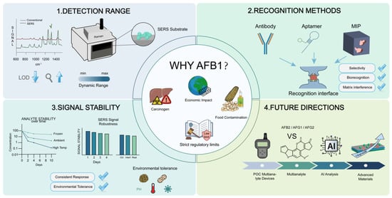
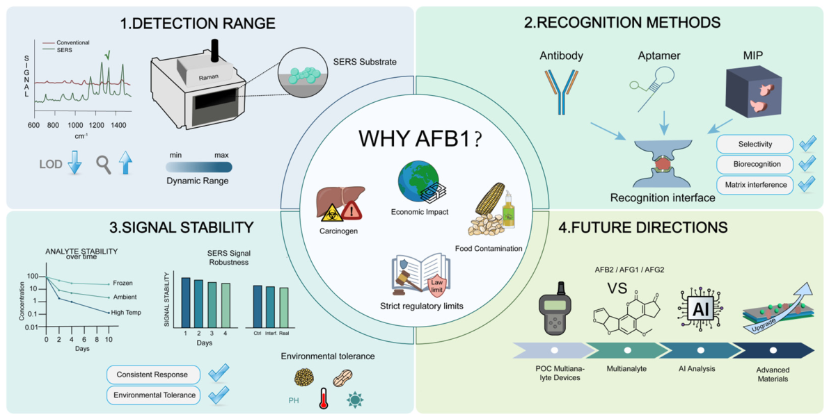
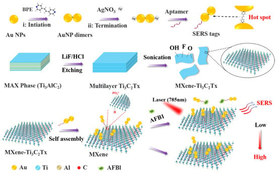
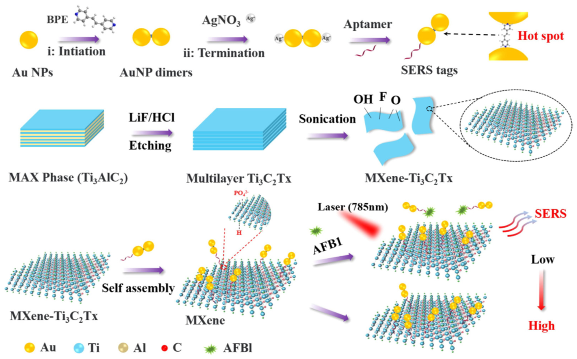
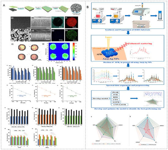
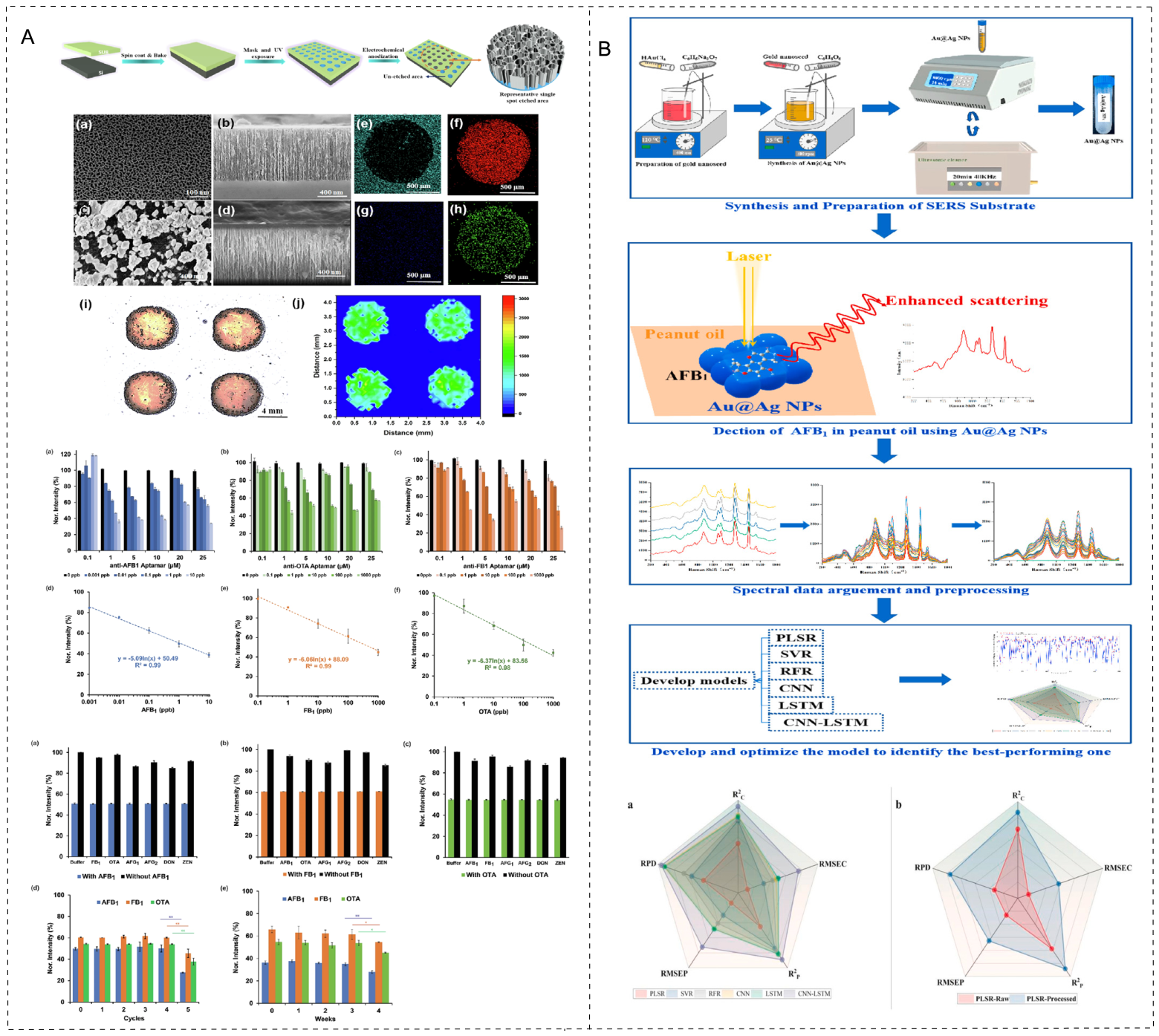

Disclaimer/Publisher’s Note: The statements, opinions and data contained in all publications are solely those of the individual author(s) and contributor(s) and not of MDPI and/or the editor(s). MDPI and/or the editor(s) disclaim responsibility for any injury to people or property resulting from any ideas, methods, instructions or products referred to in the content. |
© 2026 by the authors. Licensee MDPI, Basel, Switzerland. This article is an open access article distributed under the terms and conditions of the Creative Commons Attribution (CC BY) license.