


Abstract
Muskmelon (Cucumis melo L.) is an important cash crop primarily cultivated in the Xinjiang region, and its flesh is sweet, crisp, and juicy with a pronounced aroma. However, postharvest quality deterioration in muskmelons is associated with physiological and biochemical processes, including oxidative stress caused by the accumulation of reactive oxygen species (ROS) and the enzymatic degradation of cell wall components. These processes can compromise membrane integrity, reduce fruit firmness and nutritional quality, and consequently shorten shelf life. Temperature control has been widely used to maintain fruit quality, and low-temperature storage is a key approach for slowing ripening and extending shelf life; nevertheless, better storage temperature is highly cultivar-specific. Therefore, this study stored muskmelons at 4 °C, 10 °C, and 20 °C for 28 days, aiming to determine the effects of different storage temperatures on their quality. The results showed that, on day 28 of storage, 10 °C significantly upregulated the genes encoding superoxide dismutase (SOD), catalase (CAT), ascorbate peroxidase (APX), and peroxidase (POD), and enhanced the corresponding enzyme activities, thereby boosting ROS scavenging and reducing membrane lipid peroxidation. Concomitantly, 10 °C downregulated the genes encoding cell wall-degrading enzymes, including pectin methylesterase (PME), polygalacturonase (PG), pectin lyase (PL), β-galactosidase (β-gal), and β-glucosidase(β-glu), and inhibited their activities, thereby preserving protopectin and cellulose. On day 28 of storage, the 10 °C treatment exhibited the lowest weight loss (4.37%), maintained the highest firmness (2.67 N), and retained higher contents of total phenolics and vitamin C. In summary, storage at 10 °C maintained the postharvest quality of muskmelon by inhibiting cell wall degradation and affecting reactive oxygen species metabolism.
1. Introduction
Muskmelon (Cucumis melo L.) is an important cash crop grown mainly in the Xinjiang region of China. Its flesh is sweet, crisp, and juicy, with a pronounced aroma. However, the fruit is often harvested under high-temperature conditions, which shortens its shelf life and causes rapid quality deterioration during storage at ambient temperature [1].
Postharvest quality deterioration in muskmelon is associated with physiological and biochemical processes, including oxidative stress caused by the accumulation of ROS and the enzymatic degradation of cell wall components; these processes can compromise membrane integrity and reduce fruit firmness and nutritional quality, thereby shortening shelf life and severely constraining storage quality [2]. In ‘Jiashi’ melons stored at 20 °C, CAT and POD activities were insufficient to counteract the abnormal increases in H2O2 and MDA induced by environmental factors, which exacerbated membrane lipid peroxidation and further accelerated quality deterioration [3]. The abnormally elevated respiration rate, superoxide anion radical production rate, and MDA content in ‘Xizhoumi 17’ melons stored at 20 °C disrupted the integrity of membrane structure, thereby accelerating the postharvest senescence process of the melons [4]. In blueberries stored at 22 °C, the abnormally increased activities of PG, PME, β-glu, and cellulase (Cx) accelerated the degradation of cell wall components, leading to a continuous decline in firmness; concurrently, this was accompanied by an abnormally elevated superoxide anion radical production rate and H2O2 content, accelerating membrane lipid peroxidation and aggravating strawberry-quality deterioration [5].
Low-temperature storage is one of the primary methods for maintaining postharvest melon quality. Muskmelon is sensitive to low temperatures, and during postharvest cold storage it is highly susceptible to chilling injury (CI) [6], which in turn leads to quality decline and severely reduces its market value. Under chilling injury, the epidermis of yellow-skinned muskmelons is prone to water-soaked spots, pitting, and browning, with disrupted ROS metabolism [7], further triggering membrane lipid peroxidation and accumulate MDA production. Meanwhile, cell wall components undergo degradation under the action of cell wall-degrading enzymes, accelerating the impairment of cell wall integrity and aggravating chilling injury symptoms [8,9]. Under chilling injury, ‘Golden Empress-308’ Hami melons exhibited chilling injury symptoms such as pitting and browning, with continuously declining firmness and continuously increasing weight loss rate, aggravating fruit-quality deterioration [10]. In ‘Xizhoumi 25’ Hami melon under chilling injury, large areas of light brown spots appeared on the fruit surface as storage time increased, accompanied by fruit softening and depressions, resulting in quality deterioration [11]. Under appropriate low-temperature storage, increases in PG and Cx activity are inhibited, cell wall degradation and firmness loss are delayed, and ‘Mingzhu No. 5’ melon quality is maintained [12].
Appropriate low-temperature storage can effectively inhibit the increase in respiration rate, stabilize physiological metabolism, preserve nutritional quality, and delay quality decline, while exerting significant regulatory effects on antioxidant enzyme activities and cell wall-degrading enzyme activities. Cultivar and processing differences may affect storage dynamics; NFT treatment (0.8 °C) was the more suitable temperature condition for maintaining the quality of ‘Mingzhu 5’ melons [12]. The optimal storage temperature for Jiashi melons was 3 °C [3] and ‘Golden Empress’ Hami melons [13], 6 °C for ‘Lvbao’ melons [14], and 7–8 °C for the three thick-skinned cultivars ‘Tianlu,’ ‘Huanghemi,’ and ‘Yujinxiang’ [15]. In addition, commonly used preservation methods include melatonin [16], 1-MCP [17], combined chitosan and ethanol treatments [18], controlled pre-cooling [19], chlorogenic acid [14], ultraviolet irradiation with chitosan coating [20], ozone [21], preharvest acetylsalicylic acid [22] and low-temperature storage.
This study used ‘Huangmengcui’ muskmelons as the experimental material to systematically evaluate the effects of storage at 4 °C, 10 °C, and 20 °C on the postharvest physiology and quality attributes of thick-skinned muskmelons. Combined with the analysis of physiological and biochemical indices, the aim was to reveal the temperature-regulated postharvest quality and physiological patterns of muskmelons, clarify the relationship between quality deterioration and reactive oxygen species as well as cell wall metabolism, and provide a theoretical basis and technical support for temperature control in the cold-chain logistics of muskmelons, thereby offering a scientific reference for optimizing postharvest preservation technology and theoretical guidance and technical support for extending the shelf life.
2. Materials and Methods
2.1. Fruit Materials and Storage Treatments
Fruits of the muskmelon cultivar ‘Huangmengcui’ (Cucumis melo L.) at 90% maturity and with a soluble solids content of 16.5% were harvested from drip-irrigated crops at the experimental base of the Xinjiang Uygur Autonomous Region Facility Agriculture and Characteristic Agriculture Research and Development Center (Turpan, Shanshan County). Fruits with a uniform size and color and without visible defects or mechanical damage were selected and quickly transported to the laboratory for experiments. The surfaces of the melon fruits were first rinsed clean with tap water, then soaked in 0.02% (v/v) sodium hypochlorite solution for 2 min and air-dried at room temperature (22 ± 2 °C) until use. The fruits were then placed separately in low-temperature incubators disinfected with 75% ethanol (Model MIR-254-CR, Panasonic Healthcare Co., Ltd., Tokyo, Japan) at 4 ± 0.5 °C (relative humidity 70–75%), 10 ± 0.5 °C (relative humidity 70–75%), and 20 ± 0.5 °C (relative humidity 75–80%), with storage beginning within 24 h of harvest. Samples were collected every seven days, on days 0, 7, 14, 21, and 28, over a 28-day storage period. For each temperature setting, three independent storage chambers were used to form three biological replicates. Fruit-pulp samples from three parallel temperature-controlled storage chambers were cryopreserved in liquid nitrogen. Before measuring the various parameters, the three sets of samples were mixed and randomly divided into three portions, which were used for transcriptomic sequencing and the determination of physiological indicators such as ROS, malondialdehyde levels, and antioxidant enzyme activity.
2.2. Determination of Chilling Injury (CI) Index and Decay Index
There were three replicates in each group, and 10 fruits from each replicate were observed. Following the method described by Wang et al. [23], the chilling index was divided into four levels according to the area of chilling spots and pitting degree on the fruit surface: Grade 0 (no symptoms), Grade 1 (<10%), Grade 2 (11–25%), Grade 3 (25–50%), and Grade 4 (>50%).
Following the method described by Zhang et al. [24], fruit decay was classified into five grades according to the size of disease lesions on the fruit surface: Grade 0 (no symptoms), Grade 1 (<10%,), Grade 2 (10–30%), Grade 3 (31–50%), and Grade 4 (>50%).
CI index (%) = Σ (number of fruits of this level × CI level)/(number of total fruits × 4) × 100
Decay index (%) = Σ (number of fruits of this level × Decay level)/(number of total fruits × 4) × 100
2.3. Determination of Respiration Rate, Color Parameters, Weight Loss, and Firmness
Respiration rate was measured using the method described by Yao et al. [16]. CO2 concentrations were determined using a CO2 gas detector (Model Telaire 7001, GE Measurement & Control, St. Marys, PA, USA). Melon fruits and the infrared CO2 detector were placed in a plexiglas jar, and data were recorded at 0.5 h. Respiration intensity was expressed as the amount of CO2 released per kilogram of fruit per hour, in μg·kg−1·h−1, and calculated according to the following formula:
where: W1 is the total CO2 in the blank respiration jar (%), W2 is the total CO2 in the respiration jar after the measurement (%), V is the total volume inside the respiration jar (L), M is the molar mass of CO2 (g·mol−1), V0 is the molar volume of CO2 at the measurement temperature (L·mol−1), m is the mass of the sample (kg), and t is the measurement duration (h).
Respiration rate (μg·kg−1·h−1) = (W2 − W1) × V × M × 100/(V0 × m × t)
Color measurements were performed usinga colorimeter (Model CR8, Guangdong Sanen Time Technology Co., Ltd., Guangdong, China). At the start of the experiment, 3 fruits were randomly selected from each of the three parallel storage chambers at each temperature, numbered, and designated for color difference measurements, and three equidistant points along the equatorial region of each fruit peel were measured for L*, a*, and b* values. Schematic diagram of color difference measurement (Figure S1A).
The gravimetric determination of weight loss was performed using the weighing method, and calculated according to the following formula:
where W0 is the fruit mass before storage (kg) and Wt is the fruit mass after days of storage (kg).
Weight loss rate (%) = (W0 − Wt)/W0 × 100
Firmness was determined as described by Wang et al. [25], using a firmness tester (GY-4 digital fruit firmness tester, Zhejiang Top Instrument Co., Ltd., Hangzhou, China) with a probe dimension of 8 mm and a measurement depth of 10 mm, at three evenly spaced points around the equatorial region of each fruit, and the values were reported in newtons (N). Schematic diagram of firmness point measurement (Figure S1B).
2.4. Determination of SSC and TA
Soluble solid content (SSC) was measured using a digital refractometer (Model PAL-1, ATAGO Co., Ltd., Tokyo, Japan), and the results were expressed as %.
Titratable acidity (TA) was determined by acid–base titration [15], and the results were expressed as %.
2.5. Determination of Total Phenolic and Vitamin C Contents
Total phenolic content was determined using the method described by Wen et al. [26], with slight modifications; the sample weight was increased to 2.0 g, with units expressed as mg·g−1. Vitamin C content was measured using a colorimetric method [27].
2.6. Determination of Relative Conductivity and Malondialdehyde Content
The method of Jia et al. [28] was slightly modified. three fruits were randomly selected and sampled using an 8 mm drill bit. The samples were then cut into 2 mm thick slices of uniform thickness with a knife. After mixing the slice samples from different fruits, the measurements were performed in triplicate. A total of 20 mL of distilled water was immediately measured for electrical conductivity using a conductivity meter; this value was recorded as P1. Then, 15 slices were placed into a test tube and allowed to stand for 10 min, after which electrical conductivity was measured and recorded as P2. Tubes were boiled for 15 min in a water bath. They were then cooled to ambient temperature. Conductivity was re-measured and noted as P3. The results were calculated according to the following formula:
Relative conductivity (%) = (P2 − P1)/(P3 − P1) × 100
Malondialdehyde (MDA) content was determined using the Thio-barbituric acid method described by Tian et al. [15]. A 2.0 g sample was weighed and mixed with 5.0 mL of 10% TCA; the mixture was ground and homogenized and then centrifuged at 4 °C and 10,000 rpm for 10 min. The supernatant was collected and stored at low temperature for later use. Then, 2.0 mL of solution to be tested and 2.0 mL of 0.6% 2-thiobarbituric acid solution (Dissolved in 10% trichloroacetic acid solution) were mixed and placed in a boiling water bath for 15 min, and centrifuged at 4000 rpm for 10 min. The supernatant was collected and its absorbance at 532 nm, 600 nm and 450 nm was measured (unit: nmol·g−1). The results were calculated according to the following formula:
where: V is the total volume of the sample extract (mL); Vs is the volume of the sample extract taken at the time of measurement (mL); and W is the weight of the sample (g).
MDA content (nmol·g−1) = (6.45 × (A532 − A600) − 0.56 × A450) × V/(Vs × W × 10−3)
2.7. Determination of H2O2 Content and O2·– Generation Rate
H2O2 content was determined according to the method of Ning et al. [3], expressed in µmol·g−1. The O2·− generation rate was measured with slight modifications according to the method of Qin et al. [29], and the sample weight was increased to 2.0 g.
2.8. Assay of Antioxidant-Related Enzyme Activities
Enzymes were extracted from a 3.0 g sample. Homogenization was performed on ice in 50 mM of PBS (pH 7.8) with 1% PVPP and 1 mM of EDTA. The mixture was centrifuged at 10,000 rpm for 15 min at 4 °C. The supernatant was collected. SOD and CAT enzyme activity were determined according to the method described by Zheng et al. [30]. APX and POD activities were assayed following Xu et al. [31]. PPO activity was determined with slight modifications based on Zheng et al. [30]. Spectrophotometric readings were taken at 560, 240, 290, 470, and 398 nm for SOD, CAT, APX, POD, and PPO, respectively. All values were expressed in U·g−1.
2.9. Determination of Cell Wall Component Content
Soluble pectin and protopectin contents were determined according to the method of Liu et al. [22], with the results expressed in mg·g−1. Hemicellulose and cellulose contents were measured following Huang et al. [6] using the enthrone colorimetric assay with glucose (Glu) as the standard, and the results were expressed in mg·g−1.
2.10. Determination of Cell Wall Degrading Enzyme Activities
PME(G0707D), PG(G0701W), PL(G0702F), and Cx(G0533w4) were measured. Commercial assay kits t (Suzhou Grace Biotechnology Co., Ltd., Suzhou, China) were used. The activity units were established as follows. At 40 °C, one unit of PME was defined as the quantity of enzyme required to consume 1 μmol NaOH per milliliter of liquid per hour. PG activity was determined at 40 °C, with one unit designated as the amount of enzyme that hydrolyzes pectic acid to generate 1 mg of galacturonic acid per gram of sample per hour. At 50 °C, one unit of PL was defined as the quantity of enzyme that degrades pectin to produce 1 nmol of unsaturated galacturonic acid per gram of tissue per minute. Cx activity was measured at 40 °C, where one unit was defined as the amount of enzyme that catalyzed the production of 1 μg of reducing sugar per gram of tissue per hour.
β-gal activity was determined according to the method of You et al. [32]. A total of 1.0 g of tissue was homogenized in 4.0 mL of 40 mM sodium acetate buffer (pH 5.2) containing 5% PVP, 2% β-mercaptothion, and 0.1 M NaCl, and the homogenate was centrifuged to obtain the supernatant. For the β-gal assay, the reaction mixture comprised 0.2 mL of enzyme extract and 2 mL of 33.2 mM 2-Nitrophenyl β-d-galactopyranoside. Reactions were incubated at 37 °C for 30 min and stopped by adding 2 mL of 0.2 M Na2CO3. Absorbance was read at 400 nm. One unit was recognized as the generation of 1 μmol of 4-nitrophenol at 400 nm per minutes. All these results were presented as μmol·min−1·g−1.
β-glu activity was determined according to the method of Cao et al. [33]: 1.0 g of tissue was ground in an ice bath with pre-cooled sodium acetate buffer and centrifuged to obtain the crude enzyme solution. The crude enzyme solution was mixed with 34.9 Mm of salicin solution and reacted at 37 °C for 30 min. The reaction was terminated by adding 3,5-dinitrosalicylic acid (DNS) reagent, and the absorbance was measured at 540 nm. One unit was recognized as the generation of 1 μmol of glucose at 540 nm per minutes. Activity was expressed as μmol·min−1·g−1.
2.11. Transcriptome Analysis
Fruits were sampled on day 28 of storage. Each temperature treatment (4 °C, 10 °C, and 20 °C) at each time point comprised three biological replicates, for which fruit-pulp tissues were collected and homogenized as submitted samples, giving 3 samples and 9 libraries in total. Total RNA was extracted from the homogenized pulp tissue using TRIzol reagent, and RNA libraries were constructed for transcriptome analysis. RNA purity was assessed with a NanoDrop 2000 (Thermo Fisher Scientific Inc., Waltham, MA, USA) (OD260/280 = 1.8–2.2), and integrity was assessed using an Agilent 2100 Bioanalyzer (Agilent Technologies Inc., Santa Clara, CA, USA), with RQN/RIN ≥ 6.5 as the quality criterion. mRNA was enriched using the NEBNext® Poly(A) mRNA Magnetic Isolation Module (New England Biolabs, Beverly, MA, USA) and strand-specific libraries were constructed with the NEBNext Ultra II RNA Library Prep Kit for Illumina (both from New England Biolabs Inc., Ipswich, MA, USA), in which second-strand cDNA was synthesized with dUTP in place of dTTP. Library preparation and sequencing were performed by Peisenno Biotechnology Co., Ltd. (Shanghai, China). Sequencing was carried out on the BGI T7 platform in paired-end 150 bp (PE150) mode, with 6 Gb of data per sample.
Using the muskmelon reference genome (GCF_025177605.1_USDA_Cmelo_AY_1.0) as the reference, Fastp was used to remove sequences with 3′ adapters and reads with an average quality score below Q20, after which paired-end clean reads were aligned to the reference genome using HISAT2 (v2.1.0). Transcript assembly and quantification were performed using String Tie (v2.1.4), and expression levels were presented as FPKM (fragments per kilobase of transcript per million mapped reads). Read counts for each gene were obtained using HTSeq (v0.11.1), and FPKM was calculated based on gene length and mapped reads for gene expression normalization and inter-sample correlation assessment. Differential expression analysis between groups was performed using DESeq2, with p < 0.05 and |log2FoldChange| ≥ 1 as the screening thresholds [34]. When multiple members were present within a related gene family, genes regulating multiple temperatures were prioritized for screening, and the screening results were expressed as FPKM values.
2.12. Data Collection, Statistical Processing, and Analysis
All measurements were performed in triplicate, and the results are expressed as mean ± standard deviation (SD) (n = 3). Data were processed with Microsoft Excel 2019 (Microsoft Corporation, Redmond, WA, USA). All data were analyzed using two-way factorial ANOVA (FANOVA) via SPSS 27.0 (IBM Corporation, Armonk, NY, USA) to evaluate the temperature, storage time main effects and their interaction. Duncan’s test (p < 0.05) was applied for mean separation. Data were processed and figures were generated using Microsoft Excel 2019, GraphPad Prism 10.1 (GraphPad Software, Boston, MA, USA), and Origin 2025b (OriginLab Corporation, Northampton, MA, USA).
3. Results
3.1. Effects of Different Storage Temperatures on Postharvest Appearance, CI Index and Decay Index
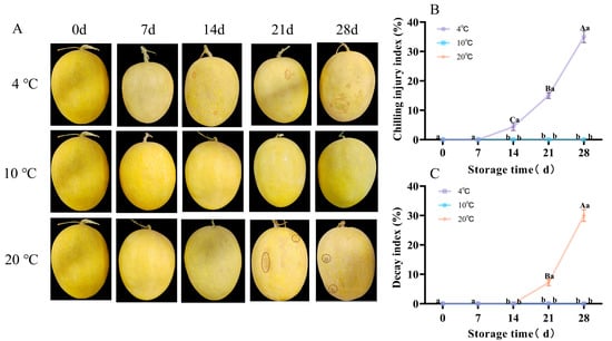
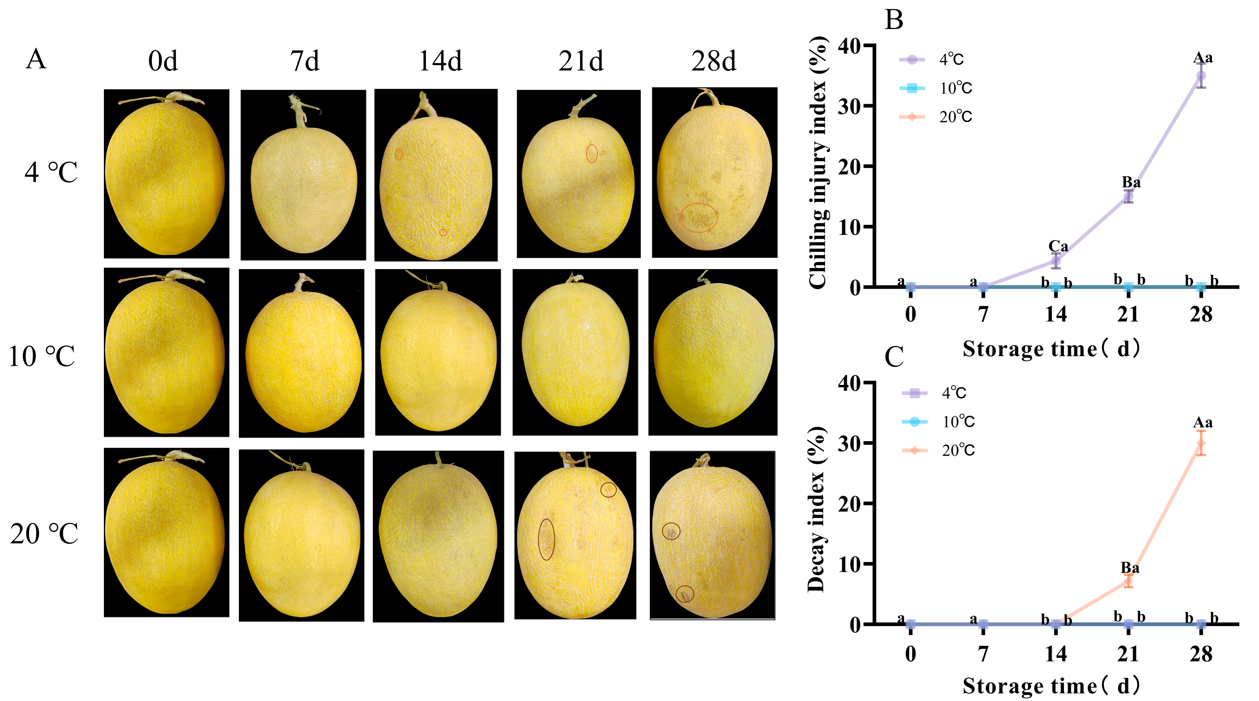
At 20 °C, on day 14 of storage the muskmelons rind exhibited pronounced depressions and spots that expanded with prolonged storage. At 4 °C, on day 14, water-soaked spots appeared on the rind, and on day 28 the surface showed extensive water-stain-like lesions, Storage at 10 °C maintained a relatively good overall appearance (Figure 1A). On day 28, the cold damage index reached 35% at 4 °C, and the decay index reached 30% at 20 °C (Figure 1B,C).
Figure 1.
Postharvest appearance (A), CI index (B), and Decay index (C) of muskmelons stored at different temperatures. In A, chilling injury symptoms at 4 °C are highlighted by small red circles, and decay symptoms at 20 °C are highlighted by small brown circles. A two-way FANOVA revealed a significant temperature × storage time interaction (p < 0.01). Differences among storage days at the same temperature are marked with uppercase letters, and differences among temperatures on the same storage day are marked with lowercase letters (p < 0.05).
3.2. Effects of Storage Temperatures on Respiration Rate and Color Difference
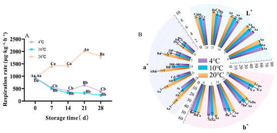
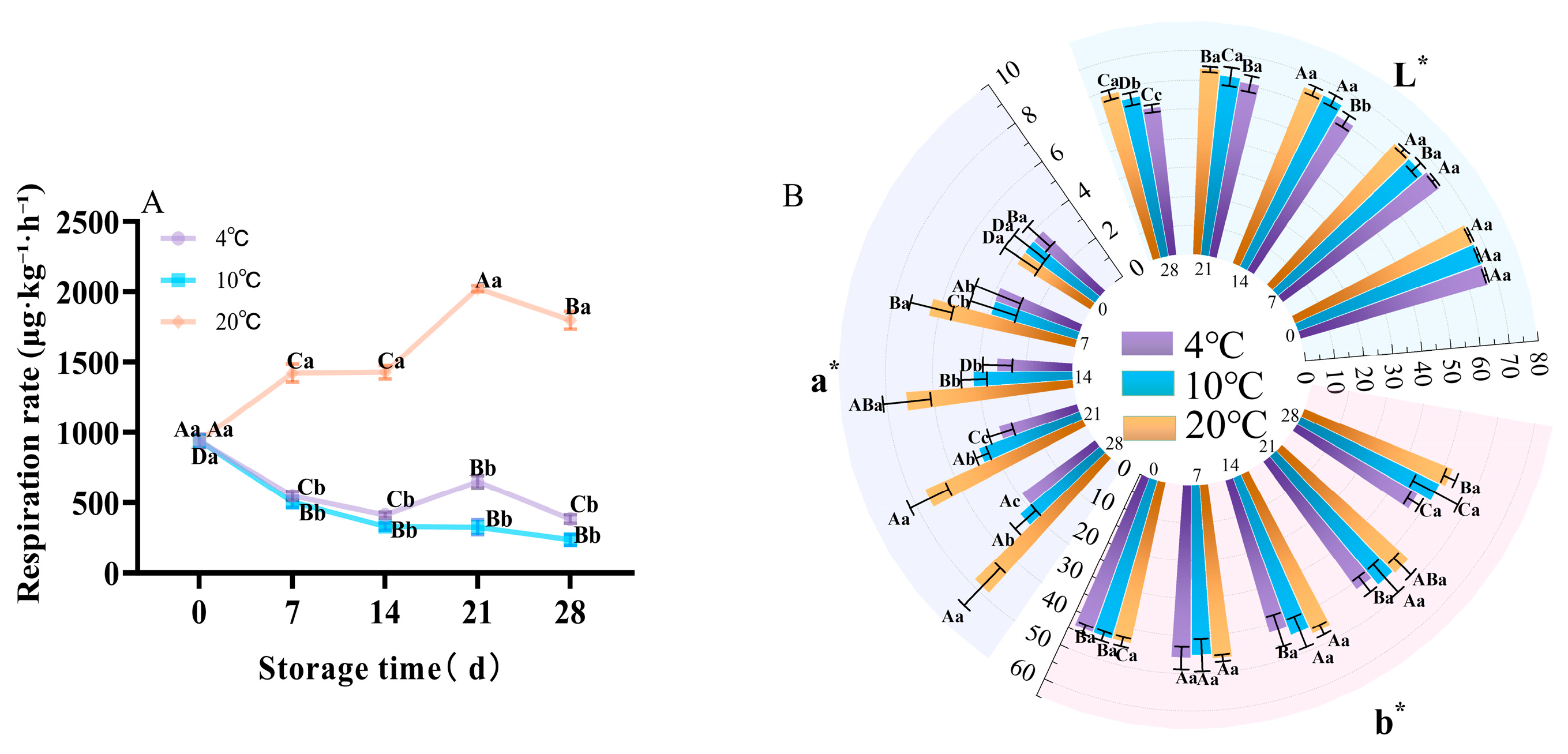
Throughout storage, muskmelons maintained relatively low respiration rates at 10 °C. On day 28 of storage, the respiration rate at 20 °C was 1799.33 μg·kg−1·h−1; at 4 °C, it was 385.20 μg·kg−1·h−1; and at 10 °C, it was 238.34 μg·kg−1·h−1. The respiration rate at 20 °C was 7.55 times that at 10 °C, and at 4 °C it was 1.62 times that at 10 °C (Figure 2A).
Figure 2.
Postharvest respiration rate (A) and color difference (L*, a*, and b*) (B) of muskmelons stored at different temperatures. A two-way FANOVA revealed a significant temperature × storage time interaction (p < 0.01). Differences among storage days at the same temperature are marked with uppercase letters, and differences among temperatures on the same storage day are marked with lowercase letters (p < 0.05).
L* denotes peel lightness, with higher values indicating greater surface gloss and more vivid coloration. As shown in the Figure 2B, peel L* of the yellow-skinned muskmelon decreased overall during storage. On day 28, the L* values at 4 °C, 10 °C, and 20 °C were 55.50, 58.60, and 51.07, respectively, representing decreases of 16.8%, 12.2%, and 23.4% relative to day 0 (p < 0.05). The largest decline occurred at 20 °C, indicating that prolonged storage at a higher temperature led to a pronounced loss of lightness. Peel a* increased overall with storage duration. On day 28, the a* values at 4 °C, 10 °C, and 20 °C were 3.84, 4.44, and 7.77, corresponding to increases of 4.6%, 20.8%, and 111.4% relative to day 0 (p < 0.05). For the yellow-skinned muskmelon, the disproportionately large increase in a* indicates atypical reddening of the peel and is an important manifestation of postharvest color deterioration (Figure 2B).
3.3. Effects of Storage Temperatures on Postharvest Quality
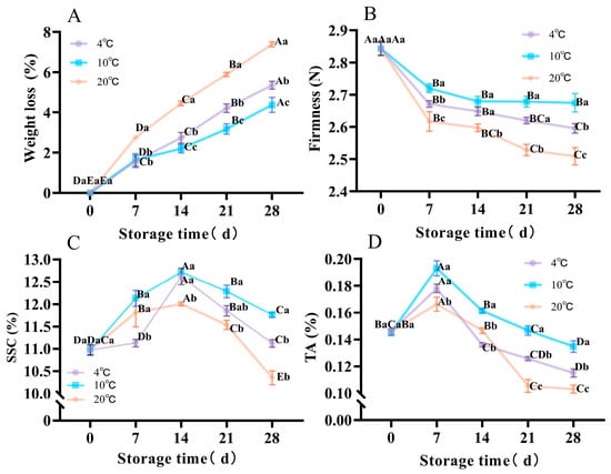
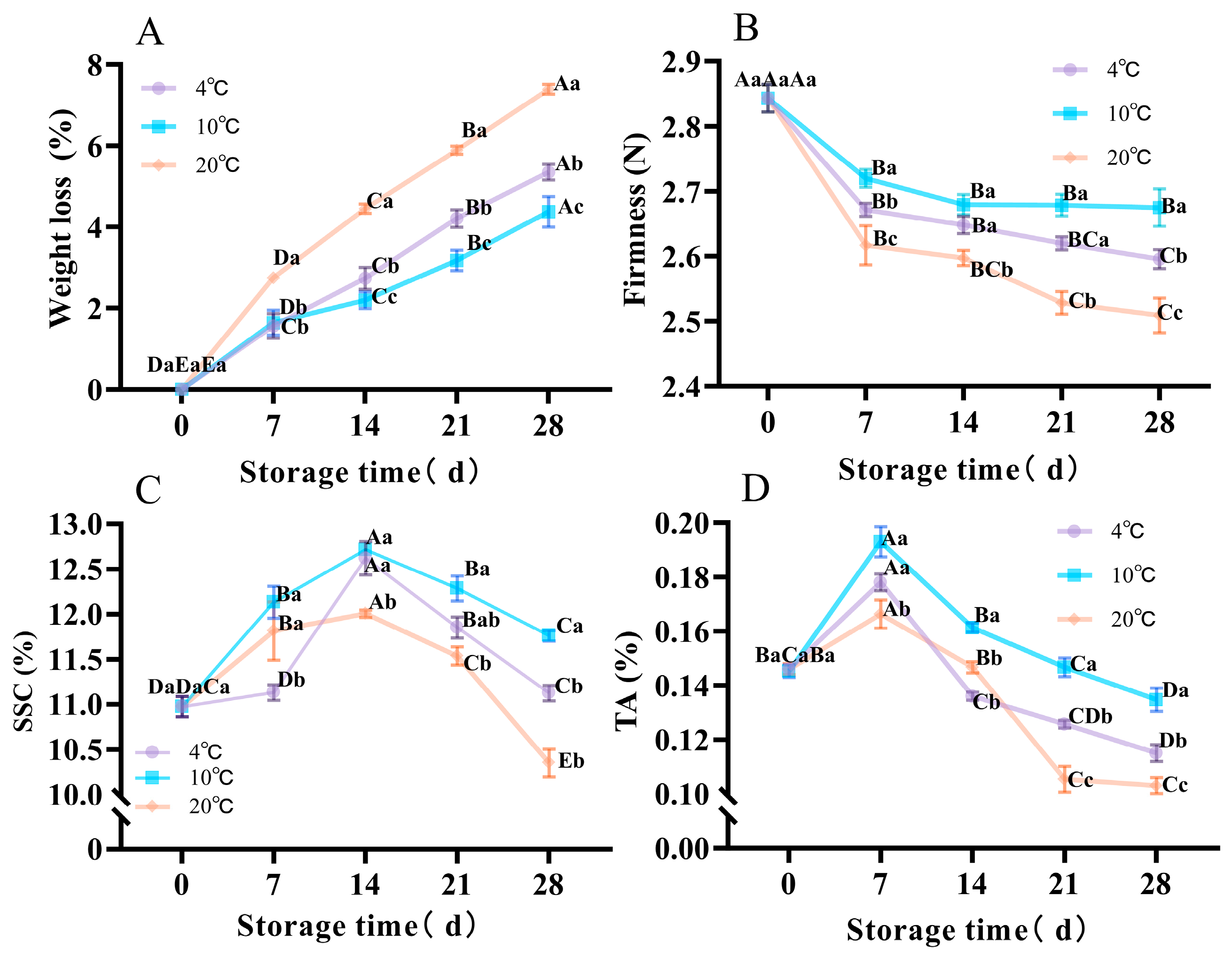
Weight loss rate is a key indicator of moisture loss during fruit storage. During the 28-day storage period, muskmelon fruits exhibited a steady increase in weight loss. On day 28, the weight loss at 4 °C was 22.43% higher than that at 10 °C, and the weight loss at 20 °C was 69.11% higher than that at 10 °C. Firmness decreased continuously throughout the 28 days. On day 28, firmness at 10 °C was 6.37% greater than at 20 °C and 2.69% greater than at 4 °C (Figure 3A,B).
Figure 3.
Postharvest weight loss (A), firmness (B), SSC (C), and TA (D) of muskmelons stored at different temperatures. A two-way FANOVA revealed a significant temperature × storage time interaction (p < 0.01). Differences among storage days at the same temperature are marked with uppercase letters, and differences among temperatures on the same storage day are marked with lowercase letters (p < 0.05).
During the 28-day storage period, the SSC and TA of muskmelon fruit rose initially and then declined. On day 28, SSC measured 11.13% at 4 °C, 11.76% at 10 °C, and 10.35% at 20 °C; the SSC at 10 °C was therefore 13.62% greater than at 20 °C and 5.66% greater than at 4 °C (Figure 3C). On the same day, TA at 10 °C exceeded that at 4 °C by 14.63% and that at 20 °C by 23.53% (Figure 3D).
3.4. Effects of Storage Temperatures on Postharvest Total Phenolic and Vitamin C Contents
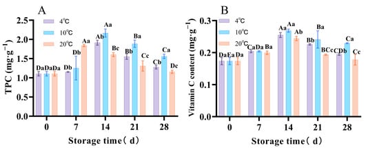
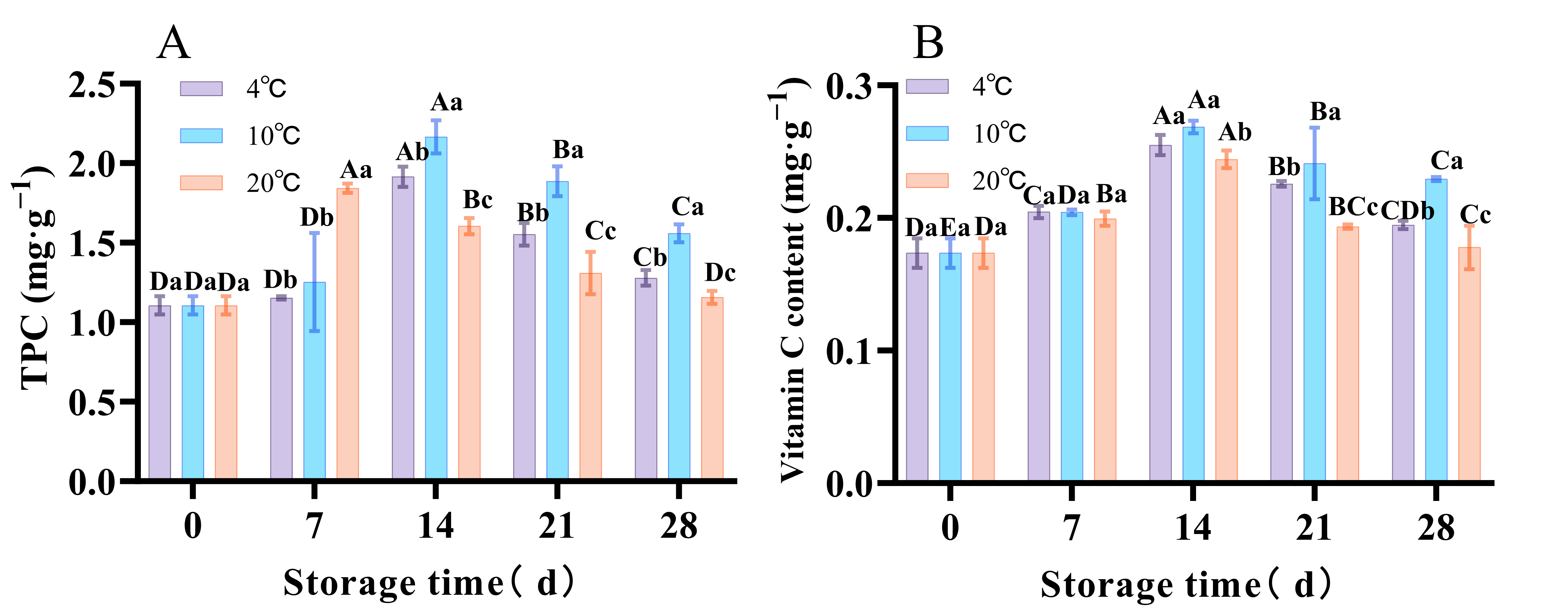
During the 28-day storage period, total phenolic and vitamin C contents first increased and then decreased. Total phenolics peaked on day 7 at 20 °C (1.84 mg·g−1), whereas at 4 °C and 10 °C they reached their maxima on day 14 (1.91 mg·g−1 and 2.17 mg·g−1, respectively). On day 28, the total phenolic content at 10 °C was 1.22 times that at 4 °C and 1.34 times that at 20 °C (Figure 4A). Vc concentrations peaked at day 14 for all temperature treatments, representing increases of 52.94%, 58.82%, and 41.18% relative to day 0 at 4 °C, 10 °C, and 20 °C, respectively. On day 28, vitamin C at 10 °C was 1.18 times that at 4 °C and 1.29 times that at 20 °C (Figure 4B). These results indicate that storage at 10 °C maintained higher levels of total phenolics and vitamin C and delayed their subsequent decline.
Figure 4.
Postharvest total phenolic (A) and vitamin C (B) contents of muskmelons stored at different temperatures. A two-way FANOVA revealed a significant temperature × storage time interaction (p < 0.01). Differences among storage days at the same temperature are marked with uppercase letters, and differences among temperatures on the same storage day are marked with lowercase letters (p < 0.05).
3.5. Effects of Storage Temperatures on Postharvest Electrolyte Leakage and MDA Content
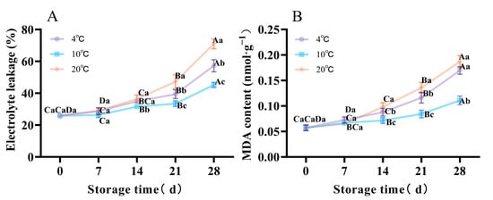
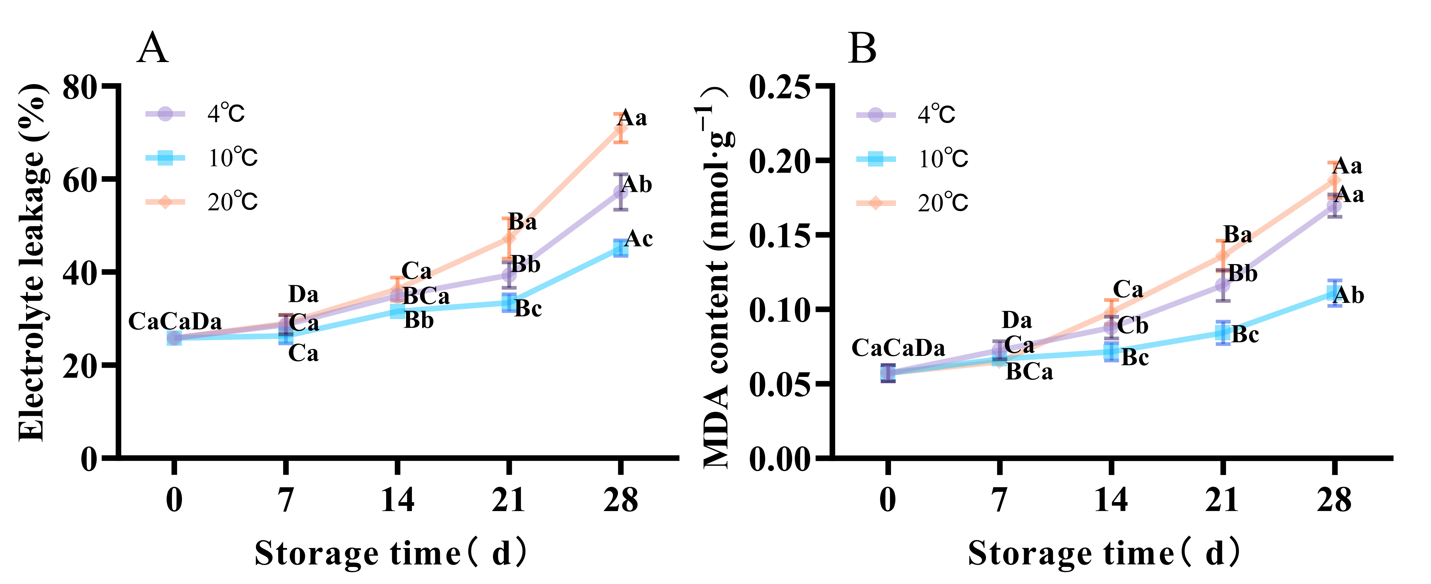
Electrolyte leakage indicates plant cell membrane health and the extent of damage due to factors such as low temperature or senescence, leading to electrolyte leakage. Throughout the 28-day storage period, both conductivity and malondialdehyde (MDA) levels rose steadily. On day 28, 20 °C gave the highest conductivity, which was 1.57 times that at 10 °C and 1.27 times that at 4 °C (Figure 5A). MDA, a marker of lipid peroxidation, reflects membrane injury. After 28 days, MDA levels at 4 °C, 10 °C, and 20 °C were 0.17, 0.11, and 0.19 nmol·g−1, respectively. The 20 °C value was 1.68 times higher than the 10 °C value, while the 4 °C value was 1.53 times higher (Figure 5B).
Figure 5.
Postharvest electrolyte leakage (A) and MDA content (B) of muskmelons stored at different temperatures. A two-way FANOVA revealed a significant temperature × storage time interaction (p < 0.01). Differences among storage days at the same temperature are marked with uppercase letters, and differences among temperatures on the same storage day are marked with lowercase letters (p < 0.05).
3.6. Effects of Storage Temperatures on Postharvest H2O2 Content and O2·– Generation Rate
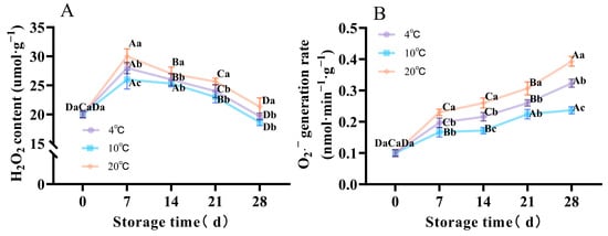
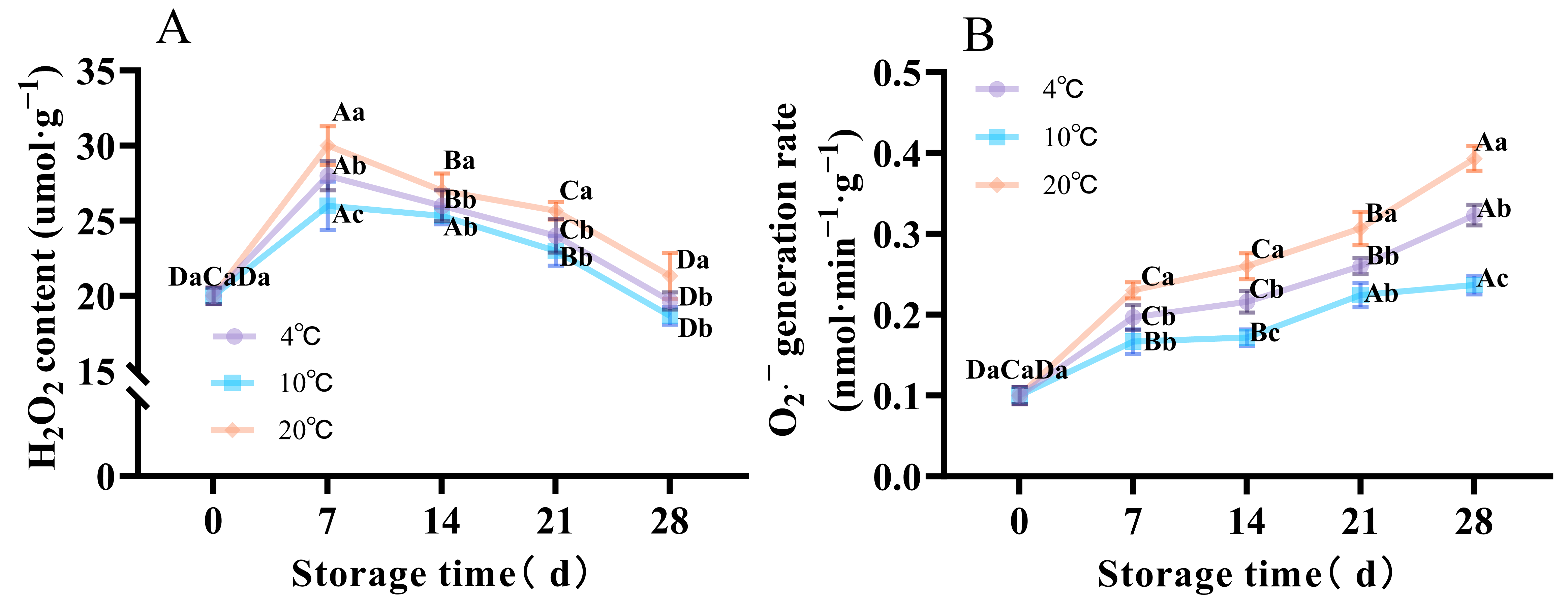
During the 28-day storage period, H2O2 content in muskmelon increased initially and then declined across all three temperature regimes. H2O2 contents peaked on day 7, reaching 28, 26, and 30 μmol·g−1 at 4 °C, 10 °C, and 20 °C, respectively. The value at 20 °C was 1.15 times that at 10 °C, and the value at 4 °C was 1.08 times that at 10 °C. These results indicate that both low and high storage temperatures promote early H2O2 accumulation, with the high-temperature effect being more pronounced. On day 28, H2O2 concentrations had fallen to relatively low values (Figure 6A).
Figure 6.
Postharvest H2O2 content (A) and (O2·–) generation rate (B) of muskmelons stored at different temperatures. A two-way FANOVA revealed a significant temperature × storage time interaction (p < 0.01). Differences among storage days at the same temperature are marked with uppercase letters, and differences among temperatures on the same storage day are marked with lowercase letters (p < 0.05).
During the 28-day storage period, O2·– generation rate in muskmelon increased steadily at all temperatures, peaking on day 28, 0.32, 0.24, and 0.39 nmol·min−1·g−1 at 4 °C, 10 °C, and 20 °C, respectively. At 20 °C, the content was 1.63 times that at 10 °C, and at 4 °C, it was 1.33 times higher than at 10 °C (Figure 6B). Content at 10 °C remained significantly lower than at 4 °C and 20 °C, indicating effective suppression of superoxide anion accumulation, maintenance of reactive oxygen metabolism balance, and mitigation of oxidative stress-induced membrane damage.
3.7. Effects of Storage Temperature on Postharvest Antioxidant Enzyme Activities
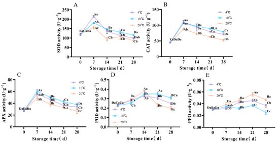
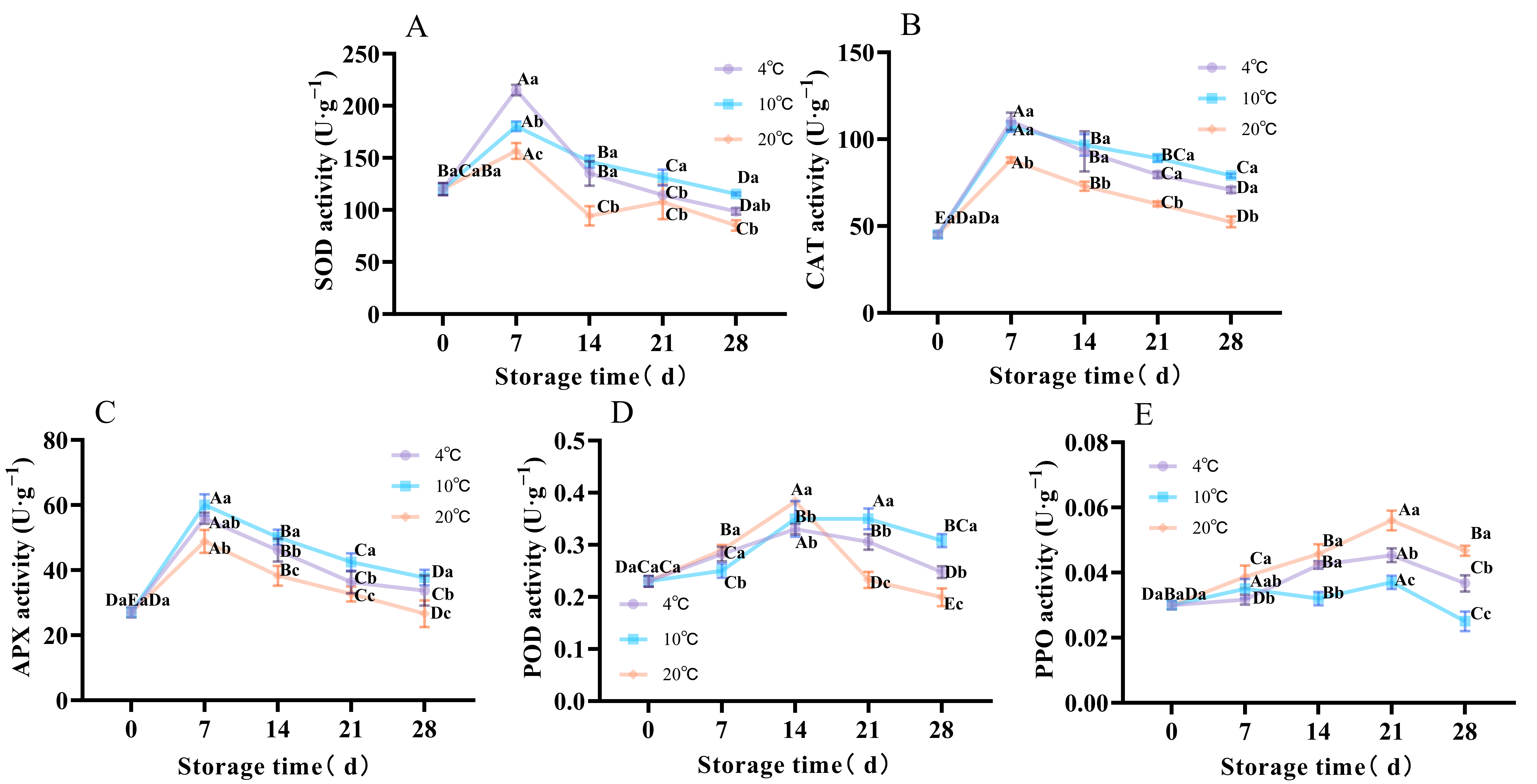
During fruit storage, ROS accumulate as the fruit matures and senesces, triggering activation of the antioxidant enzyme system (including CAT, SOD, APX) to counter oxidative stress. Prolonged storage or high temperatures can lead to reduced enzyme activities. During the 28-day period, SOD, CAT, APX, and POD activities in fruits underwent an initial increase followed by a decline at varying temperatures. After 28 days, SOD activities were 98.67, 115.33, and 85.00 U·g−1 at 4 °C, 10 °C, and 20 °C, respectively, with SOD activity at 10 °C being 1.36-fold higher than at 20 °C and 1.17-fold higher than at 4 °C (Figure 7A). CAT activities were 70.83 U·g−1 and 52.47 U·g−1 at 4 °C, and 20 °C respectively, which was 89.66% and 66.42% of the activity at 10 °C (Figure 7B). On day 7, APX activity peaked at 60.00 U·g−1 at 10 °C, exceeding the values for the 4 °C and 20 °C by 1.07-fold and 1.23-fold, respectively. On day 28, APX activities were 33.73 U·g−1 at 4 °C, 26.60 U·g−1 at 20 °C, and 37.70 U·g−1 at 10 °C, with the latter value being 1.12 and 1.42 times the values at 4 °C and 20 °C, respectively (Figure 7C). POD activity peaked on day 14 at 4 °C and 10 °C, reaching 0.33 U·g−1 and 0.38 U·g−1, respectively, before decreasing rapidly. At 20 °C, POD activity peaked on day 21 with an activity of 0.35 U·g−1. On day 28, POD activity at 10 °C was 1.24-fold higher than that at 4 °C and 1.55-fold higher than that at 20 °C (Figure 7D).
Figure 7.
Postharvest enzyme activities of SOD (A), CAT (B), APX (C), POD (D), and PPO (E) of muskmelons stored at different temperatures. A two-way FANOVA revealed a significant temperature × storage time interaction (p < 0.01). Differences among storage days at the same temperature are marked with uppercase letters, and differences among temperatures on the same storage day are marked with lowercase letters (p < 0.05).
During the 28 days of storage, PPO activity in muskmelon fruit increased initially and then decreased. On day 28, PPO activities at 4 °C, 10 °C, and 20 °C were 0.037, 0.025, and 0.047 U·g−1, respectively. The activity at 4 °C was 1.48 times higher than at 10 °C, and at 20 °C it was 1.88 times higher (Figure 7E), indicating effective suppression of PPO activity at 10 °C, which reduced browning. Storage at 10 °C also maintained higher SOD, CAT, APX, and POD activities and inhibited PPO, thereby delaying oxidative damage and preserving fruit quality.
3.8. Effects of Storage Temperatures on Postharvest Cell Wall Polysaccharides
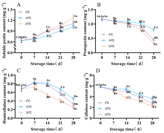
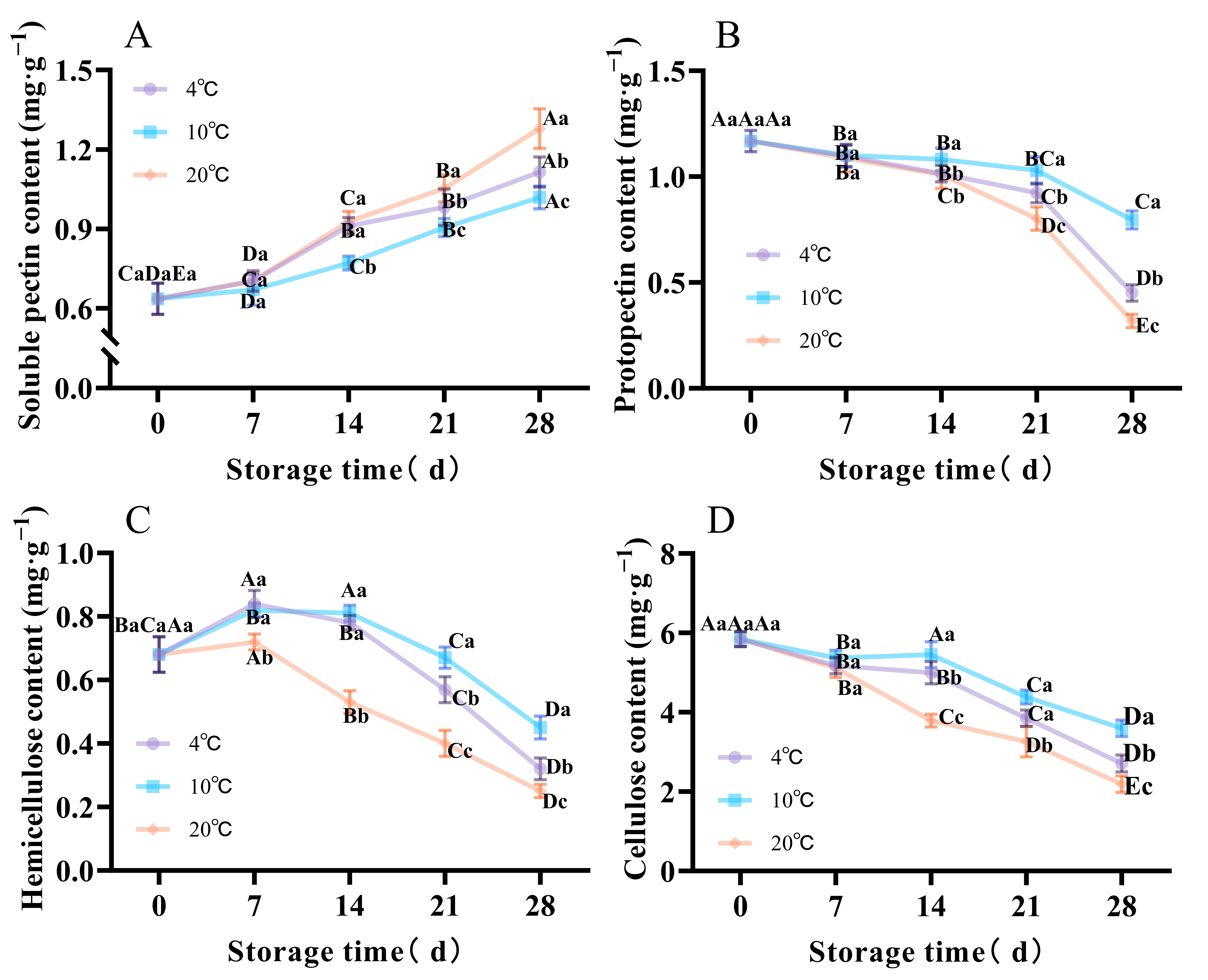
Figure 8A shows that soluble pectin content increased at all storage temperatures over time. On day 28, the soluble pectin content at 4 °C and 20 °C was 1.09 times and 1.26 times that at 10 °C, respectively (Figure 8A). In contrast, protopectin content declined steadily at all three temperatures. Compared with day 0, protopectin fell by 61.41% at 4 °C, 31.92% at 10 °C, and 72.79% at 20 °C on day 28. Consequently, the protopectin content at 10 °C was 1.76 times that at 4 °C and 2.50 times that at 20 °C (Figure 8B). Cellulose content likewise decreased continuously. On day 28 relative to day 0, cellulose had decreased by 53.66% at 4 °C, 38.42% at 10 °C, and 62.47% at 20 °C. On day 28, the cellulose content at 10 °C was 32.90% higher than at 4 °C, and 64.08% higher than at 20 °C (Figure 8D).
Figure 8.
Postharvest soluble pectin (A), protopectin (B), hemicellulose (C), and cellulose (D) contents of muskmelons stored at different temperatures. A two-way FANOVA revealed a significant temperature × storage time interaction (p < 0.01). Differences among storage days at the same temperature are marked with uppercase letters, and differences among temperatures on the same storage day are marked with lowercase letters (p < 0.05).
Hemicellulose content during storage at 4 °C, 10 °C, and 20 °C initially increased and then decreased. On day 28, hemicellulose content at 4 °C was 28.89% lower than at 10 °C, and at 20 °C it was 44.44% lower than at 10 °C (Figure 8C). Storage at 10 °C can postpone the decrease in cell wall component content, aiding in preserving cell wall stability.
3.9. Effects of Storage Temperatures on Postharvest Cell Wall Degrading Enzyme Activity
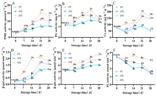
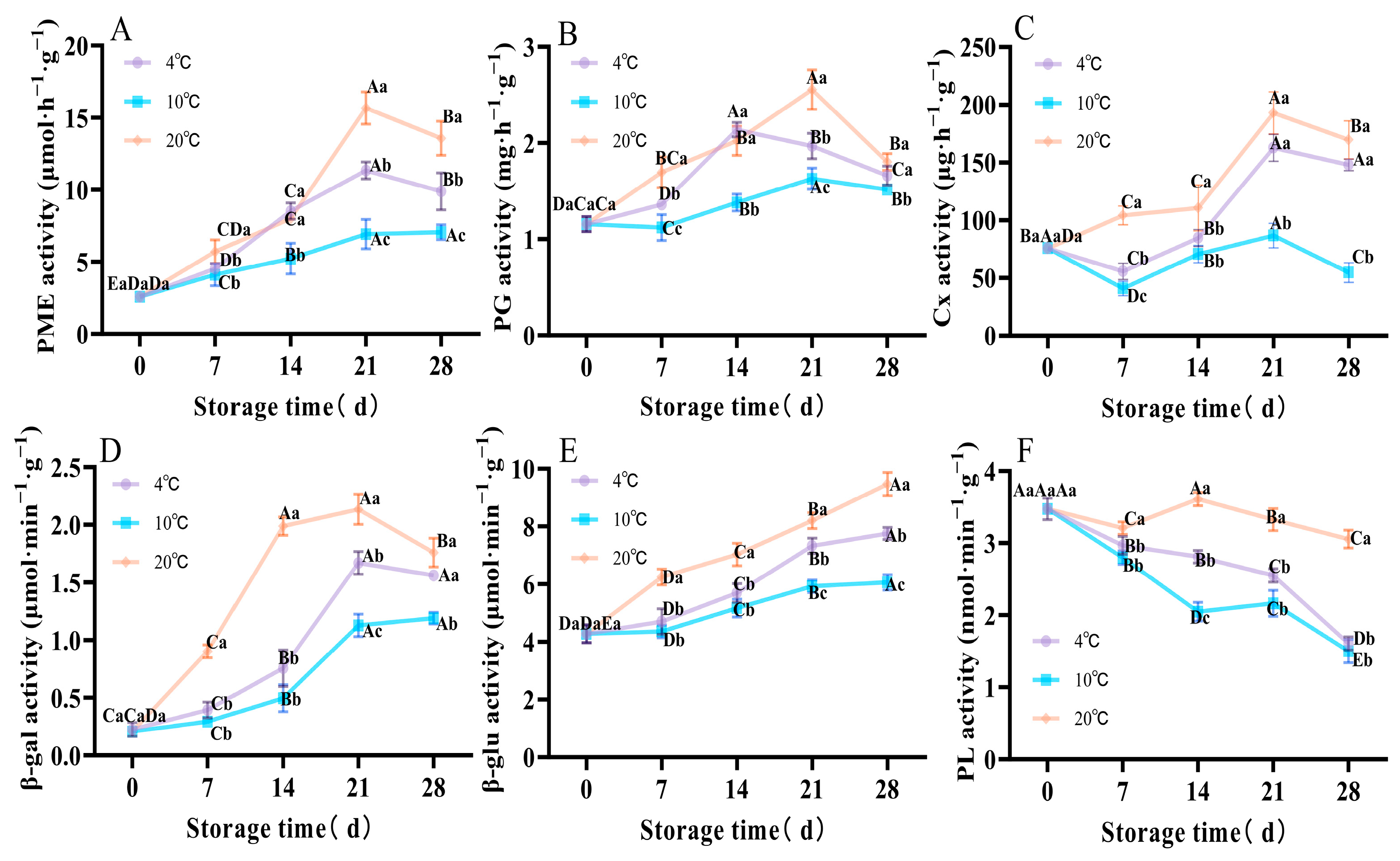
Activities of PME, PG, Cx, and β-gal all rose initially during storage and then declined. On day 28, PME activity at 4 °C exceeded that at 10 °C by 40.28%, and PME at 20 °C exceeded that at 10 °C by 92.62% (Figure 9A). On day 28, PG activities measured 1.66, 1.52, and 1.80 mg·h−1·g−1 at 4 °C, 10 °C, and 20 °C, respectively. The 4 °C value was 1.09 times that at 10 °C and the 20 °C value was 1.18 times that at 10 °C (Figure 9B). Cx activities on day 28 were 148.02, 54.79, and 169.77 μg·h−1·g−1 for 4 °C, 10 °C, and 20 °C, respectively, corresponding to 2.70 and 3.10 times increases at 4 °C and 20 °C relative to 10 °C (Figure 9C). Finally, β-gal activity on day 28 was 1.56, 1.19, and 1.76 μmol·min−1·g−1 at 4 °C, 10 °C, and 20 °C, respectively, with the 4 °C and 20 °C values equal to 1.31 and 1.48 times those of the 10 °C values, respectively (Figure 9D).
Figure 9.
Postharvest enzyme activities of PME (A), PG (B), Cx (C), β-gal (D), β-glu (E), and PL (F) of muskmelons stored at different temperatures. A two-way FANOVA revealed a significant temperature × storage time interaction (p < 0.01). Differences among storage days at the same temperature are marked with uppercase letters, and differences among temperatures on the same storage day are marked with lowercase letters (p < 0.05).
β-glu activity increased steadily over the storage period. After 28 days, activities measured 6.42, 5.19, and 8.33 μmol·min−1·g−1 at 4 °C, 10 °C, and 20 °C, respectively. Thus, the 4 °C value exceeded the 10 °C value by 1.24 times, while the 20 °C value exceeded the 10 °C value by 1.60 times (Figure 9E). In contrast, PL activity at 4 °C and 10 °C declined continuously during storage, falling to 46.3% and 43.1% of their initial activities on day 28. On day 14, PL activities were 2.81, 2.05, and 3.61 mol·min−1·g−1 at 4 °C, 10 °C, and 20 °C, respectively. The activity at 4 °C was 1.37 times that at 10 °C, and the activity at 20 °C was 1.76 times that at 10 °C (Figure 9F).
3.10. Effects of Storage Temperature on ROS and Cell Wall Metabolism Transcriptomes
Transcriptome sequencing was performed on fruit pulp samples collected on day 28 at different temperatures. After removing low-quality data, the valid data volume for each sample ranged from 8.89 to 11.39 GB. with a Q30 base percentage of over 98%, an average GC content of 42.43%, and an overall sequencing error rate of 0.0002%. These results indicate high base call accuracy, high sequencing quality, and reliable results. Details are provided in Supplementary Table S1.
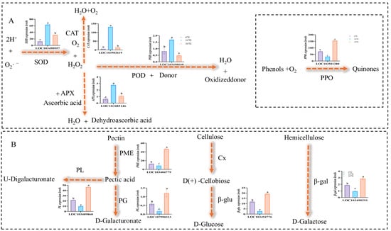
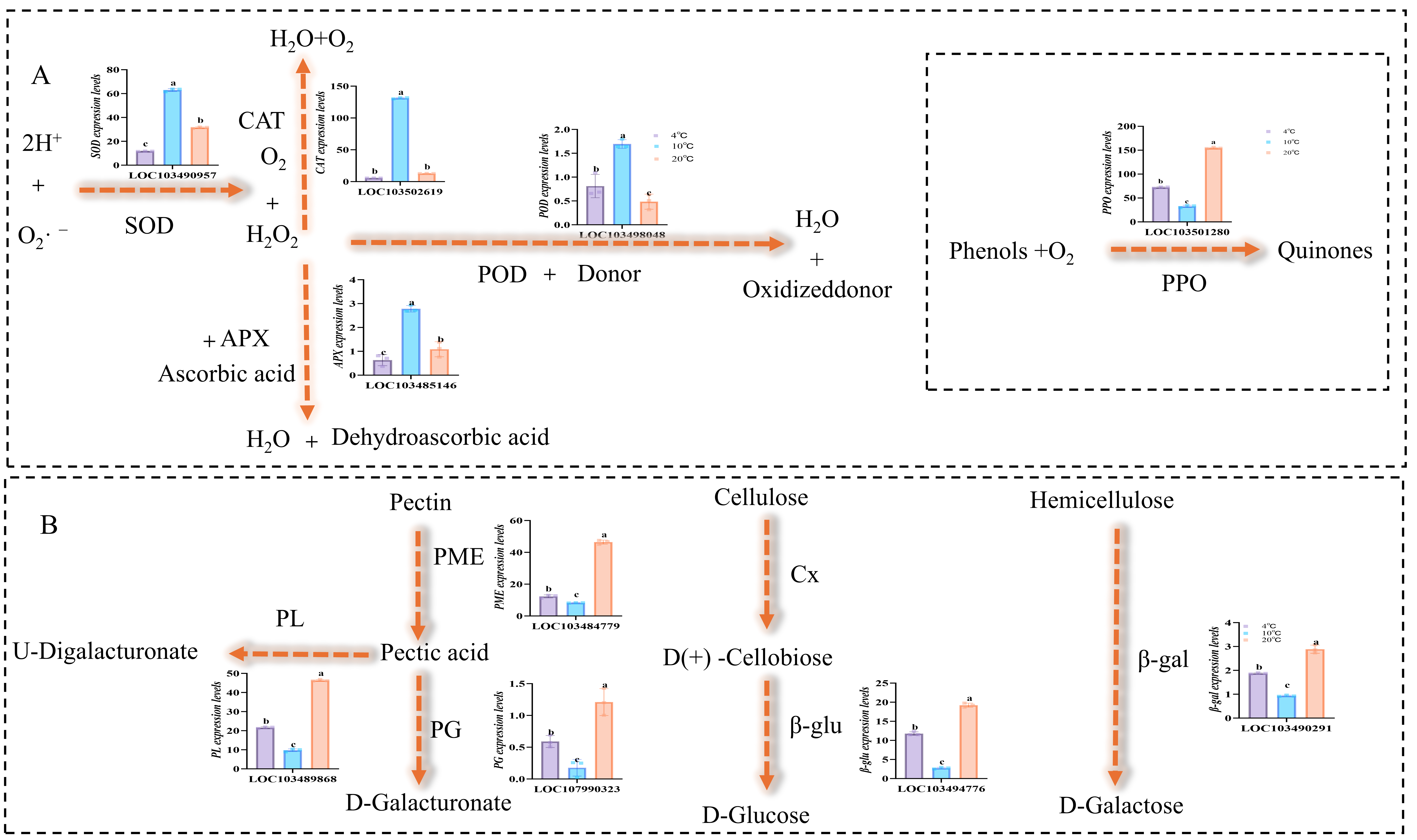
Ten differentially expressed genes associated with antioxidant activity and cell wall metabolism were identified based on p < 0.05 and |log2FoldChange| ≥ 1. Details are provided in Supplementary Table S2. Compared with 4 °C and 20 °C, storage at 10 °C significantly upregulated the relative expression levels of the antioxidant enzyme-encoding genes POD, SOD, CAT, and APX. The transcriptional abundances of the POD, SOD, CAT, and APX genes were 2.75, 6.08, 25.76, and 2.40 times those at 4 °C, respectively, and 3.28, 2.10, 9.44, and 4.64 times those at 20 °C. Storage at 10 °C downregulated the expression of the PPO-encoding gene, with its transcriptional levels being only 0.45 times that of the 4 °C group and 0.21 times that of the 20 °C (Figure 10A).
Figure 10.
Postharvest reactive oxygen species (A)and cell wall metabolism (B) pathway profiles of muskmelons at different storage temperatures. On 28 days of storage, differences in relative gene expression levels at different temperatures were indicated by lowercase letters (p < 0.05).
Storage at 10 °C significantly suppressed the expression of genes encoding cell wall-degrading enzymes—PL, PME, PG, β-glu, and β-gal—at the transcriptional level. The transcriptional levels of PL, PME, PG, β-glu, and β-gal were only 0.45, 0.67, 0.31, 0.23, and 0.58 times those at 4 °C, respectively, and 0.21, 0.18, 0.15, 0.14, and 0.37 times those at 20 °C (Figure 10B). Given the consistency between transcriptomic and enzyme activity changes, it is hypothesized that temperature may be a contributing factor to the changes in enzyme activity associated with these genes. Consequently, the rate of cell wall polysaccharide disassembly during storage was substantially decreased. In addition, it is speculated that changes in the transcriptome may be caused by differences in physiological age or tissue degeneration.
4. Discussion
Environmental stresses during fruit and vegetable storage—most notably, temperature fluctuations—disrupt ROS homeostasis and thereby trigger oxidative imbalance. When external conditions deteriorate, or the tissue progresses into senescence, intracellular ROS production exceeds the capacity for scavenging, leading to ROS accumulation [35]. Plants defend themselves with enzymatic antioxidants (for example, SOD, CAT, POD) and nonenzymatic antioxidants, but O2·– is rapidly converted by SOD into the more stable H2O2. CAT serve as the principal H2O2 removing enzymes, catalyzing H2O2 decomposition to yield H2O and O2 [36]. In the presence of an electron donor, H2O2 can be degraded by POD [37]. Under prolonged or severe stress, however, this antioxidant system is often overwhelmed and cannot match the surge in ROS. The resulting elevation in ROS levels is cytotoxic: it compromises membrane integrity, accelerates lipid peroxidation, and ultimately disrupts cellular compartmentalization [36]. Loss of membrane structure not only reduces the cell’s mechanical support—contributing directly to fruit softening—but also permits previously sequestered phenolic substrates to contact PPO. This encounter initiates quinone formation and enzymatic browning, which in turn accelerates quality decline and senescence of the fruit [24]. The results of this study are consistent with those of previous studies. The study found that proper low-temperature storage can delay the decline in antioxidant enzyme activity in cantaloupes during the later stages of storage, slow down membrane lipid peroxidation, and inhibit quality deterioration. Qin et al. [29] reported that mild cold treatment upregulates APX1 and APX4 expression in avocado, slowing the decline of APX, CAT, SOD, and POD activities during late storage. This response suppresses the accumulation of superoxide anions and H2O2, thereby inhibiting lipid peroxidation of fruit membranes. Similarly, storing Kashgar melon at appropriate temperatures increases POD and CAT activities, curbs excessive ROS accumulation, and helps preserve membrane integrity [3]. In plums, moderate low-temperature storage reduces PPO activity while delaying declines in POD and APX activities. This supports maintenance of nonenzymatic antioxidants such as total phenolics and glutathione, ultimately limiting firmness loss, weight loss, and color deterioration during storage and effectively preserving overall fruit quality [26]. Jia et al. [28] reported that when chilling injury occurred, CAT, APX, and POD exhibited parallel activity profiles during storage, each showing stress-induced peaks at specific times. Furthermore, when chilling injury occurred, antioxidant-related enzymes failed to maintain antioxidant enzyme activity in the ‘Xizhoumi 17’ melon [20] and the yellow-skinned melon [31], resulting in an inability to promptly scavenge reactive oxygen species and thereby accelerating fruit deterioration. Additionally, a similar trend was observed in watermelon seedlings [38]. In all cases, declines in antioxidant enzyme activities during late storage led to excessive ROS accumulation, inducing oxidative stress that compromised cellular function and hastened fruit softening and quality loss. At ambient temperature, longan pericarp cell membranes become compromised: increased permeability and rising relative conductivity accompany intensified lipid peroxidation. This membrane disruption facilitates interactions between PPO and phenolic substrates, accelerating browning and producing abnormal shifts in color parameters L*, a*, and b* [39]. By contrast, storage at 10 °C helps maintain membrane integrity, slows the increase in relative conductivity, and suppresses further lipid peroxidation.
Fruit softening is largely driven by cell wall degradation mediated by hydrolytic enzymes and associated proteins. PME removes methyl groups from pectin, yielding low-methyl esterified pectin that becomes susceptible to further hydrolysis by cell wall-degrading enzymes such as PG. PG cleaves the galacturonic acid linkages in the polygalacturonic acid backbone [1]. Zhang et al. [1] reported that an appropriate low temperature negatively regulated the aberrant expression of PG and PME encoding genes in ‘Gold Queen’ Hami melons when ambient and chilling injury occurred, thereby decelerating tissue softening. Tao et al. [40] found that an appropriately low storage temperature suppressed the expression of PME, β-gal, and β-glu in tomato, which delayed firmness loss and helped preserve fruit quality. Similarly, Huang et al. [6] reported that moderate cold downregulated PE, PG, PL, and β-gal–related genes in papaya; this attenuation slowed the accumulation of water-soluble pectin while reducing chelator-soluble pectin (CSP) and sodium carbonate–soluble pectin (NSP), thereby limiting cell wall component degradation and mitigating firmness decline. In avocado, exposure to suitable low temperature reduced both the expression and aberrant activities of Cx2, PG2, PME2, and PME31, inhibiting decreases in CSP and cellulose and postponing fruit softening [29]. Our findings agree with these reports. Appropriate low temperatures inhibit cell wall metabolism, slow the degradation of cell wall components, and delay the decrease in fruit firmness. Wang et al. [25] showed that storage at 6 °C for ‘Xizhoumi17’ Hami melons caused continuous upregulation of CmPME, CmPG, and Cmβ-gal, sustained high activities of cell wall-degrading enzymes (PME, PG, Cx, and β-gal) toward the end of storage led to increased levels of soluble pectin, accelerated loss of cellulose and hemicellulose and deterioration of cell wall integrity, and consequently pronounced softening and quality decline. Chilling injury induced abnormal expression of PG, Cx, and PME in mango [41] and plum [42], resulting in firmness loss and accelerated softening. Wang et al. [43] additionally reported that, at the end of cold-storage periods, PGL3 and β-gal8 were markedly upregulated in bitter gourd, thereby accelerating softening in a pattern that parallels the cold stress-induced responses observed in Hami melons in this study. Li et al. [44] found that, during storage, ‘Xizhoumi 17’ Hami melon fruits held at 22 °C exhibited pronounced increases in PG, Cx, PME, β-glu, and β-gal activities, which promoted cell wall degradation and further softening. Similarly, strawberries stored at 22 °C underwent abnormal cell wall component degradation accompanied by overactivity of PG, PME, β-glu, and Cx, which accelerated softening [5]. At 22 °C, bananas displayed significant upregulation of pectin-degrading genes such as PME, PL, and PG, leading to enhanced cell wall breakdown and increased softening and browning [45]. These reports align with the trends we observed for muskmelon stored at 20 °C.
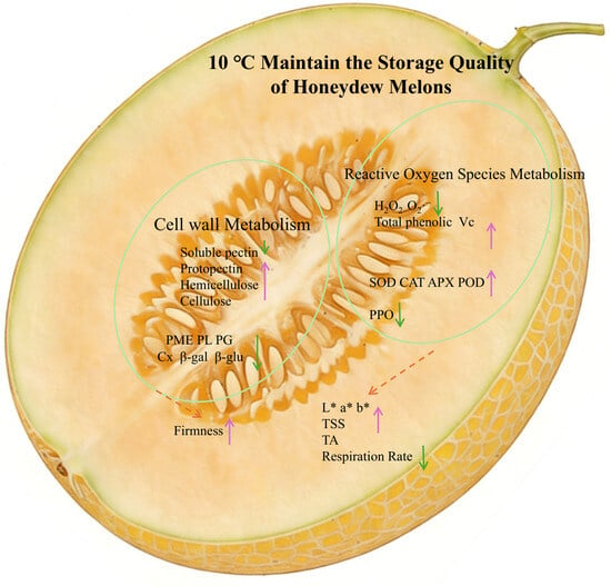
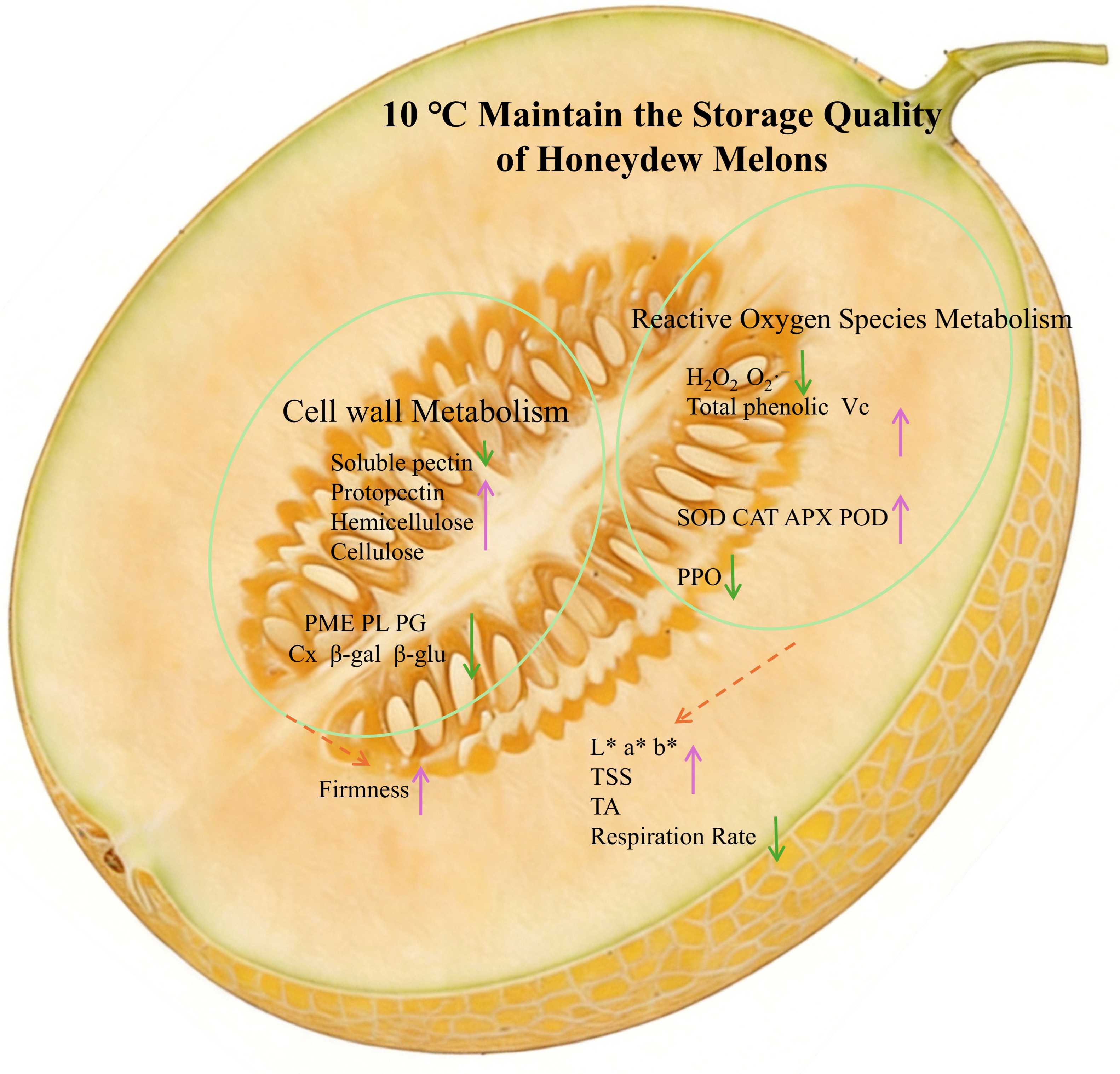
A proposed model illustrating the crosstalk between ROS and cell wall metabolism in postharvest muskmelon Aging is summarized in Figure 11.This study has several limitations. The mechanistic inferences are based largely on correlations between gene expression and enzyme activity, without proteomic analysis or functional validation; consequently, the direct regulatory roles of the differentially expressed genes in low-temperature tolerance remain to be confirmed, and the conclusion that 10 °C preserves quality by upregulating antioxidant genes and downregulating cell wall-degrading genes currently rests on expression-based correlations rather than direct evidence. In addition, this study was restricted to the single cultivar ‘Huangmengcui’ under isothermal conditions and did not address shelf life after removal from storage, temperature fluctuations, or sensory and flavor attributes. Future work integrating functional gene validation and realistic cold-chain conditions will be required to elucidate more comprehensively the mechanisms by which storage temperature regulates postharvest quality.
Figure 11.
A proposed model for maintaining the quality of muskmelons during storage at 10 °C; the upward purple arrows indicate that, compared to 4 °C and 20 °C, the levels of chemical components and enzyme activity are higher at 10 °C, while the downward green arrows indicate that, compared to 4 °C and 20 °C, the levels of chemical components and enzyme activity are lower at 10 °C.
5. Conclusions
This study demonstrates that storage at 10 °C effectively preserves the postharvest quality of ‘Huangmengcui’ muskmelons. Specifically, storage at 10 °C enhanced antioxidant enzyme activity and increased the contents of total phenolics and vitamin C, thereby reducing ROS accumulation and membrane lipid peroxidation and helping maintain cellular redox homeostasis. In addition, this storage temperature inhibited the activity of cell wall-degrading enzymes, delayed the degradation of pectin and cellulose, and consequently slowed fruit softening. These findings provide a theoretical basis for future research on the storage of thick-skinned melons and support the cold-chain transportation of this crop.
Supplementary Materials
The following supporting information can be downloaded at: https://www.mdpi.com/article/doi/s1, Figure S1: Color difference measurement points (A) and sampling points for muskmelon pulp, firmness measurement points (B); Table S1: Results of transcriptome data quality; Table S2: Transcriptomic data for 28-Day muskmelon pulp.
Author Contributions
Investigation, R.L. and H.Z.; Conceptualization, B.X. and X.G.; Methodology, R.L. and L.Z.; Validation, Z.K., X.D. and S.H.; Resources, X.G. and B.X.; Writing—original draft preparation, R.L. and L.Z.; Writing—review and editing H.Z. and B.X.; Visualization, R.L. and J.D.; Supervision, X.G. and B.X.; Project administration, C.Z.; Funding acquisition, H.Z., C.Z., X.G., and B.X. All authors have read and agreed to the published version of the manuscript.
Funding
This study was financially supported by the earmarked fund for Major Science and Technology Projects of Xinjiang Uygur Autonomous Region (2024A02007-4), Department of Science and Technology of the Xinjiang Uyghur Autonomous Region of China; Tianshan Talents-The Project for Outstanding Young Talents-Special Project for Grassroots Science and Technology Backbone Talents (2024TSYCJC0064), Department of Science and Technology of the Xinjiang Uygur Autonomous Region; China Agriculture Research System (CARS-23), Ministry of Agriculture and Rural Affairs of China; Xinjiang Agriculture Research System (XARS-06), Department of Agriculture and Rural Affairs of the Xinjiang Uyghur Autonomous Region of China; Key Research and Development Projects of Turpan (2024007), Turpan Science and Technology Bureau, Xinjiang Uyghur Autonomous Region of China; Xinjiang Uygur Autonomous Region Talent Development Fund under the ‘Tianchi Talent’ Introduction Program for Young·PhD·Researchers (Grant No. 524313001), Department of Human Resources and Social Security of the Xinjiang Uygur Autonomous Region of China; President Foundation Project of Tarim University (TDZKBS202305) of Tarim University.
Institutional Review Board Statement
Not applicable.
Informed Consent Statement
Not applicable.
Data Availability Statement
The original contributions presented in the study are included in the article/Supplementary Materials. Further inquiries can be directed to the corresponding authors.
Conflicts of Interest
The authors declare no conflicts of interest.
References
- Zhang, Q.; Shan, C.; Song, W.; Cai, W.; Zhou, F.; Ning, M.; Tang, F. Transcriptome analysis of starch and sucrose metabolism change in Gold Queen Hami melons under different storage temperatures. Postharvest Biol. Technol. 2021, 174, 111445. [Google Scholar] [CrossRef] [Scilit]
- Wang, H.-F.; Yang, Z.-Y.; Amanullah, S.; Wang, H.-L.; Liu, B.; Liu, S.; Yang, T.-T.; Wang, C.-N. Deciphering the effect of postharvest 1-MCP treatment coupled with low-temperature storage on the physiological activities and edible quality of melon. Plants 2025, 14, 586. [Google Scholar] [CrossRef] [Scilit]
- Ning, M.; Tang, F.; Chen, J.; Song, W.; Zhao, X.; Zhang, Q.; Cai, W.; Shan, C.; Li, Z. Physiological and transcriptomic analysis of postharvest Jiashi melon at different storage temperatures. Postharvest Biol. Technol. 2022, 191, 111963. [Google Scholar] [CrossRef] [Scilit]
- Meng, Y.; Dilixiati, Y.; Zhu, X.; Shi, W.; Wu, B.; Wang, J.; Wei, J. Nitric oxide fumigation maintains cantaloupe fruit quality by regulating the production of reactive oxygen species. Sci. Hortic. 2024, 338, 113801. [Google Scholar] [CrossRef] [Scilit]
- Lu, W.; Li, W.; Zhao, K.; Bai, X.; Zhang, Y.; Li, Q.; Xue, Z.; Wang, X.-X. Blue light irradiation combined with low-temperature storage further enhances postharvest quality of strawberries through improving antioxidant defense and cell wall metabolic activities. Food Chem. X 2025, 25, 102115. [Google Scholar] [CrossRef] [Scilit]
- Huang, L.; Tao, S.; Pan, Y.; Han, Z.; Chen, Y.; Tan, Y.; Yang, H. Molecular mechanisms of low temperature-induced aberrant chilling injury in papaya fruit: Physiological and transcriptomic analysis on cell wall metabolism. Sci. Hortic. 2025, 344, 114107. [Google Scholar] [CrossRef] [Scilit]
- Ning, M.; Tang, F.; Zhang, Q.; Zhao, X.; Yang, L.; Cai, W.; Shan, C. The quality of Gold Queen Hami melons stored under different temperatures. Sci. Hortic. 2019, 243, 140–147. [Google Scholar] [CrossRef] [Scilit]
- Aubert, C.; Bony, P.; Chalot, G.; Landry, P.; Lurol, S. Effects of storage temperature, storage duration, and subsequent ripening on the physicochemical characteristics, volatile compounds, and phytochemicals of western red nectarine (Prunus persica L. Batsch). J. Agric. Food Chem. 2014, 62, 4707–4724. [Google Scholar] [CrossRef] [Scilit]
- Lin, Y.; Lin, H.; Wang, H.; Lin, M.; Chen, Y.; Fan, Z.; Hung, Y.C.; Lin, Y. Effects of hydrogen peroxide treatment on pulp breakdown, softening, and cell wall polysaccharide metabolism in fresh longan fruit. Carbohydr. Polym. 2020, 242, 116427. [Google Scholar] [CrossRef] [Scilit]
- Song, W.; Zhang, P.; Zhang, H.; Xue, Y.a.; Zhang, Q.; Ning, M.; Zhao, X.; Cai, W.; Liu, X.; Zhang, X.; et al. A low concentration of exogenous salicylic acid enhances cold tolerance in Hami melons (Cucumis melo var. saccharinus) by modulating salicylic acid-response CmGST genes. Postharvest Biol. Technol. 2022, 193, 112034. [Google Scholar] [CrossRef] [Scilit]
- Jia, M.; Wang, X.; Zhang, M.; Liu, Q.; Gao, Z.; Chen, X.; Chen, H.; Zhang, Z.; Wang, J. Sodium nitroprusside reduces chilling injury in hami melons by regulating postharvest proline metabolism and energy metabolism. Sci. Hortic. 2026, 357, 114651. [Google Scholar] [CrossRef] [Scilit]
- Ren, D.; Zhang, J.; Zhang, K.; He, H.; Ma, G.; Liu, H.; Li, J. Near-freezing temperature storage maintains postharvest quality of melon through synergistic regulation of cell wall remodeling and aroma-related gene networks. Postharvest Biol. Technol. 2027, 244, 114805. [Google Scholar] [CrossRef] [Scilit]
- Peng, B.; Hao, X.; Zhang, Q.; Cai, W.; Zhao, X.; Tang, F.; Shan, C. Effects of low-temperature storage on the aroma composition and quality of Golden Empress Hami melons: A transcriptomic and metabolomic analysis. Food Biosci. 2024, 57, 103422. [Google Scholar] [CrossRef] [Scilit]
- Hao, X.; Di, J.; Han, Z. Effects of different concentrations of exogenous chlorogenic acid treatments on the storage quality of ‘lvbao melon’. Storage Process 2025, 25, 59–64. [Google Scholar]
- Tian, Y.; Cai, P.; Tang, Y.; Luo, F.; Fang, C.; Gao, J. Effects of different storage temperatures on the quality of Cucumis melo var. reticulatus chuanmicuiyu. Storage Process 2022, 22, 7–13. [Google Scholar]
- Yao, J.; Zheng, H.; Zhang, C.; Zaitouna, M.; Wang, Z.; Geng, X. Influences of exogenous melatonin on postharvest storage quality and post-ripening senescence of muskmelon. Jiangsu Agric. Sci. 2024, 52, 203–209. [Google Scholar] [CrossRef]
- Zhao, Y.; Chi, X.; Dan, J.; Wang, S.; Wang, Y.; Chen, Y. Effects of 1-MCP treatment on the storage quality and physiological characteristics of post-harvest muskmelon. North. Hortic. 2026, 8, 50–56. [Google Scholar] [CrossRef]
- Sun, H.; Wang, J.; Li, Y.; Zhang, C. Effects of chitosan and ethanol compound treatments on postharvest storage quality of thin-skinned muskmelon. North. Hortic. 2023, 14, 105–111. [Google Scholar]
- Zheng, H.; Yao, J.; Zhang, C.; Geng, X.; Wang, Z. Effects of programmed cooling on postharvest storage quality of melons. Storage Process 2024, 24, 9–13. [Google Scholar]
- Song, X.; LÜ, Y.; Wang, Y.; Chen, G.; Zhu, J.; Jiang, Y. Effect of ultraviolet irradiation combined with chitosan coating on storage quality of hami melon. Food Sci. 2023, 44, 159–165. [Google Scholar] [CrossRef]
- Zhang, Y.; Liu, P.; Zhao, X.; Chen, C.; Hou, Z. Ozone triggers network-coupled metabolic reprogramming of carbohydrate and amino acid metabolism during postharvest storage of Hami melon (Cucumis melo L.). Postharvest Biol. Technol. 2027, 243, 114654. [Google Scholar] [CrossRef] [Scilit]
- Liu, Y.; Wang, Y.; Bi, Y.; Li, S.; Jiang, H.; Zhu, Y.; Wang, B. Effect of preharvest acetylsalicylic acid treatments on ripening and softening of harvested muskmelon fruit. Sci. Agric. Sin. 2017, 50, 1865–1875. [Google Scholar] [CrossRef]
- Wang, L.; Yang, X.; Dai, B.; Wang, Y.; Du, X.; Huan, C.; Zheng, X. 1-methylcyclopropylene or methyl jasmonate-induced chilling tolerance in a stony hard peach cultivar. Sci. Hortic. 2022, 304, 111279. [Google Scholar] [CrossRef] [Scilit]
- Zhang, W.; Wang, Y.; Guo, H.; Yang, W.; Guo, M.; Chen, G. Cuticular wax removal on reactive oxygen species-related mechanisms and on the quality of Hami melon cultivars. Postharvest Biol. Technol. 2022, 193, 112060. [Google Scholar] [CrossRef] [Scilit]
- Wang, Y.; Liu, G.; Zhang, W.; Li, L.; Yang, W.; Liu, Y.; Jiang, L.; Guo, M.; Chen, G. Melatonin treatment delayed fruit softening by regulating postharvest carbohydrate metabolism of hami melon. Plant Physiol. Biochem. 2025, 219, 109328. [Google Scholar] [CrossRef] [Scilit]
- Wen, Y.; Mao, W.; Xu, B.; Dong, B.; Chen, W.; Jin, L.; Ma, W.; Cui, K. Effects of near-freezing temperature storage on phenolic substances and antioxidant activity of prune fruits. Xinjiang Agric. Sci. 2026, 63, 102–114. [Google Scholar] [CrossRef]
- Ge, L.; Zhao, D.; Wang, P.; Xie, B.; Zhang, Z.; Shangguan, X. Low temperature suppress cell wall degradation by modulating antioxidant metabolism in figs. Foods 2026, 15, 2418. [Google Scholar] [CrossRef] [Scilit]
- Jia, M.; Huang, S.; Zhang, M.; Liu, Q.; Wang, X.; Zhang, Q.; Lei, Y.; Wang, J. Effect of sodium nitroprusside on postharvest quality and reactive oxygen metabolism of hami melon. Acta Hortic. Sin. 2026, 53, 817–830. [Google Scholar] [CrossRef]
- Qin, J.; Chen, X.; Tang, X.; Shao, X.; Lai, D.; Xiao, W.; Zhuang, Q.; Wang, W.; Dong, T. Near-freezing temperature suppresses avocado (Persea americana Mill.) fruit softening and chilling injury by maintaining cell wall and reactive oxygen species metabolism during storage. Plant Physiol. Biochem. 2024, 210, 108621. [Google Scholar] [CrossRef] [Scilit]
- Zheng, Z.; Qu, H.; Zhou, H.; Yang, H.; Gao, H. Changes in quality, ultrastructure, reactive oxygen species and cell wall metabolisms of postharvest coprinus comatus stored at different temperatures. Sci. Hortic. 2022, 298, 110989. [Google Scholar] [CrossRef] [Scilit]
- Xu, B.; An, L.; Meng, X.; Zhang, T. Effect of exogenous putrescine (Put) treatments on postharvest chilling injury and reactive oxygen metabolism of yellow melon fruits. Sci. Technol. Food Ind. 2022, 43, 360–367. [Google Scholar] [CrossRef]
- You, W.; Zhang, J.; Ru, X.; Xu, F.; Wu, Z.; Jin, P.; Zheng, Y.; Cao, S. Exogenous CaCl2 delays flesh softening by inhibiting the degradation of cell wall in fresh-cut cantaloupe. Postharvest Biol. Technol. 2024, 213, 112934. [Google Scholar] [CrossRef] [Scilit]
- Cao, S.; Jin, J.; Yang, Y.; Chen, J.; Luo, D.; Ba, L. Chlorogenic acid alleviates chilling injury in peach fruit by regulating cell wall metabolism and phenylpropanoid metabolism. Postharvest Biol. Technol. 2026, 242, 114553. [Google Scholar] [CrossRef] [Scilit]
- Wei, L.; Gao, S.; Li, F.; Zhang, X.; Peng, Y.; Liu, Y.; Zhang, Y.; Qin, W. Transcriptomics and physiological indicators analyzed the delayed effect of carboxymethyl chitosan on lignification of okra during postharvest cold storage. Postharvest Biol. Technol. 2025, 222, 113394. [Google Scholar] [CrossRef] [Scilit]
- Xue, H.; Sun, Y.; Li, L.; Bi, Y.; Hussain, R.; Zhang, R.; Long, H.; Nan, M.; Pu, L. Acetylsalicylic acid (ASA) induced fusarium rot resistance and suppressed neosolaniol production by elevation of ROS metabolism in muskmelon fruit. Sci. Hortic. 2020, 265, 109264. [Google Scholar] [CrossRef] [Scilit]
- Xie, P.; Yang, Y.; Li, Y.; Wang, Y.; Bai, B.; Prusky, D.; Li, Y.; Bi, Y. Preharvest phenylalanine spraying alleviates chilling injury in harvested muskmelons by maintaining reactive oxygen species homeostasis. Food Chem. 2025, 466, 142198. [Google Scholar] [CrossRef] [Scilit]
- He, J.; Xie, S.; Zhao, X.; Fu, D. Spermidine enhances cold tolerance in tomato fruit by boosting antioxidant and energy metabolism synergy. Food Res. Int. 2025, 222, 117768. [Google Scholar] [CrossRef] [Scilit]
- Li, X.; Wang, H.; Kang, L.; Ning, N.; Yong, L.H.; Cheng, J.; Chang, G.; Zhao, W. Effects of low temperature stress on the growth, leaf membrane lipid peroxidation, and polyamine metabolism of watermelon seedlings. China Cucurbits Veg. 2026, 39, 135–140. [Google Scholar] [CrossRef]
- Ning, C.; Lin, X.; Li, J.; Xia, R.; Long, L.; Han, D.; Wu, Z.; Luo, T. Effects of preharvest gibberellin treatments on the room-temperature storability of ‘shixia’ longan fruit. Mod. Food Sci. Technol. 2025, 41, 153–166. [Google Scholar] [CrossRef]
- Tao, J.; Zuo, J.; Watkins, C.B.; Bai, C.; He, X.; Liu, S.; Han, L.; Zhao, X.; Liu, Y.; Li, J.; et al. Low storage temperature affects quality and volatile compounds in fresh tomatoes. Food Chem. 2024, 460, 140400. [Google Scholar] [CrossRef] [Scilit]
- Ajay; Gill, P.P.S.; Jawandha, S.K.; Singh, N.P.; Kaur, S.; Kaur, P. Role of gum arabic combined with cinnamic acid coating on quality and cell wall degradation enzymes of mango fruits at low temperature. Int. J. Biol. Macromol. 2024, 259, 129088. [Google Scholar] [CrossRef] [Scilit]
- Xiao, Y.; Li, Y.; Fan, Y.; Li, Z.; Zhang, X.; Liang, D.; Xia, H.; Lv, X.; Wang, X.; Wang, J.; et al. Melatonin maintains the quality and cell wall structure of plum fruits under low temperature storage. Sci. Hortic. 2024, 338, 113745. [Google Scholar] [CrossRef] [Scilit]
- Wang, H.; Li, L.; Ma, L.; Fernie, A.R.; Fu, A.; Bai, C.; Sang, Z.; Guo, S.; Zhang, F.; Wang, Q.; et al. Revealing the specific regulations of nitric oxide on the postharvest ripening and senescence of bitter melon fruit. Abiotech 2024, 5, 29–45. [Google Scholar] [CrossRef] [Scilit]
- Li, H.; Wang, X.; Bi, Y.; Wang, X.; Zhang, Q.; Lei, Y.; Huang, S.; Wang, J. Effect of walnut green peel polyphenol extract on cell wall metabolism of postharvest fruits of hami melon. Food Res. Dev. 2025, 46, 17–22. [Google Scholar] [CrossRef]
- Li, X.; Rahman, F.U.; Zhao, Y.; Lei, Q.; Chen, W.; Li, X.; Hu, H.; Zhu, X. 1-MCP and MAP treatments reduce the finger drop of banana during the shelf-life after high temperatures storage by regulating cell-wall metabolism. Postharvest Biol. Technol. 2025, 229, 113692. [Google Scholar] [CrossRef] [Scilit]
Disclaimer/Publisher’s Note: The statements, opinions and data contained in all publications are solely those of the individual author(s) and contributor(s) and not of MDPI and/or the editor(s). MDPI and/or the editor(s) disclaim responsibility for any injury to people or property resulting from any ideas, methods, instructions or products referred to in the content. |
© 2026 by the authors. Licensee MDPI, Basel, Switzerland. This article is an open access article distributed under the terms and conditions of the Creative Commons Attribution (CC BY) license.