


Abstract
This study evaluated the antagonistic activity of the cell-free supernatant of Lacticaseibacillus rhamnosus ATCC 9595 (Lcr-CFS) against Listeria monocytogenes, a major foodborne pathogen, that represents a challenge to food safety, due to its remarkable tolerance to environmental stresses and strong biofilm-forming ability. The minimum inhibitory concentration (MIC) and minimum bactericidal concentration (MBC) of Lcr-CFS against L. monocytogenes were defined as 31.25 and 62.5 mg/mL, respectively. Time-kill assays revealed dose- and time-dependent bactericidal effects. At sub-MICs, Lcr-CFS significantly reduced L. monocytogenes biofilm formation, disrupted preformed biofilms and decreased cell viability (80.3–96.7%), effects that were confirmed by 3-(4,5-dimethylthiazol-2-yl)-2,5-diphenyltetrazolium bromide (MTT) assay and fluorescence microscopy. Transmission electron microscopy showed L. monocytogenes cell wall damage, cytoplasmic leakage, and morphological alterations consistent with bactericidal effects. Additionally, exposure to 1x and 2x MIC of Lcr-CFS induced reactive oxygen species (ROS) accumulation, indicating oxidative stress as part of the mechanism by which Lcr-CFS exerts its antimicrobial activity. Gene expression analysis revealed upregulation of stress and virulence-associated genes (sigB, prfA, degU, flaA, motA, hlyA, pclA, and actA) upon exposure to 0.5x MIC suggesting a complex cross-talk network between adaptive mechanisms and environmental stresses. Although L. monocytogenes initiates a stress response, it appears unable to counteract the damage induced by Lcr-CFS, resulting in cell death. These findings highlight the antimicrobial and anti-biofilm properties of Lcr-CFS against L. monocytogenes. Given its in vitro efficacy, Lcr-CFS emerges as a promising biocontrol agent to improve food safety by mitigating the persistence of L. monocytogenes in food processing settings.
1. Introduction
Listeria monocytogenes is a Gram-positive, rod-shaped, non-spore-forming, and psychrotrophic bacterium that is ubiquitously distributed in the environment [1,2,3,4]. It is recognized as a globally significant foodborne pathogen responsible for both outbreaks and cases of listeriosis, a life-threatening infection acquired through the consumption of contaminated food [5]. Although its incidence is low compared to other foodborne bacterial diseases, listeriosis is associated with high morbidity, with approximately 90% of cases requiring hospitalization, and a mortality rate of 20–30% among high-risk populations, including children, pregnant women, the elderly, and immunocompromised individuals [6,7,8,9].
The ubiquity of L. monocytogenes can largely be attributed to the extensive stress adaptations, enabling the bacterium to survive in a variety of harsh environments [10]. Its ability to persist in diverse settings, including food processing and manufacturing facilities, makes L. monocytogenes particularly challenging to control and eradicate. As a result, it poses a significant threat to food safety, especially in ready-to-eat products [5,11].
The persistence of L. monocytogenes in food processing environments is primarily attributed to its robust biofilm-forming ability, which enhances resistance to sanitizers, heat, desiccation, and acidic stress [12,13]. These biofilms, commonly formed on equipment and surfaces, serve as reservoirs for recurring contamination and significantly hinder sanitation efforts, reinforcing the pathogen’s capacity to endure and disseminate in industrial settings [14,15,16].
The ability of biofilm-associated L. monocytogenes to withstand antimicrobial treatments and proliferate in various food matrices underscores the importance of developing effective control strategies to reduce the risk of contamination and safeguard public health [17,18]. In this context, plant-derived compounds, enzymes, bacteriophages, and bacterial metabolites, used alone or in combination with conventional methods, have been evaluated as promising alternative for biofilm control in the food industry [19,20,21,22].
Lactic acid bacteria (LAB) have been extensively investigated for their potential to prevent and control spoilage microorganisms and foodborne pathogens, due to their natural and generally recognized as safe (GRAS) status [23]. LAB are known to produce a diverse arsenal of antimicrobial compounds, such as organic acids, hydrogen peroxide, and antimicrobial peptides [24,25,26].
Lacticaseibacillus rhamnosus is a well-characterized LAB widely recognized for its health-promoting effects. Commonly found in the gastrointestinal tract and in fermented dairy products, Lc. rhamnosus has the ability to modulate the host immune response, enhance intestinal barrier function, and inhibit the growth of pathogenic microorganisms through the production of antimicrobial compounds such as organic acids, hydrogen peroxide, and bacteriocins [27]. Among its various strains, Lc. rhamnosus GG and Lc. rhamnosus GR-1 have received particular attention for their probiotic efficacy and safety profile [28,29].
This study aims to assess the unexplored potential of the cell-free supernatant of Lc. rhamnosus ATCC 9595 (Lcr-CFS) against L. monocytogenes. Specifically, we investigated the effects of Lcr-CFS on L. monocytogenes growth, biofilm formation and disruption, cell wall alterations, oxidative stress induction, and modulation of stress and virulence genes expression.
2. Materials and Methods
2.1. Bacterial Strains and Culture Conditions
Lc. rhamnosus ATCC 9595 was kindly provided by Fiocruz (Fundação Oswaldo Cruz, Rio de Janeiro, Brazil). This strain was routinely grown in Man-Rogosa-Sharpe (MRS) broth (Kasvi, Madrid, Spain), under microaerophilic conditions at 37 °C for 18–24 h. L. monocytogenes ATCC 19112, along with strains isolated from food products or food processing environments in Brazil were kindly provided by the Listeria Collection (CLIST) of the Laboratory of Bacterial Zoonoses of the Oswaldo Cruz Institute (LABZOO/Fiocruz) (Table 1). L. monocytogenes strains were cultivated in Brain Heart Infusion (BHI) broth (Himedia, Mumbai, India), under aerobic conditions at either 28 or 37 °C (depending on the experiment) for 18–24 h. All bacterial strains were preserved at −20 °C in BHI broth containing 20% glycerol.
Table 1.
Listeria monocytogenes strains: code, serotypes, year of the isolation, sources, and specimen types.
2.2. Lacticaseibacillus rhamnosus ATCC 9595 Cell-Free Supernatant (Lcr-CFS) Obtention, Inhibitory Activity Against Listeria monocytogenes and Degradation by Proteinase K
Stationary-phase cultures of Lc. rhamnosus ATCC 9595 (200 mL MRS, 37 °C, 24 h, microaerophilic conditions) [30] were centrifuged (12,500 rpm, 15 min), and the supernatants were filtered through a 0.22 μm pore-size membrane (Kasvi, São José dos Pinhais, Brazil) to obtain the cell-free supernatant (CFS). The CFS was lyophilized (SP Scientific, Warminster, PA, USA) and subsequently rehydrated in phosphate-buffered saline (PBS, pH 7.4) to obtain a stock solution at a final concentration of 500 mg/mL (corresponding to the resuspension of lyophilizate mass). This 500 mg/mL solution derived from the lyophilized CFS was designated as Lcr-CFS, stored at −20 °C, and used in all subsequent experiments.
The minimum inhibitory concentration (MIC) of Lcr-CFS against L. monocytogenes strains was determined by the broth microdilution method according to Clinical and Laboratory Standards Institute guidelines [31]. Aliquots of 50 μL of Lcr-CFS and its serial twofold dilutions (prepared from the 500 mg/mL stock solution) were dispensed into the wells of a sterile 96-well microplate containing 50 μL BHI broth (2x concentrated). Subsequently, stationary-phase L. monocytogenes cultures (106 CFU/mL; 10 μL) were added. The plates were incubated at 37 °C for 18 h under aerobic conditions [32]. Wells containing uninoculated BHI broth served as negative controls, while medium inoculated solely with L. monocytogenes served as growth controls. The MIC was defined as the lowest concentration of Lcr-CFS that completely inhibited visible bacterial growth of L. monocytogenes after 18 h of incubation at 37 °C.
To determine the minimum bactericidal concentration (MBC), aliquots (5 μL) from wells without visible growth were plated on BHI agar without Lcr-CFS and incubated at 37 °C for 48 h. The MBC was defined as the lowest concentration of Lcr-CFS yielding no visible colonies, indicating bactericidal activity [33].
To evaluate the proteinaceous nature of the antimicrobial compounds in the Lcr-CFS, a proteolytic degradation assay was performed [34], with modifications]. Aliquots of Lcr-CFS were incubated with proteinase K (1 mg/mL final concentration; Sigma-Aldrich, St. Louis, MO, USA) at 37 °C for 2 h. The enzyme was inactivated by heating at 95 °C for 10 min. The MIC of the digested Lcr-CFS against all L. monocytogenes strains was determined as described above. Untreated Lcr-CFS (without proteinase K digestion) served as a positive control for antimicrobial activity.
All experimental assays were performed in duplicate.
2.3. Time-Kill Assay
Time-kill kinetic studies were performed in BHI broth inoculated with logarithmically grown L. monocytogenes 4455 at a final concentration of approximately 106 CFU/mL. Lcr-CFS was added at 0.5x, 1x and 2x MIC. Cultures were incubated at 37 °C, and samples were collected at 0, 3, 6, 9, 12, 24, 48 and 72 h to determine viable cell counts in colony forming units per milliliter (CFU/mL) [35]. Time-kill curve was generated by plotting log10 CFU/mL versus time (h). Bactericidal activity was defined as ≥99.99% reduction relative to initial bacterial count, while bacteriostatic activity was defined as either maintenance of the initial inoculum or a reduction of <99.99% compared to the initial inoculum [36,37].
2.4. Influence of Lcr-CFS on Listeria monocytogenes Biofilm Development and Biofilm Dispersion
Biofilm-forming ability of the L. monocytogenes strains selected for this study was previously reported [38], and the bacteria were classified according to the criteria proposed by Stepanóvic et al. [39] into non-, weak, moderate, or strong biofilm producers.
To evaluate the influence of Lcr-CFS on biofilm development in 96-well polystyrene microplates, we selected the L. monocytogenes 4455, a strong biofilm producer. L. monocytogenes 4455 was co-cultured with Lcr-CFS at 0.25x and 0.5x MIC in BHI broth at 28 °C, for 72 h, in aerobic conditions. Biofilm biomass was quantified using 0.2% crystal violet staining (30 min), followed by ethanol solubilization and absorbance reading at 540 nm (TP-reader, Thermo Plate, São Paulo, Brazil). Biofilm formation inhibition rates were calculated as follows:
Inhibition rate (%) = [1 − OD540 nm (sample)/OD540 nm (positive control)] × 100.
The effect of Lcr-CFS on the bacterial cell viability in mature L. monocytogenes biofilms was determined by the 3-(4,5-dimethylthiazol-2-yl)-2,5-diphenyltetrazolium bromide (MTT) assay. After biofilm formation in polystyrene microplates (28 °C, 72 h), wells were washed with PBS, and 100 µL Lcr-CFS at different concentrations (0.25 to 2x MIC) were added. The control group received PBS instead of Lcr-CFS. Plate was incubated for 24 h at 28 °C [40]. Following incubation, the content of the wells was removed and 10 µL MTT (5 mg/mL) with 90 µL of PBS were added to each well. After 2 h incubation at 37 °C, MTT solution was discarded, and 150 µL of DMSO was immediately added to dissolve the formazan crystals. The plate was then incubated for 15 min at room temperature, and absorbance was measured at 490 nm using a microplate reader (TP-reader, Thermo Plate, São Paulo, Brazil). The experiment was performed in triplicate.
2.5. Visual Observation of Biofilm Development on Glass Surfaces in the Presence of Lcr-CFS
Optical microscopy analysis of bacterial biofilm was performed following the method described by Singh et al. [41]. In brief, sterile glass coverslips (1 × 1 cm) were deposited into each well of a 6-well plate, followed by addition of 1.5 mL of BHI broth (2x concentrated), 1.5 mL of Lcr-CFS (0.25x or 0.5x MIC), and 300 μL of stationary-phase L. monocytogenes 4455 suspension (106 CFU/mL). The plate was incubated (37 °C, 18 h), and planktonic cells were removed by PBS washing (150 μL; 3 times). The remaining biofilms were stained with 0.2% crystal violet for 30 min [42,43]. Biofilms were examined under light microscope (Nikon Instruments, New York, NY, USA) at 400× magnification for overall biofilm analysis and at 1000× for visualization of individual cell attachment. All experimental assays were performed in duplicate.
2.6. Viability of L. monocytogenes Cells in Mature Biofilms After Lcr-CFS Treatment
Mature biofilms were developed on sterile glass coverslips placed in 6-well plates, as described above, in the absence of Lcr-CFS. The mature biofilms were submerged with Lcr-CFS (0.25x to 2x MIC) for 24 h, and stained with propidium iodide (PI) (10 μg/mL, 10 min). PI selectively stains dead cells or those with compromised membranes, emitting red fluorescence [44]. Biofilms non-exposed to Lcr-CFS were used as a control. Fluorescent images were acquired using an inverted fluorescence microscope EVOS (Invitrogen, Waltham, MA, USA) equipped with a 40x phase-contrast objective and a ICX285AL CCD camera (Sony, Hod Hasharon, Israel) [40]. All tests were performed in duplicate.
2.7. Reactive Oxygen Species Production Induced by Lcr-CFS Exposure
Aliquots of 30 μL of L. monocytogenes 4455 (106 CFU/mL) were exposed to 300 μL of Lcr-CFS (0.25x to 2x MIC) for 3 h at 37 °C. After incubation, cells were harvested by centrifugation (3500 rpm, 10 min), the supernatant was discarded, and the pellet was suspended in 300 µL of PBS. Cell suspensions were stained with 0.1 μM 2′,7′-dichlorodihydrofluorescein diacetate (DCFH-DA; Sigma-Aldrich, USA) and incubated at 37 °C for 20 min. Following staining, cells were centrifuged (3500 rpm, 10 min) and washed twice using PBS to remove the extracellular dye. Aliquots of each treatment (100 μL) were transferred to polystyrene microplates and fluorescence intensity was measured using a fluorescence microplate reader EnSpire® (Perkin Elmer, Rodgau, Germany) with excitation at 485 nm and emission at 535 nm [45]. All experimental assays were performed in triplicate.
2.8. Effect of Lcr-CFS Treatment on Listeria monocytogenes Cell Structure
L. monocytogenes 4455 (106 CFU/mL) was incubated with Lcr-CFS at 0.5x and 1x MIC, at 37 °C for 24 h, to assess potential damages to the bacterial cell wall. Following incubation, cells were centrifuged (12,000 rpm, 10 min), washed three times with sodium phosphate solution (0.1 M, pH 7.2; 1 mL), and fixed in 2.5% glutaraldehyde and 4% paraformaldehyde in PBS. Post-fixation was performed with 0.1% osmium tetroxide and 0.1% ruthenium red for 3 h. Cells were dehydrated through graded ethanol series (50, 70, 90, 95, and 100% for 10 min each), followed by three washes in absolute ethanol and three washes in pure acetone for 20 min. EMbed 812 resin (EMS-14120) was added in increasing concentrations in acetone (2:1 overnight; 1:1 for 4 h; and pure resin for 48 h at 60 °C) according to the method described by Liu et al. [46]. Ultrathin sections were placed on copper grids and observed using a Zeiss EM 900 transmission electron microscope (Zeiss, Oberkochen, Germany; Electronic Microscopy Laboratory—UFTM) at 5000× magnification.
2.9. Influence of Lcr-CFS on L. monocytogenes Gene Expression
The effect of Lcr-CFS on the expression of the genes prfA, sigB, degU, flaA, motA, hlyA, pclA and actA was evaluated by real-time quantitative reverse transcription polymerase chain reaction (RT-qPCR). L. monocytogenes 4455 cultures (106 CFU/mL), and 0.5x MIC of Lcr-CFS were mixed and incubated at 28 °C for 24 h. Following incubation, cultures were centrifuged (13,500 rpm, 5 min.) and washed with PBS (1 M, pH 7.0). Total RNA was extracted using Trizol reagent (Sigma-Aldrich, St. Louis, MO, USA), and complementary DNA (cDNA) was synthesized with the High-Capacity cDNA Reverse Transcription Kit (Thermo Fisher, Waltham, MA, USA). Gene expression was quantified by RT-qPCR and performed with PowerTrackTM SYBR Green Master Mix for qPCR (Thermo Fisher, USA), following the manufacturer’s instructions. Primers used in this study were previously reported (Table 2) and the 16S rRNA gene was used as a housekeeping control.
Table 2.
Primer pairs used in real-time quantitative reverse transcription polymerase chain reactions (RT-qPCR).
Each reaction contained of the following components: 5 µL of PowerTrack SYBR Green Master Mix, 0.5 µL of forward primer, 0.5 µL of reverse primer, 0.25 µL of yellow dye, 1 µL of cDNA (20 ng), and 3.25 µL of nuclease-free water, resulting in a final reaction volume of 10 µL. Amplification was carried out using the StepOnePlusTM Real-Time PCR System (Thermo Fisher, USA). Cycling conditions were as follows: an initial denaturation step at 95 °C for 2 min, followed by 40 cycles of 95 °C for 15 s and 60 °C for 60 s. The specificity of the amplification was confirmed by melting curve analysis. Relative gene expression levels were calculated using the comparative CT method (2−ΔΔCt) [50]. All experiments were performed in biological duplicates and experimental triplicates.
2.10. Statistical Analysis
Statistical analyses were performed using GraphPad Prism 8.0 software (GraphPad Software Inc., La Jolla, CA, USA). All data were subjected to the F test and the Shapiro–Wilk test to assess variance and normality, respectively. Comparisons between groups were conducted using one-way ANOVA, followed by Dunnett’s test to evaluate the effect of Lcr-CFS on biofilm formation. Differences were considered statistically significant at p < 0.05. For the analysis of ROS production and relative gene expression, ANOVA was also employed. Genes were considered to be upregulated (2−ΔΔCt > 1) or downregulated (2−ΔΔCt < 1) in the presence of Lcr-CFS when statistically significant differences (p < 0.05) were observed in Ct values between the control and the treated groups.
3. Results
3.1. Bactericidal Activity of Lcr-CFS Against Listeria monocytogenes
The cell-free supernatant obtained from stationary-phase cultures of Lc. rhamnosus ATCC 9595 exhibited an initial pH of 4.5. The Lcr-CFS was subsequently lyophilized and suspended in PBS (pH 7.4) prior to use. The inhibitory activity of Lcr-CFS was assessed by determining its MIC against twenty-one L. monocytogenes strains. The growth of all bacterial strains was completely inhibited by Lcr-CFS, and a concentration of 31.25 mg/mL was determined as the MIC. The MBC was 62.5 mg/mL, corresponding to twice the MIC (2x MIC), suggesting a potential bactericidal effect of Lcr-CFS.
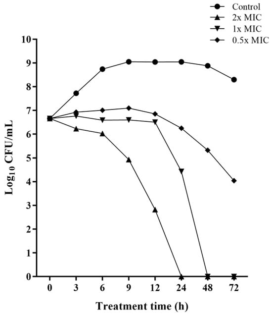
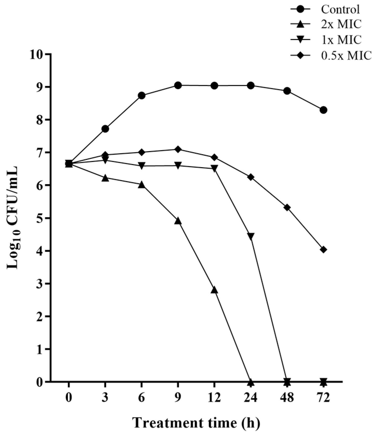
The bactericidal activity of Lcr-CFS against all L. monocytogenes strains was further confirmed by time-kill experiments, showing dose- and time-dependent kinetics. Co-incubation of L. monocytogenes with Lcr-CFS at the MBC (62.5 mg/mL) resulted in an approximately 4-log reduction in the initial bacterial population after 12 h. At the MIC (31.25 mg/mL), L. monocytogenes growth was inhibited for 12 h, with effective cell death observed at 24 h of incubation (approximately 3-log reduction), and absence of colonies after 48 h of exposure. At 0.5x MIC (15.6 mg/mL), an approximately 3-log reduction in L. monocytogenes population was observed after 72 h of exposure to Lcr-CFS (Figure 1).
Figure 1.
Time-kill curves demonstrating the bactericidal activity of Lacticaseibacillus rhamnosus cell-free supernatant (Lcr-CFS) against Listeria monocytogenes 4455. Lcr-CFS was added at time point zero (0.5x, 1x, and 2x MIC), and UFC/mL was determined for up to 72 h. The control curve represents bacterial growth in the absence of Lcr-CFS.
Lcr-CFS markedly inhibited L. monocytogenes growth, resulting in an optical density (OD600) of 0.131. In contrast, after treatment with proteinase K, Lcr-CFS lost its inhibitory activity, and the growth of L. monocytogenes was comparable to the untreated control (OD600 of 0.435 vs. 0.495, respectively). This result strongly suggests that the bioactive compound (s) synthesized by Lc. rhamnosus is proteinaceous in nature and sensitive to degradation by proteinase K.
3.2. Anti-Biofilm Effects of Lcr-CFS
As recently reported by our research group [38], L. monocytogenes 4455 (serotype 1/2a), isolated from a frozen chicken carcass in 2017, is a strong biofilm producer. Thus, strain 4455 was selected as representative of the other L. monocytogenes strains for the experiments that aimed to analyze the anti-biofilm effects of Lcr-CFS.
At 0.25x and 0.5x MIC, Lcr-CFS significantly reduced biofilm formation by L. monocytogenes 4455 compared to the untreated group (biofilm formed in the absence of Lcr-CFS) (p < 0.05). At concentrations equal or above the MIC, no bacterial growth was observed (Table 3). Lcr-CFS also disrupted pre-formed L. monocytogenes biofilms, reducing cell viability as indicated by MTT conversion relative to the untreated control biofilm (p < 0.05). It is important to highlight that the effect of Lcr-CFS on disrupting mature biofilms did not significantly differ among the tested concentrations, with reductions in cell viability ranging from 80.3 to 96.7% at 0.25x and 2x MIC, respectively (p > 0.05) (Table 3).
Table 3.
Effect of different concentrations of cell-free supernatant of Lacticaseibacillus rhamnosus ATCC 9595 (Lcr-CFS) on biofilm formation and on disruption of mature biofilm of Listeria monocytogenes 4455 on polystyrene surfaces measured by crystal violet and MTT staining, respectively.
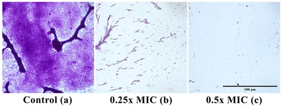
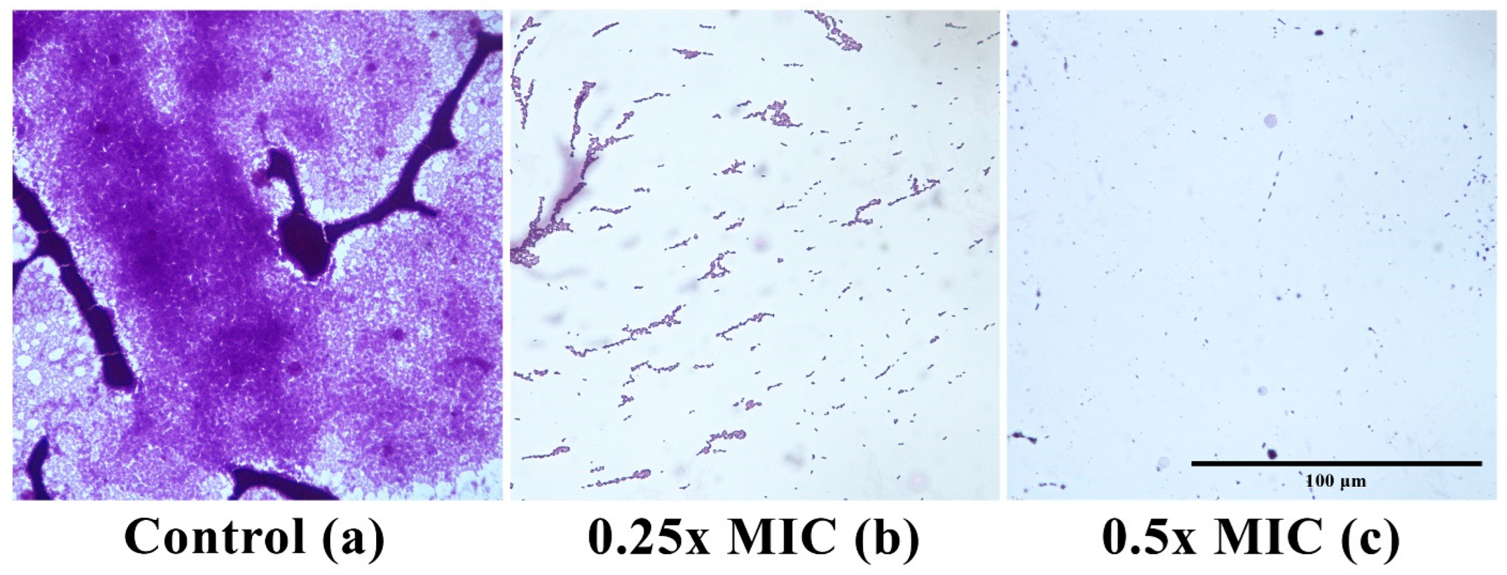
Visualization by light microscopy revealed well-formed and compact biofilms formed by L. monocytogenes 4455 on glass surfaces after 72 h of incubation at 28 °C (Figure 2a). In contrast, co-culture with Lcr-CFS at 0.25x and 0.5x MIC inhibited the development of L. monocytogenes biofilms, with only scattered cells being visualized (Figure 2b,c).
Figure 2.
Optical microscope images of 72 h Listeria monocytogenes 4455 biofilm grown on glass surfaces stained with crystal violet. (a) biofilm formed in the absence and presence of Lacticaseibacillus rhamnosus ATCC 9595 (Lcr-CFS) at 0.25x MIC (b) and 0.5x MIC (c). Magnification 400×. Scale bar is 100 µm.
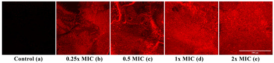
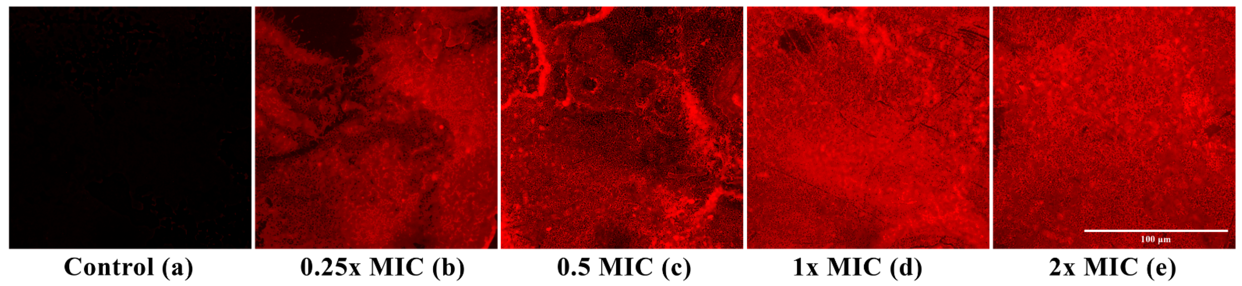
The ability of Lcr-CFS to disrupt mature L. monocytogenes biofilms formed on glass surfaces for 72 h was analyzed by fluorescence microscopy using PI, which selectively penetrates cells with compromised membranes and serves as a marker of cell death. Treatment with Lcr-CFS at 0.25x, 0.5x, 1x, and 2x MIC (Figure 3b–e) resulted in a marked increase in PI fluorescence intensity compared to the untreated control (Figure 3a), indicating bacterial cell death within the biofilm.
Figure 3.
Fluorescent microscope images of Listeria monocytogenes 4455 biofilms formed on glass surfaces after 72 h. (a) control (non-treated, non-fluorescent cells); (b–e) mature biofilms treated with Lcr-CFS (0.25x–2x MIC). Biofilms were stained with propidium iodide (PI), and red color indicates dead bacterial cells on glass surfaces. Magnification 40×. Scale bar is 100 µm.
3.3. Induction of Reactive Oxygen Species (ROS) by Lcr-CFS
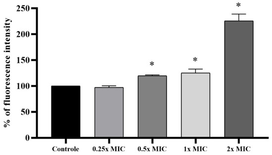
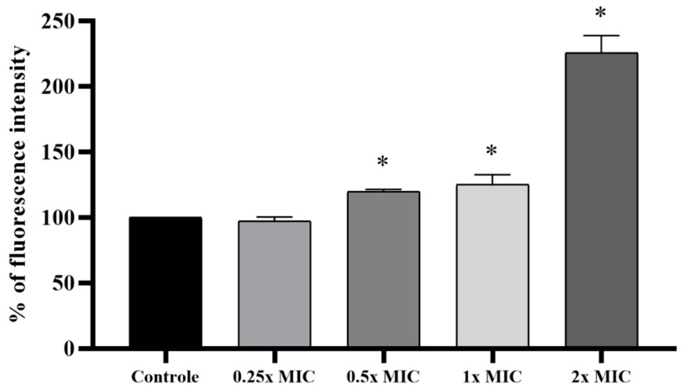
Treatment with Lcr-CFS at 1x and 2x MIC significantly enhanced ROS production in L. monocytogenes 4455 compared to the control condition (Figure 4). Notably, exposure to 2x MIC resulted in markedly higher fluorescence intensity (2.26-times fold), indicating substantial ROS generation and induction of oxidative stress. The accumulation of ROS induced by Lcr-CFS can lead to the damage of L. monocytogenes cells by either direct reaction with biomolecules such as proteins, lipids, and nucleic acids, or to indirect regulation of signaling pathways.
Figure 4.
Induction of reactive oxygen species (ROS) in Listeria monocytogenes 4455 treated with Lacticaseibacillus rhamnosus cell-free supernatant (Lcr-CFS) at concentrations of 0.25x to 2x MIC. Means marked with an asterisk (*) indicate significant differences compared to the control group, based on Dunnett’s test (p < 0.05).
3.4. Effect of Lcr-CFS Treatment on Listeria monocytogenes Cell Structure
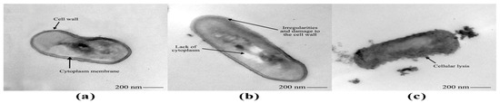
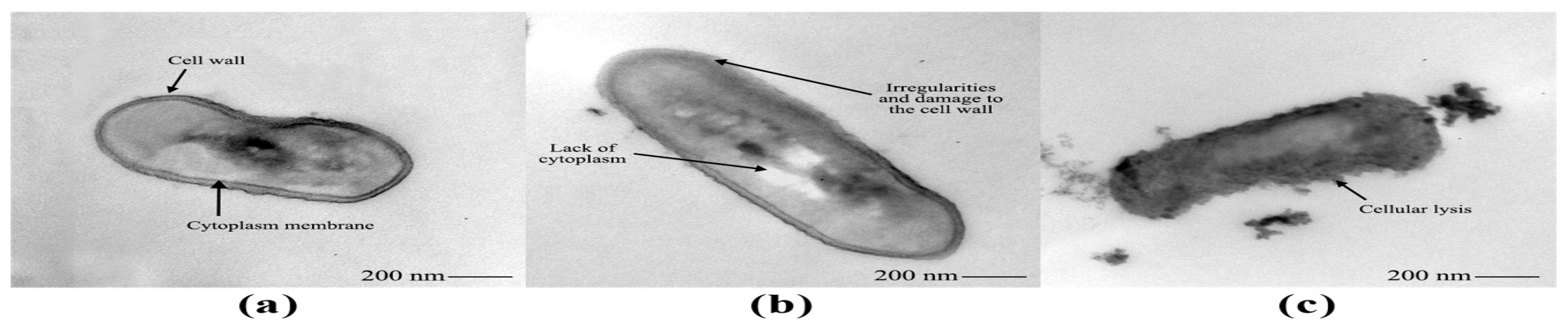
Under transmission electron microscopy, untreated L. monocytogenes 4455 cells exhibited normal rod-shaped morphology, a typical and continuous cell wall, an intact and smooth cytoplasmic membrane, as well as a cytoplasm evenly distributed along the cell (Figure 5a). After exposure to Lcr-CFS at 1x MIC for 24 h, bacterial cell lysis and leakage of intracellular material were evidenced (Figure 5c). Treatment with 0.5x MIC of Lcr-CFS caused irregularities, damage and deformation on L. monocytogenes cell wall, in addition to partial loss of cytoplasm (Figure 5b).
Figure 5.
Transmission electron microscope images of Listeria monocytogenes 4455 cells before (a) and after exposure to Lacticaseibacillus rhamnosus ATCC 9595 cell-free supernatant (Lcr-CFS) at 0.5x MIC (b) and 1x MIC (c).
3.5. Changes in Gene Expression Induced by Lcr-CFS
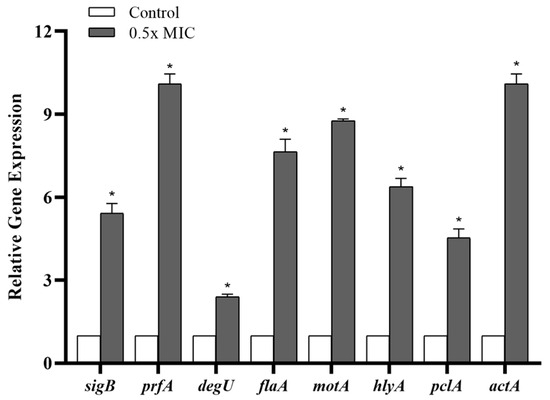
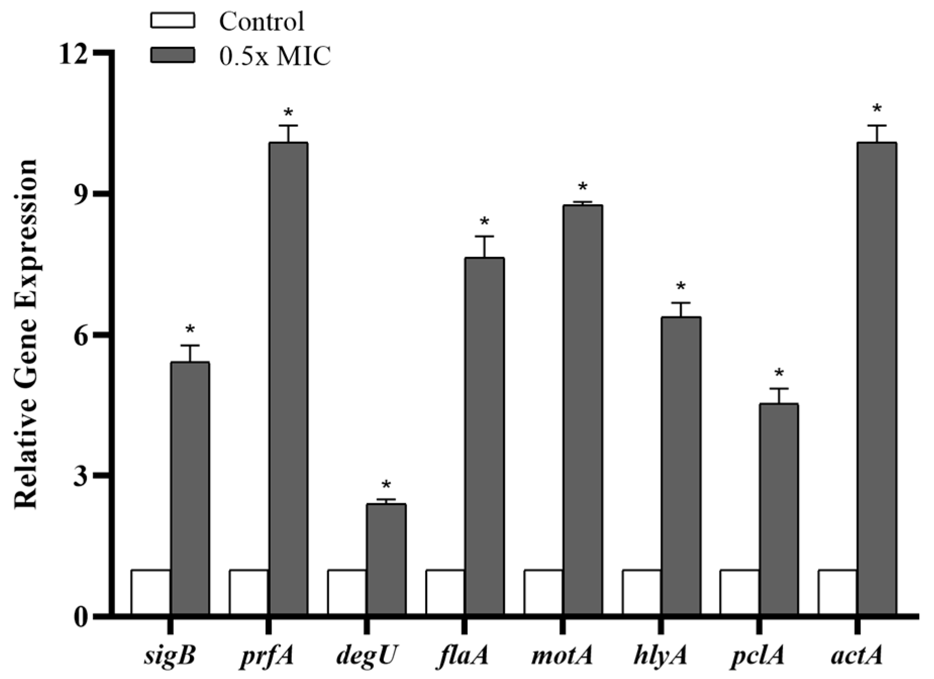
Taking into account the bactericidal activity of the Lcr-CFS at MIC-level concentrations or above, the relative expression of genes associated with response regulators (prfA, sigB, and degU), flagella (flaA, motA), and virulence factors (hlyA, pclA, and actA) was evaluated in L. monocytogenes 4455 upon exposure to 0.5x MIC of Lcr-CFS for 24 h. Compared to untreated bacterial cells, the expression of all evaluated genes was significantly upregulated in the presence of Lcr-CFS (p < 0.05). Notably, the transcription level of prfA, the main regulator of virulence factors in L. monocytogenes, and sigB, the primary stress response regulator, were significantly elevated (p < 0.05) following treatment with Lcr-CFS (Figure 6).
Figure 6.
Relative gene expression in Listeria monocytogenes 4455 after 24 h of exposure to 0.5x MIC of Lacticaseibacillus rhamnosus ATCC 9595 cell-free supernatant (Lcr-CFS). Means marked with an asterisk (*) indicate significant differences compared to the control group (non-exposed to Lcr-CFS), based on ANOVA with Tukey’s post-test (p < 0.05).
4. Discussion
Lactic acid bacteria (LAB) are recognized as effective biological control agents against the growth of undesirable microorganisms. Their protective effects primarily result from the production of antimicrobial metabolites, including organic acids, hydrogen sulfide, hydrogen peroxide, exopolysaccharides, biosurfactants, and antimicrobial peptides. Additionally, LAB can compete for nutrients [18,51] and antagonize the formation of biofilms by pathogenic microorganisms through mechanisms such as competition, exclusion, or displacement [52,53].
Several bioactive compounds produced by LAB, including antimicrobial metabolites, can be directly recovered from the culture supernatant without extensive purification processes. This highlights the potential of cell-free supernatants (CFS) as a convenient and efficient source of bioactive metabolites for diverse applications. Antimicrobial activity of CFS from different bacteria has been reported against pathogenic microorganisms [38,54,55,56,57,58,59,60,61,62].
In this study, we demonstrated the anti-Listeria activity of the CFS obtained from Lc. rhamnosus ATCC 9595. The supernatant obtained from stationary-phase cultures had an initial pH of 4.5; however, since the Lcr-CFS was lyophilized and suspended in PBS (pH 7.4), the observed anti-Listeria activity likely did not result from organic acid production. Furthermore, after treatment with proteinase K, CFS lost its anti-listerial activity, demonstrating the proteinaceous nature of the compound produced by Lc. rhamnosus.
Many LAB are known to produce ribosomally synthesized peptides, named bacteriocins, which exhibit bacteriostatic or bactericidal activity. Among these, pediocin-like class IIa bacteriocins are particularly effective against Listeria [63]. Lc. rhamnosus strains have been reported to produce bacteriocins, including compounds with anti-listerial activity [64,65,66]. The hypothesis that the anti-Listeria compound produced by Lc. rhamnosus ATCC 9595 is a bacteriocin or a bacteriocin-like substance is supported by the recent sequencing of its genome, which revealed genes encoding bacteriocin immunity proteins and accessory proteins involved in bacteriocin secretion [67].
The bactericidal activity of Lcr-CFS, observed in MBC and time-kill assays, was confirmed by transmission electron microscopy. L. monocytogenes cells exposed to 1x MIC of Lcr-CFS exhibited structural alterations, including damage to the bacterial cell envelope, membrane destabilization, and leakage of intracellular compounds. In contrast, exposure to 0.5x MIC resulted in structural changes and damage to the bacterial cell wall, indicating that, even at concentrations lower than the MIC, Lcr-CFS may disrupt vital cellular functions and ultimately cause bacterial cell death.
Antibacterial peptides produced by LAB commonly act by forming pores in the target bacterial cell membrane, which leads to membrane permeability increase, depolarization and leakage of intracellular compounds [68,69,70,71]. It has also been demonstrated that LAB and CFSs derived from LAB can elevate reactive ROS levels in target cells [72,73,74]. Recently, Wang and Zeng [45] demonstrated that the CFS from Lactobacillus pentosus L-36 exhibits antibacterial effects by damaging cell membranes and increasing ROS levels in Staphylococcus aureus.
Exposure to Lcr-CFS increased ROS production in L. monocytogenes, indicating that its lethal action derives, at least in part, from ROS-mediated oxidative stress. ROS have been associated with the lethal action of different antibiotics, regardless of their specific targets [75,76]. Large quantities of ROS can overwhelm the endogenous antioxidant defenses of pathogens, directly damaging DNA and other cellular components, leading to cell death [77,78]. Moreover, cell death can occur due to post-stress ROS-mediated toxicity if the primary damage induced by the stressor was insufficient to kill bacteria due to repair. Supporting this hypothesis, evidence from Escherichia coli was provided by Hong et al. [79], whose findings showed that even after removal of stressor, ROS continued to accumulate, leading to ongoing cell death, indicating that once a ROS level exceeds a critical threshold, the death process becomes self-driven.
Beyond its bactericidal activity, Lcr-CFS exhibited potent anti-biofilm effects against L. monocytogenes, even at sub-inhibitory concentrations (sub-MIC), inhibiting biofilm formation/development and affecting preformed biofilms. A complete inhibition of L. monocytogenes biofilm formation was achieved in the presence of 0.5x MIC Lcr-CFS, both in polystyrene microplates and on glass surfaces. This effect can be due to the ability of Lcr-CFS in reducing bacterial adhesion during the initial stages of biofilm development or to prevent the transition to the biofilm phenotype. The anti-biofilm activity of LAB has been reported in numerous studies [3,38,42,80,81,82,83,84,85,86].
Comparable anti-biofilm effects were reported by Singh et al. [53], who showed that CFS from various Lactobacillus strains effectively inhibited L. monocytogenes biofilm development. In co-incubation assays, the most notable reduction in L. monocytogenes biofilm formation was obtained using Lc. rhamnosus GG (63.01%) and Lc. rhamnosus GM18 (62.72%) supernatants. A significant reduction (more than 65.0%) in the development of biofilms by L. monocytogenes was also reported in the presence of CFS obtained from Lactobacillus acidophilus La14 150B, Lactiplantibacillus plantarum B411, and Lacticaseibacillus rhamnosus ATCC 53103 [87].
Recently, our research group demonstrated the ability of CFS of Weissella paramesenteroides UFTM 2.6.1 in inhibiting biofilm formation by L. monocytogenes: at concentrations equal to or above the MIC a complete inhibition (100.0%) of biofilm formation was observed, while a sub-MIC (0.5x MIC) reduced biofilm biomass by 59.8% to 89.5%, depending on the L. monocytogenes strain [38].
Melian et al. [88] reported that treatment of L. monocytogenes biofilms with lactocin AL705 (800 AU/mL), produced by Latilactobacillus curvatus CRL1579, for 30 min resulted in a 5-log reduction in viable cell counts and a decrease in biofilm biomass, with microscopy confirming a predominance of dead or damaged cells. Yan et al. [89] demonstrated that treatment with nisin, a bacteriocin produced by a group of Gram-positive bacteria, significantly reduced preformed L. monocytogenes biofilms. After 2 h of exposure, bacterial concentration was decreased by 23.94–35.67% compared to the untreated control.
Our findings demonstrated that Lcr-CFS significantly reduced the viability of L. monocytogenes cells within the biofilms formed in polystyrene plates, with concentrations equal to or higher than 0.25x MIC leading to a reduction of more than 80% in cell viability. Additionally, when preformed L. monocytogenes biofilms on glass surfaces were treated with Lcr-CFS, fluorescence microscopy revealed extensive cell death within the biofilm matrix, as indicated by the predominance of red-stained (nonviable) cells compared to the untreated control.
The ability of L. monocytogenes to survive in highly variable and often hostile environments relies on its finely tuned capacity to rapidly reprogram gene expression, a process largely mediated by alternative sigma factors [18,90,91]. Among these, the stress-responsive sigma factor B (SigB; σB) is crucial for enabling L. monocytogenes to adapt its transcriptional profile. Notably, the transcriptional program regulated by SigB is intricately linked with PrfA, the primary regulator of virulence genes, forming a complex network that allows L. monocytogenes to coordinate stress responses and pathogenicity simultaneously [92]. This coordinated regulation of stress adaptation and virulence maintains the bacterium’s ability to survive and persist in the natural environment, the food chain and within the host [10,93,94,95,96].
Exposure to Lcr-CFS 0.5x MIC significantly upregulated sigB and prfA genes in L. monocytogenes, reflecting a coordinated activation of stress response and virulence-associated pathways. Importantly, this transcriptional response likely represents an adaptative mechanism to cope with antimicrobial stress rather than a true increase in virulence or pathogenic potential. The observed cross-talk between sigB and prfA underscores the plasticity of L. monocytogenes regulatory networks, allowing the bacterium to modulate stress adaptation and virulence genes simultaneously in response to environmental challenges [10,95].
Upregulation of prfA concomitant with reduced biofilm formation after curcumin-mediated photodynamic inactivation of L. monocytogenes were also reported by Huang et al. [97], who suggested that the treatment increased the stress responses of the L. monocytogenes cells to resist external harsh environment. A comparable increase in sigB and prfA expressions was also observed in our recent study using the CFS of W. paramesenteroides UFTM 2.6.1, suggesting a conserved stress-related response in L. monocytogenes induced by bioactive compounds produced by LAB [38].
Additionally, the upregulation of other genes, including response regulators (degU), flagellar-associated genes (flaA and motA), and virulence factors (hlyA, pclA, and actA), following exposure to the Lcr-CFS likely reflects a bacterial survival strategy in response to antimicrobial stress, rather than an actual increase in virulence or biofilm-forming capacity, highlighting the complex and variable impact that antimicrobial agents can exert on bacterial regulatory networks. In line with our observations, Liu et al. [32] demonstrated the upregulation of genes associated with motility (degU, flaA, motB), stress response (sigB), and virulence (prfA) after treatment with natural antimicrobials, such as cinnamaldehyde, eugenol, resveratrol, and thymoquinone at sub-MICs.
Although the experiments reported in this study were conducted exclusively using in vitro models, these conditions were strategically selected to elucidate the fundamental effects of Lcr-CFS against L. monocytogenes, and findings reported provide essential preliminary evidence supporting the biocontrol potential of Lcr-CFS. The lack of experiments in more realistic food-relevant environments represents an important limitation and should be considered for validating Lcr-CFS applicability in food safety strategies.
5. Conclusions
Bioactive compounds secreted by Lc. rhamnosus ATCC 9595 (Lcr-CFS) exhibits potent bactericidal and anti-biofilm effects against L. monocytogenes. As the supernatant contains a complex mixture of bioactive compounds, the anti-Listeria effect of Lcr-CFS is likely the result of synergistic action of multiple mechanisms rather than a single dominant pathway. The bactericidal potential appears to be associated with bacterial cell wall disruption and intracellular leakage, besides induction of ROS, whereas its anti-biofilm activity likely results from reduced adhesion of planktonic cells to plastic and glass surfaces, and disruption of mature biofilms, leading to loss of L. monocytogenes cells viability. Although at concentrations lower than the MIC (0.5x), Lcr-CFS induced upregulation of genes associated with stress response and virulence in L. monocytogenes, these transcriptional changes did not translate into measurable phenotypic alterations, being insufficient to overcome Lcr-CFS bactericidal and anti-biofilm effects. Given these properties, Lc. rhamnosus represents a promising biocontrol agent in food industry settings to mitigate L. monocytogenes contamination and persistence.
Author Contributions
I.S.R.—Conceptualization; methodology; formal analysis; investigation; resources; writing—original draft preparation. H.F.P.—methodology; writing—review and editing. S.D.d.C.R.—methodology. Y.N.V.S.—writing—review and editing. M.V.d.S.—writing—review and editing. F.A.d.A.—writing—review and editing. E.A.A.N.—writing—review and editing; funding acquisition. U.M.P.—writing—review and editing; funding acquisition. A.B.F.M.—writing—review and editing. A.D.P.—Conceptualization; methodology; writing—review and editing; supervision; project administration. All authors have read and agreed to the published version of the manuscript.
Funding
This research was funded by FAPEMIG (Fundação de Amparo à Pesquisa do Estado de Minas Gerais), APQ-01597-22.
Institutional Review Board Statement
Not applicable.
Informed Consent Statement
Not applicable.
Data Availability Statement
The original contributions presented in this study are included in the article. Further inquiries can be directed to the corresponding author.
Acknowledgments
The authors thank FAPEMIG for financial support (APQ-01597-22). Isabela Sguilla Rotta was a FAPEMIG scholarship holder. During the preparation of this manuscript, the authors used ChatGPT (free version) exclusively to improve grammar and style in some sentences. All outputs were reviewed and edited by the authors, who take full responsibility for the integrity and accuracy of the content.
Conflicts of Interest
The authors declare that they have no known competing financial interests or personal relationships that could have appeared to influence the work reported in this paper. The funders had no role in the design of the study; in the collection, analyses, or interpretation of data; in the writing of the manuscript; or in the decision to publish the results.
Abbreviations
The following abbreviations are used in this manuscript:
| Lcr-CFS | Cell-Free Supernatant of Lacticaseibacillus rhamnosus ATCC 9595 |
| CFS | Cell-Free Supernatant |
| LAB | Lactic acid bacteria |
| GRAS | Generally Recognized As Safe |
| FIOCRUZ | Fundação Oswaldo Cruz |
| MRS | Man-Rogosa-Sharpe |
| CLIST | Listeria Collection |
| LABZOO | Laboratory of Bacterial Zoonoses of the Oswaldo Cruz Institute |
| BHI | Brain Heart Infusion |
| PBS | Phosphate-Buffered Saline |
| MIC | Minimum Inhibitory Concentration |
| CLSI | Clinical and Laboratory Standards Institute |
| MBC | Minimum Bactericidal Concentration |
| CFU/mL | Viable cell counts in colony forming units per milliliter |
| MTT | 3-(4,5-dimethylthiazol-2-yl)-2,5-diphenyltetrazolium bromide |
| PI | Propidium Iodide |
| DCFH-DA | 2′,7′-dichlorodihydrofluorescein diacetate |
| RT-qPCR | Real-Time Quantitative Reverse Transcription Polymerase Chain Reaction |
| RNA | Ribonucleic Acid |
| DNA | Deoxyribonucleic Acid |
| cDNA | Complementary DNA |
| ROS | Reactive oxygen species |
| OD | Optical Density |
References
- Rodriguez, C.; Taminiau, B.; García-Fuentes, E.; Daube, G.; Korsak, N. Listeria monocytogenes dissemination in farming and primary production: Sources, shendding and control measures. Food Control 2021, 120, 107540. [Google Scholar] [CrossRef] [Scilit]
- Gemmell, C.T.; Parreira, V.R.; Farber, J.M. Controlling Listeria monocytogenes growth and biofilm formation using flavonoids. J. Food Prot. 2022, 85, 639–646. [Google Scholar] [CrossRef] [Scilit] [PubMed]
- Martín, I.; Rodríguez, A.; Córdoba, J.J. Application of selected lactic-acid bacteria to control Listeria monocytogenes in soft-ripened “Torta del Casar” cheese. LWT 2022, 168, 113873. [Google Scholar] [CrossRef] [Scilit]
- Koopmans, M.M.; Brouwer, M.C.; Vásquez-Boland, J.A.; Beek, D. Human Listeriosis. Clin. Microbiol. Rev. 2023, 36, e0006019. [Google Scholar] [CrossRef] [Scilit]
- López, B.R.C.; Lozano, D.G.; Marteache, A.H.; Moreno, P.C.; García, C.R. Control of Persistent Listeria monocytogenes in the Meat Industry: From Detection to Prevention. Foods 2025, 26, 1519. [Google Scholar] [CrossRef] [Scilit]
- Vazquez-Boland, J.A.; Kuhn, M.; Berche, P.; Chakraborty, T.; Dominguez-Bernal, G.; Goebel, W.; Gonzalez-Zorn, B.; Wehland, J.; Kreft, J. Listeria pathogenesis and molecular virulence determinants. J. Clin. Microbiol. 2001, 14, 584–640. [Google Scholar] [CrossRef] [Scilit]
- Disson, O.; Moura, A.; Lacuit, M. Making sense of the biodiversity and virulence of Listeria monocytogenes. Trends Microbiol. 2021, 29, 811–822. [Google Scholar] [CrossRef] [Scilit]
- EFSA. European Food Safety Authority. The European Union One Health. Zoonoses Report. EFSA J. 2019, 17, e05926. [Google Scholar] [CrossRef] [Scilit]
- Ribeiro, A.C.; Almeida, F.A.; Medeiros, M.M.; Miranda, B.R.; Pinto, U.M.; Alves, V.F. Listeria monocytogenes: An Inconvenient Hurdle for the Dairy Industry. Dairy 2023, 4, 316–344. [Google Scholar] [CrossRef] [Scilit]
- Gaballa, A.; Guariglia-Oropeza, V.; Wiedmann, M.; Boor, K.J. Cross talk between SigB and PrfA in Listeria monocytogenes facilitates transitions between extra- and intracellular environments. Microbiol. Mol. Biol. Rev. 2019, 83, e00034-19. [Google Scholar] [CrossRef] [Scilit]
- Duma, M.N.; Ciupescu, L.M.; Dan, S.D.; Crisan-Reget, O.L.; Tabaran, A. Virulence and Antimicrobial Resistance of Listeria monocytogenes Isolated from Ready-to-Eat Food Products in Romania. Microorganisms 2024, 8, 954. [Google Scholar] [CrossRef] [Scilit]
- Davies, D. Understanding biofilm resistance to antibacterial agents. Nat. Rev. Drug Discov. 2003, 2, 114–122. [Google Scholar] [CrossRef] [Scilit]
- Penesyan, A.; Gillings, M.; Paulsen, I.T. Antibiotic discovery: Combatting bacterial resistance in cells and in biofilm communities. Molecules 2015, 20, 5286–5298. [Google Scholar] [CrossRef] [Scilit] [PubMed]
- Kadam, S.R.; Den Besten, H.M.W.; Van Der Veen, S.; Zwieterin, M.H.; Moezelaar, R.; Abee, T. Diversity assessment of Listeria monocytogenes biofilm formation: Impact of growth condition, serotype and strain origin. Int. J. Food Microbiol. 2013, 165, 259–264. [Google Scholar] [CrossRef] [Scilit] [PubMed]
- Valderrama, W.B.; Ostiguy, N.; Cutter, C.N. Multivariate analysis reveals differences in biofilm formation capacity among Listeria monocytogenes lineages. Biofouling 2014, 30, 1199–1209. [Google Scholar] [CrossRef] [Scilit]
- Doijad, S.P.; Barbuddhe, S.B.; Garg, S.; Poharkar, K.V.; Kalorey, D.R.; Kurkure, N.V.; Rawool, D.B. Biofilm-Forming Abilities of Listeria monocytogenes Serotypes Isolated from Different Sources. PLoS ONE 2015, 11, e0137046. [Google Scholar] [CrossRef] [Scilit] [PubMed]
- Oliveira, M.M.M.; Brugnera, D.D.; Alves, E.; Piccoli, R.H. Biofilm formation by Listeria monocytogenes on stainless steel surface and biotransfer potential. Braz. J. Microbiol. 2010, 41, 97–106. [Google Scholar] [CrossRef] [Scilit]
- Yang, Y.; Kong, X.; Niu, B.; Yang, J.; Chen, Q. Differences in Biofilm Formation of Listeria monocytogenes and Their Effects on Virulence and Drug Resistance of Different Strains. Foods 2024, 13, 1076. [Google Scholar] [CrossRef] [Scilit]
- Ratti, R.P.; Gomes, B.C.; Martinez, R.C.R.; Souza, V.M.; Martinis, E.C.P. Elongated cells of Listeria monocytogenes in biofilms in the presence of sucrose and bacteriocin-producing Leuconostoc mesenteroides A11. Food Sci. Technol. 2010, 30, 1011–1016. [Google Scholar] [CrossRef] [Scilit]
- Gutiérrez, D.; Rodríguez-Rubio, L.; Martínez, B.; Rodríguez, A.; García, P. Bacteriophages as Weapons Against Bacterial Biofilms in the Food Industry. Front. Microbiol. 2016, 7, 825. [Google Scholar] [CrossRef] [Scilit]
- Topka-Bielecka, G.; Dydecka, A.; Necel, A.; Bloch, S.; Nejman-Faleńczyk, B.; Węgrzyn, G.; Węgrzyn, A. Bacteriophage-Derived Depolymerases against Bacterial Biofilm. Antibiotics 2021, 10, 175. [Google Scholar] [CrossRef] [Scilit] [PubMed]
- Ammar, H.A.; Samy, R.; Reda, F.M.; Hassanein, W.A. Essential oils and Lactobacillus metabolites as alternative antibiofilm agents against foodborne bacteria and molecular analysis of biofilm regulatory genes. Sci. Rep. 2025, 15, 7576. [Google Scholar] [CrossRef] [Scilit] [PubMed]
- Food and Drug Administration (FDA). Direct Food Substances Affirmed as Generally Recognized as Safe. In 21 CFR Part 184; Food and Drug Administration (FDA): Silver Spring, MD, USA, 1977. Available online: https://www.ecfr.gov/current/title-21/chapter-I/subchapter-B/part-184 (accessed on 10 September 2025).
- Ibrahim, S.A.; Ayivi, R.D.; Zimmerman, T.; Siddiqui, S.A.; Altemimi, A.B.; Fidan, H.; Esatbeyoglu, T.; Bakhshayesh, R.V. Lactic Acid Bacteria as Antimicrobial Agents: Food Safety and Microbial Food Spoilage Prevention. Foods 2021, 10, 3131. [Google Scholar] [CrossRef] [Scilit] [PubMed]
- Almeida, J.M.; Maffei, J.T.; Gebara, C.; Minafra, C.; Toledo-Silva, B.; Gonçalves, M.C.; Langoni, H.; Neto, A.T.; Souza, F.N.; Silva, N.C.C. Exploring probiotic potential and antimicrobial properties of lactic acid bacteria from cow’s milk. Appl. Food Res. 2024, 4, 100461. [Google Scholar] [CrossRef] [Scilit]
- Qiao, Z.; Guo, X.; Shan, Z.; Luo, S.; Mao, Y.; Ren, L.; Wang, T.; Ma, Y.; Liu, Y.; Liu, J. Screening and Genome Analysis of Potential Probiotic Lactiplantibacillus plantarum with Anti-Listeria monocytogenes Activity from Traditional Fermented Foods. Microorganisms 2025, 13, 2104. [Google Scholar] [CrossRef] [Scilit]
- Xavier-Santos, D.; Scharlack, N.K.; Pena, F.L.; Aantunes, A.E. Effects of Lacticaseibacillus rhamnosus GG supplementation, via food and non-food matrices, on children’s health promotion: A scoping review. Food Res. Int. 2022, 158, 111518. [Google Scholar] [CrossRef] [Scilit]
- Westerik, N.; Kort, R.; Sybesma, W.; Reid, G. Lactobacillus rhamnosus probiotic food as a tool for empowerment across the value chain in Africa. Front. Microbiol. 2018, 9, 1501. [Google Scholar] [CrossRef] [Scilit]
- Petrova, M.I.; Reid, G.; Haar, J.A.T. Lacticaseibacillus rhamnosus GR-1, a.k.a. Lactobacillus rhamnosus GR-1: Past and future perspectives. Trends Microbiol. 2021, 29, 747–761. [Google Scholar] [CrossRef] [Scilit]
- Kuley, E.; Yazgan, H.; Özogul, Y.; Ucar, Y.; Durmus, M.; Özyurt, G.; Ayas, D. Effectiveness of Lactobacilli cell-free supernatant and propolis extract microcapsules on oxidation and microbiological growth in sardine burger. Food Biosci. 2021, 44, 101417. [Google Scholar] [CrossRef] [Scilit]
- Clinical and Laboratory Standards Institute (CLSI). Performance Standards for Antimicrobial Susceptibility Testing, 33rd ed.; CLSI Supplement M100; CLSI: Wayne, PA, USA, 2023. [Google Scholar]
- Liu, Y.; Wu, L.; Han, J.; Dong, P.; Luo, X.; Zhang, Y.; Zhu, L. Inhibition of biofilm formation and related gene expression of Listeria monocytogenes in response to four natural antimicrobial compounds and sodium hypochlorite. Front. Microbiol. 2021, 11, 617473. [Google Scholar] [CrossRef] [Scilit]
- Shamson, D.C. Antimicrobial chemotherapy—General principles. In Microbiology in Clinical Practice, 2nd ed.; United Kingdom (Chapter 3); Butterworth-Heinemann: Oxford, UK, 1989. [Google Scholar]
- Júnior, G.A.M.; Fochat, R.C.; Húngaro, H.M.; Ribeiro, J.B.; Lima, J.C.F.; Carneiro, J.C.; Vicentine, K.F.D.; Barbosa, A.A.T.; Paiva, A.D. Partial characterization of a bacteriocin-like inhibitory substance produced by Streptococcus equinus C619, a bovine rumen isolate. Concilium 2023, 23, 78–96. [Google Scholar] [CrossRef] [Scilit]
- Jiang, Q.; Lou, Z.X.; Wang, H.X.; Chen, C. Antimicrobial effect and proposed action mechanism of cordycepin against Escherichia coli and Bacillus subtilis. J. Microbiol. 2019, 57, 288–297. [Google Scholar] [CrossRef] [Scilit]
- Clinical and Laboratory Standards Institute (CLSI). Methods for Determining Bactericidal Activity of Antimicrobial Agents. In CLSI Supplement M26-A; no. 18; CLSI: Wayne, PA, USA, 1999; Volume 19. [Google Scholar]
- Ishak, A.; Mazonakis, N.; Spernovasilis, N.; Akinosoglou, K.; Tsioutis, C. Bactericidal versus bacteriostatic antibacterials: Clinical significance, differences and synergistic potential in clinical practice. J. Antimicrob. Chemother. 2025, 3, 1–17. [Google Scholar] [CrossRef] [Scilit]
- Rotta, I.S.; Rezende, S.D.C.; Perini, H.F.; da Silva, M.V.; Almeida, F.A.; Pinto, U.M.; Machado, A.B.F.; Paiva, A.D. Exploring the Potential of Weissella paramesenteroides UFTM 2.6.1 in Disrupting Quorum Sensing and Attenuating Virulence in Listeria monocytogenes. Front. Microbiol. 2025, 16, 1601203. [Google Scholar] [CrossRef] [Scilit]
- Stepanóvic, S.; Vukovic, D.; Dakic, I.; Savic, B.; Svabic-Vlahovic, M. A modified microtiter-plate test for quantification of staphylococcal biofilm formation. J. Microbiol. Methods 2000, 40, 175–179. [Google Scholar] [CrossRef] [Scilit]
- Trafny, E.A.; Lewandowski, R.; Zawistowska-Marciniakm, I.; Stepinska, M. Use of MTT assay for determination of the biofilm formation capacity of microorganisms in metalworking fluids. World J. Microbiol. Biotechnol. 2013, 29, 1635–1643. [Google Scholar] [CrossRef] [Scilit] [PubMed]
- Singh, N.; Gulhane, R.D.; Singh, A.; Goel, M.; Udelal, P.P.; Sangwan, V.; Sihag, M.K.; Goel, G.; Panwar, H.; Puniya, A.K. Exploring the antimicrobial potential of lactobacilli against early-stage and mature biofilms of Staphylococcus aureus and Pseudomonas aeruginosa. Front. Chem. 2025, 21, 1425666. [Google Scholar] [CrossRef] [Scilit] [PubMed]
- Camargo, A.C.; Todorov, S.D.; Chihib, N.E.; Drider, D.; Nero, L.A. Lactic Acid Bacteria (LAB) and Their Bacteriocins as Alternative Biotechnological Tools to Control Listeria monocytogenes Biofilms in Food Processing Facilities. Mol. Biotechnol. 2018, 60, 712–726. [Google Scholar] [CrossRef] [Scilit] [PubMed]
- Fan, Q.; Zhang, Y.; Yang, H.; Wu, Q.; Shi, C.; Zhang, C.; Xia, X.; Wang, X. Effect of coenzyme Q0 on biofilm formation and attachment-invasion efficiency of Listeria monocytogenes. Food Control 2018, 90, 274–281. [Google Scholar] [CrossRef] [Scilit]
- Perini, H.F.; Pereira, B.B.; Sousa, E.G.; Matos, B.S.; Prado, L.C.S.; Azevedo, V.A.C.; Soares, S.d.C.; da Silva, M.V. Inhibitory effect of Bacillus velezensis 1273 strain cell-free supernatant against developing and preformed biofilms of Staphylococcus aureus and MRSA. Microb. Pathog. 2024, 197, 107065. [Google Scholar] [CrossRef] [Scilit]
- Wang, G.; Zeng, H. Antibacterial Effect of Cell-Free Supernatant from Lactobacillus pentosus L-36 against Staphylococcus aureus from Bovine Mastitis. Molecules 2022, 27, 7627. [Google Scholar] [CrossRef] [Scilit]
- Liu, H.; Wang, Y.; Cao, J.; Jiang, H.; Yao, J.; Gong, G.; Chen, X.; Xu, W.; He, X. Antimicrobial activity and virulence attenuation of citral against the fish pathogen Vibrio alginolyticus. Aquaculture 2020, 15, 734578. [Google Scholar] [CrossRef] [Scilit]
- Pieta, L.; Escudero, F.L.G.; Jacobus, A.P.; Cheiran, K.P.; Gross, J.; Moya, M.L.E.; Soares, G.L.G.; Margis, R.; Frazzon, A.P.G.; Frazzon, J. Comparative transcriptomic analysis of Listeria monocytogenes reveals upregulation of stress genes and downregulation of virulence genes in response to essential oil extracted from Baccharis psiadioides. Ann. Microbiol. 2017, 67, 479–490. [Google Scholar] [CrossRef] [Scilit]
- Shi, W.; Zhang, Q.; Li, H.; Du, D.; Ma, X.; Wang, J.; Jiang, J.; Liu, C.; Kou, L.; Ren, J. Biofilm formation, motility, and virulence of Listeria monocytogenes are reduced by deletion of the gene lmo0159, a novel Listerial LPXTG surface protein. Microorganisms 2024, 12, 1354. [Google Scholar] [CrossRef] [Scilit] [PubMed]
- Jiang, X.; Ren, S.; Geng, Y.; Jiang, C.; Liu, G.; Wang, H.; Yu, T.; Liang, Y. Role of the VirSR-VirAB system in biofilm formation of Listeria monocytogenes EGD-e. Food Res. Int. 2021, 145, 110427. [Google Scholar] [CrossRef] [Scilit]
- Deatraksa, J.; Sunthornthummas, S.; Rangsiruji, A.; Sarawaneeyaruk, S.; Suwannasai, N.; Pringsulaka, O. Isolation of folate-producing Weissella spp. from Thai fermented fish (Plaa Som Fug). LWT Food Sci. Technol. 2018, 89, 388–391. [Google Scholar] [CrossRef] [Scilit]
- Leroy, F.; De Vuyst, L. Lactic acid bacteria as functional starter cultures for the food fermentation industry. Trends Food Sci. Technol. 2004, 15, 67–78. [Google Scholar] [CrossRef] [Scilit]
- Carvalho, F.M.; Teixeira-Santos, R.; Mergulhão, F.J.M.; Gomes, L.C. The Use of Probiotics to Fight Biofilms in Medical Devices: A Systematic Review and Meta-Analysis. Microorganisms 2020, 23, 27. [Google Scholar] [CrossRef] [Scilit]
- Singh, N.; Kaur, R.; Singh, B.P.; Rokana, N.; Goel, G.; Puniya, A.K.; Panwar, H. Impairment of Cronobacter sakazakii and Listeria monocytogenes biofilms by cell-free preparations of Lactobacilli of goat milk origin. Folia Microbiol. 2020, 65, 185–196. [Google Scholar] [CrossRef] [Scilit]
- Hartmann, H.A.; Wilke, T.; Erdmann, R. Efficacy of bacteriocin-containing cell-free culture supernatants from lactic acid bacteria to control Listeria monocytogenes in Food. Int. J. Food Microbiol. 2011, 146, 192–199. [Google Scholar] [CrossRef] [Scilit]
- Beristain-Bauza, S.C.; Mani-López, E.; Palou, E.; López-Malo, A. Antimicrobial activity and physical properties of protein films added with cell-free supernatant of Lactobacillus rhamnosus. Food Control 2016, 62, 44–51. [Google Scholar] [CrossRef] [Scilit]
- Tenea, G.N.; Barrigas, A. The efficacy of bacteriocin-containing cell-free supernatant from Lactobacillus plantarum Cys5-4 to control pathogenic bacteria growth in artisanal beverages. Int. Food Res. J. 2018, 25, 2131–2137. [Google Scholar]
- Pelyuntha, W.; Chaiyasut, C.; Kantachote, D.; Sirilun, S. Cell-free supernatants from cultures of lactic acid bacteria isolated from fermented grape as biocontrol against Salmonella Typhi and Salmonella Typhimurium virulence via autoinducer-2 and biofilm interference. PeerJ 2019, 7, e7555. [Google Scholar] [CrossRef] [Scilit]
- Arrioja-Bretón, D.; Mani-López, E.; Bach, H.; López-Malo, A. Antimicrobial activity of protein-containing fractions isolated from Lactobacillus plantarum NRRL B-4496 culture. Braz. J. Microbiol. 2020, 51, 1289–1296. [Google Scholar] [CrossRef] [Scilit] [PubMed]
- Kaewchomphunuch, T.; Charoenpichitnunt, T.; Thongbaiyai, V.; Ngamwongsatit, N.; Kaeoket, K. Cell-free culture supernatants of Lactobacillus spp. and Pediococcus spp. inhibit growth of pathogenic Escherichia coli isolated from pigs in Thailand. BMC Vet. Res. 2022, 18, 60. [Google Scholar] [CrossRef] [Scilit] [PubMed]
- Keeratikunakorn, K.; Kaewchomphunuch, T.; Kampon, K.; Ngamwongsatit, N. Antimicrobial activity of cell free supernatants from probiotics inhibits against pathogenic bacteria isolated from fresh boar semen. Sci. Rep. 2023, 13, 5995. [Google Scholar] [CrossRef] [Scilit]
- Park, Y.J.; Kim, Y.J.; Yu, H.H.; Lee, N.K.; Paik, H.D. Cell-free supernatants of Bacillus subtilis and Bacillus polyfermenticus inhibit Listeria monocytogenes biofilm formation. Food Control 2023, 144, 109387. [Google Scholar] [CrossRef] [Scilit]
- Poimenidou, S.V.; Skarveli, A.; Saxami, G.; Mitsou, E.K.; Kotsou, M.; Kyriacou, A. Inhibition of Listeria monocytogenes Growth, Adherence and Invasion in Caco-2 Cells by Potential Probiotic Lactic Acid Bacteria Isolated from Fecal Samples of Healthy Neonates. Microorganisms 2023, 11, 363. [Google Scholar] [CrossRef] [Scilit]
- Kjos, M.; Borrero, J.; Opsata, M.; Birri, D.J.; Holo, H.; Cintas, L.M.; Snipen, L.; Hernández, P.E.; Nes, I.F.; Diep, D.B. Target recognition, resistance, immunity and genome mining of class II bacteriocins from Gram-positive bacteria. Microbiology 2011, 157, 3256–3267. [Google Scholar] [CrossRef] [Scilit]
- Dimitrijević, R.; Stojanović, M.; Živković, I.; Petersen, A.; Jankov, R.M.; Dimitrijević, L.; Gavrović-Jankulović, M. The identification of a low molecular mass bacteriocin, rhamnosin A, produced by Lactobacillus rhamnosus strain 68. J. Appl. Microbiol. 2009, 107, 2108–2115. [Google Scholar] [CrossRef] [Scilit]
- Jeong, Y.J.; Moon, G.S. Antilisterial bacteriocin from Lactobacillus rhamnosus CJNU 0519 presenting a narrow antimicrobial spectrum. Korean J. Food Sci. Anim. Resour. 2015, 35, 137–142. [Google Scholar] [CrossRef] [Scilit]
- Oliveira, L.D.C.; Silveira, A.M.M.; Monteiro, A.D.S.; dos Santos, V.L.; Nicoli, J.R.; Azevedo, V.A.d.C.; Soares, S.d.C.; Dias-Souza, M.V.; Nardi, R.M.D. In silico prediction, in vitro antibacterial spectrum, and physicochemical properties of a putative bacteriocin produced by Lactobacillus rhamnosus strain L156.4. Front. Microbiol. 2017, 8, 876. [Google Scholar] [CrossRef] [Scilit] [PubMed]
- Benton, B.; King, S.; Greenfield, S.R.; Puthuveetil, N.; Reese, A.L.; Duncan, J.; Marlow, R.; Tabron, C.; Pierola, A.E.; Yarmosh, D.A., Jr.; et al. The ATCC Genome Portal: Microbial Genome Reference Standards with Data Provenance. Microbiol. Resour. Announc. 2021, 10, e00818-21. [Google Scholar] [CrossRef] [Scilit]
- Paiva, A.D.; Breukink, E.; Mantovani, H.C. Role of lipid II and membrane thickness in the mechanism of action of the Lantibiotic bovicin HC5. Antimicrob. Agents Chemother. 2011, 55, 5284–5293. [Google Scholar] [CrossRef] [Scilit]
- Chen, G.W.; Wu, M.; Liu, W.K.; Xie, M.M.; Zhang, W.S.; Fan, E.G.; Liu, Q. Reactive oxygen species inhibits Listeria monocytogenes invasion into HepG2 epithelial cells. Food Sci. Nutr. 2018, 29, 1501–1507. [Google Scholar] [CrossRef] [Scilit]
- Ruhland, B.R.; Reniere, M.L. Sense and sensor ability: Redox-responsive regulators in Listeria monocytogenes. Curr. Opin. Microbiol. 2019, 47, 20–25. [Google Scholar] [CrossRef] [Scilit]
- Darbandi, A.; Asadi, A.; Ari, M.M.; Ohadi, E.; Talebi, M.; Zadeh, M.H.; Emamie, A.D.; Ghanavati, R.; Kakanj, M. Bacteriocins: Properties and potential use as antimicrobials. J. Clin. Lab. Anal. 2021, 36, e24093. [Google Scholar] [CrossRef] [Scilit]
- Kim, K.W.; Kang, S.S.; Woo, S.J.; Park, O.J.; Ahn, K.B.; Song, K.D.; Lee, H.-K.; Yun, C.-H.; Han, S.H. Lipoteichoic acid of probiotic Lactobacillus plantarum attenuates poly I: C-induced IL-8 production in porcine intestinal epithelial cells. Front. Microbiol. 2017, 8, 1827. [Google Scholar] [CrossRef] [Scilit]
- Wang, H.; Zhang, L.; Xu, S.; Pan, J.; Zhang, Q.; Lu, R. Surface-layer protein from Lactobacillus acidophilus NCFM inhibits lipopolysaccharide-induced inflammation through MAPK and NF-κB signaling pathways in RAW264. 7 cells. J. Agric. Food Chem. 2018, 66, 7655–7662. [Google Scholar] [CrossRef] [Scilit] [PubMed]
- Zhao, H.; Zhang, F.; Chai, J.; Wang, J. Lactobacillus acidophilus reduces Listeria monocytogenes infection by inhibiting mitogen-activated protein kinase genes in growing rabbits. Rev. Bras. Zootec. 2020, 49, e20200054. [Google Scholar] [CrossRef] [Scilit]
- Kohanski, M.A.; Dwyer, D.J.; Hayete, B.; Lawrence, C.A.; Collins, J.J. A common mechanism of cellular death induced by bactericidal antibiotics. Cell 2007, 7, 797–810. [Google Scholar] [CrossRef] [Scilit]
- Li, H.; Zhou, X.; Huang, Y.; Liao, B.; Cheng, L.; Ren, B. Reactive Oxygen Species in Pathogen Clearance: The Killing Mechanisms, the Adaption Response, and the Side Effects. Front. Microbiol. 2020, 11, 622534. [Google Scholar] [CrossRef] [Scilit]
- Zhao, X.; Hong, Y.; Drlica, K. Moving forward with reactive oxygen species involvement in antimicrobial lethality. J. Antimicrob. Chemother. 2015, 70, 639–642. [Google Scholar] [CrossRef] [Scilit] [PubMed]
- Luan, G.; Hong, Y.; Drlica, K.; Zhao, X. Suppression of reactive oxygen species accumulation accounts for paradoxical bacterial survival at high quinolone concentration. Antimicrob. Agents Chemother. 2018, 62, e01622-17. [Google Scholar] [CrossRef] [Scilit]
- Hong, Y.; Zeng, J.; Wang, X.; Drlica, K.; Zhao, X. Post-stress bacterial cell death mediated by reactive oxygen species. Proc. Natl. Acad. Sci. USA 2019, 116, 10064–10071. [Google Scholar] [CrossRef] [Scilit]
- Guerrieri, E.; Niederhäusern, S.; Messi, P.; Sabia, C.; Iseppi, R.; Anacarso, I.; Bondi, M. Use of lactic acid bacteria (LAB) biofilms for the control of Listeria monocytogenes in a small-scale model. Food Control 2009, 20, 861–865. [Google Scholar] [CrossRef] [Scilit]
- Kasra-Kermanshahi, R.; Mobarak-Qamsari, E. Inhibition Effect of Lactic Acid Bacteria against Food Born Pathogen, Listeria monocytogenes. Appl. Food Biotechnol. 2015, 2, 11–19. [Google Scholar] [CrossRef] [Scilit]
- Gómez, N.C.; Ramiro, J.M.; Quecan, B.X.V.; Franco, B.D.G.M. Use of Potential Probiotic Lactic Acid Bacteria (LAB) Biofilms for the Control of Listeria monocytogenes, Salmonella Typhimurium, and Escherichia coli O157:H7 Biofilms Formation. Food Microbiol. 2016, 7, 863. [Google Scholar] [CrossRef] [Scilit] [PubMed]
- Jara, J.; Pérez-Ramos, A.; Solar, G.; Rodríguez, J.M.; Fernández, L.; Orgaz, B. Role of Lactobacillus biofilms in Listeria monocytogenes adhesion to glass surfaces. Int. J. Food Microbiol. 2020, 334, 108804. [Google Scholar] [CrossRef] [Scilit]
- Tatsaporn, T.; Kornkanok, K. Using Potential Lactic Acid Bacteria Biofilms and their Compounds to Control Biofilms of Foodborne Pathogens. Biotechnol. Rep. 2020, 26, e00477. [Google Scholar] [CrossRef] [Scilit]
- Elez, R.M.M.A.; Elsohaby, I.; Al-Mohammadi, A.R.; Seliem, M.; Tahoun, A.B.M.B.; Abousaty, A.; Algendy, R.M.; Mohamed, E.A.A.; El-Gazzar, N. Antibacterial and anti-biofilm activities of probiotic Lactobacillus plantarum against Listeria monocytogenes isolated from milk, chicken and pregnant women. Front. Microbiol. 2023, 14, 1201201. [Google Scholar] [CrossRef] [Scilit]
- Dinçer, E. Impact of lactic acid bacteria strains against Listeria monocytogenes biofilms on various food-contact surfaces. Arch. Microbiol. 2024, 30, 80. [Google Scholar] [CrossRef] [Scilit]
- Masebe, R.D.; Thantsha, M. Anti-biofilm activity of cell free supernatants of selected lactic acid bacteria against Listeria monocytogenes isolated from avocado and cucumber fruits, and from an avocado processing plant. Foods 2022, 11, 2872. [Google Scholar] [CrossRef] [Scilit]
- Melian, C.; Ploper, D.; Chehín, R.; Vignolo, G.; Castellano, P. Impairment of Listeria monocytogenes biofilm developed on industrial surfaces by Latilactobacillus curvatus CRL1579 bacteriocin. Food Microbiol. 2024, 121, 104491. [Google Scholar] [CrossRef] [Scilit] [PubMed]
- Yan, H.; Wu, M.; Gao, B.; Bu, X.; Dong, Q.; Hirata, T.; Li, Z. Inhibition and eradication of Listeria monocytogenes biofilm using the combined treatment with nisin and sesamol. LWT 2024, 198, 116015. [Google Scholar] [CrossRef] [Scilit]
- Guerreiro, T.; Arcari, C.P.; O’Byrne, C.P. The σB-Mediated General Stress Response of Listeria monocytogenes: Life and Death Decision Making in a Pathogen. Front. Microbiol. 2020, 11, 1505. [Google Scholar] [CrossRef] [Scilit]
- Oliveira, A.H.; Tiensuu, T.; Guerreiro, D.N.; Tükenmez, H.; Dessaux, C.; García-del Portillo, F.; O’bYrne, C.; Johansson, J. Listeria monocytogenes Requires the RsbX Protein To Prevent SigB Activation under Nonstressed Conditions. J. Bacteriol. 2022, 204, e00486-21. [Google Scholar] [CrossRef] [Scilit]
- Lebreton, A.; Cossart, P. RNA- and protein-mediated control of Listeria monocytogenes virulence gene expression. RNA Biol. 2017, 14, 460–470. [Google Scholar] [CrossRef] [Scilit]
- Dorey, A.; Marinho, C.; Piveteau, P.; O’Byrne, C. Role and regulation of the stress activated sigma factor sigma B (σB) in the saprophytic and host-associated life stages of Listeria monocytogenes. In Advances in Applied Microbiology; Elsevier: Amsterdam, The Netherlands, 2019; Volume 106, pp. 1–48. [Google Scholar] [CrossRef] [Scilit]
- Wiktorczyk-Kapischke, N.; Skowron, K.; Grudlewska-Buda, K.; Watecka-Zacharska, E.; Korkus, J.; Gospodarek-Komkowska, E. Adaptive Response of Listeria monocytogenes to the Stress Factors in the Food Processing Environment. Front. Microbiol. 2021, 14, 710085. [Google Scholar] [CrossRef] [Scilit] [PubMed]
- Sibanda, T.; Buys, E.M. Listeria monocytogenes Pathogenesis: The Role of Stress Adaptation. Microorganisms 2022, 10, 1522. [Google Scholar] [CrossRef] [Scilit]
- D’Onofrio, F.; Schirone, M.; Paparella, A.; Krasteva, I.; Tittarelli, M.; Pomilio, F.; Iannetti, L.; D’alterio, N.; Luciani, M. Stress Adaptation Responses of a Listeria monocytogenes 1/2a Strain via Proteome Profiling. Foods 2023, 12, 2166. [Google Scholar] [CrossRef] [Scilit] [PubMed]
- Huang, J.; Chen, B.; Li, H.; Zeng, Q.H.; Wang, J.J.; Liu, H.; Pan, Y.; Zhao, Y. Enhanced antibacterial and antibiofilm functions of the curcumin-mediated photodynamic inactivation against Listeria monocytogenes. Food Control 2020, 108, 106886. [Google Scholar] [CrossRef] [Scilit]

Disclaimer/Publisher’s Note: The statements, opinions and data contained in all publications are solely those of the individual author(s) and contributor(s) and not of MDPI and/or the editor(s). MDPI and/or the editor(s) disclaim responsibility for any injury to people or property resulting from any ideas, methods, instructions or products referred to in the content. |
© 2025 by the authors. Licensee MDPI, Basel, Switzerland. This article is an open access article distributed under the terms and conditions of the Creative Commons Attribution (CC BY) license (https://creativecommons.org/licenses/by/4.0/).