Next Issue
Volume 10, July
Previous Issue
Volume 10, May


Dent. J., Volume 10, Issue 6 (June 2022) – 22 articles

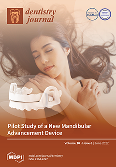
Cover Story (view full-size image): This study aimed to evaluate the effectiveness of customized mandibular advancement device (MAD) in the treatment of obstructive sleep apnea (OSA). Eight patients with a diagnosis of OSA confirmed by polysomnography (PSG) were recruited. All were treated with the new NOA® MAD by OrthoApnea (NOA®); PSG with NOA® in situ was performed (T1). The parameters were calculated at T0 and T1: AHI, supine AHI, oxygen desaturation index (ODI), percentage of recording time spent with oxygen saturation <90% (SpO2 < 90%), and mean oxygen desaturation (MeanSpO2%). The baseline values were AHI = 21.33 ± 14.79, supine AHI = 35.64 ± 12.80, ODI = 17.51 ± 13.5, SpO2 < 90% = 7.82 ± 17.08, and MeanSpO2% = 93.45 ± 1.86. After treatment with NOA®, statistically improvements in AHI and supine AHI were recorded. View this paper
  • Issues are regarded as officially published after their release is announced to the table of contents alert mailing list.
  • You may sign up for e-mail alerts to receive table of contents of newly released issues.
  • PDF is the official format for papers published in both, html and pdf forms. To view the papers in pdf format, click on the "PDF Full-text" link, and use the free Adobe Reader to open them.
Order results
Result details
Section
Select all
Export citation of selected articles as:
10 pages, 4123 KiB  
Article
Initial Displacement and Stress Distribution of Upper Central Incisor Extrusion with Clear Aligners and Various Shapes of Composite Attachments Using the Finite Element Method
by Pratchawin Laohachaiaroon, Bancha Samruajbenjakun and Ekachai Chaichanasiri
Dent. J. 2022, 10(6), 114; https://doi.org/10.3390/dj10060114 - 20 Jun 2022
Cited by 13 | Viewed by 3452
Abstract
A clear aligner is an esthetic and more comfortable option for patients who need orthodontic treatment. However, some types of tooth movement, such as extrusion, are difficult with this tool. Therefore, composite attachments have been suggested to improve tooth movement. This study aims [...] Read more.
A clear aligner is an esthetic and more comfortable option for patients who need orthodontic treatment. However, some types of tooth movement, such as extrusion, are difficult with this tool. Therefore, composite attachments have been suggested to improve tooth movement. This study aims to evaluate the initial displacement and stress distribution during upper central incisor extrusion using the conventional composite attachments. Maxillary models with the upper teeth, clear aligners, and composite attachments placed on the labial surface of the upper right central incisor were constructed. Four models were created to simulate upper central incisor extrusion: (1) without any composite attachment; (2) rectangular beveled attachment; (3) ellipsoid attachment; and (4) horizontal rectangular attachment. Clear aligners were designed to perform upper central incisor extrusion. The constructed models were analyzed using the finite element method. Initial displacement and stress distribution were analyzed. Output analysis found that the upper right central incisor in the model with a horizontal rectangular attachment had the greatest extrusive movement, followed by the model with ellipsoid attachment and the model with beveled attachment. Maximum compressive stress was seen at the cervical region of the composite attachment. Composite attachments including horizontal rectangular attachment, ellipsoid attachment, and rectangular beveled attachment can be used to perform upper central incisor extrusion. Full article
(This article belongs to the Special Issue Orthodontics and New Technologies)
Show Figures

Figure 1

13 pages, 3209 KiB  
Article
Clinical and Radiographic Evaluation of a Novel Triangular Implant Neck Design: A Case Series
by James Rudolph Collins, Brendha P. Ogando, Houlin Hong, Wei Hou and Georgios E. Romanos
Dent. J. 2022, 10(6), 113; https://doi.org/10.3390/dj10060113 - 16 Jun 2022
Viewed by 2834
Abstract
The objective of this study was to evaluate the clinical and radiographic behavior of a novel triangular neck implant configuration in partially edentulous patients. Sixteen patients with a mean age of 58.3 years, were rehabilitated with 25 implants inserted in the healed sites [...] Read more.
The objective of this study was to evaluate the clinical and radiographic behavior of a novel triangular neck implant configuration in partially edentulous patients. Sixteen patients with a mean age of 58.3 years, were rehabilitated with 25 implants inserted in the healed sites of the maxilla and mandible; implant diameter was Ø3.3 and 3.9 mm. Clinical and radiographic measurements were first performed at prosthesis delivery that served as baseline; they were further evaluated after a mean period of 15.6 months. The interproximal peri-implant bone levels were the primary outcome; the mesial and distal data were recorded and a mean value was calculated. Secondary outcomes included peri-implant probing depth (PPD) and bleeding on probing (BoP). The paired t-test was used to compare the radiographic and clinical outcomes between baseline and follow-up. The mean bone levels at the mesial and distal aspects at baseline were 0.45 (0.47) and 0.57 (0.69), respectively; at follow-up they were 0.59 (0.42) and 0.78 (0.59), respectively. The differences were not statistically significant. Similarly, no significant differences were found for the clinical parameters. Within the limitations of the present study, it could be concluded that this new triangular neck bone level implant macro-design was used successfully to treat partially edentulous patients. Larger controlled clinical studies are warranted to confirm the present radiographic and clinical findings. Full article
(This article belongs to the Special Issue Dentistry Journal: 10th Anniversary)
Show Figures

Figure 1

14 pages, 1927 KiB  
Article
In Vitro Comparison of Three Intraoral Scanners for Implant—Supported Dental Prostheses
by Vitória Costa, António Sérgio Silva, Rosana Costa, Pedro Barreiros, Joana Mendes and José Manuel Mendes
Dent. J. 2022, 10(6), 112; https://doi.org/10.3390/dj10060112 - 15 Jun 2022
Cited by 5 | Viewed by 4316
Abstract
With continuing technological developments, there have been advances in the field of fixed prosthetics, particularly in impression-taking techniques. These technological advances mean that a wide variety of diagnostic and/or rehabilitation possibilities can be explored without the need for physical models. The aim of [...] Read more.
With continuing technological developments, there have been advances in the field of fixed prosthetics, particularly in impression-taking techniques. These technological advances mean that a wide variety of diagnostic and/or rehabilitation possibilities can be explored without the need for physical models. The aim of this study was to evaluate the accuracy of three intraoral scanners used in oral implant rehabilitation using an extraoral scanner as a reference and varying the scanning area. Three models representing different clinical scenarios were scanned 15 times by each intraoral scanner and three times by the extraoral scanner. The readings were analyzed and overlaid using engineering software (Geomagic® Control X software (Artec Europe, Luxembourg)). Statistically significant differences in accuracy were found between the three intraoral scanners, iTero® (Align Technology Inc., San Jose, CA, USA), Medit® (Medit®: Seoul, Korea), and Planmeca® (Planmeca®: Helsinki, Finland). In all clinical scenarios, the iTero® scanner had the best trueness (24.4 μm), followed by the Medit® (26.4 μm) and Planmeca® (42.1 μm). The Medit® showed the best precision (18.00 μm) followed by the iTero® (19.20 μm) and Planmeca® (34.30 μm). We concluded that the iTero® scanner had the highest reproducibility and accuracy in the clinical setting. Full article
(This article belongs to the Special Issue Digital Dentures)
Show Figures

Figure 1

12 pages, 247 KiB  
Article
Factors Associated with Pre-Dental Students’ Intention and Willingness to Serve in the Underserved Community and Vulnerable Population
by Brent Lin, Jungsoo Kim, Michael Lin and Jyu-Lin Chen
Dent. J. 2022, 10(6), 111; https://doi.org/10.3390/dj10060111 - 14 Jun 2022
Cited by 1 | Viewed by 1813
Abstract
A potential solution to the problem of how to increase access to dental care for the underserved and vulnerable populations is to establish an early pipeline of underrepresented and minority college students for a career in dentistry. This study aims to explore factors [...] Read more.
A potential solution to the problem of how to increase access to dental care for the underserved and vulnerable populations is to establish an early pipeline of underrepresented and minority college students for a career in dentistry. This study aims to explore factors associated with such pre-dental students’ future intentions to serve. A cross-sectional design was utilized with 144 participants completing the questionnaire with four sections, including participants’ demographics, experience in access to dental care, psychosocial factors, and intention to serve the underserved and vulnerable populations. Descriptive statistics, chi-squared test, and logistic regression were used for statistical analyses. A positive attitude (OR = 12.03) and higher confidence towards addressing access to dental care issues (OR = 10.43) were found to be the strongest factor for higher intention to serve the underserved and vulnerable populations. Higher knowledge on the prevalence of dental caries among children (OR = 3.18) and participants who experienced difficulty in getting a dental appointment, or finding an available dentist when needed (OR = 3.43), were also associated with higher intention. Identifying key factors associated with higher intention to serve the underserved and vulnerable populations as a future dentist may facilitate workforce recruitment in the Health Profession Shortage Areas (HPSAs). Full article
11 pages, 1503 KiB  
Case Report
Post-Operative Bleeding Complications in a Periodontitis Patient Testing Positive for COVID-19
by Georgios Loukas, Madeline X. F. Kosho, Spiros Paraskevas and Bruno G. Loos
Dent. J. 2022, 10(6), 110; https://doi.org/10.3390/dj10060110 - 14 Jun 2022
Viewed by 2480
Abstract
Recent scientific evidence states that a subset of COVID-19 patients may have a risk of increased bleeding tendency. This case report presents a healthy 38-year-old woman with generalized stage III, grade C periodontitis with an abnormal post-operative blood clot formation who tested positive [...] Read more.
Recent scientific evidence states that a subset of COVID-19 patients may have a risk of increased bleeding tendency. This case report presents a healthy 38-year-old woman with generalized stage III, grade C periodontitis with an abnormal post-operative blood clot formation who tested positive for COVID-19 after a standard periodontal surgery. Previously, two periodontal surgeries proceeded without any complications and were considered standard. On day one after the third periodontal surgery the patient had no complaints. On day two the patient reported excess bleeding in the oral cavity from the operated area simultaneously with fever and loss of taste. On day three the patient was seen in our clinic; general malaise symptoms and bleeding tendency had started to decline and the patient received a COVID-19 PCR test. At day four the test resulted positive, and she reported no further complaints of intraoral bleeding. Six months later the taste of the patient was still distorted. For this patient with free medical anamnesis, we suggest that the patient had increased plasma levels of tissue plasminogen activator during the crucial postoperative period due to an acute COVID-19 infection. This led to increased plasmin levels with a hyper-fibrinolytic state as a consequence. Full article
Show Figures

Figure 1

10 pages, 2404 KiB  
Article
A 3D Digital Analysis of the Hard Palate Wound Healing after Free Gingival Graft Harvest: A Pilot Study in the Short Term
by Tiago Marques, Sara Ramos, Nuno Bernardo Malta dos Santos, Tiago Borges, Javier Montero, André Correia and Gustavo Vicentis de Oliveira Fernandes
Dent. J. 2022, 10(6), 109; https://doi.org/10.3390/dj10060109 - 13 Jun 2022
Cited by 6 | Viewed by 2436
Abstract
Purpose: Within this context, this pilot study aimed to evaluate the healing dynamics process of the hard palate after free gingival graft harvesting in the short term (3 months), utilizing digital imaging technology and tridimensional analysis software. Furthermore, assessing the results found to [...] Read more.
Purpose: Within this context, this pilot study aimed to evaluate the healing dynamics process of the hard palate after free gingival graft harvesting in the short term (3 months), utilizing digital imaging technology and tridimensional analysis software. Furthermore, assessing the results found to verify the existence of a relationship between gender or age with tissue loss. Materials and Methods: For connective-tissue harvesting, fifteen patients with gingival recessions type (RT) 1 and RT2 were selected. On the surgery day (before the procedure) and after three months, palatal impressions were taken in all patients, and cast models were done for posterior model scanning. The following variables were analyzed: mean thickness alterations (x¯ TA), maximum thickness loss (MTL), mean maximum thickness loss (x¯ MTL), and volume alterations (VA). A descriptive and bivariate analysis of the data was done. The data were submitted for statistical evaluation and were significant if p < 0.05. Results: Fifteen patients were analyzed, 11 females (73.3%) and four males (26.7%). The patients’ average age was 28 ± 8.52 years (ranging between 16 and 48 years old). The palatal wound region’s mean thickness and volume changes were −0.26 mm (±0.31) and 46.99 mm3 (±47.47 mm3) at three months. There was no statistically significant result correlating age/gender with any variable evaluated. Conclusions: Connective tissue graft harvesting promoted changes with a standard volume and thickness loss of palatal soft tissue. A 3D digital evaluation was a non-invasive method with a reproducible technique for measuring thickness or volume after connective tissue is collected. There was no relationship between age/gender and any variables analyzed. Full article
(This article belongs to the Special Issue Clinical Oral Implants and Periodontal Research)
Show Figures

Figure 1

15 pages, 324 KiB  
Article
Estimation of Factors Affecting Burnout in Greek Dentists before and during the COVID-19 Pandemic
by Maria Antoniadou
Dent. J. 2022, 10(6), 108; https://doi.org/10.3390/dj10060108 - 13 Jun 2022
Cited by 16 | Viewed by 3166
Abstract
This study is a comprehensive, cross-sectional survey in occupational burnout, career satisfaction, and quality of life conducted in March 2021 among dentists in the vast area of metropolitan Athens, Greece. Data were collected using a self-reported questionnaire based on the Copenhagen Questionnaire (CQ) [...] Read more.
This study is a comprehensive, cross-sectional survey in occupational burnout, career satisfaction, and quality of life conducted in March 2021 among dentists in the vast area of metropolitan Athens, Greece. Data were collected using a self-reported questionnaire based on the Copenhagen Questionnaire (CQ) for assessing work stress and the Maslach Burnout Inventory-Human Service Survey (MBI-HSS) for evaluating occupational burnout. Using the independent t-test, ANOVA, Pearson’s correlation, and multiple linear regression, 804 valid questionnaires were analyzed. During the pandemic, personal exhaustion was affected by gender (b = 1.862, p = 0.0001), age (b = −0.598, p = 0.0001), number of children (b = −0.886, p = 0.020) and higher degree (b = −0.450, p = 0.012). Exhaustion due to working with patients was affected by gender (b = 0.662 p = 0.0001), age (b = −0.513, p = 0.0001), number of children (b = −0.701, p = 0.0001), higher degree (b = −0.207, p = 0.028) and years in practice (b = 0.408 p = 0.0001). Males were more prone to unhappiness, dissatisfaction, and professional physical and emotional exhaustion, but personal resources through higher education, beliefs, values, and hobbies can offer a preventive shield to all dental professionals. Economic management issues can also enhance dentists’ satisfaction and feeling of safety in a rapidly changing environment. Full article
17 pages, 789 KiB  
Review
Efficiency and Safety of Dental Implantation in the Area of Hyperdense Jaw Lesions: A Narrative Review
by Kimya Taghsimi, Andrey Vyacheslavovich Vasilyev, Valeriya Sergeevna Kuznetsova, Angelina Vladimirovna Galtsova, Varditer Agabekovna Badalyan and Igor Ivanovich Babichenko
Dent. J. 2022, 10(6), 107; https://doi.org/10.3390/dj10060107 - 10 Jun 2022
Cited by 8 | Viewed by 16748
Abstract
Background. Mineralized lesions of the jaws are often found incidentally on radiographs and computed tomography. Most of them are benign, and only a few rare cases are associated with malignant transformation. However, there is little clinical data on successful rehabilitation with implants in [...] Read more.
Background. Mineralized lesions of the jaws are often found incidentally on radiographs and computed tomography. Most of them are benign, and only a few rare cases are associated with malignant transformation. However, there is little clinical data on successful rehabilitation with implants in patients with mineralized lesions. This narrative review aimed to study the efficiency and safety of dental implantation in the area of hyperdense lesions. Materials and Methods. A PubMed, Google Scholar, and Science Direct database search was carried out with keywords and manually. Results. The literature exploration identified 323 articles; only 19 of them matched the search criteria and reported cases about dental implantation in the lesion area. It has been shown that in 84.2% of described cases, dental implantation was successful: in the osteoid osteoma, odontoma, cementoblastoma, idiopathic osteosclerosis, and condensing osteitis areas dental implantation was performed without any complications. The possibility of lesion recurrence and implant failure limited the use of dental implants in the area of osteoblastoma and cemento-osseous dysplasia. Although most cases of dental implantation in hyperdense jaw lesions were successful and were not accompanied by complications, further research is needed. Full article
(This article belongs to the Special Issue Clinical Oral Implants and Periodontal Research)
Show Figures

Figure 1

12 pages, 255 KiB  
Article
Association between Periodontitis and COVID-19 Based on Severity Scores of HRCT Chest Scans
by Supriya Mishra, Vineeta Gupta, Waheda Rahman, M. P. Gazala and Sukumaran Anil
Dent. J. 2022, 10(6), 106; https://doi.org/10.3390/dj10060106 - 10 Jun 2022
Cited by 7 | Viewed by 2659
Abstract
Background: A relationship between periodontitis and COVID-19 may exist, as highlighted by several hypothetical models. However, the evidence is limited. Hence, the present study was conducted to determine whether an association exists between periodontitis and COVID-19. Methods: A cross-sectional study was carried out [...] Read more.
Background: A relationship between periodontitis and COVID-19 may exist, as highlighted by several hypothetical models. However, the evidence is limited. Hence, the present study was conducted to determine whether an association exists between periodontitis and COVID-19. Methods: A cross-sectional study was carried out with patients diagnosed with COVID-19 who were divided into three groups—mild, moderate, and severe COVID-19—based on the COVID-19 severity score of high-resolution computed tomography (HRCT) chest scans. Periodontal parameters—including the plaque index (PI), ratio of sites with gingival bleeding (BOP), pocket depth (PD), gingival recession (REC), clinical attachment loss (CAL), and mean numbers of mobile and missing teeth due to periodontitis—were recorded for all three groups. Statistical analyses were applied to the data. Results: Of 294 patients with COVID-19, approximately 50.68% (n = 149) had periodontitis, and the highest percentage (87.5%) was reported in the severe COVID-19 group. Additionally, severe and advanced stages of periodontitis (stage III–IV) were found to be significantly more frequent in subjects with severe COVID-19 than in the other two groups. The HRCT severity score (CT-SS) was moderately correlated with increased levels of periodontal parameters. Conclusions: Results of logistic regression analyses showed that the probability of developing severe COVID-19 was 2.81 times higher in patients with periodontitis. An association exists between periodontitis and severe COVID-19. Full article
13 pages, 3476 KiB  
Article
Influence on Implant Bone Healing of a Collagen Membrane Placed Subjacent the Sinus Mucosa—A Randomized Clinical Trial on Sinus Floor Elevation
by Akihiro Morimoto, Nobuhiro Kobayashi, Mauro Ferri, Giovanna Iezzi, Adriano Piattelli, Natalia Fortich Mesa and Daniele Botticelli
Dent. J. 2022, 10(6), 105; https://doi.org/10.3390/dj10060105 - 8 Jun 2022
Cited by 4 | Viewed by 2039
Abstract
Background: Perforation of the sinus mucosa is quite a frequent complication that might occur during sinus floor elevation. The perforation is often protected with a collagen membrane to avoid the extrusion of graft particles within the sinus. However, this procedure might hinder the [...] Read more.
Background: Perforation of the sinus mucosa is quite a frequent complication that might occur during sinus floor elevation. The perforation is often protected with a collagen membrane to avoid the extrusion of graft particles within the sinus. However, this procedure might hinder the innate osteogenic potential of the sinus mucosa. Hence, the aim of the study was to evaluate the influence of a placement of a collagen membrane subjacent the Schneiderian membrane during sinus floor elevation on implant bone healing. Methods: Twenty volunteers took part in the trial. Ten were randomly included in the group that received a collagen membrane subjacent the sinus mucosa (Mb group), and ten did not receive the membrane (non-Mb group). A collagenated corticocancellous porcine bone was used to fill the elevated space. Six 6 months after the sinus floor elevation, a mini implant was placed transcrestally and retrieved after a further 3 months. Histological analyses were then performed on the full body of the mini implant as well as on its coronal and apical portions. Results: The new bone apposition proportion onto the implant surface was similar in the Mb and non-Mb groups, both in the apical and coronal portions of the mini implants. A lesser amount of graft was found in contact with the surface. New bone density around the mini implants were similar both in the apical and coronal portions. However, a statistically higher proportion of graft particles was found in the Mb group compared to the non-membrane group. Conclusions: The placement of a collagen membrane subjacent the sinus mucosa did not affect bone healing at implants and bone density. Full article
Show Figures

Figure 1

11 pages, 24831 KiB  
Article
Accuracy of Mini-Implant Placement Using a Computer-Aided Designed Surgical Guide, with Information of Intraoral Scan and the Use of a Cone-Beam CT
by Georgios Vasoglou, Ioanna Stefanidaki, Konstantinos Apostolopoulos, Evmorfia Fotakidou and Michail Vasoglou
Dent. J. 2022, 10(6), 104; https://doi.org/10.3390/dj10060104 - 8 Jun 2022
Cited by 13 | Viewed by 3879
Abstract
Background: The purpose of the study was to investigate the accuracy of mini-implant placement with the use of a computer designed surgical guide derived by intraoral scanning alongside Cone-Beam Computed Tomography (CBCT) or the use of a 2D radiograph. Methods: Thirty-five mini-implants (Aarhus [...] Read more.
Background: The purpose of the study was to investigate the accuracy of mini-implant placement with the use of a computer designed surgical guide derived by intraoral scanning alongside Cone-Beam Computed Tomography (CBCT) or the use of a 2D radiograph. Methods: Thirty-five mini-implants (Aarhus® System: n = 20, Ø: 1.5 mm and AbsoAnchor®: n = 15, Ø: 1.3–1.4 mm) were placed in the maxilla and mandible of 15 orthodontic patients for anchorage purposes in cases where a CBCT was needed due to impacted teeth or for safety reasons. All were placed with the help of a computer designed surgical guide. One implant became loose and was excluded from the study. For 18 mini-implants (study group), CBCT and intraoral scanning were used for guide design, while for 16 (control group) only intraoral scanning and panoramic imaging information were used. Mini-implant position was recorded by angular and linear measurements on digital models created by combining Digital Imaging and Communications in Medicine (DICOM) and stereolithography (.stl) files. Accuracy in positioning was determined by comparing corresponding measurements for virtual and real positioned mini-implants on digital models before and after operation. The results were statistically analyzed with t-tests and the Mann-Whitney test. Results: No significant statistical differences were found for pre- and post-operational angular measurements in the study group, while significant statistical differences occurred on the same measurements for the control group (coronal angle 13.6° pre-op and 22.7° post-op, p-value = 0.002, axial angle 13.4° pre-op and 15.9° post-op, p-value = 0.034). Linear measurements pre- and post-operational for either group presented no significant statistical differences. Conclusions: A 3D designed and manufactured surgical guide with information concerning CBCT and intraoral scanning ensures accuracy on mini-implant placement while design of the guide without the use of a CBCT is less accurate, especially on inclination of the implant. Full article
(This article belongs to the Special Issue 3D Printing in Dentistry)
Show Figures

Graphical abstract

35 pages, 664 KiB  
Systematic Review
Periodontal and Peri-Implant Health Status in Traditional vs. Heat-Not-Burn Tobacco and Electronic Cigarettes Smokers: A Systematic Review
by Francesco D’Ambrosio, Massimo Pisano, Alessandra Amato, Alfredo Iandolo, Mario Caggiano and Stefano Martina
Dent. J. 2022, 10(6), 103; https://doi.org/10.3390/dj10060103 - 8 Jun 2022
Cited by 26 | Viewed by 5035
Abstract
The aim of the present systematic review was to evaluate and possibly differentiate the effects of traditional cigarettes, heat-not-burn tobacco, and electronic cigarettes on periodontal and peri-implant health status. Electronic cigarettes and heat-not-burn tobacco have become very popular in recent years and have [...] Read more.
The aim of the present systematic review was to evaluate and possibly differentiate the effects of traditional cigarettes, heat-not-burn tobacco, and electronic cigarettes on periodontal and peri-implant health status. Electronic cigarettes and heat-not-burn tobacco have become very popular in recent years and have been proposed to consumers as a safer alternative to conventional tobacco smoke, although their effect on periodontal and peri-implant health remains unclear. The study protocol was developed according to PRISMA guidelines, and the focus question was formulated according to the PICO strategy. A literature search was conducted across PubMed/MEDLINE and the COCHRANE library from 2003 to April 2022. From the 1935 titles initially identified, 18 articles were finally included in the study and extracted data were qualitatively synthesized. It may be carefully concluded that e-cigarettes may cause attenuated clinical inflammatory signs of periodontitis and, hypothetically, of peri-implantitis when compared to conventional tobacco smoke. Both alternative smoking products, containing nicotine, may likewise exert negative effects on periodontal and peri-implant health, as demonstrated by in vitro studies. Further investigations are needed to assess the impact of electronic cigarettes and heat-not-burn tobacco products on periodontal and peri-implant health status. Full article
(This article belongs to the Section Oral Hygiene, Periodontology and Peri-implant Diseases)
Show Figures

Figure 1

10 pages, 2311 KiB  
Article
Impact of Polymerization Technique and ZrO2 Nanoparticle Addition on the Fracture Load of Interim Implant-Supported Fixed Cantilevered Prostheses in Comparison to CAD/CAM Material
by Faris A. Alshahrani, Shorouq Khalid Hamid, Lujain Ali Alghamdi, Firas K. Alqarawi, Yousif A. Al-Dulaijan, Hamad S. AlRumaih, Haidar Alalawi, Maram A. Al Ghamdi, Fawaz Alzoubi and Mohammed M. Gad
Dent. J. 2022, 10(6), 102; https://doi.org/10.3390/dj10060102 - 8 Jun 2022
Cited by 6 | Viewed by 2179
Abstract
ZrO2 nanoparticles (ZNPs) have excellent physical properties. This study investigated the fracture load of implant-supported, fixed cantilevered prosthesis materials, reinforced with ZNPs and various polymerization techniques, compared with conventional and CAD/CAM materials. Sixty specimens were made from two CAD/CAM; milled (MIL) (Ceramill [...] Read more.
ZrO2 nanoparticles (ZNPs) have excellent physical properties. This study investigated the fracture load of implant-supported, fixed cantilevered prosthesis materials, reinforced with ZNPs and various polymerization techniques, compared with conventional and CAD/CAM materials. Sixty specimens were made from two CAD/CAM; milled (MIL) (Ceramill TEMP); and 3D-printed (NextDent Denture 3D+). Conventional heat-polymerized acrylic resin was used to fabricate the other specimens, which were grouped according to their polymerization technique: conventionally (HP) and autoclave-polymerized (AP); conventionally cured and reinforced with 5 wt% ZNPs (HPZNP); and autoclave reinforced with 5 wt% ZNPs (APZNP). The specimens were thermocycled (5000 cycles/30 s dwell time). Each specimen was subjected to static vertical loading (1 mm/min) using a universal Instron testing machine until fracture. Scanning electron microscopy was used for fracture surface analyses. The ANOVA showed significant fracture load differences between all the tested groups (p = 0.001). The Tukey post hoc tests indicated a significant difference in fracture load between all tested groups (p ˂ 0.001) except HP vs. HPZNP and AP vs. MIL. APZNP had the lowest mean fracture load value (380.7 ± 52.8 N), while MIL had the highest (926.6 ± 82.8 N). The CAD/CAM materials exhibited the highest fracture load values, indicating that they could be used in long-term interim prostheses. Autoclave polymerization improved fracture load performance, whereas ZrO2 nanoparticles decreased the fracture load performance of cantilevered prostheses. Full article
Show Figures

Figure 1

12 pages, 2909 KiB  
Article
Early Response and Clinical Efficacy of a Mouthwash Containing Chlorhexidine, Anti Discoloration System, Polyvinylpyrrolidone/Vinyl Acetate and Sodium DNA in Periodontitis Model: A Triple-Blind Randomized Controlled Clinical Trial
by Felice Lorusso, Gianluca Tartaglia, Francesco Inchingolo and Antonio Scarano
Dent. J. 2022, 10(6), 101; https://doi.org/10.3390/dj10060101 - 7 Jun 2022
Cited by 7 | Viewed by 2949
Abstract
Polyvinylpyrrolidone/vinyl acetate (PVP/VA) is a molecule with increased adhesion capacity, and can be associated in the bacterial plaque control with sodium DNA, a natural anti-aging molecule able to improve gingival trophism. The aim of the study is to test at two weeks the [...] Read more.
Polyvinylpyrrolidone/vinyl acetate (PVP/VA) is a molecule with increased adhesion capacity, and can be associated in the bacterial plaque control with sodium DNA, a natural anti-aging molecule able to improve gingival trophism. The aim of the study is to test at two weeks the antimicrobial and antiplaque properties, subjects affected by chronic periodontitis, showed by a mouthwash containing Chlorhexidine (CHX) 0.2% with Anti Discoloration System (ADS), PVP-VA, and Sodium DNA in comparison with a placebo mouthwash. A single center randomized controlled trial was conducted on a total of fifty-four (54) subjects. In the test Group (n = 27) patients were treated by a 0.2% Chlorhexidine (CHX) mouthwash with ADS, PVP-VA, and Sodium DNA, while a placebo mouthwash was used in the control Group (B). The full mouth plaque score (FMPS), full mouth bleeding score (FMBS), and gingival index (GI) were assessed at baseline, and at 1 and 2 weeks after treatment. FMPS score recorded at baseline (V2) was 52.7 ± 9.2 in the Group Test and 58.2 ± 6.1 in the Group Control (p > 0.05). After 1 week (V3), FMPS was 13.3 ± 5.6 in the Group Test and 18.7 ± 4.3 in the Group Control (p < 0.05), while at V4 (2 weeks), FMPS was 14.2 ± 4.1 in the Group Test and 20.3 ± 5.2 in the Group Control (p < 0.05). FMBS score recorded at baseline (V2) was 46.7 ± 8.7 in the Group Test and 49.2 ± 6.2 in the Group Control (p > 0.05). After 1 week (V3), FMBS was 12.7 ± 4.2 in the test Group Test and 18.5 ± 5.9 in the control Group Control (p < 0.05), while after 2 weeks (V4), it was 13.1 ± 3.2 in the Group Test and 19.8 ± 4.9 Group Control (p < 0.05). This trial has clinically showed the efficacy of a new formulation of chlorhexidine mouthwash in reducing bacterial plaque and gingival inflammation. A significant reduction of inflammation and bleeding scores was found in periodontal patients treated by a mouthwash containing CHX 0.2% with ADS, PVP-VA, and Sodium DNA compared to those treated with a placebo mouthwash. Full article
Show Figures

Graphical abstract

10 pages, 941 KiB  
Article
Pilot Study of a New Mandibular Advancement Device
by Marzia Segù, Giovanna Campagnoli, Marco Di Blasio, Antonio Santagostini, Matteo Pollis and Luca Levrini
Dent. J. 2022, 10(6), 99; https://doi.org/10.3390/dj10060099 - 6 Jun 2022
Cited by 9 | Viewed by 3140
Abstract
This study was conducted to determine the efficacy of a customized mandibular advancement device (MAD) in the treatment of obstructive sleep apnea (OSA). Eight patients (M = 3; F = 5; mean age = 56.3 ± 9.4) with a diagnosis of OSA confirmed [...] Read more.
This study was conducted to determine the efficacy of a customized mandibular advancement device (MAD) in the treatment of obstructive sleep apnea (OSA). Eight patients (M = 3; F = 5; mean age = 56.3 ± 9.4) with a diagnosis of OSA confirmed by polysomnography (PSG) were recruited on the basis of the following inclusion criteria: apnea-hypopnea index (AHI) > 5, age between 18 and 75 years, body mass index (BMI) < 25, and PSG data available at baseline (T0). All were treated with the new NOA® MAD by OrthoApnea (NOA®) for at least 3 months; PSG with NOA in situ was performed after 3 months of treatment (T1). The following parameters were calculated at T0 and T1: AHI, supine AHI, oxygen desaturation index (ODI), percentage of recording time spent with oxygen saturation <90% (SpO2 < 90%), and mean oxygen desaturation (MeanSpO2%). Data were submitted for statistical analysis. The baseline values were AHI = 21.33 ± 14.79, supine AHI = 35.64 ± 12.80, ODI = 17.51 ± 13.5, SpO2 < 90% = 7.82 ± 17.08, and MeanSpO2% = 93.45 ± 1.86. Four patients had mild OSA (5 > AHI < 15), one moderate OSA (15 > AHI < 30), and three severe OSA (AHI > 30). After treatment with NOA®, statistically significant improvements in AHI (8.6 ± 4.21) and supine AHI (11.21 ± 7.26) were recorded. OrthoApnea NOA® could be an effective alternative in the treatment of OSA: the device improved the PSG parameters assessed. Full article
(This article belongs to the Special Issue Orthodontics and New Technologies)
Show Figures

Figure 1

10 pages, 2007 KiB  
Article
Shear Bond Strength of Ah26 to Human Dentin Treated with Dimethyl Sulfoxide (Dmso)
by Fotis Dimopoulos, Konstantinos Kodonas and Christos Gogos
Dent. J. 2022, 10(6), 100; https://doi.org/10.3390/dj10060100 - 6 Jun 2022
Viewed by 1932
Abstract
The purpose of this study was to examine the bond strength of AH26 to human coronal dentin exposed to DMSO. A total of 70 dentin specimens were equally divided into two groups. Each dentin surface was pre-treated with 2 mL of 2.5% NaOCl, [...] Read more.
The purpose of this study was to examine the bond strength of AH26 to human coronal dentin exposed to DMSO. A total of 70 dentin specimens were equally divided into two groups. Each dentin surface was pre-treated with 2 mL of 2.5% NaOCl, 3 mL of EDTA 17%, and distilled water. One group was finally rinsed with 50% DMSO. Following the AH26 application, the bond strength was tested by subjecting the samples to a shear load at a crosshead speed of 0.5 mm/min using universal testing equipment. The results according to paired samples t-test indicated that there was a statistically insignificant difference between the two groups. Therefore, DMSO had no effect on the bond strength of AH26 root sealer to dentin. Full article
(This article belongs to the Topic Advances in Biomaterials)
Show Figures

Graphical abstract

15 pages, 777 KiB  
Concept Paper
Dental Academic Degrees in Germany Compared to the USA
by Nikoletta Vargas and Georgios E. Romanos
Dent. J. 2022, 10(6), 98; https://doi.org/10.3390/dj10060098 - 2 Jun 2022
Viewed by 2736
Abstract
There are different avenues for obtaining postgraduate doctoral/Ph.D. degrees in Germany and abroad. Depending on their interests and career plans, candidates can choose a postgraduate doctorate/Ph.D. that focuses on a career in academia or a doctorate that does not involve all elements of [...] Read more.
There are different avenues for obtaining postgraduate doctoral/Ph.D. degrees in Germany and abroad. Depending on their interests and career plans, candidates can choose a postgraduate doctorate/Ph.D. that focuses on a career in academia or a doctorate that does not involve all elements of a Ph.D. and is obtained for the title’s sake. Germany offers this type of diversity and flexibility, whereas the USA postgraduate doctorate model presents a more structured doctorate. The current article provides insight regarding various and more flexible pathways for obtaining a postgraduate doctorate by comparing the German and the American model. The diversity of academic degrees in dentistry and medicine, such as postgraduate doctoral degrees and the higher postdoctoral degrees available in Germany for graduates interested in academia, makes educational evaluation processes and credentials recognition challenging. The lack of transparency and a systematic approach for the academic acknowledgment of the different scientific values of each doctorate type is creating confusion, primarily when German postgraduate doctorate holders pursue academic careers internationally. The current article aims to enhance the knowledge about the different academic degrees and facilitate the educational evaluations, specialty applications, and employment processes. Understanding the additional scientific value of each doctorate type offered in Germany is imperative for their credential recognition internationally. Full article
Show Figures

Figure 1

9 pages, 1108 KiB  
Review
CAMBRA Protocol Efficacy: A Systematic Review and Critical Appraisal
by Ana Coelho, Inês Amaro, Tainá Iunes, Anabela Paula, Carlos Miguel Marto, José Saraiva, Manuel Marques Ferreira and Eunice Carrilho
Dent. J. 2022, 10(6), 97; https://doi.org/10.3390/dj10060097 - 1 Jun 2022
Cited by 2 | Viewed by 4604
Abstract
The Caries Management by Risk Assessment (CAMBRA) protocol consists of analyzing the patient’s profile by assessing the risk and protective factors and assigning a risk level to the patient to allow an individualized treatment plan, which combines restorative treatments with a preventive chemical [...] Read more.
The Caries Management by Risk Assessment (CAMBRA) protocol consists of analyzing the patient’s profile by assessing the risk and protective factors and assigning a risk level to the patient to allow an individualized treatment plan, which combines restorative treatments with a preventive chemical therapy. This systematic review and critical appraisal aim to evaluate the effectiveness of the CAMBRA protocol in reducing the incidence of caries lesions and oral bacterial load. An electronic search was carried out in the Cochrane Library, PubMed, Web of Science, Scopus, and Embase databases. Clinical studies evaluating the incidence of dental caries lesions and/or the reduction of cariogenic bacterial load (Streptococcus mutans and/or Lactobacilli spp.), with at least an intervention group that follows the CAMBRA protocol and a control group published up until January 2022, were included. The methodological quality assessment of the included clinical studies was assessed through the Revised Cochrane risk-of-bias tool for randomized trials (RoB 2). The quality of the case-control study was evaluated according to the Methodological Index for Non-Randomized Studies (ROBINS-I). There is currently no scientific evidence to recommend the integration of the CAMBRA protocol into clinical practice. The results reported by the studies included in the systematic review do not suggest a decrease in the incidence of dental caries lesions and/or bacterial load. There is a clear need for the development of new clinical studies with an adequate methodology and follow-up, both for the CAMBRA protocol and for its components individually. Full article
Show Figures

Figure 1

16 pages, 2082 KiB  
Article
The Effect of MI Varnish™ on Caries Increment and Dietary Habits among 6- and 12-Year-Old Children in Riga, Latvia: A 3-Year Randomized Controlled Trial
by Jekaterina Gudkina, Bennett T. Amaechi, Stephen H. Abrams, Anda Brinkmane and Eva Petrosina
Dent. J. 2022, 10(6), 96; https://doi.org/10.3390/dj10060096 - 1 Jun 2022
Cited by 1 | Viewed by 3017
Abstract
Aims: This randomized controlled trial investigated the effect of MI Varnish™ (5% NaF/CPP-ACP) on caries increment in 6- and 12-year-old children in Riga, Latvia within 36 months. Methods: Forty-eight 6-year-old children (Group 1) and forty-seven 12-year-old children (Group 3) received quarterly varnish application, [...] Read more.
Aims: This randomized controlled trial investigated the effect of MI Varnish™ (5% NaF/CPP-ACP) on caries increment in 6- and 12-year-old children in Riga, Latvia within 36 months. Methods: Forty-eight 6-year-old children (Group 1) and forty-seven 12-year-old children (Group 3) received quarterly varnish application, while forty-eight 6-year-old children (Group 2) and thirty-seven 12-year-old children (Group 4) did not have varnish applied. All children/parents received the same preventive advice. All children were visually examined using ICDAS-II criteria. Questionnaires on dietary habits were completed by the children/parents at baseline and after 36 months. DMFS and dfs were calculated from ICDAS data. The statistical analysis was performed (α = 0.05) using a Chi-squared test, paired t-test (Welch test) and the Pearson correlation coefficient. The trial registration number is ISRCTN10584414. Results: In Group 1 versus Group 2, the DMFS(SD) (Baseline/36 months) values were 5.02(5.85)/13.21(6.67) (p < 0.001) versus 2.65(4.54)/10.81(6.14) (p < 0.001), respectively; the dfs(SD) (Baseline/36 months) values were 36.75(12.96)/24.04(12.9) (p < 0.001) versus 33.67(12.74)/23.88(11.91) (p < 0.001), respectively. In Group 3 versus Group 4, the DMFS(SD) (Baseline/36 months) values were 48.62(23.18)/70.96(23.28) (p < 0.001) versus 34.73(17.99)/54.95(16.09) (p < 0.001), respectively; the dfs(SD) (Baseline/36 months) values were 1.7(4.4)/0 (p < 0.05) versus 2(6.39)/0 (p = 0.06), respectively. The prevalence of caries (dfs + DMFS) decreased by 4.52 (p < 0.001) and 1.63 (p < 0.001) in Groups 1 and 2, respectively, but increased by 20.64 (p < 0.001) and 18.22 (p < 0.001) in Groups 3 and 4, respectively. An analysis of the questionnaires indicated the habitual, frequent consumption of a sugary diet by all the children. A significant correlation (r = 0.321; p < 0.05) was observed between caries increment and the frequency of daily intake of sugary snacks, soft drinks and tea with sugar at baseline only in Group 1. Conclusions: A quarterly application of MI varnish (CPP-ACP/fluoride) reduced caries increment in 6- and 12-year-old children in Riga, Latvia. Full article
Show Figures

Graphical abstract

8 pages, 424 KiB  
Article
Assessment of Knowledge towards Radiation Protection Measures among Newly Graduated Dentists from Egypt and the Kingdom of Saudi Arabia: A Questionnaire-Based Cross-Sectional Study
by Soha Mohamed Ali Basha, Munerah Saleh BinShabaib and Shatha Subhi ALHarthi
Dent. J. 2022, 10(6), 95; https://doi.org/10.3390/dj10060095 - 1 Jun 2022
Cited by 2 | Viewed by 2278
Abstract
Background: It is hypothesized that knowledge towards ionizing radiation (IR) protection measures is lacking among newly graduated dentists from Egypt and Saudi Arabia. The purpose of the present questionnaire-based study was to compare the IR protection knowledge among newly graduated dentists from [...] Read more.
Background: It is hypothesized that knowledge towards ionizing radiation (IR) protection measures is lacking among newly graduated dentists from Egypt and Saudi Arabia. The purpose of the present questionnaire-based study was to compare the IR protection knowledge among newly graduated dentists from Egypt and the Kingdom of Saudi Arabia (KSA). Methods: The present investigation was a questionnaire-based study and had a cross-sectional observational analytical design. Newly graduated dentists were defined as individuals who had graduated from a credentialed dental institution within the past 36-months. The questionnaire was related to knowledge about radiation safety, awareness and practices. The questionnaire comprised of 17 multiple choice questions. Questionnaires in which all the 17 questions were not answered or had missing pages were excluded. Odds ratios were computed for the number of correct and incorrect responses and 95% confidence intervals were determined. Individuals that provided up to 40%, 41% to 70% and >70% correct answers were categorized as having “low competence”; “moderate competence” and “high competence” in IR knowledge. p-values less than 0.05 were considered statistically significant. Results: The mean percentage of correct answers provided by dentists from Egypt and KSA were 56.9% and 67.4%, respectively. There was no statistically significant difference in the percentages of correct responses for the 17 questions submitted by respondents from Egypt and KSA. The overall percentage of correct responses ranged between 40.2–71.2% and 45.4–81.4% for respondents from Egypt and KSA, respectively. The odds ratios and 95% confidence intervals showed no statistically significant correlation in the responses from respondents from Egypt and KSA for each of the 17 questions addressed. Conclusion: Newly graduated dentists from Egypt and KSA are moderately competent regarding IR doses and related safety measures. It is recommended that modifications in the undergraduate dental education curriculum with emphasis on IR safety and practice would help enhance the knowledge and competence of students and newly graduated dentists. Moreover, routine continuing dental education seminars/programs may help enhance the IR knowledge of dental students and dentists. Full article
(This article belongs to the Collection Feature Papers in Dental Education)
Show Figures

Figure 1

13 pages, 572 KiB  
Review
Does Low-Taper Root Canal Shaping Decrease the Risk of Root Fracture? A Systematic Review
by Francesco Puleio, Giuseppe Lo Giudice, Angela Militi, Ugo Bellezza and Roberto Lo Giudice
Dent. J. 2022, 10(6), 94; https://doi.org/10.3390/dj10060094 - 1 Jun 2022
Cited by 7 | Viewed by 2756
Abstract
Minimal root-canal preparation has been suggested to reduce the risk of root fracture, but as a result, satisfactory cleaning and shaping do not take place. Large-scale taper instrumentation can contribute to removing infected tissue; however, it may weaken the tooth structure. The aim [...] Read more.
Minimal root-canal preparation has been suggested to reduce the risk of root fracture, but as a result, satisfactory cleaning and shaping do not take place. Large-scale taper instrumentation can contribute to removing infected tissue; however, it may weaken the tooth structure. The aim of this systematic review is to evaluate whether root-canal shaping with low-taper instruments decreases the risk of root fracture, compared to high-conicity shaping. A search was performed on Ovid MEDLINE, PubMed, and the Web of Science. The inclusion criteria were: studies comparing the root fracture resistance of endodontically treated teeth, shaped with low- and high-conicity taper instruments, in human trials, and via in vitro study. The review includes all types of endodontically treated teeth, with various instrument tapers. The scientific search engines produced 328 results. Only 20 of the results were evaluated after screening. Based on the articles analyzed, it is not clear whether a taper difference can determine differences in root fracture resistance. No randomized controlled trial (RCTs) with long follow-ups have been published to date. It must also be taken into account that the in vitro studies do not consider the numerous differences that there are between in vitro and clinical evaluation. The review was registered on the PROSPERO website, with the protocol number CRD42020151451. Full article
(This article belongs to the Special Issue New Endodontic Materials)
Show Figures

Figure 1

12 pages, 836 KiB  
Systematic Review
Does Ultraviolet Radiation Exhibit Antimicrobial Effect against Oral Pathogens Attached on Various Dental Implant Surfaces? A Systematic Review
by Fahad A. Abdullatif and Mansour Al-Askar
Dent. J. 2022, 10(6), 93; https://doi.org/10.3390/dj10060093 - 31 May 2022
Cited by 4 | Viewed by 2674
Abstract
Background: Dental implant therapy is currently identified as the most effective treatment for edentulous patient. However, peri-implant inflammations were found to be one of the most common complications that leads to the loss and failure of dental implantation. Ultraviolet (UV) radiation has [...] Read more.
Background: Dental implant therapy is currently identified as the most effective treatment for edentulous patient. However, peri-implant inflammations were found to be one of the most common complications that leads to the loss and failure of dental implantation. Ultraviolet (UV) radiation has been proposed to enhance bone integration and reduce bacterial attachment. In this study, we aimed to systematically review the current evidence regarding the antimicrobial effect of UV on different dental implant surfaces. Methods: Five databases including PubMed, Scopus, Web of science, VHL, and Cochran Library were searched to retrieve relevant articles. All original reports that examined the effect of the application of UV radiation on dental implants were included in our study. Results: A total of 16 in vitro studies were included in this systematic review. Polymethyl methacrylate UV radiation has induced a significant decrease in bacterial survival in PMMA materials, with an increased effect by modification with 2.5% and 5% TiO2 nanotubes. UV-C showed a superior effect to UV-A in reducing bacterial attachment and accumulation. UV wavelength of 265 and 285 nm showed powerful bactericidal effects. UV of 365 nm for 24 h had the highest inhibition of bacterial growth in ZnO coated magnesium alloys. In UV-irradiated commercially pure titanium surfaces treated with plasma electrolytic oxidation, silver ion application, heat or alkali had shown significant higher bactericidal effect vs non-irradiated treated surfaces than the treatment with any of them alone. UVC and gamma-ray irradiation increased the hydrophilicity of zirconia surface, compared to the dry heat. Conclusion: UV radiation on Ti surfaces exhibited significant antibacterial effects demonstrated through the reduction in bacterial attachment and biofilm formation with suppression of bacterial cells growth. Combination of UV and treated surfaces with alkali, plasma electrolytic oxidation, silver ion application or heat enhance the overall photocatalytic antimicrobial effect. Full article
Show Figures

Figure 1

Previous Issue
Next Issue
Back to TopTop