


Abstract
In this study, we present new data about the cytotoxic activity of metal complexes of salinomycin with Co(II), Cu(II) and Zn(II) against human cervical cancer (HeLa) and melanoma (A375, SH-4) cell lines. The effect of the compounds on cell viability and proliferation was evaluated in short-term experiments (up to 72 h) with monolayer cultures using the 3-(4,5-dimethylthiazol-2-yl)-2,5-diphenyltetrazolium bromide (MTT) test, neutral red uptake (NR), crystal violet staining (CV) and double staining with acridine orange (AO) and propidium iodide (PI). The cytotoxic effect of the metal complexes of salinomycin was found to be comparable and even superior to that of the commercial antitumor agents cisplatin and oxaliplatin. Long-term experiments revealed the ability of the compounds to completely suppress 3D cell growth when applied at concentrations ≥ 3.1 μM (for HeLa cells) and ≥6.2 µM (for A375 cells). Embryonic Lep-3 cells are highly sensitive to the influence of the complexes investigated, whereas non-tumor HaCaT human keratinocytes exhibit relatively higher resistance to their cytotoxic effect compared to tumor cell lines. The Zn(II) disalinomycinate exerted the highest selectivity index among the tested compounds against melanoma cells, whereas the non-coordinated antibiotic showed pronounced selectivity toward HeLa cells.
1. Introduction
Despite the undoubted successes in the diagnosis, prevention and treatment of some neoplasms, cancer continues to be a major global health and societal challenge in the 21st century due to its high and increasing incidence (driven by population growth and aging, as well as changes in exposure to risk factors such as obesity, air pollution, etc.), its significant impact on life expectancy and the burden it places on health systems [1,2]. In 2022, approximately 20 million new cases of cancer and 9.7 million deaths were registered worldwide [3].
The treatment of cancer is very difficult, since malignantly transformed cells are not foreign pathogens to the body, but modified self-cells that must be killed or physically removed [4]. Some of the main problems facing modern cancer therapy are: tumor cell heterogeneity; multiple drug resistance; the inability of a large number of anticancer drugs to overcome existing physiological barriers; unwanted side/toxic effects (some of which may be life-threatening and dose-limiting); still limited knowledge of the molecular basis (genetic and epigenetic) of tumor cell biology and behavior.
Cancer stem cells (CSCs) deserve special attention. Cancer stem cells are a small population of tumor cells that drive cancer by initiating tumors, resisting treatments, and promoting recurrence and metastasis. These cells are difficult to target because they possess intrinsic drug resistance, utilizing mechanisms like efficient DNA repair, drug efflux pumps, and the ability to enter and exist in a latent/dormant state [5,6,7].
The search for new, highly effective antitumor drugs, including those against cancer stem cells, with good biological tolerability, is one of the main challenges facing modern biomedical science.
Salinomycin (SalH) belongs to the group of the carboxylate ion carriers and possesses pronounced selectivity toward potassium ions (K+). In 2009, it was found that among 16,000 compounds, salinomycin demonstrated the highest activity against breast cancer stem cells [8]. Later studies revealed the antitumor activity of this ionophore against different types of cancer stem cells, more specifically: colorectal cancer, pancreatic cancer, bile duct cancer and cervical cancer [9,10,11,12,13]. Promising cytotoxic activity of salinomycin has also been reported in melanoma cell lines [14]. Of interest is the ability of salinomycin to attack cancer stem cells and overcome multidrug resistance by inhibiting key mechanisms, such as ABC transporters. Additionally, it suppresses cancer stem cell activity by inhibiting Wnt and hedgehog signaling pathways, as well as ALDH1 (aldehyde dehydrogenase 1) activity [15].
The reported toxicity of salinomycin, however, hinders its clinical application as an antitumor agent [16,17,18]. Many derivatives of SalH have been synthesized in an attempt to improve its biological activity [19,20,21,22,23,24,25,26]. Metal complexes of salinomycin have been shown to exhibit higher antitumor activity against human cancer cell lines compared to the uncoordinated compound [27,28]. Moreover, Zn(II) disalinomycinate (SalZn) exerted lower in vivo toxicity compared to Sal [29]. Previously published data revealed that the metal complexes of salinomycin with K(I), Co(II), Mn(II) and Ni(II) could be successfully loaded into liposomes [30]. The liposomal formulations of the metal complexes of salinomycin demonstrated pronounced antitumor activity against human leukemic cell lines with inhibitory concentration 50 (IC50) in the submircomolar concentration range [30]. The study demonstrated the potency of the coordinated compounds of salinomycin in the targeted chemotherapy. Up to now, data on the antitumor effects of the coordinated compounds of salinomycin are limited (for cervical carcinoma) [31,32] or not found in the available literature (in the case of melanoma).
Based on the promising results for the biological activity of the complexes of salinomycin with Co(II), Cu(II), and Zn(II), we initiated a comprehensive in vitro study aimed at the evaluation of the cytotoxicity of these compounds as potential antitumor agents against human cell lines established from cervical cancer (HeLa) and primary and metastatic melanoma (A375 and SH-4).
2. Results
2.1. Short-Term Experiments on 2D Monolayer Cell Cultures
When conducting cytotoxicity tests, the substances were applied in the following concentrations: 0.1, 0.5, 1, 5, 10, 25, 50, and 100 µg/mL for SalH, Co(II) disalinomycinate (SalCo), Cu(II) disalinomycinate (SalCu) and SalZn; 0.1, 0.5, 1, 5, 10, 50, 100 and 200 µg/mL for cisplatin (CisPt) and oxaliplatin (OxPt). The experimental data were obtained mainly by the MTT test, recognized as the gold standard for such experiments. Neutral red uptake assay and crystal violet staining were also performed on some of the cell lines (HeLa, A375) treated with salinomycin and its metal complexes for additional/comparative information (Table 1).
Table 1.
Half-maximal inhibitory concentration (IC50, μM), for SalH, SalCo, SalCu, and SalZn, evaluated by MTT, NR, CV. Time of exposure—72 h.
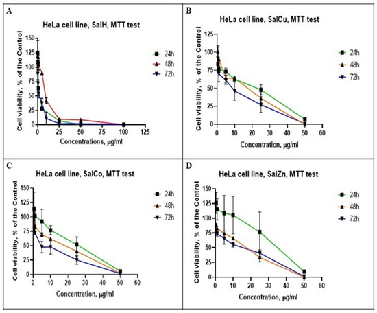
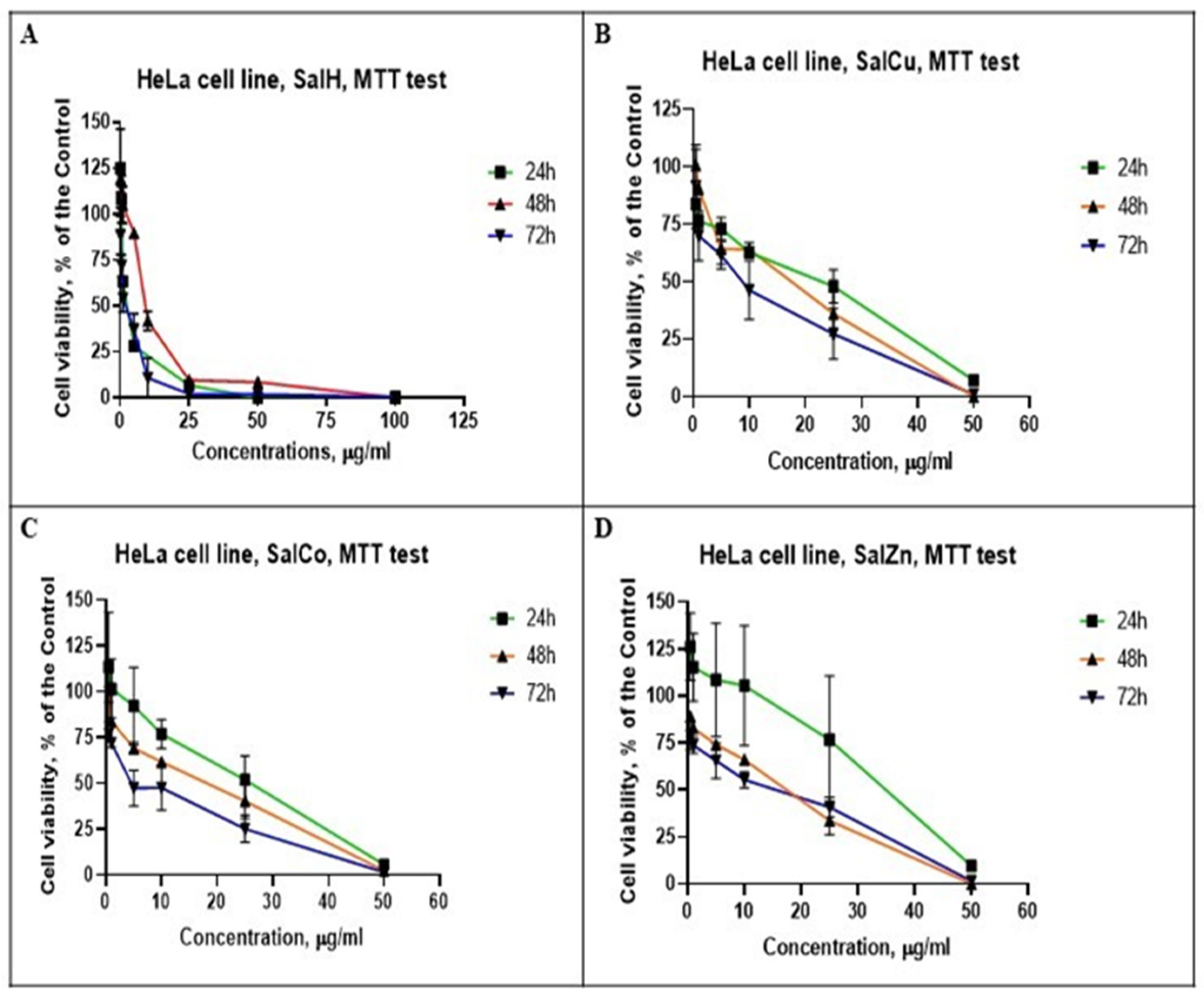
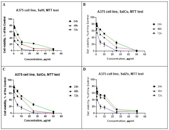
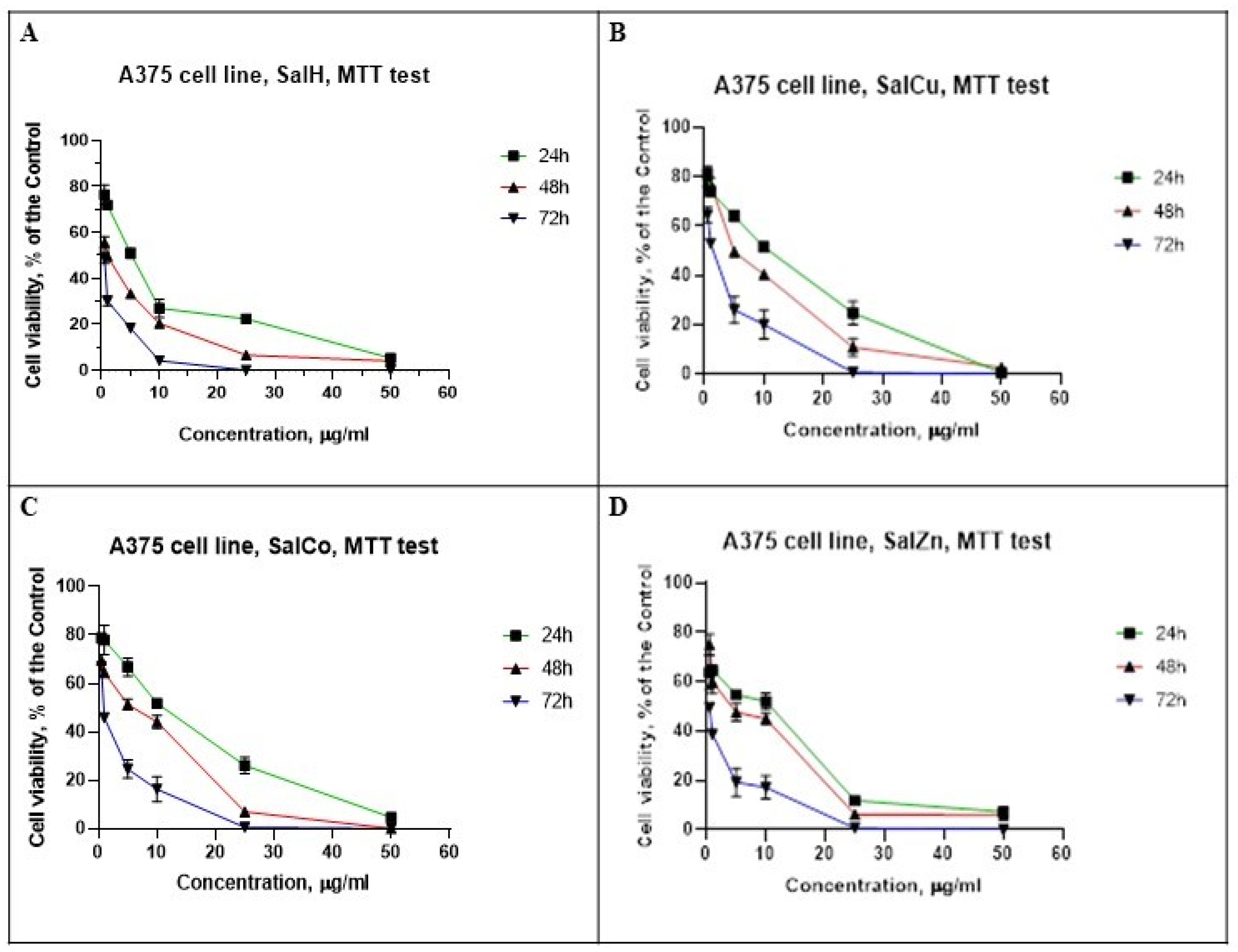
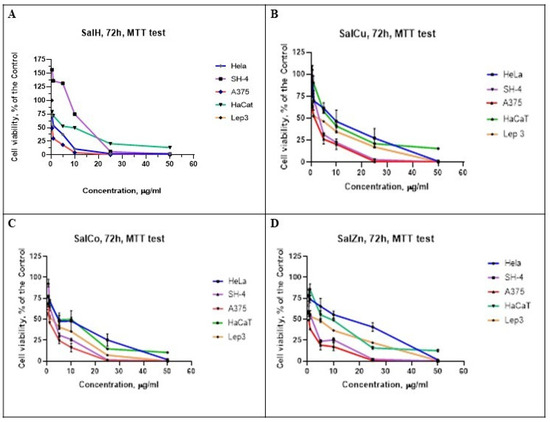
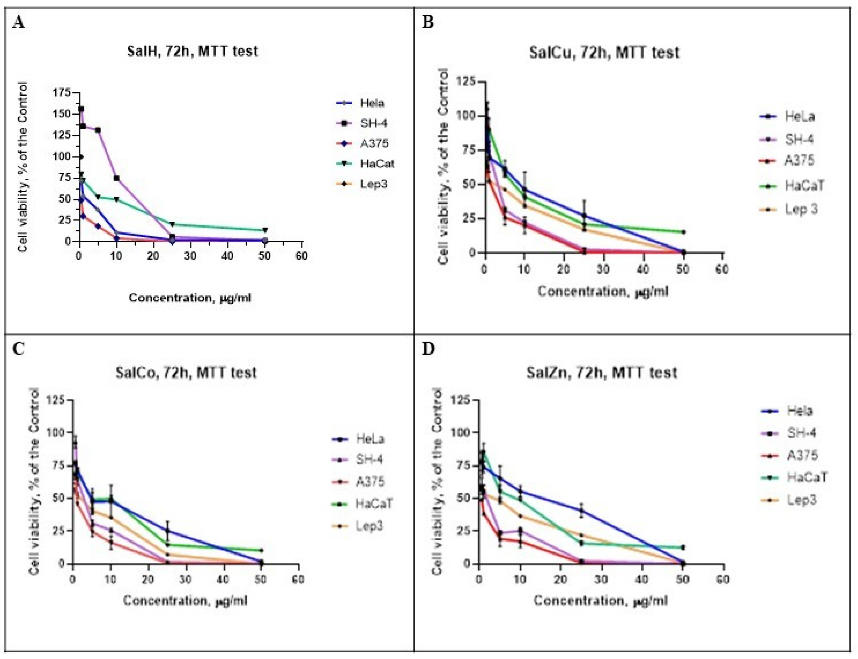
Figure 1 and Figure 2 depict the influence of time of incubation of the HeLa and A375 cells with the tested compounds on cell viability. The concentration–response curves constructed using the experimental data are presented in Figure 3.
Figure 1.
Effects of SalH (A) and the tested coordinated compounds, SalCu (B), SalCo (C) and SalZn (D), on cell viability of human cervical carcinoma cell line (HeLa) at 24 h, 48 h, and 72 h of incubation. The results are obtained by the MTT test.
Figure 2.
Effects of SalH (A) and the tested coordinated compounds, SalCu (B), SalCo (C) and SalZn (D), on cell viability of human melanoma A375 cell line at 24 h, 48 h, and 72 h of incubation. The results are obtained by the MTT test.
Figure 3.
Effects of SalH (A) and the tested coordinated compounds, SalCu (B), SalCo (C) and SalZn (D), on cell viability of HeLa, SH-4, A375, HaCaT and Lep3 cell lines at 72 h of incubation. The results are obtained by the MTT test.
Table 2 presents the hierarchical ranks of salinomycin, its metal complexes, and the antitumor agents cisplatin and oxaliplatin based on their ability to reduce cell viability and/or proliferation.
Table 2.
Hierarchical orders (descending, starting from the most cytotoxic compound) of SalH, its Cu(II), Co(II) and Zn(II) complexes, and antitumor controls (CisPt, OxPt), ranked by IC50 (μM) values determined via MTT, NR, and CV assays after 72 h of treatment in tumor and non-tumor cells.
Table 3 demonstrates hierarchical orders of sensitivity for human tumor and non-tumor cell lines, treated with SalH and its Cu(II), Co(II) and Zn(II) complexes, as well as antitumor agents (CisPt, OxPt), determined by the MTT test (IC50, µM) after 72 h of treatment.
Table 3.
Cytotoxic sensitivity (at 72 h) of human tumor and non-tumor cell lines to SalH and its Cu(II), Co(II), and Zn(II) complexes compared to CisPt/OxPt. Hierarchical order (descending).
The selectivity index values of the compounds for HeLa, A375 and SH-4 cells are given in Table 4.
Table 4.
Selectivity index values (SI) of the tested compounds.
The results obtained reveal that:
- (i)
- Cell viability decreased with increasing concentrations of the metal complexes and exposure times (Figure 1, Figure 2 and Figure 3). The maximum effect was established for 72 h of incubation of the HeLa and A375 cells with the investigated compounds. Therefore, all other short-term experiments for evaluation of the ability of the metal complexes to affect cell viability were performed at 72 h of treatment.
- (ii)
- The cytotoxic activity of salinomycin and its metal complexes was demonstrated using all three methods: the MTT test, neutral red uptake assay, and crystal violet staining (Table 1). A cell-specific response is observed. For example, both melanoma cell lines (A375, SH-4) were most sensitive to SalZn, while in cervical carcinoma cells (HeLa) and non-tumor cells, Lep-3 and HaCAT, SalZn is at the middle or at the bottom of the hierarchical order. SalCo and SalCu reduce the survival of human keratinocytes (HaCaT line) and the embryonic fibroblastoid cells (Lep-3 line) to the highest extent (Table 2).
- (iii)
- Under the conditions of the experiments conducted in our study, the cytotoxic effect of the metal complexes of salinomycin was either lower than the effect of the uncoordinated antibiotic (HeLa cells) or more pronounced (A375, SH-4) and surpasses that of the commercial antitumor agents cisplatin and oxaliplatin (Table 1 and Table 2).
The comparison of the sensitivity of the cell lines to the cytotoxic effect of the tested substances is also of interest (Table 3). Non-tumor HaCat keratinocytes are least susceptible to the cytotoxic effects of metal complexes SalZn, SalCu, and SalCo (Table 3). SalCo is more toxic to A375 cells than Lep-3 cells, and both melanoma cell lines (A375 and SH-4) are relatively more sensitive to the effects of SalZn compared to non-tumor Lep-3 cells (Table 3). Zn(II) disalinomycinate demonstrated the highest selectivity against melanoma (A375 and SH-4) cells (Table 3). A high selectivity index against the A375 cell line was also established for SalH and SalCo (Table 4). The selectivity index values of SalH, SalZn, and SalCo were higher than the calculated selectivity of cisplatin (Table 4). SalH exerted the highest selectivity against HeLa cells (Table 4).
2.2. Cytopathological Studies
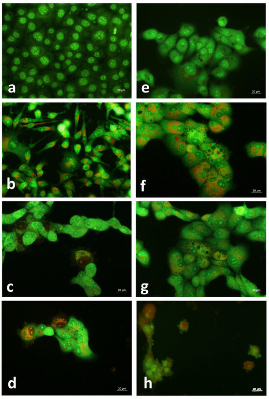
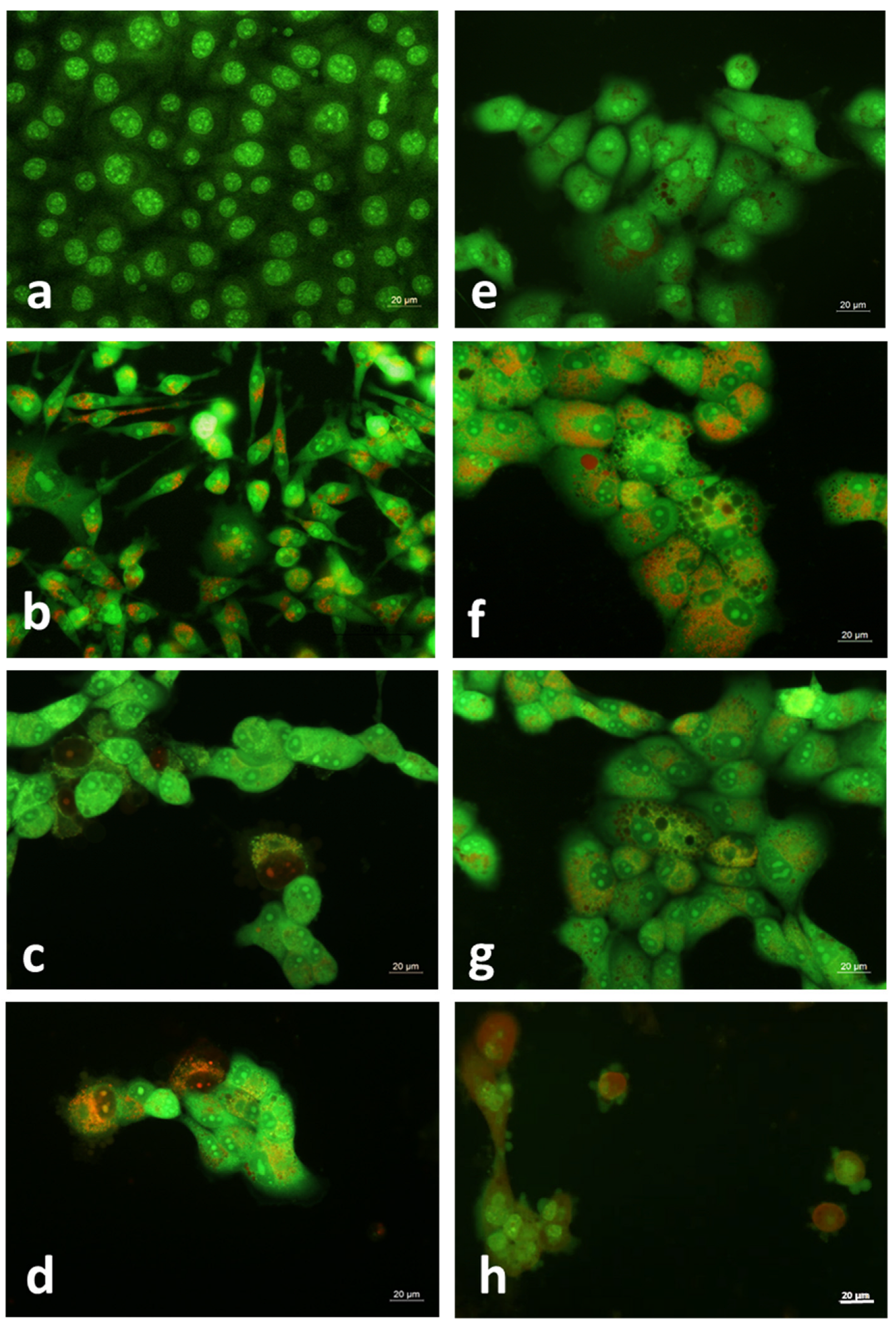
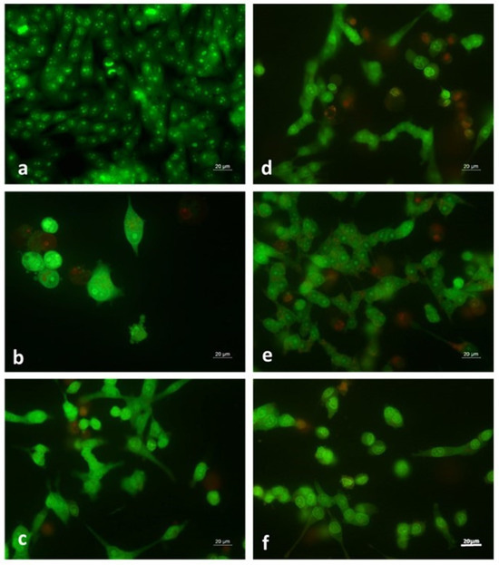
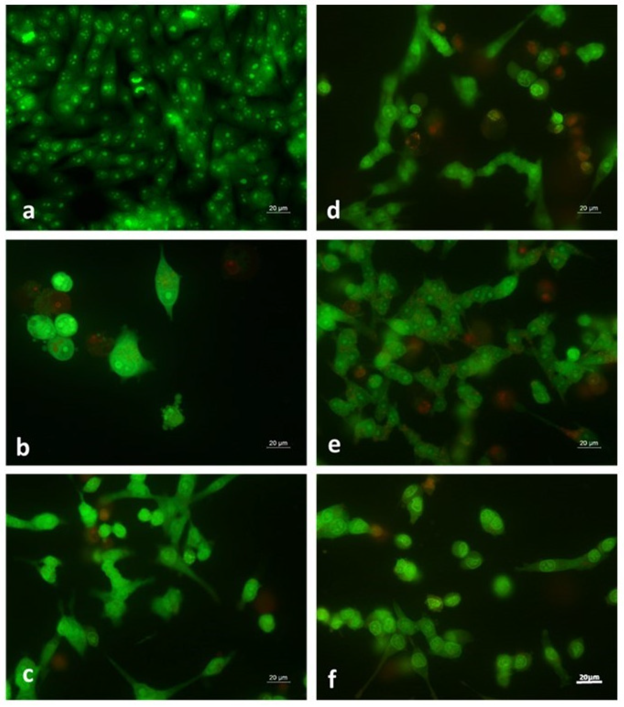
The cytotoxic effect of salinomycin metal complexes and their ability to induce cytopathological changes in treated cells was also confirmed by double staining with acridine orange and propidium iodide. Along with the reduced cell monolayer at 72 h, cytomorphological changes, such as cell swelling, vacuolization, chromatin condensation, and the formation of apoptotic bodies, were observed (Figure 4 and Figure 5).
Figure 4.
Cytopathological alterations induced by coordinated compounds of salinomycin in HeLa cells. (a)—untreated control HeLa cells; (b)—treated with 6.7 µM SalH; (c)—treated with 3.1 µM SalCu; (d)—HeLa cells, treated with 6.1 µM SalCu; (e)—HeLa cells, treated with 3.1 µM SalCo; (f)—HeLa cells, treated with 6.1 µM SalCo; (g)—HeLa cells, treated with 6.2 µM SalZn; (h)—HeLa cells, treated with 15.6 µM SalZn. Double staining with AO and PI, microscope Leika DM 500B (Wetzlar, Germany), Bar = 20 µm.
Figure 5.
Cytopathological alterations induced by coordinated compounds of salinomycin in A375 cells. (a)—untreated control A375 cells; (b)—treated with 10 μM SalH; (c)—A375 cells, treated with 0.6 μM SalCu; (d)—A375 cells, treated with 0.6 μM SalCo; (e)—A375 cells, treated with 0.3 µM SalZn; (f)—A375 cells, treated with 0.6 μM SalZn. Double staining with AO and PI, microscope Leika DM 500B (Wetzlar, Germany), Bar = 20 µm.
2.3. Long-Term Experiments on 3D Cell Colonies
To better understand the fate of A375 and HeLa cell lines cultured in the presence of salinomycin metal complexes and whether their cytotoxic effect is reversible, a 3D colony-forming assay was performed. Table 5 lists the effective concentrations that completely inhibit 3D tumor cell growth in a semi-liquid medium. The results show that 32 days after the start of the experiment, no 3D colonies were detected when the examined compounds were applied at concentrations ≥ 1.3 μM (salinomycin, HeLa), ≥3.1 μM (metal complexes, HeLa), and ≥6.1 μM (metal complexes, A375).
Table 5.
Effect of SalH, SalCu, SalCo and SalZn on the ability of HeLa and A375 cells to form 3D colonies in a semi-solid medium.
3. Discussion
Cervical cancer is one of the most common cancers in women worldwide and causes 41,000 deaths annually [33]. It has been reported that up to 50% of patients diagnosed with cervical cancer develop metastasis despite all available methods for treatment of this disease [34]. Cervical cancer stem cells (CCSCs) have been shown to be of major significance for failed therapy, metastasis and disease recurrence [35].
The literature data indicate that melanoma is another very common malignancy with a rapid increase in the incidence rate in Europe, the USA and Australia [36]. The successful treatment and survival rate of this disease depends strongly on its stage. While melanoma in stages I and II can be successfully cured by surgery, immunotherapy or targeted therapy, the successful treatment of advanced or metastatic melanoma is much more problematic [36]. When all protocols fail, chemotherapy is the only available option to slow down disease progression and to relieve the symptoms [37]. Unfortunately, the administration of the approved drugs for treatment of melanoma is associated with severe side effects [37].
The development of new and safer cytotoxic agents with high selectivity toward cancer cells is of crucial significance for the successful treatment of cancer, especially in the advanced stage.
In this study we provide the first data on the in vitro antitumor activity of complexes of the antineoplastic agent salinomycin with Co(II), Cu(II) and Zn(II) against cervical cancer (HeLa) cells and primary (A375) and metastatic melanoma (SH-4) cells. The cytotoxicity of the compounds on non-tumor cells (HaCAT and Lep-3) was also evaluated. To gain more detailed information about the antitumor activity of the tested complexes, we used a combination of assays with different cellular targets (molecules, organelles) and mechanisms of action—MTT test, neutral red uptake test and crystal violet staining. The MTT test has been accepted as a golden standard for estimation of cytotoxicity of new compounds. However, this method is only related with the effect of the tested compounds on cellular metabolic activity, mainly the ability of succinate dehydrogenase and other mitochondrial enzymes to reduce MTT(3-(4,5-dimethylthiazolyl-2)-2,5-diphenyltetrazolium bromide) into insoluble formazan crystals [38]. The NR method quantifies the toxic effects of compounds by measuring the ability of lysosomes in living cells to incorporate the supravitally dye neutral red, while the CV is directly related with cell survival, since it stains DNA and proteins [39,40,41]. The experimental data obtained by all three methods (MTT, NR, CV) reveal the capacity of the examined metal complexes to decrease viability and proliferation of the treated cells in a time- and concentration-dependent manner. Significant cytotoxic activity of the metal complexes of salinomycin and the non-coordinated antibiotic was observed at very low concentrations (below 5 μg/mL). At these concentrations, DMSO content was maintained below 0.5% (v/v), which is recognized as a non-cytotoxic concentration [42,43]. Even at the established IC50 values for the complexes of salinomycin, DMSO levels remained well below the toxic threshold [42,43], demonstrating that the solvent did not confound the results presented in Table 1. Previously published data indicate that the metal salts—Co(II) nitrate, Cu(II) acetate, and Zn(II) nitrate—are less active against leukemic tumor cells than the corresponding metal complexes of salinomycin with Co(II), Cu(II), and Zn(II) [27]. The reported results confirmed that the complexes remain intact and do not undergo dissociation in the cell culture medium. Therefore, the stability of the complexes of salinomycin in Co(II), Cu(II), and Zn(II) was not further investigated in this study.
The tested metal complexes of salinomycin exhibited different effects on the cellular metabolic activity in HeLa cells (Table 1). The lowest inhibitory concentration (IC50, μM), estimated by MTT assay was found for Co(II) disalinomycinate, demonstrating that this compound altered the cellular metabolic activity of these cells to the highest extent compared to the other two metal complexes. It should be pointed out that the complexes of salinomycin with Co(II), Cu(II) and Zn(II) have the same structure, as discussed in detail by Ivanova et al. [27]. Hence, the observed differences for the effects of the examined metal complexes on the cellular metabolic activity in HeLa cells could be related to the different biological roles of the divalent metal ions Co(II), Cu(II) and Zn(II) in the studied cell line. Studies in the literature demonstrated that Co(II) induced severe toxicity on mitochondria by inhibiting the activity of enzymes involved in mitochondrial respiration [44]. Moreover, this metal ion can affect mitochondrial membrane potential. It has been shown that Co(II) displaces Zn(II) and Cu(II) from their binding sites in the enzyme superoxide dismutase, decreasing its activity and the ability of cells to neutralize reactive oxygen species (ROS) [45,46,47,48]. All of this could contribute to the enhanced effect of Co(II) disalinomycinate on the cellular metabolic activity in HeLa cells compared to the other two metal-coordinated compounds. It is worth mentioning that some compounds could increase the NADPH level or the activity of LDH (lactate dehydrogenase) resulting in the elevated production of formazan [40,49,50,51,52]. The elevated IC50 value for Zn(II) disalinomycinate in cervical cancer cells (MTT assay) may stem from either a protective effect of Zn(II) on mitochondria or compound-induced alterations in cellular processes that increase formazan accumulation. Further investigation is required to elucidate the precise mechanisms behind this observation. The data for IC50 of the tested compounds in the HeLa cells obtained by NR assay may be influenced by a lower degree of permeability of the metal complexes of salinomycin across the lysosomal membrane compared to the uncoordinated antibiotic. The values for IC50 of the compounds in HeLa cells, estimated by CV staining, were lower compared to the values obtained by the MTT test. An exception was observed only for the uncoordinated antibiotic.
In contrast to HeLa cells, our results regarding the cytotoxic activity of the tested compounds against A375 cells indicated that the lowest IC50 values were obtained using the classical MTT test. An exception was observed for the SalCu complex only. The observed cell-specific response can be explained by the specific features of the individual cell lines, the examined compounds, and the methods used. The cell lines applied as model systems in our study have different origins (from primary and metastatic melanoma, cervical carcinoma, non-tumor keratinocytes, and embryonic cells). The cells have undergone changes during their cultivation in laboratory conditions. It is also known that each cell line is a unique system. Embryonic cells are characterized by high sensitivity to adverse factors, including chemical signals. In contrast, cancer cells are selected during tumor progression due to genetic instability and evolutionary pressure to become better adapted to their microenvironment [53,54].
Atjanasuppat et al. 2009 [55] classified potential antitumor agents based on their IC50 values as follows. IC50 values below or equal to 20 μg/mL show that the compound is active. IC50 values in the range from 20 to 100 μg/mL demonstrate that the tested agent is moderately active. When the calculated IC50 concentration is in the range 100–1000 μg/mL, the compounds must be considered as weakly active and values of IC50 higher than 1000 μg/mL signal no activity. This scale has also been used by other authors [56]. The IC50 values obtained in our studies revealed that all tested compounds must be considered as active against cervical cancer (HeLa), primary melanoma (A375) and metastatic melanoma (SH-4) cells. Compared to other metal complexes of Co(II) and Cu(II) with organic ligands, SalCo and SalCu demonstrated more pronounced activity in HeLa cells, evaluated by the MTT assay [57]. We found only two published studies about in vitro antitumor activity of the metal complex of salinomycin against HeLa cells [31,32]. It has been demonstrated that the Ce(IV) complex of salinomycin exerted pronounced antitumor activity in HeLa cells with IC50 of 2.73 μM, calculated by the MTT method [31]. Similar to SalCo, SalCu and SalZn, [Ce(Sal)(η2-NO3)2OH] exerted a lower effect on cellular metabolic activity in HeLa cells compared to salinomycin [31]. The metal complexes of salinomycin, however, have shown much higher activity than cisplatin, epirubicin and oxaliplatin [31]. Literature data revealed that the cytotoxicity of the thallium (I) complex of salinomycin, evaluated by CV assay, was comparable to that of the uncoordinated antibiotic [32]. In A375 and SH-4 cells, the highest activity was observed for Zn(II) disalinomycinate. To the best of our knowledge, there is no published information about the cytotoxicity of metal complexes of salinomycin against human melanoma.
The selectivity index is one of the important markers in drug discovery since it provides information about the selectivity of the tested compounds toward tumor cells [58]. A selectivity index higher than three indicates high selectivity and refers to the new substance as suitable for further in vivo studies [58]. The selectivity index values given in Table 4 demonstrated that Zn(II) disalinomycinate exerted pronounced selectivity against melanoma. Remarkable selectivity against primary melanoma (A375) was also established for SalH and SalCo. The selectivity of the conventional drug cisplatin against primary melanoma was lower compared to the values found for SalH, SalCo and SalZn.
The observed severe cytotoxicity of the tested compounds in this study was further confirmed by cytopathological studies. All coordinated compounds induced morphological alterations in HeLa and A375 cells at very low concentrations (from 3 μM to 15 μM for HeLa cells and from 0.3 μM to 10 μM for A375 cells). A more detailed study is needed to understand the exact mechanism of the antitumor activity of SalCo, SalCu and SalZn, and to attribute the observed structural modifications to apoptosis.
To further evaluate the cytotoxic effect of the metal complexes of salinomycin on the tumor cells, long-term experiments with 3D cultures were performed. Compared to the 2D model systems, the 3D models could mimic the real behavior of the tumors [59] and provide more significant results about the potency of new compounds as antitumor agents in vivo. The tested compounds demonstrated a pronounced inhibitory effect on the ability of the studied tumor cells to form 3D colonies.
4. Materials and Methods
4.1. Chemicals
Salinomycin sodium (C42H69O11Na; SalNa, p.a.) was delivered by Biovet Ltd. (Peshtera, Bulgaria). The metal complexes of salinomycin (SalH) were of composition [Co(C42H69O11)2(H2O)2]·2H2O (MW = 1631 g/mol) (SalCo), [Cu(C42H69O11)2(H2O)2]·2H2O (MW = 1635.5 g/mol) (SalCu), and [Zn(C42H69O11)2(H2O)2] (MW = 1601.4 g/mol) (SalZn), correspondingly. The procedure for the synthesis of the compounds, purity and structures was published previously by Ivanova et al. [27]. Gibco-Invitrogen UK (Paisley, UK) supplied Dulbecco’s modified Eagle’s medium (DMEM) and fetal bovine serum. Dimethyl sulfoxide (DMSO) and trypsin were obtained from AppliChem (Darmstadt, Germany). Purified agarose, thiazolyl blue tetrazolium bromide (MTT), 3-amino-7-dimethylamino-2-methylphenazine hydrochloride (neutral red), crystal violet, cisplatin and oxaliplatin were delivered by Sigma-Aldrich Chemie GmbH (Taufkirchen, Germany). Antibiotics (penicillin and streptomycin) for cell cultures were bought from Lonza (Verviers, Belgium). Ethylenediaminetetraacetic acid (EDTA) and all other chemicals of the highest purity were obtained from local suppliers.
4.2. Antitumor Agents
Cisplatin and oxaliplatin were applied as positive controls in some of the experiments. They were included in the study because they are used in the treatment of a wide range of cancers, including cervical cancer and melanoma, and platinum is the only metal, the compounds of which have entered clinical oncology [60,61,62,63].
4.3. Cell Culture Studies
The human cell lines HeLa (cervical carcinoma), A375 (primary melanoma), and SH-4 (derived from pleural effusion of a patient with metastatic melanoma), as well as non-tumor embryo cells (Lep-3) and keratinocytes (HaCaT) were used as model systems. The cells were provided by the Cell Bank/Cell Culture Collection of the Institute of the Experimental Morphology and Pathology with Museum—Bulgarian Academy of Sciences. The institute follows the established quality assurance protocols to guarantee the authenticity, purity and long-term viability of the cells.
Cell lines were routinely cultured under standard conditions (37 °C, 5% carbon dioxide and air humidity provided by a thermostat, Thermo Scientific, Hepa class 100, Waltham, MA, USA) and passaged using 0.05% trypsin and 0.02% ethylenediaminetetraacetic acid.
More detailed information about the cell cultivation conditions was published by Ivanova et al. [64].
4.3.1. MTT Assay
The cells were seeded in 96-well microplates at a cell density of 1 × 10⁴ per well. Treatment began 24 h later, with fresh medium containing the compounds investigated at a given concentration (0.1–100 µg/mL for SalH, SalCo, SalCu and SalZn; 0.1–200 for cisplatin and oxaliplatin) being added (each concentration was tested in four replicates). Cells cultured in compound-free medium served as controls. The ability of the compounds to affect cell viability was determined after incubation for 24, 48 or 72 h. For this purpose, the culture medium was removed and replaced with an MTT solution (5 mg MTT in 10 mL DMEM) after which the microplates were put in the CO2 incubator for 3 h. The procedure ended with extraction of the formed blue formazan in metabolically active, living cells using a 1:1 (v/v) mixture of 100% ethanol and DMSO, and the absorbance was measured as described by Mosmann, 1983 [65].
4.3.2. Neutral Red Uptake Test (NR)
The NR assay has been developed on the basis of Borenfreund and Puerner’s method [66]. A mixture of 1 mL 0.4% solution of NR in 80 mL medium was added to each well with the matching cells. The plate was kept in a CO2 incubator for three hours to secure the absorption of the essential dye. After that, the medium, containing NR, was removed, and the cells were rinsed with phosphate-buffered saline (PBS, 7.2–7.4, 0.2 mL/well). In total, 0.1 mL of a solution containing 96% ethanol (50 parts), distilled H2O (49 parts) and acetic acid (1 part) (50:49:1, v:v:v) was used to extract the dye from the cells.
4.3.3. Crystal Violet Staining (CV)
Crystal violet staining assay was performed according to instructions by Sotome et al. [67]. After the incubation of the treated cells and controls for 72 h at 37 °C in a CO2 incubator, the media were removed, the cells were rinsed with distilled water, and they were stained for 30 min with a 0.4% methanolic solution of crystal violet.
The optical density at λ = 540/620 nm (MTT and CV) and λ = 540 nm (NR) was detected by a TECAN SunriseTM automatic microplate reader (Grödig, Austria). Relative cell viability was calculated as the percentage of the untreated control (100% viability). The half-maximal inhibitory cytotoxic concentration (IC50) of the compounds, causing a 50% decrease in cell viability, was calculated from the concentration–response curves. The results are presented as the mean values from three independent assays. The selectivity index, calculated as the ratio of IC50 value for the non-tumor cells divided by IC50 for tumor cells, was determined.
4.3.4. Double Staining with Acridine Orange (AO) and Propidium Iodide (PI)
Cells were seeded into 6-well plates at a concentration of 3–3.5 × 10⁵ cells per well and incubated with the compounds for 24–72 h, alongside untreated control samples. Subsequently, the coverslips were washed with phosphate-buffered saline (PBS, pH 7.2–7.4) for two minutes. To avoid signal loss, stained cells were analyzed within 30 min using a Leika DM 500B fluorescence microscope (Wetzlar, Germany) [68].
4.3.5. Three-Dimensional Cell Colony-Forming Assay
A 3D colony-forming assay was used to evaluate the ability of the compounds to prevent the tumor cells from producing 3D colonies [69]. Tumor cells (approximately 103 cells/well) were placed and suspended in 0.45% pure agarose in 2X DMEM containing different concentrations of the compounds under investigation. The cell growth was observed for 32 days using an inverted microscope (Carl Zeiss, Jena, Germany). The effective inhibitory concentration (µM) of each compound that completely inhibited 3D cancer cell colony formation in the semi-solid medium was calculated.
4.4. Statistical Analysis
The arithmetic mean (x) and its error (SEM) were calculated by the method of analysis of variance. The individual groups were compared using one-way analysis of variance (ANOVA) followed by the Dunnet test. The difference between groups was considered significant at the p < 0.05 level of significance. The statistical processing of the experimental data was conducted by the computer program GraphPad Prism, Version 8.0.2 (263).
5. Limitation of the Study
This research aimed to provide the first data regarding the in vitro antitumor activity of metal complexes of salinomycin with Co(II), Cu(II), and Zn(II) against cervical carcinoma and primary and metastatic melanoma. The present research does not demonstrate details about the exact mechanism of the antitumor activity of the tested compound in cervical cancer and melanoma. Further quantitative studies should be conducted to assess the ability of these compounds to induce apoptosis in primary and metastatic melanoma and cervical carcinoma. Studies on a broader panel of non-tumor cell lines are needed for a more comprehensive evaluation of the selectivity of the tested compounds.
6. Conclusions
In this study, we demonstrated for the first time that the metal complexes of salinomycin with Co(II), Cu(II), and Zn(II) exerted pronounced cytotoxicity in human cervical cells (HeLa) and melanoma cells (A375, SH-4), decreasing their viability and 2D/3D growth. The pronounced selectivity index of Zn(II) disalinomycinate against melanoma cells (both primary and metastatic), of Co(II) disalinomycinate against primary melanoma along with the established high selectivity index of salinomycinic acid (SalH) toward HeLa cells suggests that these compounds are suitable for future investigations with a panel of tumor and non-tumor cell lines and primary cell cultures to better clarify their potency as antitumor agents for the treatment of cervical cancer and melanoma. Further studies on the mechanism of antitumor activity of salinomycin and its metal complexes with Co(II), Cu(II), and Zn(II) are warranted.
Author Contributions
Conceptualization, R.A. and J.I.; methodology, T.Z., H.H., R.A., A.A. and L.D.; validation, T.Z., H.H., R.A., A.A. and L.D.; formal analysis, T.Z., L.D. and R.A.; investigation, T.Z., H.H., R.A., L.D., P.D. and J.I.; resources, R.A. and P.D.; writing—original draft preparation, R.A. and J.I.; writing—review and editing, T.Z., H.H., R.A., A.A., L.D., P.D. and J.I.; project administration, R.A.; funding acquisition, R.A. All authors have read and agreed to the published version of the manuscript.
Funding
The financial support by the European Union Next Generation EU through the National Recovery and Resilience Plan of the Republic of Bulgaria, project no. BG-RRP-2.004-0008 as well as by the National Science Fund in Bulgaria, grant no KП-06-KOCT/18 from 12.08.2024 is acknowledged. H.H. was supported by the Bulgarian Ministry of Education and Science under the National Research Program “Young scientists and postdoctoral students-2”, approved by DCM 206/07.04.2022.
Institutional Review Board Statement
Not applicable.
Informed Consent Statement
Not applicable.
Data Availability Statement
All data are included in the manuscript.
Conflicts of Interest
The authors declare no conflicts of interest. Peter Dorkov is employed in BIOVET JSC. The company supplied salinomycin sodium, used as a starting material for the synthesis of salinomycinic acid (salinomycin), and for the synthesis of metal complexes of salinomycin with Co(II), Cu(II), and Zn(II). The company did not participate in the experimental design, collection, analysis, interpretation of data, the writing of this article, or decision for publication.
References
- The global challenge of cancer. Nat. Cancer 2020, 1, 1–2. [CrossRef] [Scilit]
- Piña-Sánchez, P.; Chávez-González, A.; Ruiz-Tachiquín, M.; Vadillo, E.; Monroy-García, A.; Montesinos, J.J.; Grajales, R.; Gutiérrez de la Barrera, M.; Mayani, H. Cancer Biology, Epidemiology, and Treatment in the 21st Century: Current Status and Future Challenges From a Biomedical Perspective. Cancer Control. 2021, 28. [Google Scholar] [CrossRef] [Scilit]
- World Health Organization (WHO). 1 February 2024. Available online: https://www.who.int/news/item/01-02-2024-global-cancer-burden-growing--amidst-mounting-need-for-services (accessed on 14 April 2026).
- Levi, M.S.; Borne, R.F.; Williamson, J.S. A review of cancer chemopreventive agents. Curr. Med. Chem. 2001, 8, 1349–1362. [Google Scholar] [CrossRef] [Scilit]
- Truskowski, K.; Amend, S.R.; Pienta, K.J. Dormant cancer cells: Programmed quiescence, senescence, or both? Cancer Metastasis Rev. 2023, 42, 37–47. [Google Scholar] [CrossRef] [Scilit]
- El-Tanani, M.; Rabbani, S.A.; Satyam, S.M.; Rangraze, I.R.; Wali, A.F.; El-Tanani, Y.; Aljabali, A.A.A. Deciphering the Role of Cancer Stem Cells: Drivers of Tumor Evolution, Therapeutic Resistance, and Precision Medicine Strategies. Cancers 2025, 17, 382. [Google Scholar] [CrossRef] [Scilit]
- Lee, H.; Kim, B.; Park, J.; Park, S.; Yoo, G.; Yum, S.; Kang, W.; Lee, J.; Yoon, H.; Yoon, B. Cancer stem cells: Landscape, challenges and emerging therapeutic innovations. Sig. Transduct. Target Ther. 2025, 10, 248. [Google Scholar] [CrossRef] [Scilit]
- Gupta, P.B.; Onder, T.T.; Jiang, G.; Tao, K.; Kuperwasser, C.; Weinberg, R.A.; Lander, E.S. Identification of selective inhibitors of cancer stem cells by high-throughput screening. Cell 2009, 138, 645–659. [Google Scholar] [CrossRef] [Scilit] [PubMed]
- Zhou, J.; Li, P.; Xue, X.; He, S.; Kuangm, Y.; Zhao, H.; Chen, S.; Zhi, Q.; Guo, X. Salinomycin induces apoptosis in cisplatin-resistant colorectal cancer cells by accumulation of reactive oxygen species. Toxicol. Lett. 2013, 2222, 139–145. [Google Scholar] [CrossRef] [Scilit] [PubMed]
- Zhang, G.N.; Liang, Y.; Zhou, L.J.; Chen, S.P.; Chen, G.; Zhang, T.P.; Kang, T.; Zhao, Y.P. Combination of salinomycin and gemcitabine eliminates pancreatic cancer cells. Cancer Lett. 2011, 313, 137–144. [Google Scholar] [CrossRef] [Scilit] [PubMed]
- Klose, J.; Guerlevik, E.; Trostel, T.; Kuehnel, F.; Schmidt, T.H.; Schneider, M.; Ulrich, A. Salinomycin inhibits cholangiocarcinoma growth by inhibition of autophagic flux. Oncotarget 2018, 9, 3619–3630. [Google Scholar] [CrossRef] [Scilit]
- Wang, Q.; Yen, Y.T.; Xie, C.; Liu, F.; Liu, Q.; Wei, J.; Liu, B. Combined delivery of salinomycin and docetaxel by dual-targeting gelatinase nanoparticles effectively inhibits cervical cancer cells and cancer stem cells. Drug Deliv. 2021, 28, 510–519. [Google Scholar] [CrossRef] [Scilit]
- Wang, Q.; Liu, F.; Wang, L.; Xie, C.; Wu, P.; Du, S.; Zhou, S.; Sun, Z.; Liu, Q.; Yu, L.; et al. Enhanced and Prolonged Antitumor Effect of Salinomycin-Loaded Gelatinase-Responsive Nanoparticles via Targeted Drug Delivery and Inhibition of Cervical Cancer Stem Cells. Int. J. Nanomed. 2020, 15, 1283–1295. [Google Scholar] [CrossRef] [Scilit]
- Liu, Y.; Hao, Y.; Li, Y.; Zheng, Y.; Dai, J.; Zhong, F.; Wei, W.; Fang, Z. Salinomycin induces autophagic cell death in salinomycin-sensitive melanoma cells through inhibition of autophagic flux. Sci Rep. 2020, 10, 18515. [Google Scholar] [CrossRef] [Scilit]
- Qi, D.; Liu, Y.; Li, J.; Huang, J.H.; Hu, X.; Wu, E. Salinomycin as a potent anticancer stem cell agent: State of the art and future directions. Med. Res. Rev. 2022, 42, 1037–1063. [Google Scholar] [CrossRef] [Scilit] [PubMed]
- Dorne, J.L.; Fernandez-Cruz, M.L.; Bertelsen, U.; Renshaw, D.W.; Peltonen, K.; Anadon, A.; Feil, A.; Sanders, P.; Wester, P.; Fink-Gremmels, J. Risk assessment of coccidostatics during feed cross-contamination: Animal and human health aspects. Toxicol. Appl. Pharmacol. 2013, 270, 196–208. [Google Scholar] [CrossRef] [Scilit] [PubMed]
- Boehmerle, W.; Muenzfeld, H.; Springer, A.; Huehnchen, P.; Endres, M. Specific targeting of neurotoxic side effects and pharmacological profile of the novel cancer stem cell drug salinomycin in mice. J. Mol. Med. 2014, 92, 889–900. [Google Scholar] [CrossRef] [Scilit] [PubMed]
- Story, P.; Doube, A. A case of human poisoning by salinomycin, an agricultural antibiotic. N. Z. Med. J. 2004, 117, U799. [Google Scholar]
- Antoszczak, M. A comprehensive review of salinomycin derivatives as potent anticancer and anti-CSCs agents. Eur. J. Med. Chem. 2019, 166, 48–64. [Google Scholar] [CrossRef] [Scilit]
- Antoszczak, M.; Huczyński, A. Salinomycin and its derivatives—A new class of multiple-targeted “magic bullets”. Eur. J. Med. Chem. 2019, 176, 208–227. [Google Scholar] [CrossRef] [Scilit]
- Kuran, D.; Flis, S.; Antoszczak, M.; Piskorek, M.; Huczyński, A. Ester derivatives of salinomycin efficiently eliminate breast cancer cells via ER-stress-induced apoptosis. Eur. J. Pharmacol. 2021, 893, 173824. [Google Scholar] [CrossRef] [Scilit]
- Czerwonka, D.; Müller, S.; Cañeque, T.; Colombeau, L.; Huczyński, A.; Antoszczak, M.; Rodriguez, R. Expeditive Synthesis of Potent C20-epi-Amino Derivatives of Salinomycin against Cancer Stem-Like Cells. ACS Org. Inorg. Au 2022, 2, 214–221. [Google Scholar] [CrossRef] [Scilit]
- Li, B.; Wu, J.; Tang, L.; Lian, X.; Li, Z.; Duan, W.; Qin, T.; Zhao, X.; Hu, Y.; Zhang, C.; et al. Synthesis and anti-tumor activity evaluation of salinomycin C20-O-alkyl/benzyl oxime derivatives. Org. Biomol. Chem. 2022, 20, 870–876. [Google Scholar] [CrossRef] [Scilit]
- Jiang, R.; Zhang, X.; Li, N.; Mao, Y.; Chen, H.; Deng, Z.; Wang, W.; Jiang, Z.X.; Xu, L.; Yang, Z. Effective Synthesis of C20-Epi-Isothiocyanato-Salinomycin and its Thiourea Derivatives as Potential Anticancer Agents. Chemistry 2024, 30, e202402483. [Google Scholar] [CrossRef] [Scilit]
- Alqahtani, T.; Kumarasamy, V.M.; Huczyński, A.; Sun, D. Salinomycin and its derivatives as potent RET transcriptional inhibitors for the treatment of medullary thyroid carcinoma. Int. J. Oncol. 2020, 56, 348–358. [Google Scholar] [CrossRef] [Scilit]
- Antoszczak, M.; Mielczarek-Puta, M.; Struga, M.; Huczynski, A. Urea and Thiourea Derivatives of Salinomycin as Agents Targeting Malignant Colon Cancer Cells. Anti-Cancer Agents Med. Chem. 2025, 25, 330–338. [Google Scholar] [CrossRef] [Scilit]
- Ivanova, J.; Pantcheva, I.N.; Zhorova, R.; Momekov, G.; Simova, S.; Stoyanova, R.; Zhecheva, E.; Ivanova, S.; Mitewa, M. Synthesis, spectral properties, antibacterial and antitumor activity of salinomycin complexes with Co(II), Ni(II), Cu(II) and Zn(II) transition metal ions. J. Chem. Chem. Eng. 2012, 6, 551–562. [Google Scholar]
- Pashkunova-Martic, I.; Kukeva, R.; Stoyanova, R.; Pantcheva, I.; Dorkov, P.; Friske, J.; Hejl, M.; Jakupec, M.; Hohagen, M.; Legin, A.; et al. Novel Salinomycin-Based Paramagnetic Complexes-First Evaluation of Their Potential Theranostic Properties. Pharmaceutics 2022, 14, 2319. [Google Scholar] [CrossRef] [Scilit]
- Atanasov, V.; Stoykova, S.; Goranova, Y.; Nedzhib, A.; Tancheva, L.; Ivanova, J.; Pantcheva, I. Preliminary study on in vivo toxicity of monensin, salinomycin and their metal complexes. Bul. Chem. Commun. 2014, 46, 233–273. [Google Scholar]
- Momekova, D.; Momekov, G.; Ivanova, J.; Pantcheva, I.; Drakalska, E.; Stoyanov, N.; Guenova, M.; Michova, A.; Balashev, K.; Arpadjan, S.; et al. Sterically stabilized liposomes as a platform for salinomycin metal coordination compounds: Physicochemical characterization and in vitro evaluation. J. Drug Deliv. Sci. Technol. 2013, 23, 215–223. [Google Scholar] [CrossRef] [Scilit]
- Petkov, N.; Pantcheva, I.; Ivanova, A.; Stoyanova, R.; Kukeva, R.; Alexandrova, R.; Abudalleh, A.; Dorkov, P. Novel Cerium(IV) Coordination Compounds of Monensin and Salinomycin. Molecules 2023, 28, 4676. [Google Scholar] [CrossRef] [Scilit] [PubMed]
- Petkov, N.; Dorkov, P.; Ugrinov, A.; Encheva, E.; Abrashev, M.; Zasheva, D.; Daneva, T.; Pantcheva, I.N. Crystal Structure and Properties of Thallium(I) Salinomycinate. Int. J. Mol. Sci. 2025, 26, 6504. [Google Scholar] [CrossRef] [Scilit]
- Bray, F.; Ferlay, J.; Soerjomataram, I.; Siegel, R.L.; Torre, L.A.; Jemal, A. Global cancer statistics 2018: GLOBOCAN estimates of incidence and mortality worldwide for 36 cancers in 185 countries. CA Cancer J. Clin. 2018, 68, 394–424. [Google Scholar] [CrossRef] [Scilit]
- Kumar, L.; Harish, P.; Malik, P.S.; Khurana, S. Chemotherapy and targeted therapy in the management of cervical cancer. Curr. Probl. Cancer 2018, 42, 120–128. [Google Scholar] [CrossRef] [Scilit] [PubMed]
- Di Fiore, R.; Suleiman, S.; Drago-Ferrante, R.; Subbannayya, Y.; Pentimalli, F.; Giordano, A.; Calleja-Agius, J. Cancer Stem Cells and Their Possible Implications in Cervical Cancer: A Short Review. Int. J. Mol. Sci. 2022, 23, 5167. [Google Scholar] [CrossRef] [Scilit]
- Waseh, S.; Lee, J.B. Advances in melanoma: Epidemiology, diagnosis, and prognosis. Front. Med. 2023, 10, 2023. [Google Scholar] [CrossRef] [Scilit]
- Simon, A.; Kourie, H.R.; Kerger, J. Is there still a role for cytotoxic chemotherapy after targeted therapy and immunotherapy in metastatic melanoma? A case report and literature review. Chin. J. Cancer 2017, 36, 10. [Google Scholar] [CrossRef] [Scilit] [PubMed]
- Ghasemi, M.; Turnbull, T.; Sebastian, S.; Kempson, I. The MTT Assay: Utility, Limitations, Pitfalls, and Interpretation in Bulk and Single-Cell Analysis. Int. J. Mol. Sci. 2021, 22, 12827. [Google Scholar] [CrossRef] [Scilit]
- Bacanli, M.; Anlar, H.G.; Başaran, A.A.; Başaran, N. Assessment of Cytotoxicity Profiles of Different Phytochemicals: Comparison of Neutral Red and MTT Assays in Different Cells in Different Time Periods. Turk J. Pharm. Sci. 2017, 14, 95–107. [Google Scholar] [CrossRef] [Scilit]
- Śliwka, L.; Wiktorska, K.; Suchocki, P.; Milczarek, M.; Mielczarek, S.; Lubelska, K.; Cierpiał, T.; Łyżwa, P.; Kiełbasiński, P.; Jaromin, A.; et al. The Comparison of MTT and CVS Assays for the Assessment of Anticancer Agent Interactions. PLoS ONE 2016, 11, e0155772. [Google Scholar] [CrossRef] [Scilit]
- Feoktistova, M.; Geserick, P.; Leverkus, M. Crystal Violet Assay for Determining Viability of Cultured Cells. Cold Spring. Harb. Protoc. 2016, 2016, pdb-prot087379. [Google Scholar] [CrossRef] [Scilit]
- Asiri, A.; Tasleem, M.; Al Said, M.; Asiri, A.; Al Qarni, A.A.; Bakillah, A. Optimizing Cell Density and Unveiling Cytotoxic Profiles of DMSO and Ethanol in Six Cancer Cell Lines: Experimental and In Silico Insights. Methods Protoc. 2025, 8, 93. [Google Scholar] [CrossRef] [Scilit] [PubMed]
- Culita, D.-C.; Dyakova, L.; Marinescu, G.; Zhivkova, T.; Spasov, R.; Patron, L.; Alexandrova, R.; Oprea, O. Synthesis, Characterization and Cytotoxic Activity of Co(II), Ni(II), Cu(II), and Zn(II) Complexes with Nonsteroidal Antiinflamatory Drug Isoxicam as Ligand. J. Inorg. Organomet. Polym. Mater. 2019, 29, 580–591. [Google Scholar] [CrossRef] [Scilit]
- Barceloux, D.G. Chromium. J. Toxicol. Clin. Toxicol. 1999, 37, 173–194. [Google Scholar] [CrossRef] [Scilit]
- Karovic, O.; Tonazzini, I.; Robola, N.; Edstrom, E.; Lovdahl, C.; Fredholm, B.B. Toxic effects of cobalt in primary cultures of mouse astrocytes. Similarities with hypoxia and role of HIF-1α. Biochem. Pharmacol. 2007, 73, 694–708. [Google Scholar] [CrossRef] [Scilit]
- Battaglia, V.; Compagnone, A.; Bandino, A.; Bragadin, M.; Rosii, C.A.; Zanetti, F.; Colombatto, S.; Grillo, M.A.; Toninello, A. Cobalt induces oxidative stress in isolated liver mitochondria responsible for permeability transition and intrinsic apoptosis in hepatocyte primary cultures. Int. J. Biochem. Cell. Biol. 2009, 41, 586–594. [Google Scholar] [CrossRef] [Scilit]
- Mou, Y.H.; Yang, J.Y.; Cui, N.; Wang, J.M.; Hou, Y.; Song, S.; Wu, C.F. Effects of cobalt chloriden nitric oxide and cytokines/chemokines production in microglia. Int. Immunopharm. 2012, 13, 120–125. [Google Scholar] [CrossRef] [Scilit]
- Czarnek, K.; Siwicki, A.K. Influence of chromium (III), cobalt (II) and their mixtures on cell metabolic activity. Curr. Issues Pharm. Med. Sci. 2021, 34, 87–93. [Google Scholar] [CrossRef] [Scilit]
- Vistica, D.T.; Skehan, P.; Scudiero, D.; Monks, A.; Pittman, A.; Boyd, M.R. Tetrazolium-based assays for cellular viability: A critical examination of selected parameters affecting formazan production. Cancer Res. 1991, 1, 2515–2520. [Google Scholar]
- Shoemaker, M.; Cohen, I.; Campbell, M. Reduction of MTT by aqueous herbal extracts in the absence of cells. J. Ethnopharmacol. 2004, 93, 381–384. [Google Scholar] [CrossRef] [Scilit] [PubMed]
- Wang, P.; Henning, S.M.; Heber, D. Limitations of MTT and MTS-Based Assays for Measurement of Antiproliferative Activity of Green Tea Polyphenols. PLoS ONE 2010, 5, e10202. [Google Scholar] [CrossRef] [Scilit] [PubMed]
- Bruggisser, R.; von Daeniken, K.; Jundt, G.; Schaffner, W.; Tullberg-Reinert, H. Interference of plant extracts, phytoestrogens and antioxidants with the MTT tetrazolium assay. Planta Med. 2002, 68, 445–448. [Google Scholar] [CrossRef] [Scilit]
- Li, B.; Pan, Y.K.; Jin, X.; Chan, H.M. Effects of an environmental chemical mixture on early-stage embryo development: In vitro evidence from human embryonic stem cells. Toxicol. Vitr. 2025, 108, 106101. [Google Scholar] [CrossRef] [Scilit] [PubMed]
- Baldassarre, G.; de la Serna, I.L.; Vallette, F.M. Death-ision: The link between cellular resilience and cancer resistance to treatments. Mol. Cancer 2025, 24, 144. [Google Scholar] [CrossRef] [Scilit]
- Atjanasuppat, K.; Wongkham, W.; Meepowpan, P.; Kittakoop, P.; Sobhon, P.; Bartlett, A.; Whitfield, P.J. In vitro screening for antihelminthic and antitumour activity of ethnomedicinal plants from Thailand. J. Ethnoparmacol. 2009, 123, 475–482. [Google Scholar] [CrossRef] [Scilit]
- Fithrotunnisa, Q.; Arsianti, A.; Kurniawan, G.; Qorina, F.; Tejaputri, N.A.; Azizah, N.N. In Vitro Cytotoxicity of Hibiscus sabdariffa Linn Extracts on A549 Lung Cancer Cell Line. Pharmacogn. J. 2020, 12, 14–19. [Google Scholar] [CrossRef] [Scilit]
- Dinev, D.; Popova, K.B.; Zhivkova, T.; Dyakova, L.; Abudalleh, A.; Alexandrova, R.; Culita, D.C.; Mocanu, T.; Maxim, C.; Marinescu, G. Synthesis, structural characterization, and cytotoxic activity in tumor cells of Cu(II) and Co(II) complexes with o-vanillin amino acids Schiff bases. Appl. Organomet. Chem. 2022, 36, e6862. [Google Scholar] [CrossRef] [Scilit]
- Bieszczad, B.; Garbicz, D.; Świtalska, M.; Dudek, J.M.; Warszycki, D.; Wietrzyk, J.; Grzesiuk, E.; Mieczkowski, A. Improved HDAC Inhibition, Stronger Cytotoxic Effect and Higher Selectivity against Leukemias and Lymphomas of Novel, Tricyclic Vorinostat Analogues. Pharmaceuticals 2021, 14, 851. [Google Scholar] [CrossRef] [Scilit] [PubMed]
- Abbas, Z.N.; Al-Saffar, A.Z.; Jasim, S.M.; Sulaiman, G.M. Comparative analysis between 2D and 3D colorectal cancer culture models for insights into cellular morphological and transcriptomic variations. Sci. Rep. 2023, 13, 18380. [Google Scholar] [CrossRef] [Scilit] [PubMed]
- Soengas, M.S.; Lowe, S.W. Apoptosis and melanoma chemoresistance. Oncogene 2003, 22, 3138–3151. [Google Scholar] [CrossRef] [Scilit]
- Geisler, J.P.; Swathirajan, J.; Wood, K.L.; Manahan, K.J. Treatment of advanced or recurrent cervical cancer with Cisplatin or Cisplatin containing regimens: A cost effective analysis. J. Cancer 2012, 3, 454–458. [Google Scholar] [CrossRef] [Scilit]
- Kolomeyevskaya, N.V.; Lele, S.B.; Miller, A.; Riebandt, G.C.; Blum, B.L.; Odunsi, K.O.; Frederick, P.J. Oxaliplatin is a safe alternative option for patients with recurrent gynecologic cancers after hypersensitivity reaction to Carboplatin. Int. J. Gynecol. Cancer 2015, 25, 42–48. [Google Scholar] [CrossRef] [Scilit] [PubMed]
- Weng, C.H.; Wu, C.S.; Wu, J.C.; Kung, M.L.; Wu, M.H.; Tai, M.H. Cisplatin-Induced Giant Cells Formation Is Involved in Chemoresistance of Melanoma Cells. Int. J. Mol. Sci. 2020, 21, 7892. [Google Scholar] [CrossRef] [Scilit] [PubMed]
- Ivanova, J.; Kukeva, R.; Stoyanova, R.; Zhivkova, T.; Abudalleh, A.; Dyakova, L.; Alexandrova, R.; Pashkunova-Martic, I.; Theiner, J.; Dorkov, P.; et al. New Iron(III)-Containing Composite of Salinomycinic Acid with Antitumor Activity—Synthesis and Characterization. Inorganics 2024, 12, 206. [Google Scholar] [CrossRef] [Scilit]
- Mosmann, T. Rapid colorimetric assay for cellular growth and survival: Application to proliferation and cytotoxicity assays. J. Immunol. Methods 1983, 65, 55–63. [Google Scholar] [CrossRef] [Scilit]
- Borenfreund, E.; Puerner, J. Toxicity determination in vitro by morphological alterations and neural red absorption. Toxicol. Lett. 1985, 24, 119–124. [Google Scholar] [CrossRef] [Scilit]
- Saotome, K.; Morita, H.; Umeda, M. Cytotoxicity test with simplified crystal violet staining method using microtitre plates and its application to injection drugs. Toxicol. Vitr. 1989, 3, 317–321. [Google Scholar] [CrossRef] [Scilit]
- Abdel Wahab, S.I.; Abdul, A.B.; Alzubairi, A.S.; Mohamed Elhassan, M.; Mohan, S. In Vitro Ultramorphological Assessment of Apoptosis Induced by Zerumbone on (HeLa). J. Biomed. Biotechnol. 2009, 2009, 769568. [Google Scholar] [CrossRef] [Scilit]
- Abudalleh, A.M.; Alexandrov, M.; Alexandrova, R. Ammonium vanadate decreases viability and proliferation activity of cultured virus-transformed rat sarcoma cells. Compt. Rend Acad. Bulg. Sci. 2013, 66, 61–66. [Google Scholar] [CrossRef] [Scilit]

Disclaimer/Publisher’s Note: The statements, opinions and data contained in all publications are solely those of the individual author(s) and contributor(s) and not of MDPI and/or the editor(s). MDPI and/or the editor(s) disclaim responsibility for any injury to people or property resulting from any ideas, methods, instructions or products referred to in the content. |
© 2026 by the authors. Licensee MDPI, Basel, Switzerland. This article is an open access article distributed under the terms and conditions of the Creative Commons Attribution (CC BY) license.