


Abstract
Steam explosion-assisted vacuum extraction (SVE), an integrated extraction strategy, was developed to improve the yields of bioactive constituents from Perilla frutescens and to explore the potential mechanisms responsible for the observed performance enhancement. Compared with conventional Reflux extraction (RE), extracts obtained using the integrated SVE process contained higher abundances of major bioactive compounds. The contents of ferulic acid, scutellarin, and rosmarinic acid extracted from P. frutescens leaves rose from 3.126 to 4.025 mg per g of dried leaves (increased 1.29-fold), 0.236 to 0.536 mg/g (increased 2.27-fold), and 0.614 to 2.386 mg/g (increased 3.89-fold), respectively, while the total content of the four quantified bioactive compounds rose from 4.574 to 7.521 mg/g (increased 1.64-fold). Scanning electron microscopy revealed that SVE-treated samples exhibited microstructural alterations characterized by greater surface roughness and visible pore formation, which may enhance solvent accessibility and facilitate mass transfer. Metabolomic profiling further indicated higher levels of hydroxycinnamic acids, flavonoid glycosides, fatty acids, and triterpenoids in SVE-processed samples, reflecting compositional changes in P. frutescens extracts. These favorable changes may be partly attributed to the combined effects of structural modification induced during SVE pretreatment and low-temperature vacuum extraction; such combined effects may facilitate mass transfer and mitigate thermal degradation of heat-sensitive constituents. Overall, this study provides preliminary insights into the possible mechanisms underlying this integrated extraction strategy and may support the development of efficient extraction approaches for P. frutescens.
1. Introduction
Perilla frutescens (L.) Britt. is an annual herb of the Lamiaceae family and is widely recognized as a medicinal-food plant in East Asia [1]. Owing to its rich phytochemical composition, including phenolic acids, flavonoids, terpenoids, and volatile constituents, P. frutescens exhibits a broad spectrum of biological activities, such as anti-inflammatory, lipid metabolism-regulating, gut microbiota-modulating, and antidepressant effects [2,3,4,5,6,7,8,9]. These properties have contributed to its application in traditional medicines, functional foods, nutraceuticals, and natural product development [10].
Its major bioactive constituents, particularly phenolic compounds and volatile essential oils, are susceptible to thermal degradation, oxidation, and volatilization during processing and extraction, resulting in the loss of bioactive components and deterioration of extract quality. With the increasing demand for value-added utilization of P. frutescens [11], developing efficient extraction technologies for maximizing the recovery and preservation of its bioactive compounds has become a key research focus.
The extraction process is a key determinant of the recovery efficiency of bioactive constituents and the overall quality of P. frutescens extracts. Conventional extraction techniques, such as reflux extraction (RE) and vacuum extraction (VE), have been widely applied for the recovery of bioactive compounds from P. frutescens. However, prolonged heating during RE may contribute to the degradation of thermolabile phenolic compounds, oxidation of susceptible components, and evaporation of volatile essential oils, thereby compromising both extraction efficiency and extract quality. While vacuum extraction alleviates thermal degradation under low pressure and mild heating, intact plant cell walls remain a major barrier to extraction, blocking solvent penetration and suppressing the liberation of intracellular bioactive compounds [12,13].
Steam explosion-assisted vacuum extraction (SVE) has recently emerged as a potential extraction strategy, particularly suitable for plant materials rich in thermolabile and volatile phytochemicals. The instantaneous depressurization following high-pressure steam treatment generates mechanical forces that may disrupt plant cell wall architecture, increase tissue porosity, and facilitate the release of intracellular bioactive constituents [14,15,16,17]. Nevertheless, SVE pretreatment mainly improves tissue accessibility, whereas vacuum extraction (VE) provides relatively mild extraction conditions that may help preserve thermolabile compounds. Therefore, integrating steam explosion pretreatment with VE may represent a potential strategy to improve mass transfer while maintaining favorable conditions for heat-sensitive constituents during extraction.
Despite this potential, the application of SVE to P. frutescens has not been systematically investigated. In particular, whether SVE treatment can promote bioactive constituent enrichment and alter the chemical composition of P. frutescens extracts remains unclear. Therefore, this study established an SVE extraction method and systematically evaluated its effects on the enrichment of bioactive constituents and changes in the chemical composition of P. frutescens. Furthermore, possible mechanisms underlying the observed changes associated with the integrated extraction strategy were explored. These findings may provide insights into extraction strategies for aromatic medicinal plants and medicinal–food resources.
2. Materials and Methods
2.1. Materials and Reagents
Dried leaves of Perilla frutescens (L.) Britt. were purchased from Jiangxi Jiangzhong Chinese Herbal Pieces Co., Ltd. (Nanchang, China; Batch No. 240301). According to the supplier’s quality specification, the material consisted of mature leaves. Caffeic acid (Batch No. M28HB18194) was obtained from Shanghai Yuanye Bio-Technology Co., Ltd. (Shanghai, China). Rosmarinic acid (Batch No. MUST-24040202), ferulic acid (Batch No. MUST-2401002), and scutellarin (Batch No. MUST-24040202) were purchased from Chengdu Must Bio-Technology Co., Ltd. (Chengdu, China).
HPLC-grade methanol and acetonitrile were used for chromatographic analyses. Ultrapure water was prepared using a UPC-I-30T ultrapure water purification system (Wuhan Youpu Instrument Equipment Co., Ltd., Wuhan, China). All other reagents were of analytical grade.
2.2. Instruments
The principal instruments used in this study included a QBS-200 steam explosion device (Suzhou Qingzheng Ecological Technology Co., Ltd., Suzhou, China), DZTW thermostatic heating mantle (Beijing Yongguangming Medical Instrument Co., Ltd., Beijing, China), KQ-500E ultrasonic cleaner (Kunshan Ultrasonic Instrument Co., Ltd., Kunshan, China), HH-ZK8 thermostatic water bath (Gongyi Yuhua Instrument Co., Ltd., Gongyi, China), SHB-III circulating water vacuum pump (Zhengzhou Greatwall Scientific Industrial and Trade Co., Ltd., Zhengzhou, China), N-1001 rotary evaporator (Shanghai Yarong Biochemical Instrument Factory, Shanghai, China), TGL-20MC refrigerated centrifuge (Hunan Xiangxin Instrument Co., Ltd., Changsha, China), BSA224S analytical balance (Sartorius Scientific Instruments Co., Ltd., Beijing, China), UPC-I-30T ultrapure water purification system (Wuhan Youpu Instrument Equipment Co., Ltd., Wuhan, China), Nano-S particle size and zeta potential analyzer (Malvern Instruments Ltd., Malvern, UK), FE28 pH meter (Mettler-Toledo, Shanghai, China), and Regulus 8100 scanning electron microscope (Hitachi, Tokyo, Japan).
2.3. Preparation of P. frutescens Extracts
Three independent extraction replicates were prepared for each extraction method. The obtained extracts were used for subsequent physicochemical, chemical, and metabolomic analyses.
2.3.1. Steam Explosion-Assisted Vacuum Extraction (SVE)
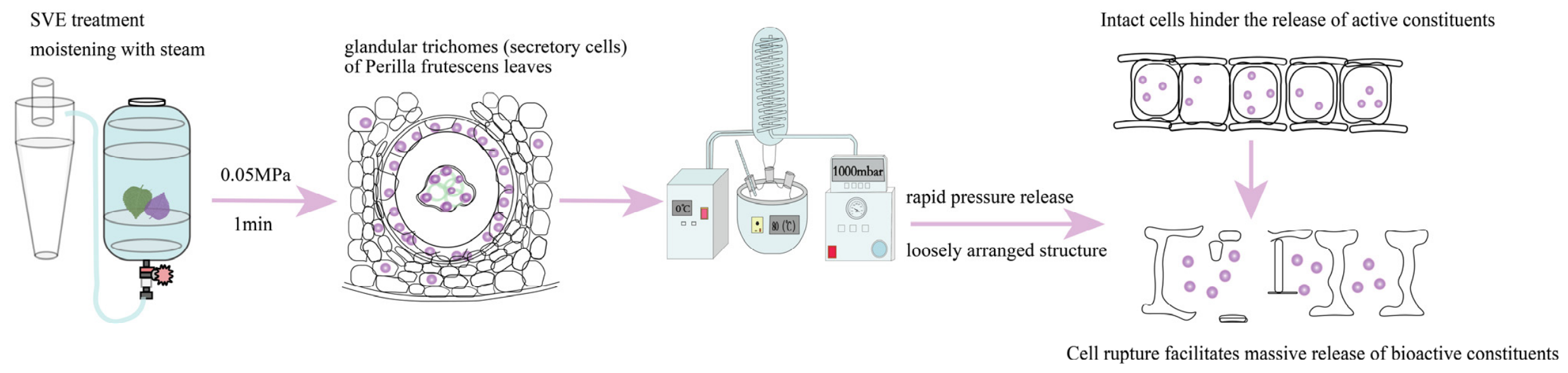
P. frutescens leaves (100 g) were loaded into the 5 L steam explosion chamber with a fill factor of 30% (Figure 1). The samples were treated with saturated steam at 0.05 MPa (gauge pressure) for a holding time of 1 min [18,19], with a heating ramp time of 90 s, followed by instantaneous full depressurization. After steam explosion pretreatment, the treated samples were dried to constant weight at 45 °C. Subsequently, 100 g of the dried treated material was immersed in 1000 mL of ultrapure water for 30 min and then extracted at 80 °C under a vacuum pressure of 0.05 MPa for 40 min. All analyte contents were calculated on a dry-weight basis.
Figure 1.
Schematic diagram of the SVE process principle.
2.3.2. Reflux Extraction (RE)
One hundred grams of untreated P. frutescens leaves were soaked in 1000 mL of ultrapure water for 30 min and then extracted under reflux at 100 ± 2 °C for 40 min.
2.3.3. Vacuum Extraction (VE)
Untreated P. frutescens leaves (100 g) were immersed in 1000 mL of ultrapure water for 30 min and subsequently extracted at 80 °C under a vacuum pressure of 0.05 MPa for 40 min.
2.4. Physicochemical Characterization of Extracts
2.4.1. pH Determination
The pH values of the extracts obtained by different extraction methods were measured using an E28 pH meter at room temperature. All measurements were performed in triplicate.
2.4.2. Particle Size and Zeta Potential Analysis
An appropriate volume of P. frutescens extract was diluted with ultrapure water. The particle size and zeta potential were determined using a Nano-S particle size analyzer (Malvern Instruments Ltd., Malvern, UK). All measurements were performed in triplicate.
2.4.3. Scanning Electron Microscopy (SEM)
The treated P. frutescens extraction residues were observed using a Regulus 8100 scanning electron microscope (Hitachi, Tokyo, Japan). The samples were sputter-coated with gold. Micro-morphological features were examined and photographed under an accelerating voltage of 15.0 kV at magnifications of 500×, 1000×, and 2000×. Representative images were selected according to consistent observations from multiple fields of view and independently prepared samples.
2.5. Determination of Major Bioactive Compounds in P. frutescens Extract by HPLC
2.5.1. Chromatographic Conditions
Quantitative analysis was performed using an Agilent 1260 high-performance liquid chromatography (HPLC, Agilent, Santa Clara, CA, USA) system equipped with an Agilent Eclipse Plus C18 column (250 mm × 4.6 mm, 5.0 μm).
The mobile phase consisted of acetonitrile (A) and 0.1% phosphoric acid aqueous solution (B). Gradient elution was performed as follows: 0–50 min, 12–19% A; 50–60 min, 19–22% A. The flow rate was maintained at 1.0 mL min−1. The column temperature was set at 30 °C, the detection wavelength was 325 nm, and the injection volume was 10 μL.
2.5.2. Quantification of Major Bioactive Compounds
The contents of caffeic acid, ferulic acid, rosmarinic acid, and scutellarin in the extracts were determined using the external standard method. Standard calibration curves were established using mixed reference solutions. All samples were filtered through a 0.22 μm membrane filter before HPLC analysis.
The contents of bioactive compounds were calculated based on the initial dry weight of P. frutescens leaves and expressed as mg/g dried leaves.
2.6. LC-MS/MS Analysis
2.6.1. Chromatographic and Mass Spectrometric Conditions
Comprehensive chemical profiling of P. frutescens extracts was performed using an ultra-performance liquid chromatography-Tandem Mass Spectrometry (LC-MS/MS, Thermo Fisher Scientific, Waltham, MA, USA) system.
The mobile phase consisted of 0.01% acetic acid aqueous solution (A) and isopropanol/acetonitrile (1:1, v/v) (B). Chromatographic separation was performed on a Phenomenex Kinetex C18 column (2.1 mm × 100 mm, 2.6 μm, Phenomenex, Torrance, CA USA). The column temperature was set at 25 °C, and the flow rate was kept constant at 0.30 mL/min. The gradient elution program was as follows: 0.00–1.00 min, 1% B; 1.00–8.00 min, increased to 99% B; 8.00–9.00 min, held at 99% B; 9.00–9.10 min, decreased to 1% B; 9.10–12.00 min, held at 1% B for column re-equilibration. The autosampler temperature was maintained at 4 °C and the injection volume was 2 μL.
The Orbitrap Exploris 120 mass spectrometer (Thermo Fisher Scientific, Waltham, MA, USA) was used for its ability to acquire MS/MS spectra on data-dependent acquisition (DDA) mode under the control of the acquisition software (Xcalibur, Thermo Fisher Scientific, version 4.4). The ESI source conditions were set as follows: sheath gas flow rate as 50 Arb, Aux gas flow rate as 15 Arb, capillary temperature 320 °C, full MS resolution as 60,000, MS/MS resolution as 15,000, collision energy: SNCE 20/30/40, spray voltage as 3.8 kV (positive) or −3.4 kV (negative), respectively.
Raw LC–MS data were processed using the XCMS package (version 3.20.0) for peak detection, retention time correction, peak alignment, and peak integration. Peak detection was performed using the centWave algorithm (ppm = 10, peakwidth = 5–30 s, snthresh = 3), and retention time correction was conducted using the obiwarp algorithm. Peak areas were normalized prior to statistical analysis. Quality control (QC) samples were prepared by pooling equal aliquots of all samples and were periodically injected throughout the analytical sequence to monitor the stability and reproducibility of the analytical system. Metabolite identification was performed based on accurate mass (MS1), MS/MS fragmentation spectra, and chromatographic retention time. Metabolite annotation was conducted using the Biotree platform by matching the acquired data against the Human Metabolome Database (HMDB), the PubChem database, and the Biotree in-house reference library. Annotation confidence was assigned according to the annotation strategy implemented in the Biotree platform, and the annotation level and MS2 score were used to evaluate the confidence of metabolite identification [20]. For metabolites with identical precursor ions or molecular formulas, metabolite annotation was further supported by characteristic MS/MS fragmentation patterns together with chromatographic retention behavior to improve the reliability of compound identification.
2.6.2. Sample Preparation
Extract solutions obtained from the SVE, VE, and RE groups were centrifuged at 12,000 rpm for 15 min at 4 °C. Subsequently, 300 μL of the supernatant was transferred to a centrifuge tube and mixed with 1000 μL of extraction solvent containing an internal standard (methanol/acetonitrile/water = 2:2:1, v/v/v). The mixture was vortexed for 30 s, sonicated in an ice-water bath for 5 min, and stored at −20 °C for 1 h to precipitate proteins.
The samples were then centrifuged again at 12,000 rpm for 15 min at 4 °C. The supernatants were collected and filtered through a 0.22 μm membrane prior to LC-MS/MS analysis.
2.7. Multivariate Statistical Analysis
Principal component analysis (PCA) and orthogonal partial least squares-discriminant analysis (OPLS-DA) were employed to evaluate differences in chemical composition among extracts obtained by different extraction methods. Differential metabolites were screened according to the criteria of variable importance in projection (VIP) > 1 and p < 0.05.
2.8. Statistical Analysis
All experiments were performed using three independent extraction samples for each group. The results are expressed as mean ± standard deviation (SD). Here, n = 3 represents three independent extraction replicates rather than technical replicates. Statistical analyses were conducted using SPSS 26.0 software (IBM Corp., Armonk, NY, USA). Significant differences among groups were evaluated using one-way analysis of variance (ANOVA), and p < 0.05 was considered statistically significant.
3. Results
3.1. Effects of Different Extraction Methods on pH, Zeta Potential and Particle Size
As shown in Table 1, the pH values of extracts obtained by different extraction methods ranged from 5 to 6 within the weak-acid region. Statistically significant differences in pH were observed among groups, while the absolute variation was relatively small. The particle size and zeta potential of extracts exhibited obvious differences across treatments. The average particle sizes of VE and SVE extracts were 376.10 nm and 386.23 nm, respectively, both markedly lower than that of the RE extract. The zeta-potential values of all groups were approximately −8 to −9 mV.
Table 1.
Physical properties of P. frutescens extract by different extraction methods (mean ± SD, n = 3).
3.2. SEM Analysis
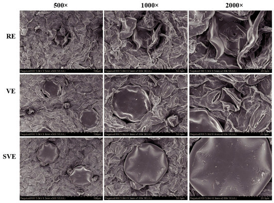
SEM observations suggested that the SVE-treated samples exhibited a relatively rougher surface and more visible surface pores than the RE group, indicating that the SVE pretreatment step appears to alter the microstructure of the plant tissues. These structural features may improve solvent accessibility; however, their contribution to mass transfer was not directly quantified in the present study. SEM observations revealed distinct microstructural differences among the extraction treatments (Figure 2). The RE-treated samples exhibited surface wrinkling and localized fissures, whereas the VE-treated samples retained a relatively smoother tissue morphology. Compared with both RE-treated samples, the SVE-treated samples exhibited a rougher surface with more visible pores and irregular structural features, suggesting that the SVE pretreatment step may induce noticeable microstructural alterations in the plant tissues. The observed structural differences suggest that steam explosion modified the tissue architecture of P. frutescens compared with RE.
Figure 2.
SEM images of extraction residues from P. frutescens treated with different extraction methods (RE, VE, SVE) (magnification: 500×, 1000×, 2000×).
3.3. Quantitative Analysis of Major Bioactive Compounds by HPLC
Significant differences in the contents of major bioactive compounds were observed among the three extraction methods (Table 2, Tables S1–S5 in Supplementary Materials). Compared with the RE group, SVE treatment markedly increased the concentrations of ferulic acid, scutellarin, and rosmarinic acid. Among these compounds, rosmarinic acid exhibited the most pronounced increase, rising from 0.614 mg/g to 2.386 mg/g, representing a 3.89-fold enhancement. Furthermore, the total content of the four quantified bioactive compounds increased from 4.574 mg/g to 7.521 mg/g, corresponding to an approximately 1.64-fold increase.
Table 2.
Contents of four components in P. frutescens extracts obtained by different extraction methods (mean ± SD, n = 3).
Rosmarinic acid is one of the principal phenolic acids in P. frutescens and serves as an important contributor to its anti-inflammatory activities. The increased level of rosmarinic acid suggests that the SVE pretreatment step may contribute to the enhanced recovery of phenylpropanoid-derived phenolic compounds during extraction. Similarly, the increased abundance of scutellarin and other flavonoid glycosides indicates that the porous tissue structure generated during SVE pretreatment may facilitate the extraction of cell-wall-associated flavonoids.
Importantly, the integration of steam explosion pretreatment with vacuum extraction may contribute to both improved accessibility of plant tissues and the preservation of heat-sensitive constituents under relatively mild extraction conditions. Therefore, the enhanced enrichment of bioactive compounds observed in the SVE group may be associated with improved mass transfer resulting from structural modification, together with the potential protective effect of reduced-temperature extraction conditions.
3.4. Quality Control Assessment of LC–MS/MS Analysis
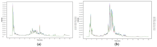
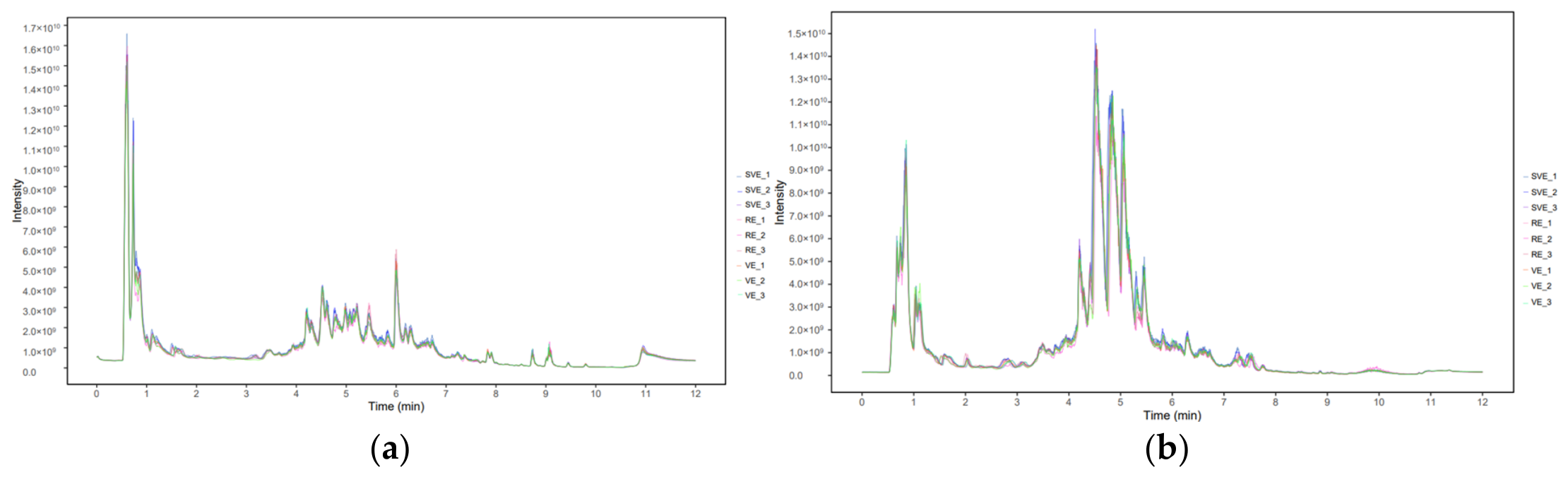
To evaluate the stability of the analytical platform and the reliability of the metabolomic data, equal aliquots of the SVE, RE, and VE extracts were pooled to prepare quality control (QC) samples. The QC samples were analyzed by LC–MS/MS, and the total ion chromatograms (TICs) acquired in both positive and negative ion modes are presented in Figure 3 (Figures S1–S6 in Supplementary Materials). The overlaid TICs of the QC samples exhibited a high degree of overlap under both ionization modes, with consistent retention times, chromatographic peak shapes, and signal intensities throughout the analytical sequence. The major metabolites were primarily eluted between 4 and 6 min, where repeated injections showed stable peak profiles and comparable signal responses without obvious chromatographic drift. These results indicate that the LC–MS/MS analytical system exhibited good instrumental stability and analytical reproducibility, providing reliable data quality for subsequent multivariate statistical analysis and differential metabolite screening.
Figure 3.
TIC plots of P. frutescens samples under positive and negative modes ((a) Positive ions; (b) Negative ions).
3.5. OPLS-DA Analysis
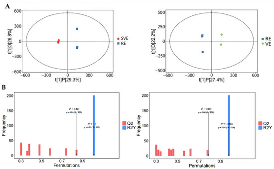
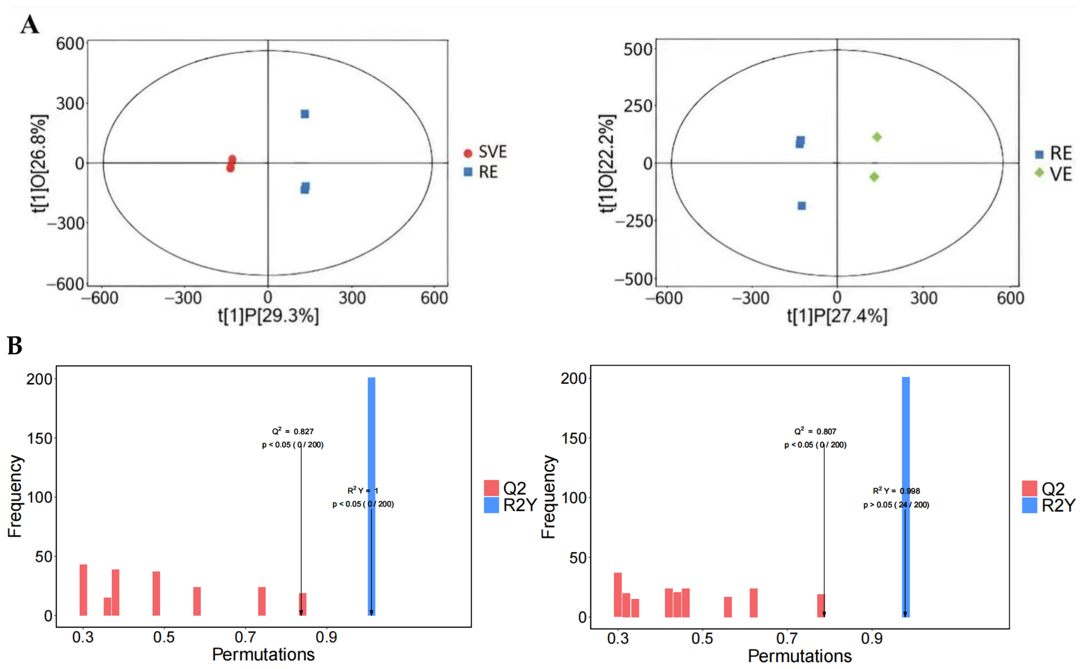
To further characterize the compositional differences among extraction methods and assess model robustness, OPLS-DA models were established for the SVE–RE and VE–RE comparison groups. As shown in Figure 4A, both models exhibited clear group separation, indicating pronounced differences in chemical composition among the extracts.
Figure 4.
OPLS-DA analysis and model validation of P. frutescens extracts obtained by different extraction methods. (A) OPLS-DA score plots; (B) results of 200 permutation tests.
The SVE–RE model yielded R2Y and Q2 values of 0.998 and 0.827, respectively, while the VE–RE model exhibited corresponding values of 1.000 and 0.807. Furthermore, the 200-permutation tests confirmed that the original models possessed significantly higher R2Y and Q2 values than the randomized models (p < 0.05), suggesting that the established models showed acceptable predictive performance without obvious evidence of overfitting.
Collectively, these findings confirm that different extraction strategies lead to signif icant and reproducible variations in the chemical composition of P. frutescens extracts, with SVE exerting the most pronounced influence on metabolite profiles.
3.6. Screening and Identification of Differential Chemical Constituents
Chemical constituent profiling showed that SVE predominantly enriched hydroxycinnamic acids, flavonoids and isoflavonoids relative to RE. The upregulated compounds included the phenylpropanoid pathway metabolite 4-O-p-coumaroylquinic acid, flavonoid glycosides (vicenin II, tiliroside, 7,3′,4′-trimethoxyquercetin), and the isoflavonoid 5,7,3′,4′-tetrahydroxy-6,8-diprenylisoflavone. Previous studies have reported that these compounds are associated with various biological activities, including anti-inflammatory effects.
Compared with VE, SVE significantly increased the contents of lipophilic bioactive components, including fatty acids (linoleic acid, 16-hydroxypalmitic acid, 2,4-dihydroxybutanoic acid) and the triterpenoid ursolic acid. Existing studies have demonstrated that these lipophilic metabolites possess multiple biological functions, including antibacterial and lipid metabolism-modulating properties.
Taken together, these results suggest that the integrated SVE process not only appears to facilitate the release of hydrophilic phenolic acids and flavonoids but also enhances the extraction of lipophilic fatty acids and triterpenoids. Therefore, SVE demonstrates a broad enhancement of extraction efficiency across multiple compound classes and is particularly advantageous for the simultaneous recovery of multiple classes of bioactive compounds from P. frutescens.
4. Discussion
This study compared the integrated steam explosion–vacuum extraction (SVE) with conventional reflux extraction (RE) and single vacuum extraction (VE) to efficiently obtain bioactive substances from P. frutescens leaves. Through microscopic structural observation, quantitative analysis of bioactive components, untargeted metabolomics and physicochemical characterization, the combined effects of the integrated SVE process were characterized. The mechanisms associated with improved extraction efficiency and altered colloidal properties of SVE extracts are described as follows.
SEM observation (Figure 2) showed that SVE-treated P. frutescens residues exhibited a rougher surface with abundant visible pores, in sharp contrast to the wrinkled surface and localized fissures in the RE group. Such pore-dominated microstructural changes are typical outcomes of rapid depressurization and mechanical shear inherent to steam explosion, consistent with documented effects of steam explosion on lignocellulosic and other plant materials [17,18]. Previous studies have verified that steam explosion disrupts the cellulose network of cell walls and increases tissue porosity, thereby expanding the contact interface between solvent and matrix, shortening diffusion pathways and promoting the outward release of intracellular components [15,16]. A large number of phenolic acids and flavonoids in plant tissues bind to cell-wall matrices or are stored in subcellular compartments, making their liberation difficult under conventional extraction systems. Therefore, the structural remodeling observed in this work offers a potential mechanistic explanation for the elevated yields of these bioactive analytes quantified by HPLC (Table 2). Complementary to this physical release effect, vacuum extraction creates mild thermal environments under reduced pressure to reduce thermal degradation of heat-sensitive compounds such as rosmarinic acid [13]. The improved recovery of bioactive components under SVE may be linked to a combination of tissue structural modification, enhanced mass transfer and thermal protection of thermolabile metabolites. It should be noted, however, that the SEM observation in this study is only qualitative; quantitative characterization of pore size and porosity will help establish a clearer correlation between structural modification and extraction efficiency.
Benefiting from tissue disruption and thermoprotection offered by the integrated SVE technique, the extraction recovery of bioactive components in P. frutescens extracts was markedly improved. As shown in Table 2, SVE treatment greatly enhanced the recovery of characteristic bioactive compounds from P. frutescens, including rosmarinic acid, ferulic acid, scutellarin and the total content of four quantified analytes. This enrichment pattern was further validated by untargeted metabolomic analysis (Table 3 and Table 4). Compared with RE, SVE increased the contents of a series of phenylpropanoid metabolites, including hydroxycinnamic acids, flavonoid glycosides and isoflavonoids. These results demonstrate that the integrated SVE strategy delivers dual advantages: steam-explosion-triggered tissue fragmentation facilitates the release of hydrophilic and lipophilic compounds embedded in plant matrices, while mild thermal conditions during vacuum extraction preserve thermolabile phenolics. Existing reports on steam explosion pretreatment of medicinal herbs support our findings, confirming that combining physical tissue fragmentation with mild low-temperature extraction broadens the scope of retrievable bioactive constituents from medicinal-food homologous plants.
Table 3.
Differences in chemical composition between the SVE and RE comparison groups.
Table 4.
Differences in chemical composition between the SVE and VE comparison groups.
In addition, different extraction approaches substantially altered the physicochemical and colloidal characteristics of final extracts. Although statistically significant intergroup differences in pH were detected, their absolute variations were minor, indicating that extraction methods only exert limited practical influence on extract acidity. Particle size and zeta-potential results reveal that extraction conditions can shape the physicochemical features of extracts. VE and SVE extracts possessed comparable particle sizes, both significantly smaller than those of RE extracts, suggesting that vacuum extraction conditions, rather than steam explosion pretreatment, dominate the particle-size characteristics of final extracts. This is most likely because the reduced-pressure mild mass-transfer environment favors the formation of fine and uniformly dispersed particles. The absolute zeta-potential values of all groups were below 10 mV (approximately −8 to −9 mV), indicating weak electrostatic repulsion within the system. According to the DLVO theory, surface charges of this magnitude cannot maintain colloidal stability relying solely on electrostatic interactions; hence, zeta potential cannot be used alone as an indicator for judging dispersion stability. Moreover, the colloidal behavior of plant extracts is co-regulated by multiple factors, including steric hindrance, soluble macromolecules (e.g., polyphenols and polysaccharides), and ionic strength. Accordingly, the particle-size and zeta-potential data from this study only demonstrate that different extraction protocols modify the physical properties of the dispersed phase and should not be directly extrapolated to storage stability or dispersion performance under practical conditions. Further accelerated storage tests and sedimentation observations are required to more reliably evaluate the colloidal properties of P. frutescens extracts.
Unsupervised PCA models were built using LC–MS/MS datasets to preliminarily evaluate the effects of extraction methods on the chemical composition of P. frutescens extracts. The PCA score plot revealed clear separation between SVE and RE, as well as between VE and RE samples. The first two principal components explained 53.1% (SVE–RE) and 56.7% (VE–RE) of the total variance (Figure S23 in Supplementary Materials), consistent with the reported range (45–60%) for untargeted metabolomic studies of herbal plants, demonstrating that the PCA models reflect global metabolic differences. Tight clustering within each group indicates good reproducibility. PCA loadings show that phenolic acids, flavonoids and terpenoids are the major compounds responsible for sample separation: flavonoid derivatives dominate separation along PC1, while phenolic acids contribute most to the variance along PC2 (Figures S10–S17, S19 and S20 in Supplementary Materials). The distinct separation among groups indicates that extraction methods alter the overall metabolic profiles of P. frutescens extracts.
Based on the OPLS-DA classification results in Figure 4 (Figures S7–S9, S18, S21 and S22 in Supplementary Materials), VIP values were calculated to assess the contribution of individual metabolites to group discrimination. Most differential metabolites from SVE–RE and VE–RE comparisons had VIP > 1. Metabolites satisfying both VIP > 1 and the univariate threshold of p < 0.05 were retained for annotation and subsequent comparative analysis.
Differences in metabolite abundance among SVE, VE and RE extracts should be attributed to distinct extraction behaviors rather than intrinsic metabolic reprogramming of plant tissues. As discussed above, structural modification occurs during SVE pretreatment, and the subsequent vacuum-extraction step may confer thermoprotective effects. Metabolomic results further corroborate that the integrated SVE workflow reshapes extract metabolic profiles by modulating compound release and stability during extraction, instead of altering the plant’s inherent biosynthetic pathways.
Compared with other emerging extraction techniques, SVE represents an alternative strategy coupling physical tissue modification with mild extraction conditions. Ultrasound-assisted extraction improves mass transfer primarily via cavitation effects, while microwave-assisted extraction depends on volumetric heating, which carries risks of localized overheating for thermolabile bioactive constituents. By contrast, SVE physically disrupts plant tissue architecture under reduced-pressure and low-temperature conditions, promoting compound release while preserving heat-sensitive components. This concept aligns with published reviews highlighting that combined extraction strategies can achieve synergistic improvements in extraction efficiency [21,22,23,24]. Even so, head-to-head comparative experiments under identical processing parameters are still needed to fully characterize the strengths and drawbacks of SVE relative to ultrasound-, microwave-assisted and other innovative extraction techniques, which is essential for supporting its industrial translation.
From an application standpoint, SVE shows promising prospects for processing medicinal-food homologous plants abundant in thermolabile constituents. However, steam-explosion pretreatment raises upfront equipment investment. Its practical industrial merit needs to be further verified through systematic evaluation of process economics, energy consumption, environmental footprint and large-scale operational performance. Several limitations of this study should be noted. First, only a single steam-explosion condition was tested, and no steam-explosion-only group (without subsequent vacuum extraction) was set up; accordingly, the respective contributions of steam explosion and vacuum extraction, as well as optimal process parameters, remain undefined. Second, SEM analysis was largely qualitative, and quantitative image analysis would offer more robust evidence for tissue structural alterations. Third, the metabolomic analysis relied on three independent extraction replicates per group without separate validation datasets. Future research should prioritize steam-explosion parameter optimization, quantitative structural characterization, independent metabolomic validation, and comprehensive assessment of economic performance and scale-up feasibility.
5. Conclusions
This study demonstrates that steam explosion-assisted vacuum extraction (SVE) is an effective strategy to improve the recovery of bioactive compounds from P. frutescens. Comparative analysis among SVE, VE and RE reveals that the elevated contents of phenolic acids, flavonoids and key differential metabolites are associated with the combined outcomes of SVE pretreatment-induced tissue structural modification and the mild thermal environment offered by vacuum extraction within this integrated workflow. Within the SVE workflow, pretreatment-related structural changes may improve solvent accessibility, while vacuum-extraction conditions may help limit thermal degradation of thermolabile bioactive constituents. These coupled phenomena may collectively contribute to the superior extraction performance observed for the integrated SVE approach.
These findings advance our mechanistic understanding of combined pretreatment-extraction technologies for medicinal-food homologous plants. The SVE-derived P. frutescens extracts rich in phenolic bioactives show promising potential for functional food ingredients, natural antioxidant additives, and herbal pharmaceutical raw materials. Nevertheless, industrial translation still faces challenges regarding equipment cost, energy consumption and scale-up feasibility. This work provides a fundamental reference for developing efficient and gentle extraction processes for aromatic medicinal plant resources.
Supplementary Materials
The following supporting information can be downloaded at: https://www.mdpi.com/article/10.3390/separations13090268/s1, Figure S1: Overlaid total ion chromatograms (TIC) of RE samples under negative ionization mode; Figure S2: Overlaid total ion chromatograms (TIC) of VE samples under negative ionization mode; Figure S3: Overlaid total ion chromatograms (TIC) of SVE samples under negative ionization mode; Figure S4: Overlaid total ion chromatograms (TIC) of RE samples under positive ionization mode; Figure S5: Overlaid total ion chromatograms (TIC) of VE samples under positive ionization mode; Figure S6: Overlaid total ion chromatograms (TIC) of SVE samples under positive ionization mode; Figure S7: VIP plot of differential metabolites from the OPLS-DA model for RE vs. VE comparison; Figure S8: VIP plot of differential metabolites from the OPLS-DA model for VE vs. SVE comparison; Figure S9: VIP plot of differential metabolites from the OPLS-DA model for RE vs. SVE comparison; Figure S10: PCA biplot of RE and VE sample groups; Figure S11: PCA biplot of VE and SVE sample groups; Figure S12: PCA biplot of RE and SVE sample groups; Figure S13: PCA biplot of RE and VE groups; Figure S14: PCA biplot of VE and SVE groups; Figure S15: PCA biplot of RE and SVE groups; Figure S16: Permutation plot of the OPLS-DA model (RE vs. VE); Figure S17: Z-score distribution plot of differential metabolites between RE and VE groups; Figure S18: Volcano plot of metabolites between RE and VE groups; Figure S19: Permutation plot of the OPLS-DA model (RE vs. SVE); Figure S20: Z-score distribution plot of differential metabolites between RE and SVE groups; Figure S21: Volcano plot of metabolites between RE and SVE groups; Figure S22: OPLS-DA Variable importance in projection (VIP) plots derived from the OPLS-DA models; Figure S23: PCA score plots of LC–MS/MS data from perilla extracts obtained by different extraction methods. (a) SVE vs. RE; (b) VE vs. RE; Table S1: Validation of the HPLC method for determination of four bioactive compounds; Table S2: Precision of the HPLC method (n = 6); Table S3: Repeatability (n = 6); Table S4: Stability (24 h); Table S5: Recovery test (n = 3).
Author Contributions
Conceptualization, B.Z.; methodology, B.Z.; Writing—original draft, B.Z.; investigation, B.Z. and H.Z.; formal analysis, Q.W.; data curation, Q.W. and H.Z.; supervision, Y.W.; project administration, Y.W.; funding acquisition, Y.W.; Writing—review & editing, Y.W. All authors have read and agreed to the published version of the manuscript.
Funding
This research was funded by the Natural Science Foundation of Jiangxi Province, grant number 20232BAB206168.
Institutional Review Board Statement
Not applicable.
Informed Consent Statement
Not applicable.
Data Availability Statement
The original contributions presented in this study are included in the article/Supplementary Material. Further inquiries can be directed to the corresponding author.
Acknowledgments
The authors thank the staff of the Jiangxi University of Traditional Chinese Medicine for their administrative and technical support during the experiments. The authors also appreciate Z.F. Wu for resource provision, T. Yao for visualization work, and M. Yang for manuscript review-and-editing.
Conflicts of Interest
The authors declare no conflicts of interest.
Abbreviations
The following abbreviations are used in this manuscript:
| SVE | Steam explosion-assisted Vacuum Extraction |
| VE | Vacuum Extraction |
| RE | Reflux Extraction |
| SEM | Scanning Electron Microscopy |
| HPLC | High Performance Liquid Chromatography |
| LC-MS/MS | Liquid Chromatography-Tandem Mass Spectrometry |
| PCA | Principal Component Analysis |
| OPLS-DA | Orthogonal Partial Least Squares Discriminant Analysis |
References
- Wu, X.L.; Dong, S.T.; Chen, H.Y.; Guo, M.X.; Sun, Z.Y.; Luo, H.M. Perilla frutescens: A traditional medicine and food homologous plant. Chin. Herb. Med. 2023, 15, 369–375. [Google Scholar] [CrossRef] [Scilit] [PubMed]
- Kang, Q.L.; Li, Z.Z.; Fan, S.S.; Rong, R.; Jiang, H.Q.; Jiang, X.L.; Zhang, X.Q.; Gong, L.L. Chemical constituents analysis of Perilla e Folium and Perilla e Caulis based on UPLC-Q-Exactive-Orbitrap-MS. Chin. J. Exp. Tradit. Med. Formulae 2020, 26, 156–162. [Google Scholar] [CrossRef]
- Ahmed, H.M. Ethnomedicinal, phytochemical and pharmacological investigations of Perilla frutescens (L.) Britt. Molecules 2019, 24, 102. [Google Scholar] [CrossRef] [Scilit] [PubMed]
- Zhang, D.Y.; Zhou, W.C.; Shen, A.; Chen, Y.Z.; Mu, H.Y.; Wang, S.X.; Wei, J.T. Analysis of active constituents in Perilla frutescens leaves from different origins. Food Res. Dev. 2025, 46, 142–147. [Google Scholar]
- Xu, Y.; He, D.X.; Zhao, L.P.; Zhang, T.T.; Diao, T.T.; Cai, J.; Huang, Y.Q.; Bi, J.J.; Chen, Q.; Zhang, Q.Y.; et al. Optimization of extraction process and antioxidant activity analysis of polysaccharides from Perilla frutescens leaves. Chin. Agric. Sci. Bull. 2025, 27, 170–179. [Google Scholar]
- Yang, H.; Ma, P.; Lin, M.B.; Hou, Q. Research progress on chemical constituents, anti-inflammatory effects and mechanisms of Perilla frutescens leaves. Chin. J. Pharmacol. Toxicol. 2017, 31, 279–286. [Google Scholar]
- Huang, S.Z.; Liu, Y.F.; Wang, L.; Ni, M.M.; Li, X.M.; Zhang, L.S.; Chen, J.Y. Effects of egg yolk phospholipids combined with perilla seed oil on maintaining healthy blood lipid levels in rats. China Oils Fats 2024, 49, 56–61. [Google Scholar]
- Hu, T.T.; Ye, M.Y.; Zhu, J.; Yang, M.; Wang, Y.Q. Research progress on heat-clearing and detoxifying medicines in treating pulmonary inflammatory diseases based on the lung–gut axis. Lishizhen Med. Mater. Med. Res. 2024, 35, 173–177. [Google Scholar]
- Liu, R.Q.; Li, Y.Q.; Xiao, S.H.; Zheng, Q.; Zhong, Y.; Hu, G.T.; Hu, P.Y. Research progress on antidepressant effects and mechanisms of qi-regulating traditional Chinese medicines. Chin. Tradit. Herb. Drugs 2024, 55, 3558–3568. [Google Scholar]
- Li, Y.; Zhang, Y.; Wang, Y.Y.; Li, X.; Zhou, L.; Yang, J.; Guo, L.P. Metabolites and chemometric study of Perilla (Perilla frutescens) from different varieties and geographical origins. J. Food Sci. 2022, 87, 5240–5251. [Google Scholar] [CrossRef] [Scilit] [PubMed]
- Ye, X.S.; Tian, W.J.; Wang, G.H.; Lin, K.; Zhu, S.X.; Xia, Y.Y.; Sun, B.L.; Shu, X.J.; Liu, W.; Chen, H.F. The food and medicine homologous Chinese Medicine from Leguminosae species: A comprehensive review on bioactive constituents with neuroprotective effects on nervous system. Food Med. Homol. 2025, 2, 9420033. [Google Scholar] [CrossRef] [Scilit]
- Zhang, S.S.; Hua, L.P.; Chen, J.T.; Ding, M.H.; Shi, S.L. Optimization of volatile oil extraction process, composition and antioxidant activity of aromatic traditional Chinese medicine prescriptions. Chin. Tradit. Pat. Med. 2023, 45, 1261–1264. [Google Scholar]
- Wang, Y.Q.; Wu, Z.F.; Ke, G.; Yang, M. An effective vacuum assisted extraction method for the optimization of labdane diterpenoids from Andrographis paniculata by response surface methodology. Molecules 2015, 20, 430–445. [Google Scholar] [CrossRef] [Scilit] [PubMed]
- Zhang, H.N.; Liu, Y.; Zhang, H.L.; Wan, Q.Z.; Wang, Y.Q. Effect of different drying and grinding techniques on the physicochemical properties and biological activities of fungal polysaccharides. Food Med. Homol. 2025, 2, 9420045. [Google Scholar] [CrossRef] [Scilit]
- Wan, F.C.; Feng, C.F.; Luo, K.Y.; Cui, W.Y.; Xia, Z.H.; Cheng, A.W. Effect of steam explosion on phenolics and antioxidant activity in plants: A review. Trends Food Sci. Technol. 2022, 124, 13–24. [Google Scholar] [CrossRef] [Scilit]
- Xue, X.Y.; Qi, Z.W.; Zhang, C.W.; Kong, X.H.; Lei, J.D.; Zhou, H.; Wang, C.Z. Effects of Steam Explosion Pretreatment on the Physicochemical Properties, Phenolic Profile and Its Antioxidant Activities of Baphicacanthus cusia (Nees) Bremek. Leaves. LWT 2024, 214, 117126. [Google Scholar] [CrossRef] [Scilit]
- Jacquet, N.; Maniet, G.; Vanderghem, C.; Delvigne, F.; Richel, A. Application of steam explosion as pretreatment on lignocellulosic material: A review. Ind. Eng. Chem. Res. 2015, 54, 2593–2598. [Google Scholar] [CrossRef] [Scilit]
- Wang, D.L.; Liu, Z.B.; Su, W.Y.; Lu, Y.; Feng, X.X.; Liu, Y.; Lin, J.S.; Wen, B.; Xiao, M.J.; Ni, L. Improving the utilization of summer tea leaves through steam explosion technology: Flavor transformation and cold-brewing feasibility. Food Chem. 2026, 498, 147224. [Google Scholar] [CrossRef] [Scilit] [PubMed]
- Zeng, X.F.; Ge, Z.X.; Yan, M. Effect of Steam Explosion Pretreatment on the Quality and Flavor of Pressed Perilla Seed Oil. Food Sci. 2025, 46, 257–263. [Google Scholar] [CrossRef]
- Zhou, Z.W.; Luo, M.D.; Zhang, H.S.; Yin, Y.D.; Cai, Y.P.; Zhu, Z.J. Metabolite Annotation from Knowns to Unknowns through Knowledge-Guided Multi-Layer Metabolic Networking. Nat. Commun. 2022, 13, 6656. [Google Scholar] [CrossRef] [Scilit] [PubMed]
- Belwal, T.; Ezzat, S.M.; Rastrelli, L.; Bhatt, I.D.; Daglia, M.; Baldi, A.; Devkota, H.P.; Orhan, I.E.; Patra, J.K.; Das, G.; et al. A Critical Analysis of Extraction Techniques Used for Botanicals: Trends, Priorities, Industrial Uses and Optimization Strategies. TrAC Trends Anal. Chem. 2018, 100, 82–102. [Google Scholar] [CrossRef] [Scilit]
- Azmir, J.; Zaidul, I.S.M.; Rahman, M.M.; Sharif, K.M.; Mohamed, A.; Sahena, F.; Jahurul, M.H.A.; Ghafoor, K.; Norulaini, N.A.N.; Omar, A.K.M. Techniques for Extraction of Bioactive Compounds from Plant Materials: A Review. J. Food Eng. 2013, 117, 426–436. [Google Scholar] [CrossRef] [Scilit]
- Mustafa, A.; Turner, C. Pressurized Liquid Extraction as a Green Approach in Food and Herbal Plants Extraction: A Review. Anal. Chim. Acta 2011, 703, 8–18. [Google Scholar] [CrossRef] [Scilit] [PubMed]
- Lefebvre, T.; Destandau, E.; Lesellier, E. Selective Extraction of Bioactive Compounds from Plants Using Recent Extraction Techniques: A Review. J. Chromatogr. A 2021, 1635, 461770. [Google Scholar] [CrossRef] [Scilit] [PubMed]

Disclaimer/Publisher’s Note: The statements, opinions and data contained in all publications are solely those of the individual author(s) and contributor(s) and not of MDPI and/or the editor(s). MDPI and/or the editor(s) disclaim responsibility for any injury to people or property resulting from any ideas, methods, instructions or products referred to in the content. |
© 2026 by the authors. Licensee MDPI, Basel, Switzerland. This article is an open access article distributed under the terms and conditions of the Creative Commons Attribution (CC BY) license.