Skip to Content
  • Article
  • Open Access

28 May 2026

Phytochemical Profiling and Bioactivity Correlation of Acacia nilotica and Acacia seyal Using HPLC-DAD Analysis


Department of Chemistry, College of Science, Qassim University, Buraydah 51452, Saudi Arabia

Abstract

This study provides a comprehensive phytochemical and biological evaluation of Acacia nilotica and Acacia seyal samples collected from Khartoum State, Sudan. Methanolic extracts of A. nilotica leaves (SN), A. seyal leaves (TL), and A. nilotica seeds (S) were evaluated for total phenolic content (TPC), total flavonoid content (TFC), antioxidant activity (DPPH radical scavenging and reducing power), and antimicrobial activity against selected pathogenic microorganisms. High-performance liquid chromatography coupled with diode array detection (HPLC-DAD) was used to identify and quantify individual phenolic compounds. The SN sample exhibited the highest TPC (287.527 mg GAE/g), DPPH scavenging activity (96.815%), and reducing power (2.356), whereas TL showed the highest flavonoid content (155.296 mg QE/g). All samples demonstrated considerable antifungal activity against Candida albicans, while antibacterial activity varied according to species and extract type. HPLC-DAD analysis revealed the presence of major phenolic compounds, including naringenin, gallic acid, catechin, and methyl gallate, with notable variations among the investigated samples. In addition, several unidentified peaks were observed in the chromatographic profiles, suggesting the presence of additional phytochemical constituents under the applied analytical conditions. A strong positive correlation was observed between total phenolic content and antioxidant activity (R2 > 0.97), indicating the possible contribution of phenolic compounds to the observed bioactivity. Overall, this study provides further insight into the relationship between phenolic composition and biological activity in Acacia species and highlights their potential as natural sources of bioactive compounds for pharmaceutical and food applications.

1. Introduction

The Leguminosae family’s Acacia genus includes over 1350 species that span a wide range of ecological amplitudes [1]. The taxonomy of Acacia has undergone several phylogenetic revisions over time due to the morphological and genetic diversity within the group. Recent classifications recognize multiple related genera, including Acacia, Senegalia, and Vachellia, particularly for African species previously classified under Acacia sensu lato [2,3]. Scientific studies have thoroughly evaluated numerous species, highlighting their significant importance in the field of ethnopharmacology. For instance, researchers have deemed preparations from numerous species beneficial for external application in the treatment of scabies, wounds, injuries, and sores [4]. For centuries, people have used them as ethnomedicinal remedies for skin, sexual, gastrointestinal, and tooth issues. Currently, numerous herbal products, including toothpaste and conditioners, are available in the market, either in their entirety or in combination with other ingredients [5]. Additionally, people typically use the bark, leaves, pods, gums, roots, and flowers of Acacia species as a decoction, infusion, paste, or poultice. Folk medicine has used these parts as an astringent, emollient, stimulant, antidote to poisonous bites and stings, and as a remedy for diarrhea and dysentery [6].
Several scientific reports [7,8,9] have confirmed that many species of the genus Acacia display important pharmacological actions such as anti-inflammatory, hepatoprotective, and antioxidant activities, thereby demonstrating their beneficial effects on human health. Additionally, studies have found that they act as effective anticancer agents, with certain species demonstrating significant cytoselectivity against cancer cells [10,11,12].
The wide applications of Acacia species in the treatment and management of numerous ailments have prompted many researchers worldwide to characterize the chemical compounds in these plants, in addition to elucidating their pharmacological potential. Flavonoids, phenolic acids, and terpenoids, primarily found in leaves, pods, and stem barks, are the main compounds isolated from Acacia species. These compounds have demonstrated multiple therapeutic activities, including antifungal, antibacterial, antioxidant, anticancer, antiparasitic, cytotoxic, and immunomodulatory properties [13]. Moreover, Acacia species are a rich source of some glycosides, such as cyanogenic glycosides, which have strong antioxidant activity [7]. Among the various Acacia species, Acacia nilotica and Acacia seyal are widely recognized for their phytochemical richness and biological activities. The species Acacia nilotica extends extensively throughout subtropical and tropical Africa, from Egypt to Mauritania and southward to South Africa [14,15,16]. The plant has a lot of useful secondary substances, involving ascorbic acid, carotene, terpenes, phenolic glycosides, volatile essential oils, gallic acid, isoquercetin, calcium, magnesium, selenium, and extras [13,17]. Ali et al. [18] characterize Nilotica as a medicinal and pharmaceutical plant that serves various purposes. Traditional medicine employs A. nilotica to treat a variety of ailments, including tuberculosis, pneumonia, gonorrhea, and smallpox. A. nilotica exhibited potent antimicrobial properties against both bacteria and fungi [5]. The methanolic extract of A. nilotica leaves and the ethanolic extract of stem bark against Gram-positive and Gram-negative microorganisms were tested. The extracts exhibited antimicrobial activity against both categories of bacteria, as indicated by the results [19,20]. A. nilotica leaves’ ethanolic extract demonstrated antimicrobial activity against goat-isolated Campylobacter coli [21]. Saini et al. [5] investigated the antimicrobial activity of five Acacia species. The findings suggested that A. nilotica exhibited the most potent antifungal activity against Candida albicans and Aspergillus niger. Mahesh and Satish [19] say that the methanolic leaf extract of A. nilotica was very good at killing Aspergillus flavus, Drechslera turcica, and Fusarium verticillioides. A. nilotica bark extract mitigates liver toxicity and prevents the formation of hepatic malondialdehyde [22]. We have assessed the antihypertensive and antispasmodic properties of A. nilotica legumes. The methanolic extract of A. nilotica inhibited the spontaneous contraction of rabbit jejunum [23].
Tropical Africa is almost exclusively home to the Acacia seyal tree. The bark of A. seyal was extracted with ethanol and showed antimycobacterial and cyclooxygenase inhibition [24], antibacterial [25], antimalarial [26], and anticancer properties [27]. The examination of specialized metabolites in the leaves, seeds, and flowers of A. seyal uncovered a substantial quantity of saponosides and flavonoids [7].
Recent studies, including Ahmed et al. [28], have investigated the phenolic composition and biological activities of different parts of Acacia nilotica using HPLC-DAD analysis. However, comparative studies involving different Acacia species and plant parts remain limited. Therefore, the present study aimed to comparatively investigate the phenolic composition of Acacia nilotica seeds and leaves and Acacia seyal leaves using HPLC-DAD analysis, in addition to evaluating their antioxidant and antimicrobial activities. This approach provides further insight into species- and tissue-related phytochemical variation and its possible association with biological activity. HPLC-DAD profiling was combined with bioactivity assays to better understand the relationship between phenolic composition and antioxidant and antimicrobial effects. Furthermore, several unidentified peaks were observed in the chromatographic profiles, suggesting the presence of additional phytochemical constituents under the applied analytical conditions. Further characterization studies may be required to identify these compounds and evaluate their possible biological significance.

2. Materials and Methods

All solvents used for extraction and HPLC analysis were of HPLC grade and obtained from Fisher Scientific (Waltham, MA, USA).

2.1. Samples

Dry leaves and seeds of Acacia nilotica and leaves of Acacia seyal were collected from the Sunut forest (Mogran area at White Nile-Khartoum) and from Al-Kalakla White Nile (south of Khartoum) (Table 1). Following collection, an electric grinder was used to turn the material into a fine powder, which was then kept clean until needed.
Table 1. Sample information.

2.2. Determination of Flavonoids and Phenolic Acids by HPLC

2.2.1. Sample Preparation

One gram of each sample was mixed with 20 mL of methanol, sonicated for 15 min and filtered through a 0.45 µm membrane filter prior to HPLC injection.

2.2.2. HPLC Conditions

The HPLC investigation was carried out using the Agilent 1260 series (Agilent Technologies, Santa Clara, CA, USA). Zorbax Eclipse C8 (Agilent Technologies, Santa Clara, CA, USA) (25 × 0.46 cm, 5 µm) was used as the reverse-phase column. The mobile phase consisted of water containing 0.05% trifluoroacetic acid (solvent A) and acetonitrile (solvent B). Table 2 shows the gradient system for the mobile phase, which maintains a constant solvent flow rate of 0.9 mL/min. The detector was monitored at 280 nanometers. The injection volume was 5 µL. The column temperature was maintained at 25 °C. Phenolic compounds were identified by comparing the retention times of sample peaks to those of external standards tested under identical chromatographic conditions. Compound concentrations were determined by comparing the peak areas of sample compounds to those of authentic external standards evaluated under the same chromatographic conditions.
Table 2. Gradient system of mobile phase.

2.3. Assessment of Total Phenolic Contents (TPCs)

The spectrophotometric technique and Folin–Ciocalteu reagent were used to determine the total phenolic contents, as explained by Mythili et al. [29]. A total of 1 g of each sample was dissolved in 20 mL of methanol; the extract was filtered using Whatman filter No. 1 (Grade 589/2; Whatman International Ltd., Maidstone, UK). Folin–Ciocalteu solution (1 mL diluted with distilled water 1:10) was mixed with 1 mL of the extract sample for three minutes, then 3 mL of sodium carbonate (2%) was added and maintained for 15 min at 25 °C; the total phenols were determined using a UV–Vis spectrophotometer (V-730, JASCO Corporation, Tokyo, Japan) at 765 nm. The results were expressed as gallic acid equivalents (mg GAE/g sample). The gallic acid content of the samples was calculated using the standard calibration equation (Y = 1.0752X + 0.0002; R2 = 0.999). The calibration curve was prepared using gallic acid standard solutions in the concentration range of 0.01–0.50 mg/mL. The calibration curve was prepared using gallic acid standard solutions in the concentration range of 0.01–0.50 mg/mL.

2.4. Assessment of Total Flavonoids Content (TFC)

The method described by Ebrahimzadeh et al. [30] and Nabavi et al. [31] was used to determine the total flavonoids of the samples. A total of 1.0 g of each sample was dissolved in 20 mL of methanol and filtered. A total of 1 mL of sample was dissolved in 1.5 mL of methanol, 0.1 mL of aluminum chloride (10%), 0.1 mL of potassium acetate (1 M), and 2.8 mL of distilled water. The mixture was kept at room temperature for 10 min, and the absorbance was measured at 415 nm using a UV–Vis spectrophotometer (JASCO V-730, Tokyo, Japan).
The quercetin concentration in the test samples was quantified using the usual linear equation (A = 0.022X + 0.006; R2 = 0.999). The calibration curve was prepared using quercetin standard solutions in the concentration range of 0–70 µg/mL.

2.5. Evaluation of DPPH Radical Scavenging Activity

The DPPH radical scavenging activity was evaluated according to the method described by Burits and Bucar [32]. Briefly, 1.0 g of each sample was dissolved in 20 mL of 98% methanol and filtered using Whatman No. 1 filter paper (Grade 589/2). One milliliter of the extract was mixed with 1 mL of 0.2 mM DPPH solution and kept at room temperature for 30 min. The absorbance was then measured at 517 nm using a UV–Vis spectrophotometer (JASCO V-730, Tokyo, Japan).
%   D P P H   r a d i c a l   s c a v e n g i n g   a c t i v i t y = A c A s A c × 100
where A c is the absorbance of the control and A s is the absorbance of the sample.

2.6. Reduction Power (RP)

The reducing power (RP) of the samples was determined using the method described by Oyaizu [33]. Briefly, 1.0 g of each sample was dissolved in 20 mL of 98% methanol and filtered using Whatman No. 1 filter paper (Grade 589/2). One milliliter of the extract was mixed with 5 mL of 0.2 M sodium phosphate buffer (pH 6.6) and 2.5 mL of 1% potassium ferricyanide. The mixture was incubated at 50 °C for 20 min, followed by the addition of 5 mL of 1% trichloroacetic acid and centrifugation at 2000 rpm for 10 min. The absorbance of the resulting solution was measured at 700 nm using a UV–Vis spectrophotometer (JASCO V-730, Tokyo, Japan).

2.7. Antimicrobial Activity

2.7.1. Preparation of Extracts for Antimicrobial Assay

Methanolic extracts were prepared by overnight shaking of dried plant samples in methanol (10% w/v). After filtration, the supernatant evaporated to dryness using a rotary evaporator. The dried extracts were re-dissolved in DMSO at a concentration of 100 mg/mL prior to antimicrobial testing.

2.7.2. Tested Microorganisms

Three bacterial strains and one yeast strain were used to evaluate the antimicrobial activity of the methanolic extracts of A. nilotica leaves (SN), A. seyal leaves (TL), and A. nilotica seeds (S). The tested microorganisms included one Gram-positive bacterium, Staphylococcus aureus ATCC 13565 (ATCC, Manassas, VA, USA), two Gram-negative bacteria, Escherichia coli O157:H7 ATCC 51659 and Salmonella enterica, and the yeast Candida albicans ATCC 10231. The strains were cultured on nutrient agar plates at 37 °C for 24 h and stored at 4 °C until use.

2.7.3. Disk Diffusion Technique

Following the recommendations made by the European Committee for Antimicrobial Susceptibility Testing [34], an antibacterial test was carried out. A total of 5 mL of tryptone-soy broth was added to a normal colony that had been cultured for the entire night. The broth culture was incubated at 35 °C until it showed signs of turbidity, which is the same as a 0.5 “McFarland” standard solution. After an overnight incubation period, nutritional agar plates (25 mL agar/9 cm plate or comparable) were inoculated in three distinct orientations using sterile cotton swabs, resulting in a semi-confluent growth. After 15 min, disks containing the chemicals being tested were placed on the dried surface of the infected agar plates, and 20 h of incubation at 35 °C resulted in the measurement of the inhibitory zone diameters (mm).

2.7.4. Statistical Analysis

Statistical analyses for TPC, TFC, DPPH, and reducing power (RP) assays were performed using SPSS software (Version 21.0, SPSS Inc., Chicago, IL, USA). Results were expressed as mean ± standard deviation (SD) of three replicates (n = 3). One-way analysis of variance (ANOVA) followed by Duncan’s Multiple Range Test was used to determine significant differences among samples at p < 0.05 [35].
For antimicrobial activity analysis, statistical significance was evaluated using Minitab 18 software. Data were expressed as mean ± standard error (SE), and one-way ANOVA followed by Fisher’s Least Significant Difference (LSD) test was applied at p < 0.05 [36].

3. Results and Discussion

3.1. HPLC Analysis of Flavonoids and Phenolic Acids

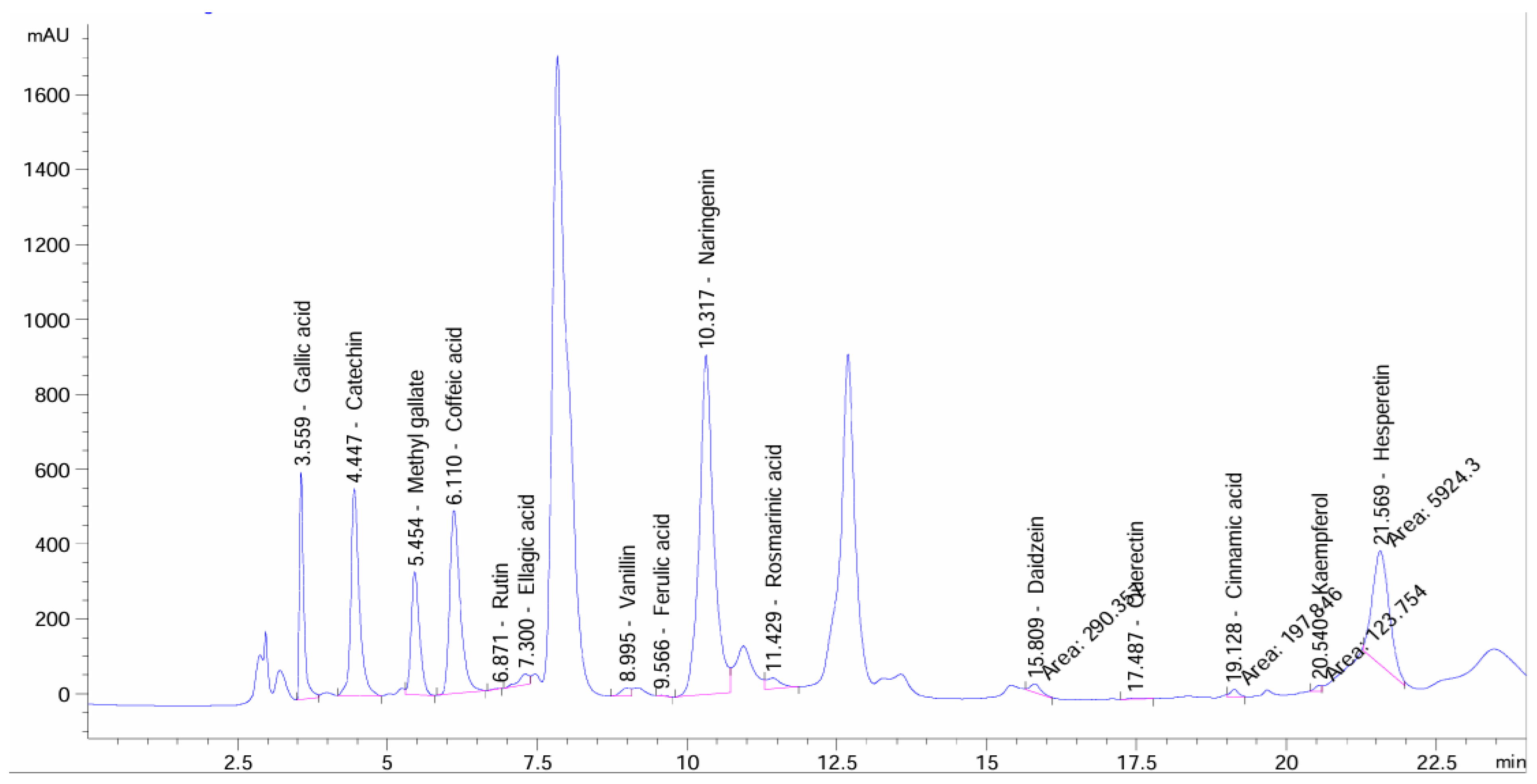
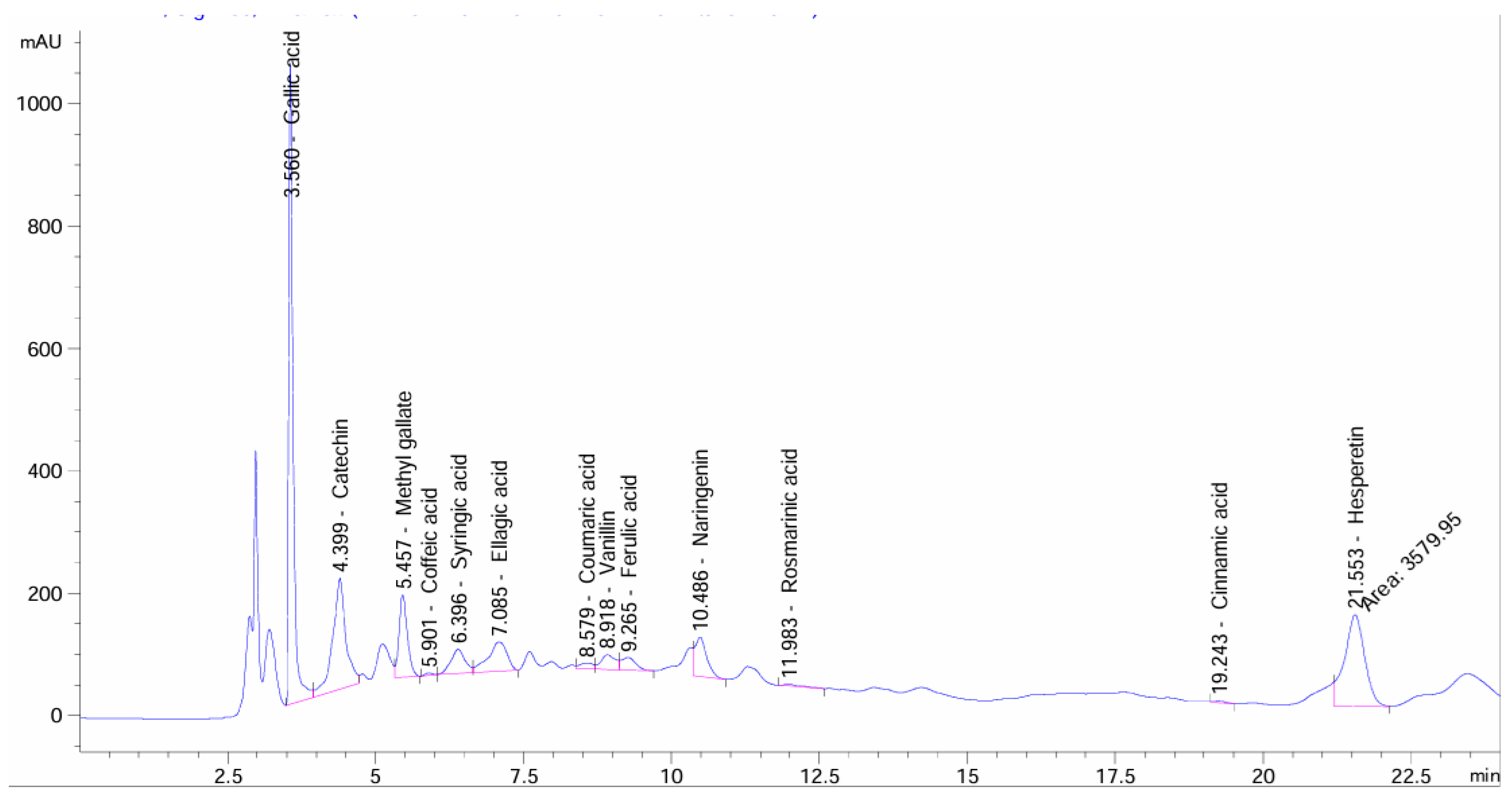
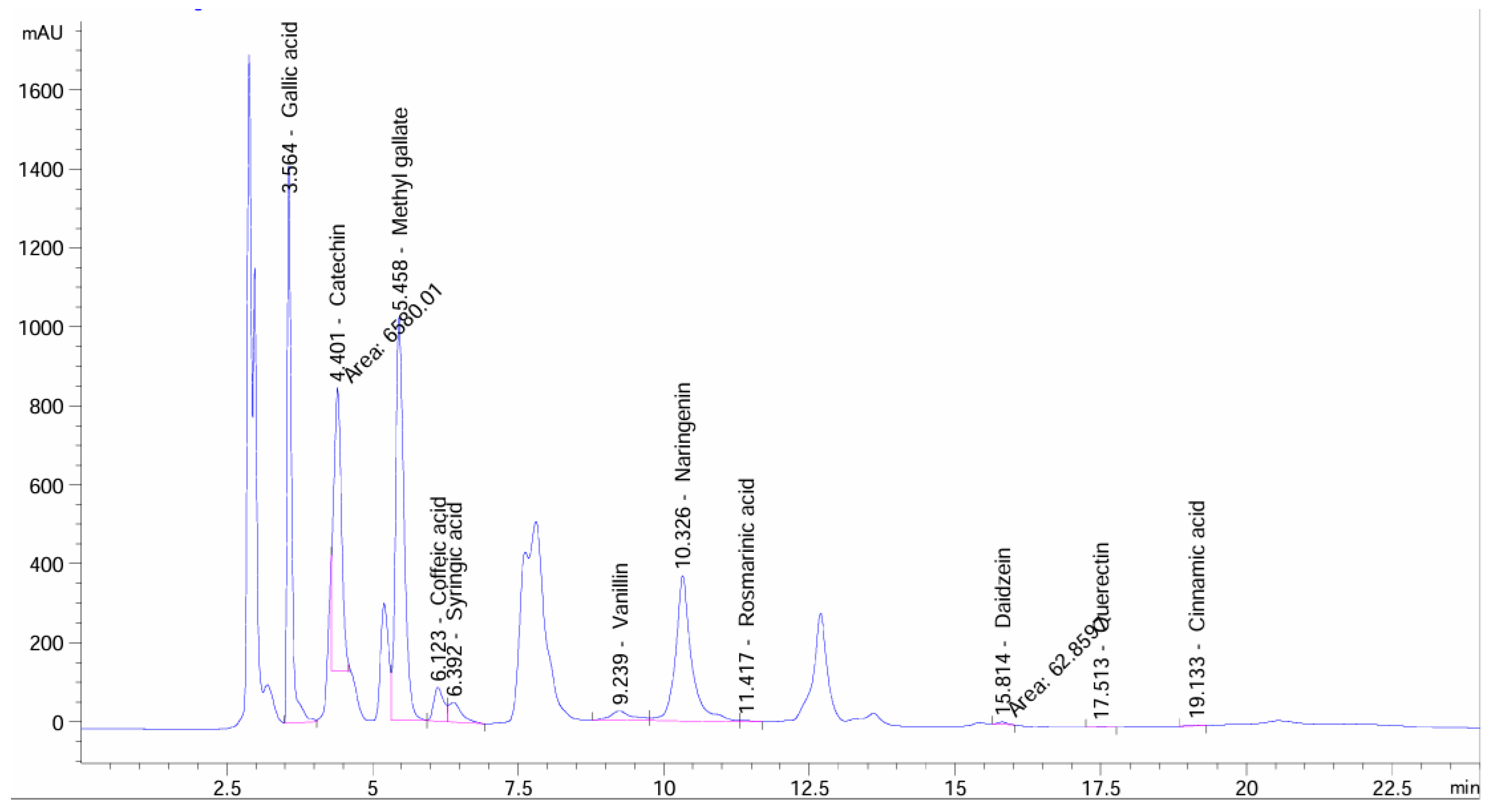
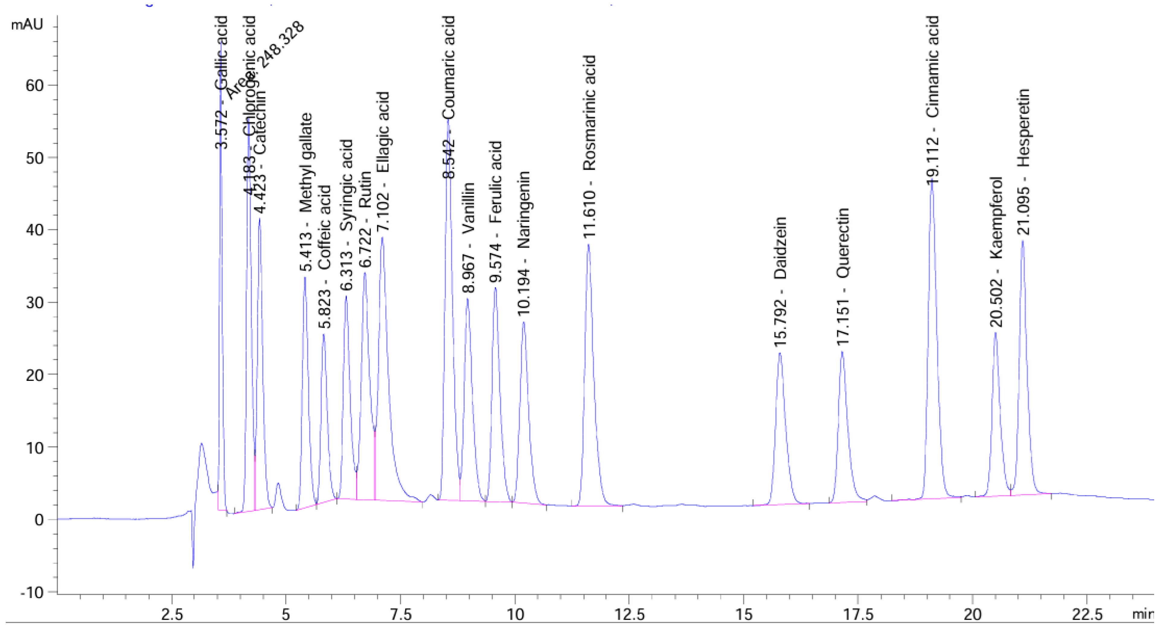
HPLC-DAD analysis revealed qualitative and quantitative differences in phenolic compounds identified based on comparison with external standards among the Acacia samples investigated (Figure 1). The SN sample (Table 3 and Figure 2) was characterized by a high concentration of naringenin, which is well known for its strong antioxidant properties, including radical scavenging and metal chelation [37]. In contrast, TL (Figure 3) and S samples (Figure 4) showed higher levels of gallic acid, catechin, and methyl gallate (Table 3), which have been reported to exhibit strong antimicrobial activity [38,39,40,41,42].
Figure 1. HPLC chromatogram of flavonoid and phenolic acid standards.
Table 3. Phenolic compounds identified by HPLC-DAD based on comparison with external standards.
Figure 2. HPLC chromatogram of flavonoid and phenolic acid levels in SN sample (A. nilotica leaves).
Figure 3. HPLC chromatogram of flavonoid and phenolic acid levels in TL sample (A. seyal leaves).
Figure 4. HPLC chromatogram of flavonoids and phenolic acid levels in S sample (A. nilotica seeds).
These variations highlight the influence of plant species and plant parts on phytochemical composition. Similar variations have been reported in previous studies on Acacia species [28,43]. However, the higher concentrations detected in this study suggest that environmental factors or extraction conditions may have enhanced compound recovery. Environmental factors such as soil composition, temperature, water availability, and sunlight exposure may influence secondary metabolite biosynthesis in Acacia species. In addition, extraction efficiency can be affected by solvent polarity and extraction conditions, which may contribute to variations in compound recovery.
Importantly, the chromatographic profiles also revealed several unidentified peaks, particularly in SN and S samples, suggesting the presence of additional phytochemical constituents under the applied analytical conditions. Variations in the occurrence of these peaks among the samples may reflect differences in phytochemical composition. Further characterization using advanced analytical techniques such as LC-MS/MS and NMR spectroscopy may be required to identify these compounds and evaluate their potential biological significance.

3.2. Phenolic Compounds and Their Biological Activities

The results of DPPH radical scavenging activity, reducing power (RP), total phenolic content, and total flavonoid content are presented in Table 4. The samples investigated exhibited considerable antioxidant activity, with sample SN showing the highest phenolic content compared with TL and S samples. This finding suggests a possible contribution of phenolic compounds to the observed antioxidant activity. In contrast, Abdel-Farid et al. [7] reported higher phenolic content and antioxidant activity in A. seyal compared with A. nilotica. A strong positive linear relationship was observed between total phenolic content and antioxidant activity. The correlation between TPC and DPPH radical scavenging activity was described by the regression equation y = 0.1897x + 42.872 (R2 = 0.9955), while the relationship between TPC and reducing power followed the equation y = 0.0137x − 1.6889 (R2 = 0.9758). Several studies [44,45,46,47] have reported a positive correlation between total phenolic content and free radical scavenging activity. In the present study, the SN sample exhibited higher phenolic content and antioxidant activity compared with those reported by Abdel-Farid et al. [7], El-Chaghaby et al. [48], and Sulaiman et al. [49].
Table 4. Total phenolic content, total flavonoid content, and antioxidant activities of SN, TL, and S samples.
Compared with the findings of Abdel-Farid et al. [7], the TL (A. seyal) sample exhibited higher phenolic content and antioxidant activity. However, the total phenolic content was lower than that reported by Ramde-Tiendrebeogo et al. [50] and Magnini et al. [51], whereas the total flavonoid content was comparatively higher. In contrast, Vadivel and Biesalski [52] reported higher phenolic content and antioxidant activity in their samples.
Plant extracts commonly exhibit antibacterial activity due to the presence of various phytochemical constituents, including phenolics, flavonoids, alkaloids, tannins, and glycosides [53]. Polyphenolic compounds may contribute to this activity through interactions with bacterial cell membranes, leading to increased membrane permeability and disruption of cellular integrity. According to Olatunde et al. [54], bacterial cell death may result from the excessive leakage of essential intracellular ions and molecules following membrane damage.
Table 5 presents the antimicrobial activity of the investigated samples, which showed strong activity against the fungal strain C. albicans. Satish et al. [55] reported that the ethanolic leaf extract of A. nilotica exhibited considerable antifungal activity against Candida albicans and Trichophyton rubrum. Similarly, Jodi et al. [56] reported inhibition zones ranging from 9 to 35 mm for A. nilotica. In the present study, the methanolic leaf extract of A. nilotica (SN sample) exhibited an inhibition zone of 16.67 mm against C. albicans, which was higher than the values reported by Attahiru et al. [57] and comparable to those reported by Jodi et al. [56]. The TL sample showed an inhibition zone of 16.33 mm, whereas sample S exhibited the highest antifungal activity (17.67 mm), exceeding the value reported by Sulayli et al. [58] and Swami Narsingh et al. [59]. The relatively larger inhibition zones observed against Candida albicans compared with the positive control may be influenced by differences in diffusion behavior and the complex composition of bioactive compounds in the plant extracts under the assay conditions used. Therefore, these findings should not be interpreted as direct superiority over the standard antifungal agent. The SN and S samples showed considerable antibacterial activity against Staphylococcus aureus, while the TL and S samples exhibited higher activity against Salmonella enterica and Escherichia coli. Although the SN sample showed the highest total phenolic content and antioxidant activity, sample S demonstrated stronger antibacterial activity against Gram-negative bacteria. This observation suggests that antimicrobial activity may not depend solely on total phenolic concentration but may also be influenced by other phytochemical constituents such as tannins, saponins, alkaloids, terpenoids, glycosides, or synergistic interactions among bioactive compounds.
Table 5. Antimicrobial activity of SN, TL, and S methanolic extracts.

3.3. Overall Interpretation and Significance

Overall, the present study suggests that variations in phenolic composition may contribute to the biological activities of Acacia species. The integration of HPLC-DAD profiling with antioxidant and antimicrobial assays provided additional insight into the possible relationship between phytochemical composition and biological activity. Furthermore, several unidentified peaks were observed in the chromatographic profiles, suggesting the presence of additional phytochemical constituents under the applied analytical conditions. Further characterization studies may be required to identify these compounds and evaluate their possible biological significance. These findings highlight the potential of Acacia species as natural sources of bioactive compounds for pharmaceutical, nutraceutical, and food applications.

4. Conclusions

Methanolic extracts of Acacia nilotica and Acacia seyal exhibited significant antioxidant and antimicrobial activities, which may be associated with differences in phenolic composition. Among the investigated samples, A. nilotica leaves showed the highest antioxidant activity, whereas A. nilotica seeds demonstrated comparatively stronger antibacterial activity despite their lower total phenolic content, suggesting that antimicrobial effects may involve additional phytochemical constituents beyond total phenolics. HPLC-DAD analysis revealed qualitative and quantitative differences in phenolic compounds among the investigated samples, in addition to the presence of several unidentified peaks under the applied analytical conditions. Further studies are recommended to identify these unknown compounds and evaluate their possible biological significance using advanced analytical techniques.
The present study was limited by the relatively small sample size, limited geographical coverage within Khartoum State, and the absence of seasonal variation assessment. Therefore, further studies involving larger sample sizes, wider geographical sampling, and different seasonal conditions are recommended to better understand phytochemical variation and biological activity.

Funding

This research received no external funding.

Data Availability Statement

The original contributions presented in this study are included in the article. Further inquiries can be directed to the corresponding author.

Acknowledgments

The Researchers would like to thank the Deanship of Graduate Studies and Scientific Research at Qassim University for financial support (APC-QU-2026).

Conflicts of Interest

The author declares no conflicts of interest.

References

  1. Seigler, D.S. Phytochemistry of Acacia-sensu lato. Biochem. Syst. Ecol. 2003, 31, 845–873. [Google Scholar] [CrossRef]
  2. Kyalangalilwa, B.; Boatwright, J.S.; Daru, B.H.; Maurin, O.; Van der Bank, M. Phylogenetic position and revised classification of Acacia s.l. (Fabaceae: Mimosoideae) in Africa, including new combinations in Vachellia and Senegalia. Bot. J. Linn. Soc. 2013, 172, 500–523. [Google Scholar] [CrossRef]
  3. Dyer, C. New names for the African Acacia species in Vachellia and Senegalia. South. For. 2014, 76, iii. [Google Scholar] [CrossRef][Green Version]
  4. Wickens, K.; Pennacchio, M. A search for novel biologically active compounds in the phyllodes of Acacia species. Conserv. Sci. West. Aust. 2002, 4, 139–144. [Google Scholar]
  5. Saini, R.; Roy, S.; Kumar, A. Comparative pharmacognostical and antimicrobial studies of Acacia species (Mimosaceae). J. Med. Plants Res. 2008, 2, 378–386. [Google Scholar]
  6. Deshmukh, S.P.; Bhajipale, N.S. A review on Acacia species of therapeutics importance. Int. J. Pharm. Biol. Sci. Arch. 2018, 6, 24–34. [Google Scholar]
  7. Abdel-Farid, I.; Sheded, M.; Mohamed, E. Metabolomic profiling and antioxidant activity of some Acacia species. Saudi J. Biol. Sci. 2014, 21, 400–408. [Google Scholar] [CrossRef]
  8. Hemashree, J.; Lakshmi, T. Anti-inflammatory action of Acacia catechu seed extract. Adv. Pharm. Educ. Res. 2018, 8, 92–95. [Google Scholar]
  9. Lakshmi, T.; Sri, R.B.; Senthilkumar, S.; Haribalan, P.; Parameshwari, R.; Vijayaraghavan, R.; Rajeshkumar, S. Seed and bark extracts of Acacia catechu protect liver from acetaminophen-induced hepatotoxicity by modulating oxidative stress, antioxidant enzymes and liver function enzymes in Wistar rat model. Biomed. Pharmacother. 2018, 108, 838–844. [Google Scholar] [CrossRef]
  10. Afsar, T.; Trembley, J.H.; Salomon, C.E.; Razak, S.; Khan, M.R.; Ahmed, K. Growth inhibition and apoptosis in cancer cells induced by polyphenolic compounds of Acacia hydaspica: Involvement of multiple signal transduction pathways. Sci. Rep. 2016, 6, 23077. [Google Scholar] [CrossRef] [PubMed]
  11. Ngaffo, C.M.; Tchangna, R.S.; Mbaveng, A.T.; Kamga, J.; Harvey, F.M.; Ngadjui, B.T.; Bochet, C.G.; Kuete, V. Botanicals from the leaves of Acacia sieberiana had better cytotoxic effects than isolated phytochemicals towards MDR cancer cell lines. Heliyon 2020, 6, e05412. [Google Scholar] [CrossRef]
  12. Ranasinghe, A.; Lakshmi, T. In vitro anticancer activity of Acacia heartwood extract. Plant Cell Biotechnol. Mol. Biol. 2020, 21, 41–47. [Google Scholar]
  13. Aremu, O.; Olayemi, O.; Ajala, T.; Isimi, Y.; Oladosu, P.; Ekere, K.; John, J.; Emeje, M. Antibacterial evaluation of Acacia nilotica Lam. (Mimosaceae) seed extract in dermatological preparations. J. Res. Pharm. 2020, 24, 170–181. [Google Scholar] [CrossRef]
  14. Abdalla, A.A.; Mustafa, M.I.; Makhawi, A.M. Phytochemical screening and antimicrobial activities studies of Acacia nilotica fruit cover. bioRxiv 2020. [Google Scholar] [CrossRef]
  15. Ahmed, D.A.; Esraa, A.; Svenning, J.C.; Mohamed, B.; El-Shaltout, K. Wild plant species in Egyptian gardens of the Nile region: Conservation viewpoint. Egypt. J. Bot. 2020, 60, 719–732. [Google Scholar] [CrossRef]
  16. Gaara, A.H.; El-Toumy, S.A.; Farrag, A.R.H.; Ahmed, N.M. Antidiabetic, antihyperlipidemic and antioxidant activities of Acacia albida in streptozotocin-induced diabetes in rats and its metabolites. Egypt. J. Chem. 2020, 63, 337–348. [Google Scholar] [CrossRef]
  17. Badry, M.O.; Al-Hawshabi, O.S.S.; Osman, A.K. Flora and phytochorology of Lahij Governorate of Yemen: Systematic revision of wild legumes of the family Fabaceae. Egypt. J. Bot. 2021, 61, 591–610. [Google Scholar] [CrossRef]
  18. Ali, A.; Akhtar, N.; Khan, B.A.; Khan, M.S.; Rasul, A.; Uz-Zaman, S.; Khalid, N.; Waseem, K.; Mahmood, T.; Ali, L. Acacia nilotica: A plant of multipurpose medicinal uses. J. Med. Plants Res. 2012, 6, 1492–1496. [Google Scholar] [CrossRef]
  19. Mahesh, B.; Satish, S. Antimicrobial activity of some important medicinal plants against plant and human pathogens. World J. Agric. Sci. 2008, 4, 839–843. [Google Scholar]
  20. Banso, A. Phytochemical and antibacterial investigation of bark extracts of Acacia nilotica. J. Med. Plants Res. 2009, 3, 82–85. [Google Scholar]
  21. Solomon-Wisdom, G.O.; Shittu, G.A. In vitro antimicrobial and phytochemical activities of Acacia nilotica leaf extract. J. Med. Plants Res. 2010, 4, 1232–1234. [Google Scholar]
  22. Singh, B.N.; Singh, B.R.; Singh, R.L. Antioxidant and anti quorum sensing activities of green pod of Acacia nilotica L. Food Chem. Toxicol. 2009, 47, 778–786. [Google Scholar] [CrossRef]
  23. Gilani, A.H.; Shaheen, F.; Zaman, M. Studies on antihypertensive and antispasmodic activities of methanol extract of Acacia nilotica pods. Phytother. Res. 1999, 13, 665–669. [Google Scholar] [CrossRef]
  24. Eldeen, I.M.S.; Van Staden, J. Cyclooxygenase inhibition and antimycobacterial effects of extracts from Sudanese medicinal plants. S. Afr. J. Bot. 2008, 74, 225–229. [Google Scholar] [CrossRef][Green Version]
  25. Eldeen, I.M.S.; Van Staden, J. In vitro pharmacological investigation of extracts from some trees used in Sudanese traditional medicine. S. Afr. J. Bot. 2007, 73, 435–440. [Google Scholar] [CrossRef]
  26. Muthaura, C.N.; Keriko, J.M.; Mutai, C.; Yenesew, A.; Gathirwa, J.W.; Irungu, B.N.; Nyangacha, R.; Mungai, G.M.; Derese, S. Antiplasmodial potential of traditional antimalarial phytotherapy remedies used by the Kwale community of the Kenyan Coast. J. Ethnopharmacol. 2015, 170, 148–157. [Google Scholar] [CrossRef]
  27. Saeed, M.E.M.; Abdelgadir, H.; Sugimoto, Y.; Khalid, H.E.; Efferth, T. Cytotoxicity of 35 medicinal plants from Sudan towards sensitive and multidrug-resistant cancer cells. J. Ethnopharmacol. 2015, 174, 644–658. [Google Scholar] [CrossRef]
  28. Ahmed, N.; Kandhro, A.A.; Chaudhry, A.W.; Shaikh, A.R.; Qureshi, A.U.; Hayat, A.; Basit, A.; Memon, H.D.; Sidhu, A.R.; Zahid, S. Identification and quantification of phenolic compounds by HPLC-DAD and bioactivity assay of Acacia nilotica. Erwerbs-Obstbau 2023, 66, 247–255. [Google Scholar] [CrossRef]
  29. Mythili, K.; Umamaheswara Reddy, C.; Chamundeeswari, D.; Manna, P.K. Determination of total phenol, alkaloid, flavonoid and tannin in different extracts of Calanthe triplicata. J. Pharmacogn. Phytochem. 2014, 2, 40–44. [Google Scholar]
  30. Ebrahimzadeh, M.A.; Nabavi, S.M.; Nabavi, S.F.; Bahramian, F.; Bekhradnia, A.R. Antioxidant and free radical scavenging activity of H. officinalis L. var. angustifolius, V. odorata, B. hyrcana and C. speciosum. Pak. J. Pharm. Sci. 2010, 23, 29–34. [Google Scholar] [PubMed]
  31. Nabavi, S.M.; Ebrahimzadeh, M.A.; Nabavi, S.F.; Fazelian, M.; Eslami, B. In vitro antioxidant and free radical scavenging activity of Diospyros lotus and Pyrus boissieriana growing in Iran. Phcog. Mag. 2009, 5, 122–126. [Google Scholar]
  32. Burits, M.; Bucar, F. Antioxidant activity of Nigella sativa essential oil. Phytother. Res. 2000, 14, 323–328. [Google Scholar] [CrossRef]
  33. Oyaizu, M. Studies on products of browning reactions: Antioxidative activities of products of browning reaction prepared from glucosamine. Jpn. J. Nutr. 1986, 44, 307–315. [Google Scholar] [CrossRef]
  34. European Committee for Antimicrobial Susceptibility Testing of the European Society of Clinical Microbiology and Infectious Diseases. Determination of minimum inhibitory concentrations (MICs) of antibacterial agents by agar dilution. Clin. Microbiol. Infect. 2000, 6, 509–515. [Google Scholar] [CrossRef]
  35. Duncan, D.B. Multiple range and multiple F tests. Biometrics 1955, 11, 1–42. [Google Scholar] [CrossRef]
  36. Fisher, R.A. The Design of Experiments; Oliver and Boyd: Edinburgh, UK, 1935. [Google Scholar]
  37. Kim, J.H.; Lee, J.K. Naringenin enhances NK cell lysis activity by increasing the expression of NKG2D ligands on Burkitt’s lymphoma cells. Arch. Pharm. Res. 2015, 38, 2042–2048. [Google Scholar] [CrossRef]
  38. Rodríguez, M.; Alberto, M.; Manca de Nadra, M. Antibacterial effect of phenolic compounds from different wines. Food Control 2007, 18, 93–101. [Google Scholar] [CrossRef]
  39. Kang, J.; Li, Q.; Liu, L.; Jin, W.; Wang, J.; Sun, Y. The specific effect of gallic acid on Escherichia coli biofilm formation by regulating pgaABCD genes expression. Appl. Microbiol. Biotechnol. 2018, 102, 1837–1846. [Google Scholar] [CrossRef]
  40. Choi, J.G.; Kang, O.H.; Lee, Y.S.; Oh, Y.C.; Chae, H.S. Antibacterial activity of methyl gallate isolated from Galla Rhois or carvacrol combined with nalidixic acid against nalidixic acid resistant bacteria. Molecules 2009, 14, 1773–1780. [Google Scholar] [CrossRef] [PubMed]
  41. Ahn, Y.J.; Lee, C.O.; Kweon, J.H.; Ahn, J.W.; Park, J.H. Growth-inhibitory effects of Galla Rhois-derived tannins on intestinal bacteria. J. Appl. Microbiol. 1998, 84, 439–443. [Google Scholar] [CrossRef] [PubMed]
  42. Ahn, Y.J.; Lee, H.S.; Oh, H.S.; Kim, H.T.; Lee, Y.H. Antifungal activity and mode of action of Galla Rhois-derived phenolics against phytopathogenic fungi. Pestic. Biochem. Physiol. 2005, 81, 105–112. [Google Scholar] [CrossRef]
  43. Elshamy, S.; Handoussa, H.; El-Shazly, M.; Mohammed, E.D.; Kuhnert, N. Metabolomic profiling and quantification of polyphenols from leaves of seven Acacia species by UHPLC-QTOF-ESI-MS. Fitoterapia 2023, 172, 105741. [Google Scholar] [CrossRef]
  44. Cai, Y.; Luo, Q.; Sun, M.; Corke, H. Antioxidant activity and phenolic compounds of 112 traditional Chinese medicinal plants associated with anticancer. Life Sci. 2004, 74, 2157–2184. [Google Scholar] [CrossRef]
  45. Djeridane, A.; Yousfi, M.; Nadjemi, B.; Boutassouna, D.; Stocker, P.; Vidal, N. Antioxidant activity of some Algerian medicinal plant extracts containing phenolic compounds. Food Chem. 2006, 97, 654–660. [Google Scholar] [CrossRef]
  46. Katalinic, V.; Milos, M.; Jukic, M. Screening of 70 medicinal plant extracts for antioxidant capacity and total phenols. Food Chem. 2006, 94, 550–557. [Google Scholar] [CrossRef]
  47. Katsube, T.; Tabata, H.; Ohta, Y.; Yamasaki, Y.; Anuurad, E.; Shiwaku, K. Screening for antioxidant activity in edible plant products: Comparison of low-density lipoprotein oxidation assay, DPPH radical scavenging assay, and Folin–Ciocalteu assay. J. Agric. Food Chem. 2004, 52, 2391–2396. [Google Scholar] [CrossRef] [PubMed]
  48. El-Chaghaby, G.A.; Rashad, S.; Abdel-Kader, S.F.; Rawash, E.S.A.; Moneem, M.A. Assessment of phytochemical components, proximate composition and antioxidant properties of Scenedesmus obliquus, Chlorella vulgaris and Spirulina platensis algae extracts. Egypt. J. Aquat. Biol. Fish. 2019, 23, 521–526. [Google Scholar] [CrossRef]
  49. Sulaiman, C.T.; Sadashiva, C.T.; George, S.; Goplakrishnan, V.K.; Balachandran, I. Chromatographic studies and in vitro screening for acetylcholinesterase inhibition and antioxidant activity of three Acacia species from South India. Anal. Chem. Lett. 2013, 3, 111–118. [Google Scholar] [CrossRef]
  50. Ramde-Tiendrebeogo, A.; Tibiri, A.; Hilou, A.; Lompo, M.; Millogo-Kone, H.; Nacoulma, O.; Guissou, I. Antioxidative and antibacterial activities of phenolic compounds from Ficus sur Forssk. and Ficus sycomorus L. (Moraceae): Potential for sickle cell disease treatment in Burkina Faso. Int. J. Biol. Chem. Sci. 2012, 6, 328–336. [Google Scholar] [CrossRef]
  51. Magnini, R.D.; Hilou, A.; Millogo-Koné, H.; Pagès, J.M.; Davin-Regli, A. Acacia senegal extract rejuvenates the activity of phenicols on selected Enterobacteriaceae multidrug-resistant strains. Antibiotics 2020, 9, 323. [Google Scholar] [CrossRef]
  52. Vadivel, V.; Biesalski, H.K. Total phenolic content, in vitro antioxidant activity and type II diabetes relevant enzyme inhibition properties of methanolic extract of traditionally processed underutilized food legume, Acacia nilotica (L.) Willd ex Delile. Int. Food Res. J. 2012, 19, 593–601. [Google Scholar]
  53. Debalke, D.; Birhan, M.; Kinubeh, A.; Yayeh, M. Assessments of antibacterial effects of aqueous-ethanolic extracts of Sida rhombifolia aerial part. Sci. World J. 2018, 2018, 8429809. [Google Scholar] [CrossRef]
  54. Olatunde, O.O.; Tan, S.L.D.; Shiekh, K.A.; Benjakul, S.; Nirmal, N.P. Ethanolic guava leaf extracts with different chlorophyll removal processes: Anti-melanosis, antibacterial properties and the impact on qualities of Pacific white shrimp during refrigerated storage. Food Chem. 2021, 341, 128251. [Google Scholar] [CrossRef]
  55. Satish, S.; Raghavendra, M.P.; Raveesha, K.A. Evaluation of the antibacterial potential of some plants against human pathogenic bacteria. Adv. Biol. Res. 2008, 2, 44–48. [Google Scholar]
  56. Jodi, S.M.; Omoniyi, S.; Ungo-Kore, H.Y.; Ngwai, Y.B. Antifungal activities of honey and Acacia nilotica extracts. FUDMA J. Sci. 2020, 4, 779–783. [Google Scholar]
  57. Attahiru, A.; Haruna, Y.; Muhammad, A.; Gana, A. Phytochemical analysis and antifungal activity of methanol extract of Acacia nilotica leaves. Int. J. Life Sci. Res. 2021, 9, 10–15. [Google Scholar]
  58. Sulayli, A.I.; Moustafa, M.F.; Eid, E.M. Genetic variability, antimicrobial activity and natural water-soluble vitamins contents of five Acacia species growing in Jazan Region, Saudi Arabia. Pak. J. Agric. Sci. 2019, 56, 289–300. [Google Scholar]
  59. Dev, S.N.C.; De, K.; Singh, S. Antimicrobial activity and phytochemical analysis of Acacia nilotica (L.) Del. Indian J. Appl. Pure Biol. 2014, 29, 331–332. [Google Scholar]
Disclaimer/Publisher’s Note: The statements, opinions and data contained in all publications are solely those of the individual author(s) and contributor(s) and not of MDPI and/or the editor(s). MDPI and/or the editor(s) disclaim responsibility for any injury to people or property resulting from any ideas, methods, instructions or products referred to in the content.

Article Metrics

Citations

Article Access Statistics

Multiple requests from the same IP address are counted as one view.