


Abstract
Stone coal is an important vanadium-bearing resource and a potential source of rare earth elements (REEs). Previous studies have mainly focused on the bulk occurrence, resource potential, and leaching behavior of V or REEs in stone coal, whereas the microscale spatial relationships between V and REEs and their evolution during leaching remain poorly constrained. In this study, three representative stone coal samples were analyzed by synchrotron radiation micro-X-ray fluorescence (μXRF) to characterize the microscale distributions of V and REEs in raw samples and corresponding leaching residues. Pearson correlation analysis was further used to quantify changes in V–REE spatial relationships during leaching. The results showed that V–REE relationships were generally weak and were modified to different extents after leaching. In the GZ sample, the V–Eu correlation coefficient decreased from 0.63 to 0.34, indicating that the migration of V and REEs was not fully synchronized. The three samples also showed different REE distribution tendencies after leaching: GZ showed partial transfer of REEs to the leachate with residual retention, PX showed mixed behavior with appreciable retention in the residue, whereas PZ retained REEs predominantly in the residue. These results suggest that the integrated utilization of V and REEs in stone coal can be better achieved through a staged recovery route, in which the REE recovery pathway is determined according to their actual distribution between the leachate and the residue after V leaching. This study provides a microscale basis for the comprehensive utilization of coal-related critical metal resources.
1. Introduction
Stone coal is an important vanadium (V)-bearing resource that also contains rare earth elements (REEs) [1,2,3,4]. Traditionally, its utilization has focused primarily on V extraction because of V’s dominant economic value [5,6,7]. Previous studies on stone coal have mainly focused on V occurrence, roasting or acid-leaching mechanisms, and process optimization for improving V extraction efficiency [8,9,10]. However, the rapidly growing global demand for critical metals has increased interest in the comprehensive utilization of stone coal, particularly the recovery of REEs [9,10,11]. In parallel, increasing attention has been paid to REEs in coal-related resources because of their strategic importance and potential recoverability [4]. Despite this potential, REEs in stone coal generally occur at relatively low concentrations and exhibit pronounced microscale heterogeneity.
In coal-related mineral resources, REEs commonly include light and middle REEs such as La, Ce, Nd, Sm, Eu, Gd, and Dy, whose occurrence may be controlled by discrete REE-bearing minerals, adsorption onto clay minerals or organic matter, phosphate phases, or fine-grained aluminosilicate and oxide matrices. In V-bearing stone coal, these REEs may exhibit different degrees of spatial association with V depending on the mineralogical composition and geochemical characteristics of the ore [12]. As a result, the spatial relationships between vanadium and REEs, as well as their possible evolution during leaching, remain insufficiently understood [13,14]. This uncertainty limits the evaluation of whether REEs can be effectively recovered together with vanadium during conventional extraction processes.
Synchrotron radiation micro-X-ray fluorescence (μXRF) enables direct visualization of elemental distributions at high spatial resolution and has been widely applied to trace-element characterization in heterogeneous materials [15,16,17]. This technique is particularly suitable for stone coal, in which microscale elemental heterogeneity cannot be adequately resolved by bulk compositional analysis alone [18,19]. μXRF can reveal localized enrichment, dispersion, and spatial overlap of trace elements within complex mineral matrices [20]. Recent microscale and synchrotron-based studies have further shown that the spatial distribution and speciation of REEs can strongly influence their extraction behavior in coal-related materials [21]. When combined with Pearson correlation analysis, μXRF mapping can also provide a quantitative description of inter-element spatial co-variation before and after leaching. However, most existing studies have been based on bulk chemical analysis, leaching behavior, process optimization, or general mineralogical characterization, and therefore provide limited information on the microscale spatial relationships between V and REEs in stone coal. In particular, whether V and REEs are spatially associated before leaching and whether their relationships are preserved, weakened, or reorganized during acid leaching remain poorly understood. Therefore, a microscale investigation of V–REE spatial relationships and their leaching-related evolution is necessary for developing more rational strategies for the integrated utilization of V- and REE-bearing stone coal.
In this study, three representative stone coal samples from Guzhang, Pingxiang, and Pengze, China, were selected to compare sample-dependent V–REE occurrence and redistribution during leaching. Synchrotron radiation μXRF was used to map the distributions of V and REEs in raw samples and corresponding leaching residues, and Pearson correlation analysis was applied to quantify changes in V–REE spatial co-variation. The novelty of this study lies in tracking the leaching-related evolution of REE distribution and V–REE spatial relationships in stone coal at the microscale using synchrotron-based elemental mapping. The objectives were to: (i) characterize the microscale distributions of V and REEs; (ii) determine how V–REE spatial relationships change after leaching; and (iii) assess the implications for staged recovery of V and REEs. The results provide a microscale basis for the comprehensive utilization of V- and REE-bearing stone coal.
2. Materials and Methods
2.1. Material
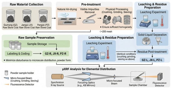
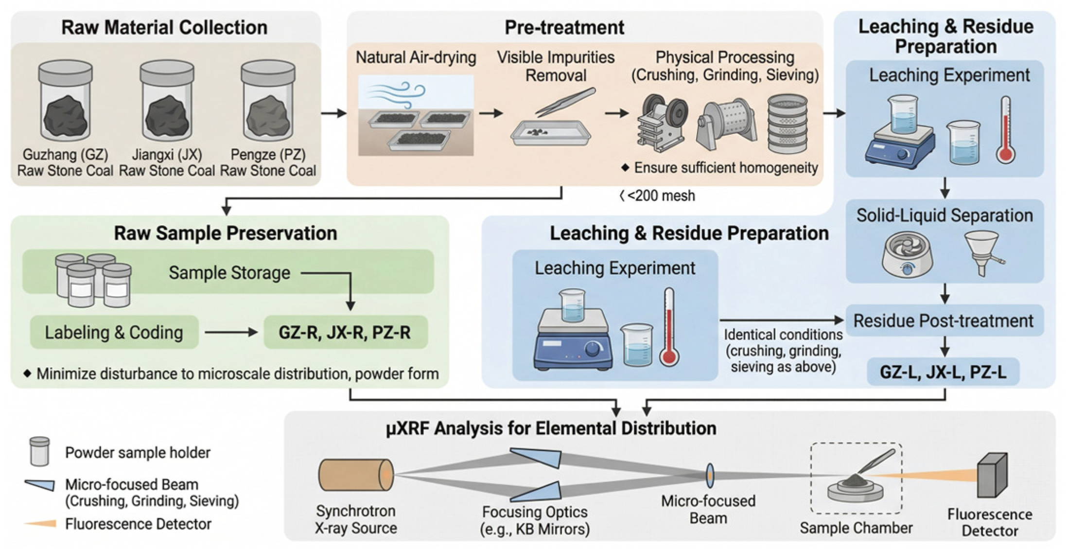
Three stone coal samples were used in this study and were collected from Guzhang, Pingxiang, and Pengze, China. The samples were denoted as GZ, PX, and PZ according to their sampling regions. Each sample was prepared as a composite sample from multiple subsamples collected from the same mineralized horizon. After collection, the samples were air-dried, manually cleaned to remove visible impurities, crushed, ground, and sieved before leaching experiments and μXRF analysis. The initial V and total REE contents of the three raw stone coal samples are summarized in Table 1. For μXRF analysis, both the raw samples and the corresponding leaching residues were prepared as powders using the same procedure to ensure consistency and comparability. The raw samples were designated GZ-R, PX-R, and PZ-R, and the corresponding leaching residues were designated GZ-L, PX-L, and PZ-L. A schematic overview of sample preparation, leaching, μXRF mapping, and correlation analysis is shown in Figure 1.
Table 1.
Initial V and total REE contents of the three raw stone coal samples (wt.%).
Figure 1.
Comprehensive workflow of sample preparation, leaching, μXRF mapping, and correlation analysis for the stone coal samples.
2.2. Leaching Procedure
Leaching experiments were conducted under identical conditions for the three stone coal samples to enable direct comparison of elemental behavior after treatment. Before leaching, the stone coal samples were crushed, ground, and sieved to a particle size fraction of <74 μm. The leaching experiments were conducted in a 250 mL thermostatic glass reactor equipped with mechanical stirring and temperature control. For each experiment, 10 g of stone coal was mixed with 100 mL of 2.0 mol/L H2SO4, corresponding to a liquid-to-solid ratio of 10:1 mL/g, and leached at 80 °C with a stirring speed of 300 r/min for 4 h. The stirring speed of 300 r/min was selected to maintain stable slurry suspension and reproducible leaching conditions in the present reactor setup, and comparable agitation conditions have been used in previous acid-leaching studies of vanadium-bearing stone coal or shale systems [22]. No external oxidant or reductant was added, and the redox condition was not externally controlled during leaching. After leaching, the slurry was vacuum-filtered using a 0.45 μm membrane filter to separate the leachate from the solid residue. The filtrate was collected for subsequent chemical analysis, while the solid residue was washed with deionized water to near-neutral pH, dried, and retained for subsequent μXRF analysis.
2.3. Synchrotron Radiation μXRF Measurements
Synchrotron radiation μXRF measurements were performed at beamline 4W1B of the Beijing Synchrotron Radiation Facility (BSRF) [23]. The storage ring was operated at 2.5 GeV with a beam current of 150–250 mA. The incident X-rays were monochromatized to 15 keV using a W/B4C double-multilayer monochromator (BSRF, Beijing, China) and focused to a spot size of approximately 50–100 μm using a polycapillary lens (BSRF, Beijing, China). Powdered samples were mounted on custom-made glass slides and sealed on both sides with Scotch adhesive tape. The μXRF mapping was performed on selected representative areas of the powder-mounted samples to characterize the microscale spatial distributions of V and REEs. The mapping area and spatial resolution were selected to capture local elemental heterogeneity while maintaining comparable analytical conditions among the raw samples and their corresponding leaching residues. Two-dimensional elemental maps were collected in step-scanning mode with a step size of 0.1 mm. Fluorescence signals were recorded using a Si(Li) solid-state detector (PGT/Princeton Gamma-Tech Inc., Princeton, NJ, USA), and the detector dead time was maintained below 30%. The spectra were processed using PyMca (version 5.9.6) for energy calibration, nonlinear fitting, and batch analysis. The net peak intensities of selected elements were normalized to the ion chamber counts and then used as semi-quantitative indicators for elemental mapping and correlation analysis. It should be noted that the resulting maps reflect relative intensity distributions rather than absolute elemental concentrations.
2.4. Data Processing and Correlation Analysis
Pearson correlation analysis was performed using the pixel-intensity datasets derived from the μXRF elemental maps to evaluate spatial co-variation between V and individual REEs, as well as among different REEs [24,25]. Pearson correlation analysis was performed using the pixel-intensity datasets derived from the μXRF elemental maps to evaluate spatial co-variation between V and individual REEs, as well as among different REEs [26,27]. The Pearson correlation coefficient was calculated according to Equation (1):
where rxy is the Pearson correlation coefficient between elements x and y, xi and yi are the normalized pixel intensities of the two elements at pixel, and are the mean intensities of the two elements within the selected mapping area, and n is the total number of pixels used in the calculation [28].
Correlation coefficients were calculated from the pixel-intensity datasets of individual elements within the selected mapping areas, and the resulting heat maps were used to compare similarities and differences in elemental associations among the three stone coal samples before and after leaching. For comparative discussion, the REEs were grouped into light, middle, and heavy rare earth elements according to commonly used geochemical classifications [29,30,31]. Positive correlation coefficients indicate similar spatial variation trends within the mapped area, whereas weak or negative coefficients indicate limited spatial correspondence or relative spatial separation. Because the μXRF data used for correlation analysis are semi-quantitative, the Pearson correlation coefficients were interpreted only as indicators of relative spatial co-variation within the selected mapped areas, rather than as quantitative correlations of absolute elemental concentrations or direct evidence of common mineral phases. Pm was retained as a Pm-labelled fluorescence channel resolved during spectral fitting; however, because Pm has no stable isotopes, Pm-related correlations were interpreted with particular caution and were not used to support the main geochemical conclusions.
3. Results and Discussion
3.1. Microscale Distribution Characteristics of V and REEs in Raw Stone Coal Samples
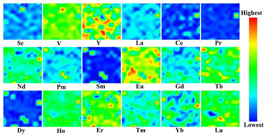
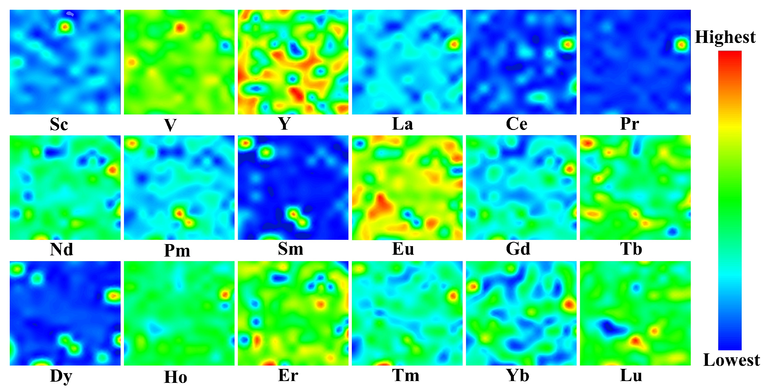
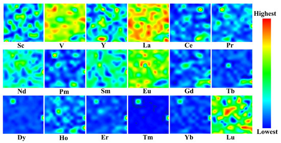
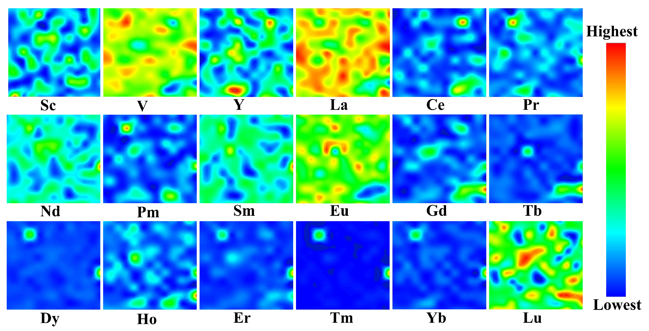
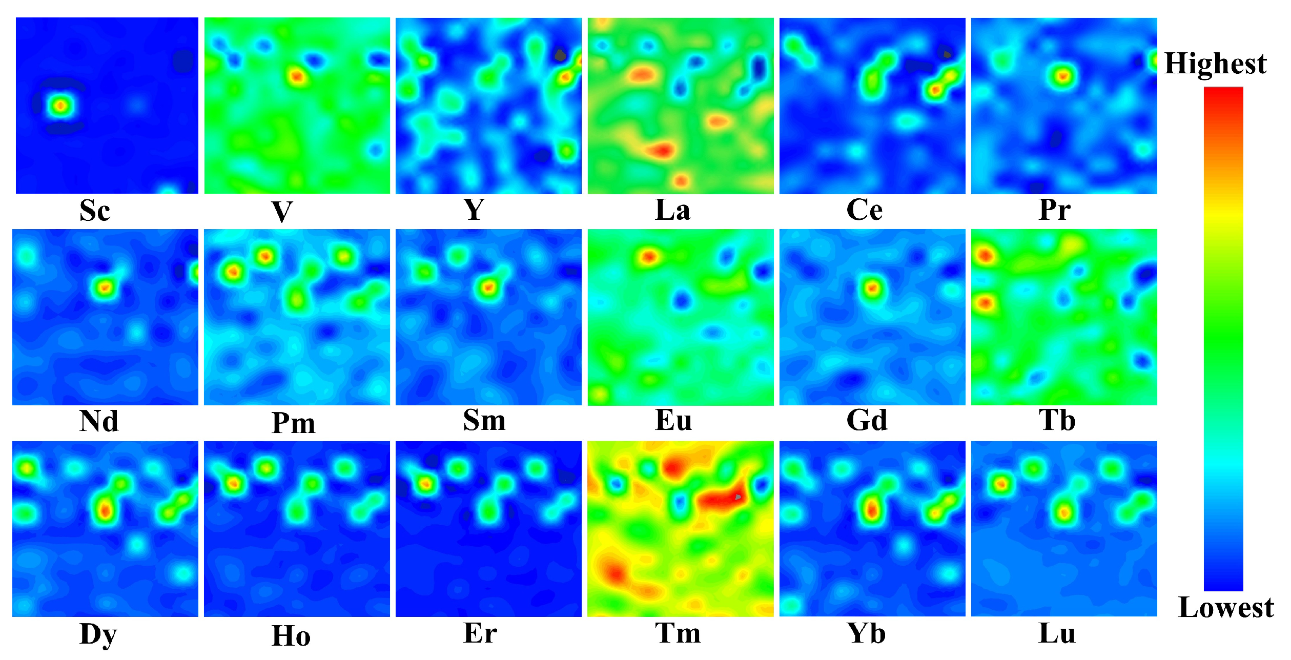
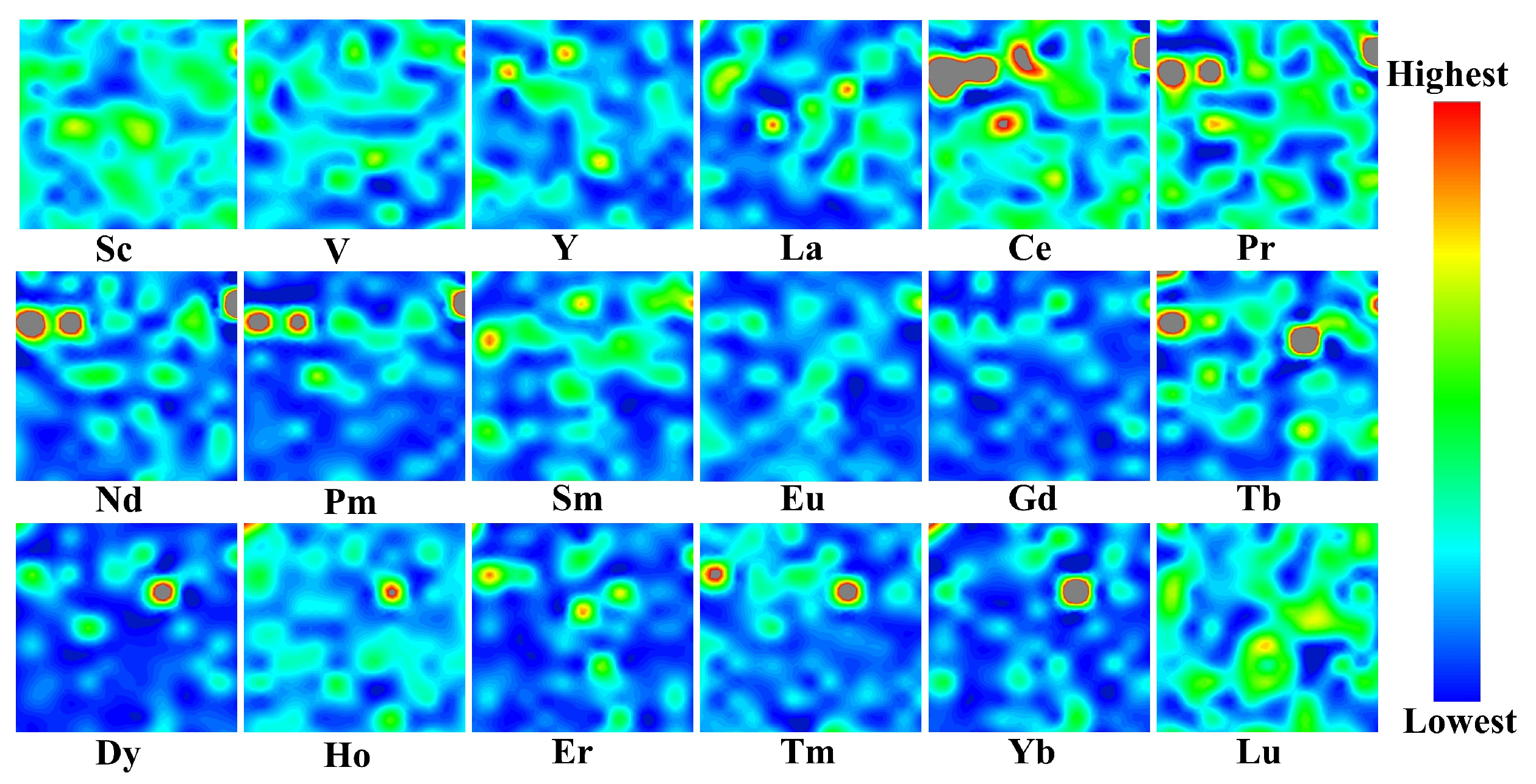
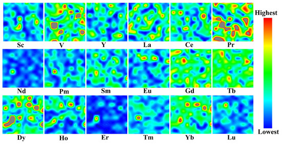
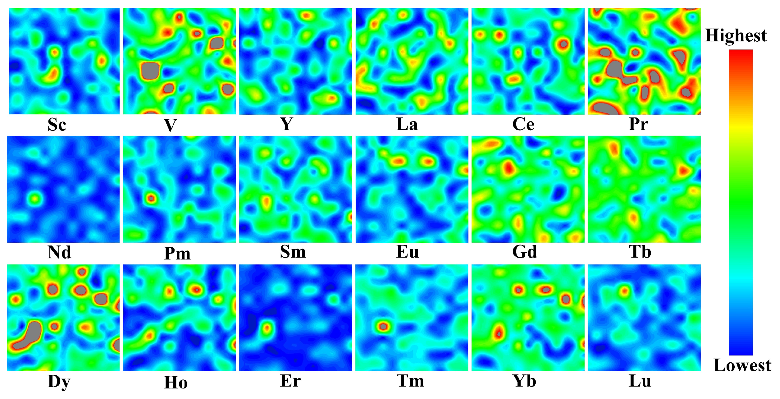
Synchrotron-based μXRF elemental mapping was carried out on three raw stone coal samples (GZ-R, PX-R, and PZ-R) to reveal the microscale distributions of V and REEs. In Figure 2, Figure 3 and Figure 4, the color scale from blue to red represents increasing fluorescence intensity and thus higher relative elemental abundance within the mapped regions. These spatial intensity maps allow direct visualization of whether V and REEs occur as diffuse background signals, localized hotspots, or partially overlapping enrichment domains.
Figure 2.
μXRF elemental maps of V and REEs in the GZ-R.
Figure 3.
μXRF elemental maps of V and REEs in the PX-R.
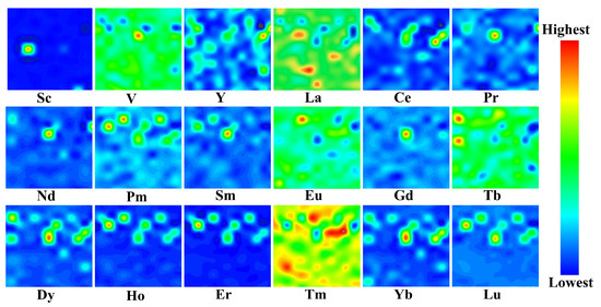
Figure 4.
μXRF elemental maps of V and REEs in the PZ-R.
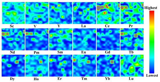
In the Guzhang sample (GZ-R, Figure 2), V is distributed across most of the mapped area and appears mainly as a diffuse background, with only a few local enrichment spots. Most REEs are confined to smaller regions. La, Ce, and Nd overlap to a large extent, suggesting a relatively close spatial association among these light REEs, whereas Sm is enriched in nearby but slightly shifted areas. Eu shows a broader and more diffuse distribution than the other REEs, with fewer distinct hotspots and a higher background signal. Among the examined REEs, Eu appears spatially closest to V, whereas the main enriched regions of La, Ce, and Nd are largely separated from the V-bearing areas.
The Pingxiang sample (PX-R, Figure 3) shows a clear separation between the light REEs and the middle-heavy REEs. The light REEs (La, Ce, Pr, and Nd) are concentrated in several localized regions, whereas the middle-heavy REEs (Dy, Ho, Er, Tm, Yb, and Lu) occur mainly along the sample margins and in isolated enriched domains. Spatial consistency within the middle-heavy REE group is relatively strong. V still appears mainly as a broadly distributed background, but, compared with GZ-R, it is spatially closer to La than to the other REEs. Therefore, the distribution pattern of PX-R is defined mainly by separation within the REE assemblage rather than by any obvious correspondence between V and the REEs.
The Pengze sample (PZ-R, Figure 4) exhibits a markedly different pattern and the most distinct separation between V and the REEs. V is widely dispersed throughout the mapped area and shows only minor local enrichments without obvious clustering. The REEs, by contrast, are concentrated in discrete hotspots. The middle-heavy REEs, including Dy, Ho, Er, Yb, and Lu, remain closely associated in space, and their enriched regions commonly overlap or occur in adjacent areas. Although the light REEs (for example, La, Ce, Pr, and Nd) are also locally enriched, their internal spatial correspondence is weaker and their overlap with the middle-heavy REEs is limited. None of the individual REEs in this sample shows a clear spatial association with V.
Overall, the three raw stone coal samples display distinct microscale distribution patterns. A common feature, however, is that V mainly occurs as a dispersed background signal, whereas REEs are more commonly concentrated in localized hotspots. Direct spatial overlap between V and individual REEs is generally limited, although some sample-specific proximity is observed, such as V-Eu in GZ-R and V-La in PX-R. In addition, the REEs themselves show internal differentiation, particularly between the light and middle-heavy subgroups. These observations indicate that the occurrence of V and REEs in stone coal is heterogeneous and not characterized by a consistent spatial association across different ore types.
3.2. Microscale Distribution Characteristics of V and REEs in Leaching Residues
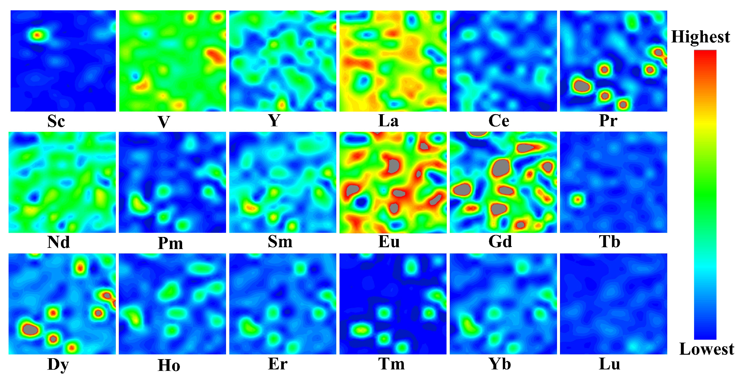
Figure 5, Figure 6 and Figure 7 show the synchrotron-based μXRF elemental maps of V and REEs in the corresponding leaching residues (GZ-L, PX-L, and PZ-L). In GZ-L (Figure 5), V remains distributed across most of the mapped area and appears mainly as a dispersed background rather than distinct enrichment zones. After leaching, several REEs are retained in localized regions. La, Ce, and Nd remain concentrated in relatively restricted areas, and Sm is also preserved in specific parts of the residue. Some middle-heavy REEs become more diffuse than in the raw sample, which may reflect partial dissolution or alteration of their original host phases followed by short-range redistribution, adsorption onto residual surfaces, or local reprecipitation. Because μXRF mapping does not directly determine mineral hosts or binding states, this interpretation should be regarded as a plausible geochemical explanation rather than definitive mineralogical evidence. The principal REE-enriched regions remain largely separated from the V-bearing background. Although Eu remains closer to V than most other REEs, its spatial relationship with V in the residue is visibly weaker than that observed in GZ-R.
Figure 5.
μXRF elemental maps of V and REEs in the GZ-L.
Figure 6.
μXRF elemental maps of V and REEs in the PX-L.
Figure 7.
μXRF elemental maps of V and REEs in the PZ-L.
The elemental maps of PX-L (Figure 6) show a different residual distribution pattern. V still appears mainly as a broadly distributed background, whereas the retained REEs are distributed more unevenly. Eu, Gd, and Dy persist in distinct enriched domains, indicating relatively clear residual enrichment of these elements under the applied leaching conditions. Some middle-heavy REEs still occur in related areas, but this relationship is expressed mainly within the REE group rather than between REEs and V. Accordingly, V shows little direct overlap with the REE-enriched regions.
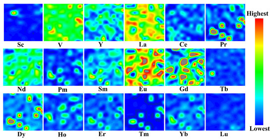
In PZ-L (Figure 7), the residue is characterized by the persistent enrichment of middle-heavy REEs. Elements such as Dy, Ho, and Yb retain pronounced hotspots and remain closely associated in space, indicating strong retention of this subgroup after acid leaching. By comparison, V is still present primarily as a dispersed background and overlaps only slightly with these enriched domains. Therefore, the spatial separation between V and the retained REEs is even more evident in PZ-L than in the other two residues. The elemental maps of the leaching residues show that, although leaching modified the distribution patterns of several REEs, the overall spatial separation between V and most REEs remained evident in the solid phase. In contrast, some REE subgroups, particularly part of the middle-heavy REEs, still preserved localized enrichment after leaching. These observations suggest that the leaching responses of V and REEs were not uniformly coupled under the tested conditions. To quantify these changes in spatial co-variation, Pearson correlation analysis was further performed and is discussed in Section 3.3.
3.3. Comparison of Inter-Element Relationships of REEs Before and After Leaching
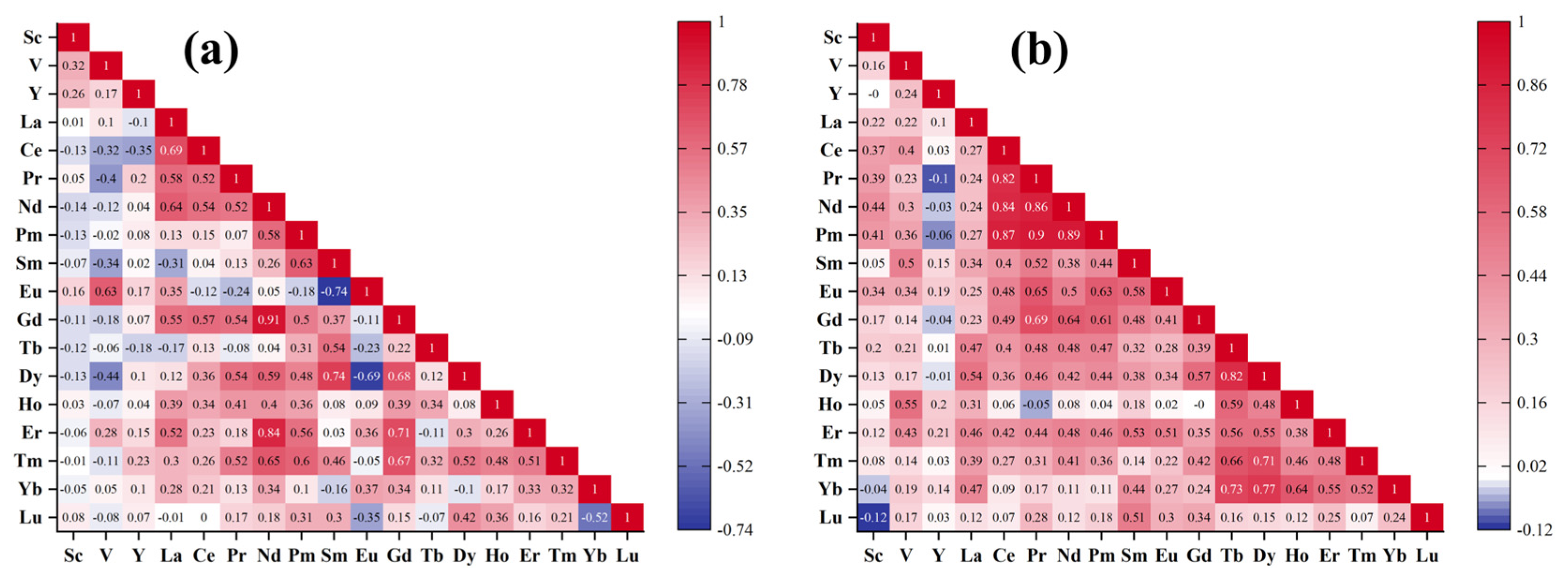
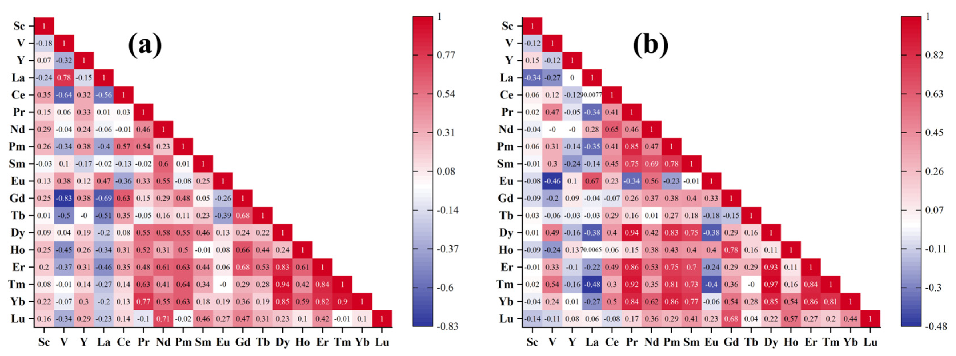
Figure 8, Figure 9 and Figure 10 present Pearson correlation matrices that quantify the spatial co-variation between V and individual REEs in the raw stone coal samples and their corresponding leaching residues [24]. As described in Section 2.4, the correlation coefficients are used as relative indicators of spatial co-variation within the mapped areas, rather than as quantitative concentration correlations or direct mineralogical evidence. These matrices are complementary to the μXRF elemental maps: the maps show where each element is distributed, whereas the matrices describe how closely the spatial intensity patterns of V and individual REEs, as well as those among different REEs, covary before and after leaching.
Figure 8.
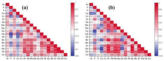
Correlation matrix of the selected elements in (a) GZ-R and (b) GZ-L.
Figure 9.
Correlation matrix of the selected elements in (a) PX-R and (b) PX-L.
Figure 10.
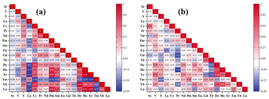
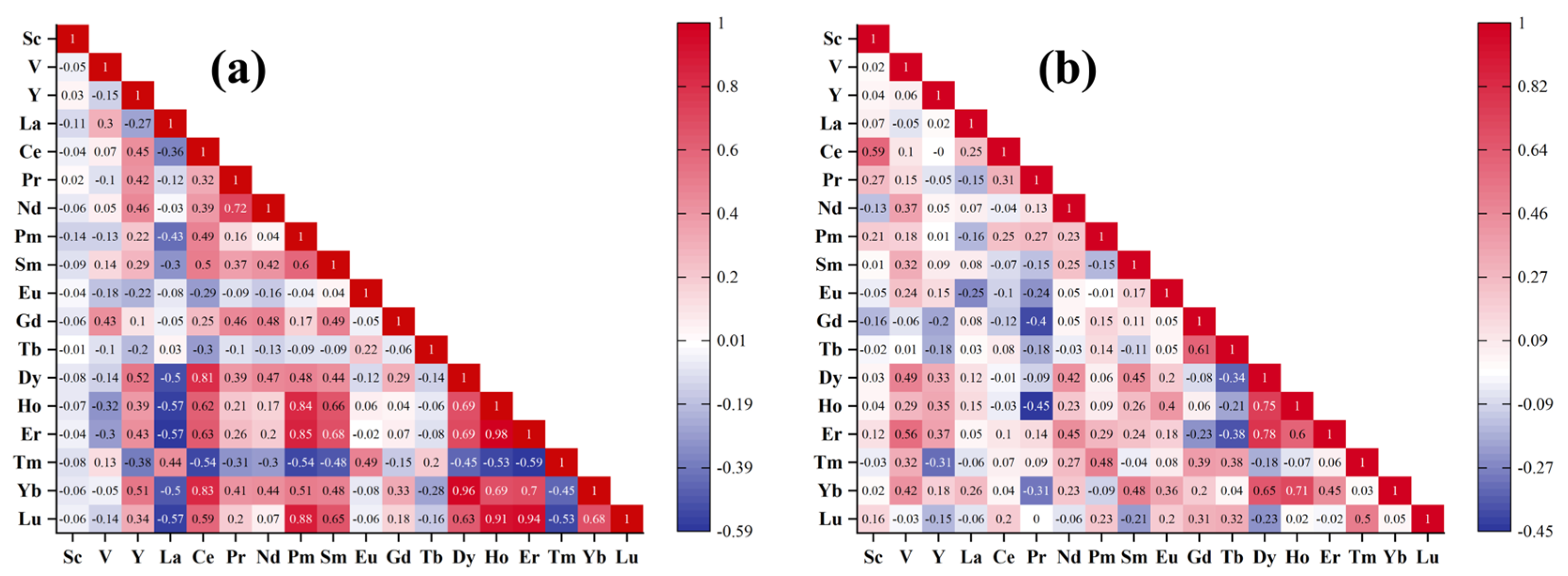
Correlation matrix of the selected elements in (a) PZ-R and (b) PZ-L.
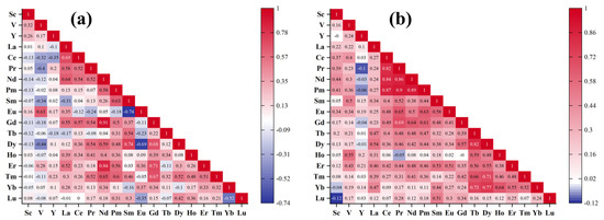
In the GZ sample, Eu shows the highest correlation with V in the raw sample (GZ-R, Figure 8a), with a coefficient of 0.63. By contrast, V-Ce (−0.32) and V-Pr (−0.40) are weakly or negatively correlated, indicating limited correspondence between V and most light REEs. Within the REE group, some element pairs remain closely related, such as Nd-Gd (0.91), whereas Sm and Eu are negatively correlated (−0.74), pointing to clear differentiation within the REE distribution pattern. After leaching, the V-Eu correlation decreases to 0.34, although Eu remains the REE most closely related to V in the residue (GZ-L, Figure 8b). At the same time, moderate positive correlations appear for V-Sm (0.50), V-Ho (0.55), and V-Er (0.43). This suggests that the V-REE relationships in GZ were modified after leaching and partly redistributed.
In the PX sample, the raw material (PX-R, Figure 9a) is characterized by a strong positive correlation between V and La (0.78), whereas V shows negative correlations with Ce (−0.64), Gd (−0.83), Tb (−0.50), and Ho (−0.45). This is consistent with the μXRF maps, which reveal separation between the light REEs and the middle-heavy REEs. Within the REE group, the middle-heavy REEs remain relatively close to one another, as reflected by Dy-Yb (0.85) and Ho-Er (0.61). After leaching (PX-L, Figure 9b), the strong positive and negative correlations involving V become less prominent. Moderate positive correlations appear for V-Pr (0.47), V-Dy (0.49), and V-Tm (0.54), whereas the V-La correlation changes to −0.27. At the same time, correlations among the middle-heavy REEs become stronger, including Dy-Er (0.93), Dy-Tm (0.97), and Er-Yb (0.86). These results show that the V-La relationship observed in the raw sample was not preserved after leaching.
In the PZ sample, the raw sample (PZ-R, Figure 10a) shows the weakest correlations between V and REEs. Most V-REE pairs are close to zero, with only weak positive correlations for V-La (0.30) and V-Gd (0.43), indicating that V is largely separated from the REEs in spatial distribution. In contrast, the middle-heavy REEs show close internal relationships, such as Ho-Er (0.98), Dy-Yb (0.96), and Yb-Lu (0.68), reflecting a relatively coherent subgroup. After leaching (PZ-L, Figure 10b), moderate positive correlations appear between V and several REEs, including Nd (0.37), Dy (0.49), Er (0.56), and Yb (0.42). At the same time, relationships among the middle-heavy REEs become weaker, with relatively high correlations retained only for Dy-Ho (0.75) and Ho-Yb (0.71).
Taken together, the μXRF observations and correlation matrices show that V and REEs did not exhibit a uniform spatial response to acid leaching across the three stone coal samples. In GZ, the relatively stronger V–Eu relationship weakened after leaching; in PX, the V–La association observed in the raw sample was not preserved in the residue; and in PZ, moderate post-leaching correlations appeared for some REEs without clear visual evidence of direct co-enrichment with V. These results indicate that leaching altered V–REE spatial relationships in a strongly sample-dependent manner, rather than producing synchronous extraction of V and REEs. A decrease in V–REE correlation after leaching is consistent with non-synchronous mobilization or retention, whereas persistent or enhanced positive correlations may reflect local co-retention, redistribution, or co-enrichment within the residue. Such behavior may be related to differences in original host phases and acid solubility, as well as adsorption onto newly exposed residual surfaces or local reprecipitation during leaching. However, because μXRF mapping and correlation analysis do not directly identify mineral hosts or chemical binding states, definitive interpretation of the controlling mechanisms requires further mineralogical and spectroscopic characterization, such as XRD, SEM-EDS, or X-ray absorption spectroscopy.
3.4. Conceptual Integrated Flowsheet for the Recovery of V and REEs from Stone Coal
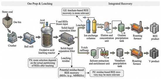
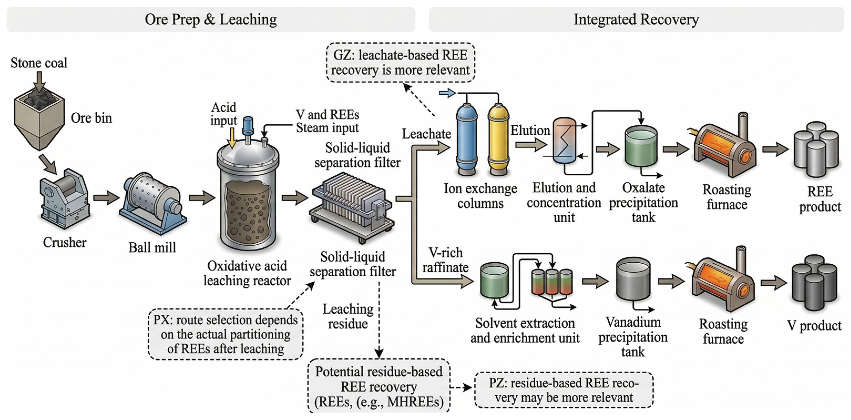
The μXRF elemental maps and Pearson correlation results collectively indicate that V and REEs do not exhibit a consistent or universally coupled leaching response in the three stone coal samples. In the raw materials, V shows only weak or selective spatial associations with most REEs, and these relationships are modified after leaching in a sample-dependent manner. In addition, some REEs, especially parts of the middle-heavy REE group, remain locally enriched in the solid residues. The post-leaching distribution tendency of REEs also differs among the three samples. In GZ, part of the REEs appears to be transferred into the leachate, while some REEs are still retained in the residue. In PX, the REEs show mixed behavior, with appreciable retention in the solid residue. In PZ, the μXRF maps show that REEs, especially several middle-heavy REEs, are still mainly concentrated in the residue after leaching. These differences indicate that REE recovery from stone coal should not be treated simply as an automatic extension of V extraction. Instead, the downstream recovery strategy should be selected according to whether REEs are mainly transferred into the leachate, retained in the residue, or distributed between both phases after V leaching. Based on these observations, Figure 11 is presented as a conceptual flowsheet for illustrating possible downstream REE recovery routes after V leaching.
Figure 11.
A conceptual staged recovery route for stone coal.
In this flowsheet, REE-bearing stone coal V ore first undergoes crushing and grinding, followed by acid leaching to mobilize V together with part of the REEs. The slurry is then separated into leachate and solid residue. At this stage, the distribution of individual REEs should be identified from experimental results rather than inferred only from the leaching behavior of V, because the present μXRF and correlation results show that REE redistribution during leaching is not necessarily consistent with that of V. For REEs transferred into the leachate, a solution-based recovery route can be considered. Ion exchange may be used for preliminary REE separation, followed by elution and concentration, oxalate precipitation, and roasting to obtain REE products. After REE recovery, V remaining in the solution can then be recovered by solvent extraction, V precipitation, and roasting [32,33].
A staged recovery route can therefore be considered in which REEs are separated either from the leachate or from the residue according to their post-leaching distribution tendency, followed by V recovery from the corresponding process stream [34,35]. This route may be suitable when some REEs show migration behavior relatively similar to that of V during leaching, as observed for Eu in the GZ sample. However, the present results indicate that such a route is not generally applicable to all stone coal samples. In the PX sample, no REE showed a stable relationship with V before and after leaching, suggesting that direct recovery of REEs from the V-bearing leachate may not always be appropriate. For samples in which a large fraction of REEs remains in the solid phase, such as PZ, the leaching residue should be regarded as a potential REE resource rather than as final waste. Although Nd, Dy, Er, and Yb show stronger correlations with V after leaching in PZ, the μXRF results indicate that these elements are still mainly concentrated in the residue. Their recovery should therefore be considered from the solid phase rather than from the leachate.
In summary, the key issue is not whether V and REEs are recovered at the same stage, but where the REEs are distributed after acid leaching. The proposed flowsheet is therefore conceptual rather than a validated industrial process. In practical terms, the front-end leaching and solid–liquid separation steps can remain unchanged, while the downstream REE recovery route may be adjusted according to the actual post-leaching distribution tendency of REEs. Further process-level validation is still required to optimize this recovery strategy. Nevertheless, this flexible approach retains V extraction as the main process while providing a potential pathway for improving the comprehensive utilization of chemically heterogeneous stone coal.
4. Conclusions
This study combined synchrotron radiation μXRF elemental mapping with Pearson correlation analysis to investigate the microscale relationships between V and REEs in three representative stone coal samples and their corresponding leaching residues. The main conclusions are as follows:
- (1)
- In the raw stone coal samples, V generally occurred as a dispersed background signal, whereas REEs were more commonly concentrated in localized hotspots. Spatial associations between V and most REEs were weak, while stronger relationships were more often observed within specific REE subgroups.
- (2)
- Leaching altered the spatial relationships between V and REEs in a sample-dependent manner. In the GZ sample, Eu showed the closest spatial relationship with V both before and after leaching, although the corresponding correlation coefficient decreased from 0.63 to 0.34. In the PX sample, the V-La association observed in the raw sample was not retained after leaching. In the PZ sample, moderate positive correlations between V and several REEs appeared after leaching, but these REEs were still mainly retained in the residue.
- (3)
- The results do not support a general assumption of synchronous leaching of REEs with V during acid leaching of stone coal. Changes in V-REE spatial co-variation after leaching more likely reflect ore-specific redistribution behavior than universal co-transfer into solution.
- (4)
- The recovery of REEs from stone coal should be designed according to their actual post-leaching distribution tendency between the leachate and the residue after V extraction. A staged recovery strategy is more appropriate than a fixed co-recovery assumption for the comprehensive utilization of V- and REE-bearing stone coal. In practical terms, the proposed flexible flowsheet does not require frequent modification of the front-end leaching process. Instead, the downstream REE recovery route can be selected according to the actual post-leaching distribution tendency of REEs between the leachate and the residue, allowing REE recovery to be directed to the stream in which they are preferentially enriched, thereby improving process feasibility and cost effectiveness.
Author Contributions
Writing—original draft, validation, writing—review and editing, H.-H.T., C.-Y.L., X.-X.Z. and Y.C.; methodology, formal analysis, supervision, L.W. and Q.-J.G.; resources, formal analysis, investigation, validation, writing—review and editing, supervision, W.S. All authors have read and agreed to the published version of the manuscript.
Funding
This research was supported by the National Natural Science Foundation of China (52474310), the Science and Technology Innovation Program of Hunan Province (2023RC3067) and the National Natural Science Foundation of Hunan Province, China (2023JJ20071).
Data Availability Statement
The original contributions presented in this study are included in the article. Further inquiries can be directed to the corresponding author.
Acknowledgments
The μ-XRF beam time was granted by the 4W1B end station of the Beijing Synchrotron Radiation Facility, Institute of High Energy Physics, Chinese Academy of Sciences. The staff members of 4W1B are acknowledged for their support in measurements and data reduction.
Conflicts of Interest
The authors declare no conflict of interest.
References
- Wang, Y.; Wang, Y.; Li, Y.; Wu, C.; Han, X.; Zhao, N.; Zhang, Z.; Dai, L.; Wang, L.; He, Z. A Review on Vanadium Extraction Techniques from Major Vanadium-containing Resources. Rare Met. 2024, 43, 4115–4131. [Google Scholar] [CrossRef] [Scilit]
- Choudhary, A.K.S.; Kumar, S.; Maity, S. A Study on Speciation and Enrichment of Rare Earth Elements (REE) by Sequential Extraction from a Potential Coal Fly Ash Resource and Its Role in REE Extractability. Hydrometallurgy 2024, 224, 106256. [Google Scholar] [CrossRef] [Scilit]
- Zhou, Z.; Sun, S.; Sun, Y.; Yang, Z.; Xiao, F.; Sui, C.; Yu, K.; Tu, G. A Sustainable Process for Vanadium Recovery from Stone Coal: Efficient Enrichment via Alkali Melting–Carbothermal Reduction. Miner. Eng. 2025, 234, 109710. [Google Scholar] [CrossRef] [Scilit]
- Agrawal, R.; Ragauskas, A.J. Sustainable Recovery of Rare Earth Elements (REEs) from Coal and Coal Ash through Urban Mining: A Nature Based Solution (NBS) for Circular Economy. J. Environ. Manag. 2025, 384, 125411. [Google Scholar] [CrossRef] [Scilit]
- Yang, J.; Wang, Y.; Gao, X.; Zuo, R.; Song, L.; Jin, C.; Wang, J.; Teng, Y. Vanadium: A Review of Different Extraction Methods to Evaluate Bioavailability and Speciation. Minerals 2022, 12, 642. [Google Scholar] [CrossRef] [Scilit]
- Jammulamadaka, H.; Pisupati, S.V. A Critical Review of Extraction Methods for Vanadium from Petcoke Ash. Fuels 2023, 4, 58–74. [Google Scholar] [CrossRef] [Scilit]
- Zhang, Y.-M.; Bao, S.-X.; Liu, T.; Chen, T.-J.; Huang, J. The Technology of Extracting Vanadium from Stone Coal in China: History, Current Status and Future Prospects. Hydrometallurgy 2011, 109, 116–124. [Google Scholar] [CrossRef] [Scilit]
- Wang, M.; Huang, S.; Chen, B.; Wang, X. A Review of Processing Technologies for Vanadium Extraction from Stone Coal. Miner. Process. Extr. Metall. 2020, 41, 249–264. [Google Scholar] [CrossRef] [Scilit]
- Zhao, Y.; Chen, L.; Yi, H.; Zhang, Y.; Song, S.; Bao, S. Vanadium transitions during roasting-leaching process of vanadium extraction from stone coal. Minerals 2018, 8, 63. [Google Scholar] [CrossRef] [Scilit]
- Chen, F.; Zhang, Y.; Huang, J.; Liu, T.; Xue, N. Mechanism of enhancing extraction of vanadium from stone coal by roasting with MgO. Minerals 2017, 7, 33. [Google Scholar] [CrossRef] [Scilit]
- Graedel, T.E.; Miatto, A. Vanadium: A U.S. Perspective on an Understudied Metal. Environ. Sci. Technol. 2023, 57, 8933–8942. [Google Scholar] [CrossRef] [Scilit]
- Stuckman, M.Y.; Lopano, C.L.; Granite, E.J. Distribution and speciation of rare earth elements in coal combustion by-products via synchrotron microscopy and spectroscopy. Int. J. Coal Geol. 2018, 195, 125–138. [Google Scholar] [CrossRef] [Scilit]
- Modi, P.; Jamal, A.; Varshney, R.; Rahi, I.C.; Siddiqui, M.A. Rare Earth Elements Mobility, Leaching and Recovery by Different Chemicals Treatment on Coal Samples and Calcined Samples of Sohagpur Coalfield, Madhya Pradesh, India. Int. J. Coal Prep. Util. 2023, 43, 148–168. [Google Scholar] [CrossRef] [Scilit]
- Zhang, W.; Noble, A.; Yang, X.; Honaker, R. A Comprehensive Review of Rare Earth Elements Recovery from Coal-Related Materials. Minerals 2020, 10, 451. [Google Scholar] [CrossRef] [Scilit]
- Menon, M.; Smalley, A.; Geraki, K.; Babaei, M.; Ranawana, V. A Comparison of Micronutrient Elements in Cooked White, Brown and Parboiled Rice Using Synchrotron Micro-X-Ray Fluorescence Imaging. Food Chem. 2025, 495, 146421. [Google Scholar] [CrossRef] [Scilit]
- Ysnaga, O.E. Studies About Atomic Compositions based on Synchrotron Radiation X- ray Microfluorescence (SR-μXRF) Analysis of Materials (at Micro- and Nano-Scale) Obtained by Sol-Gel Processes. Am. J. Phys. Appl. 2024, 12, 88–103. [Google Scholar] [CrossRef] [Scilit]
- Lorentz, K.O.; De Nolf, W.; Cotte, M.; Ioannou, G.; Foruzanfar, F.; Zaruri, M.R.; Sajjadi, S.M.S. Synchrotron Radiation Micro X-Ray Fluorescence (SR-μXRF) Elemental Mapping of Ancient Hair: Metals and Health at 3rd Millennium BCE Shahr-i Sokhta, Iran. J. Archaeol. Sci. 2020, 120, 105193. [Google Scholar] [CrossRef] [Scilit]
- Dai, S.; Ren, D.; Chou, C.-L.; Finkelman, R.B.; Seredin, V.V.; Zhou, Y. Geochemistry of Trace Elements in Chinese Coals: A Review of Abundances, Genetic Types, Impacts on Human Health, and Industrial Utilization. Int. J. Coal Geol. 2012, 94, 3–21. [Google Scholar] [CrossRef] [Scilit]
- Jaiswal, H.; Singh, A.K. Coal as a Non-Conventional Resource of Rare Earth Elements (REEs) and Trace Elements: Benefits and Impacts on Environment and Human Health—A Review. Results Earth Sci. 2025, 3, 100064. [Google Scholar] [CrossRef] [Scilit]
- Tsuji, K.; Nakano, K.; Takahashi, Y.; Hayashi, K.; Ro, C.-U. X-Ray Spectrometry. Anal. Chem. 2012, 84, 636–668. [Google Scholar] [CrossRef] [Scilit] [PubMed]
- Jiu, B.; Jin, Z.; Wang, Z. Multiscale In-Situ Elemental Characterization of Critical Elements in Low Rank Coal, Implications for Modes of Occurrence. Fuel 2023, 349, 128632. [Google Scholar] [CrossRef] [Scilit]
- Wang, J.; Zhang, Y.; Liu, T.; Huang, J. Evaluation of Microwave Intensified Vanadium Bearing-High Carbonaceous Shale Acid Extraction Process. Minerals 2018, 8, 113. [Google Scholar] [CrossRef] [Scilit]
- Solé, V.A.; Papillon, E.; Cotte, M.; Walter, P.; Susini, J. A Multiplatform Code for the Analysis of Energy-Dispersive X-Ray Fluorescence Spectra. Spectrochim. Acta Part B At. Spectrosc. 2007, 62, 63–68. [Google Scholar] [CrossRef] [Scilit]
- Pushie, M.J.; Pickering, I.J.; Korbas, M.; Hackett, M.J.; George, G.N. Elemental and chemically specific X-ray fluorescence imaging of biological systems. Chem. Rev. 2014, 114, 8499–8541. [Google Scholar] [CrossRef] [Scilit]
- Pattammattel, A.; Tappero, R.; Ge, M.; Zhang, H.; Aronstein, P.; Forman, H.J.; O’DAy, P.A.; Yan, H.; Chu, Y.S. Multimodal X-ray nano-spectromicroscopy analysis of chemically heterogeneous materials. Sci. Rep. 2022, 12, 17054. [Google Scholar] [CrossRef] [Scilit]
- Hettiarachchi, G.M.; Scheckel, K.G.; Ryan, J.A.; Sutton, S.R.; Newville, M. μ-XANES and μ-XRF Investigations of Metal Binding Mechanisms in Biosolids. J. Environ. Qual. 2006, 35, 342–351. [Google Scholar] [CrossRef] [Scilit]
- Dao, E.; Clavijo Jordan, M.V.; Geraki, K.; Martins, A.F.; Chirayil, S.; Sherry, A.D.; Farquharson, M.J. Using Micro-Synchrotron Radiation X-Ray Fluorescence (µ-SRXRF) for Trace Metal Imaging in the Development of MRI Contrast Agents for Prostate Cancer Imaging. J. Trace Elem. Med. Biol. 2022, 74, 127054. [Google Scholar] [CrossRef] [Scilit] [PubMed]
- Limbeck, A.; Brunnbauer, L.; Lohninger, H.; Pořízka, P.; Modlitbová, P.; Kaiser, J.; Janovszky, P.; Kéri, A.; Galbács, G. Methodology and Applications of Elemental Mapping by Laser Induced Breakdown Spectroscopy. Anal. Chim. Acta 2021, 1147, 72–98. [Google Scholar] [CrossRef] [Scilit] [PubMed]
- Balaram, V. Rare Earth Elements: A Review of Applications, Occurrence, Exploration, Analysis, Recycling, and Environmental Impact. Geosci. Front. 2019, 10, 1285–1303. [Google Scholar] [CrossRef] [Scilit]
- Hidayah, N.N.; Abidin, S.Z. Extraction of Light, Medium and Heavy Rare-Earth Elements Using Synergist Extractants Developed from Ionic Liquid and Conventional Extractants. C. R. Chim. 2019, 22, 728–744. [Google Scholar] [CrossRef] [Scilit]
- Hatje, V.; Schijf, J.; Johannesson, K.H.; Andrade, R.; Caetano, M.; Brito, P.; Haley, B.A.; Lagarde, M.; Jeandel, C. The Global Biogeochemical Cycle of the Rare Earth Elements. Glob. Biogeochem. Cycles 2024, 38, e2024GB008125. [Google Scholar] [CrossRef] [Scilit]
- Zeng, L.; Li, Q.; Xiao, L. Extraction of Vanadium from the Leach Solution of Stone Coal Using Ion Exchange Resin. Hydrometallurgy 2009, 97, 194–197. [Google Scholar] [CrossRef] [Scilit]
- Ng, L.Y.; Choo, E.H.; Chiang, A.K.M.; Ng, C.Y. Recovery of Rare Earth Elements: A Comprehensive Review from the Perspective of Secondary Sources and Potentially Greener Leachants. Sep. Purif. Technol. 2026, 382, 135752. [Google Scholar] [CrossRef] [Scilit]
- Li, X.; Deng, Z.; Wei, C.; Li, C.; Li, M.; Fan, G.; Huang, H. Solvent Extraction of Vanadium from a Stone Coal Acidic Leach Solution Using D2EHPA/TBP: Continuous Testing. Hydrometallurgy 2015, 154, 40–46. [Google Scholar] [CrossRef] [Scilit]
- Sobri, N.A.M.; Harun, N.; Yunus, M.Y.M. A Review of the Ion Exchange Leaching Method for Extracting Rare Earth Elements from Ion Adsorption Clay. Chem. Eng. Res. Des. 2024, 208, 94–114. [Google Scholar] [CrossRef] [Scilit]

Disclaimer/Publisher’s Note: The statements, opinions and data contained in all publications are solely those of the individual author(s) and contributor(s) and not of MDPI and/or the editor(s). MDPI and/or the editor(s) disclaim responsibility for any injury to people or property resulting from any ideas, methods, instructions or products referred to in the content. |
© 2026 by the authors. Licensee MDPI, Basel, Switzerland. This article is an open access article distributed under the terms and conditions of the Creative Commons Attribution (CC BY) license.