


Summary
Burgunder J-M, Tan EK, Wirthmüller U, Jung HH. LRRK2 mutations are not frequent in Swiss patients with Parkinson’s disease. Schweiz Arch Neurol Psychiatr. 2008;159:412–8.
A wealth of recent data has shed light on the genetics of Parkinson’s disease. Mutations of specific genes have been discovered in a number of autosomal dominant and recessive forms of the disease, and polymorphisms in the same or other genes found to be associated with its occurrence in sporadic forms. Mutations in the gene encoding leucine-rich repeat kinase 2 (LRRK2) cause an autosomal dominant form of Parkinson’s disease (PARK8) and have been found in patients with Parkinson’s disease with a highly variable frequency in different populations in the world. In this study we have examined the most common mutations so far described in this large gene:p.R1441C (together with p.R1441G and p.R1441H), p.Y1699C and p.G2019S. Seventy-three patients from Switzerland with Parkinson’s disease diagnosed according to Brain Bank criteria with the exception of positive family history were included in this study.The three exons, in which the above mutations had been found, were sequenced after amplification. There were no sequence differences compared with the published wild-type sequences in all patients.These data point at the low incidence of LRRK2 mutation in the Swiss (regions of Berne and Zurich) populations, similar to findings in other central European populations, but at great variance to Mediterranean populations.
Introduction
Parkinson’s disease is a frequent disorder, 1% of people older than 60 years in Western countries are estimated to be affected, and the average age at onset is 60 [1].With an age of 85, the prevalence even increases to 4% [2]. Environmental factors, including industrial pollutants, farming, living in a rural area, drinking well water, pesticide and herbicide exposure and head trauma [3,4,5,6,7,8,9] have been associated with an increased risk to develop the disorder. The best-established example of an environmental factor leading to Parkinson’s disease is MPTP, an oxidant present in artisanal drug preparations, which had caused an epidemic of the disorder in young patients in the eighties of the last century, and which is now commonly used to induce Parkinson’s disease in animal models [10]. Other non-genetic causes for Parkinson’s disease have been reviewed recently [11].
However, a positive family history has consistently been reported as a strong risk factor for the disease [3,12], and a large family from Grisons has been reported in the early literature in the field [13]. Up to 27% of patients with the disorder report a relative with Parkinson’s disease and up to 16% its presence in a parent or sibling.The risk of firstdegree relatives to get Parkinson’s disease is up to 9.5 times greater as compared to relatives of controls [8,12,14,15]. In a large twin study monozygotic twin pairs with onset in at least one person before the age of 50 had a significant higher risk of 6 times as compared to dizygotic twin pairs [16]. When the involvement of the dopaminergic function was examined using PET, the concordance in a group of 18 monozygotic twin pairs was significant at baseline, as compared to 16 dizygotic twin pairs [17]. Furthermore, the proportion became even higher in monozygotic twins during follow-up [17]. In a more recent study similarity for age at onset among sibling pairs with Parkinson’s disease and the greater risk for biological relatives as compared to spouses have again supported the presence of a genetic component [18]. Risk of Parkinson’s disease was high in first relatives of both early and late-onset patients, the risk for siblings was increased only for those of early onset patients [19]. Other studies have started to examine the interaction between genetic and environmental factors. For example, there is a significant increase in risk in cases with positive family history and smoking [20].
Table 1.
Selected genes and loci associated with Parkinson’s disease and parkinsonism.
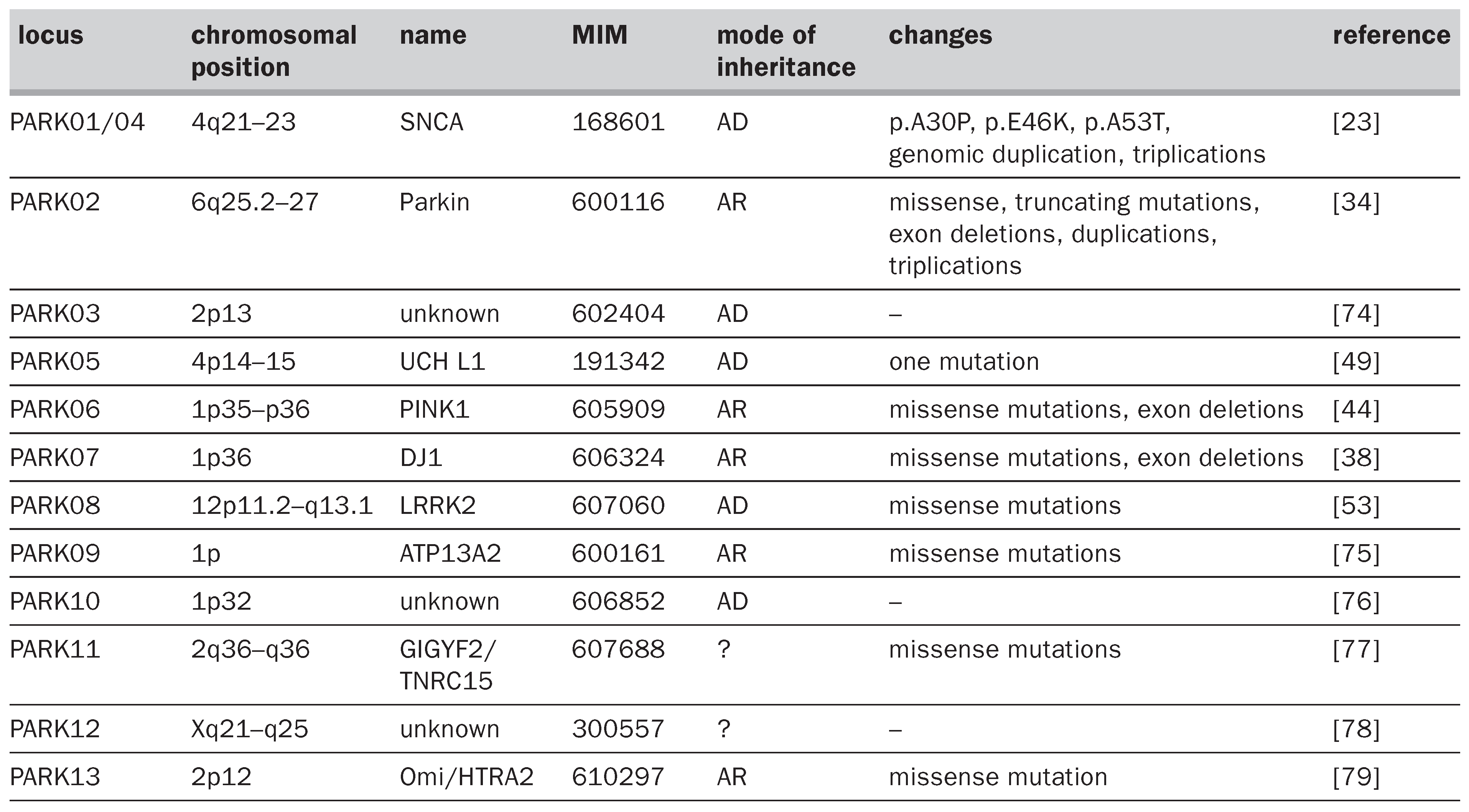
The strongest evidence for a genetic causation of Parkinson’s disease has come from the observation of families with a clear-cut autosomal dominant or recessive inheritance. The investigations in these families with the performance of linkage studies have allowed the discovery of different genes (table 1) [21,22]; furthermore, additional loci are known, and in time more genes with mutation directly causing the disorder are expected to be discovered.
α-synuclein was the first gene with mutations causing Parkinson’s disease [23] and a p.A53T was found in one large family of Italian origin and three smaller pedigrees from Greece [23]. Later a p.A30P [24] and a p.E46K [25] mutation were found in other families of German or Spanish origin, respectively. Duplication or triplication of the region containing the gene, or of individual exons was found in additional families [26], with dementia in addition to Parkinsonian features, and a more severe phenotype in patients with triplication [27], pointing at a gene dose effect with more prominent α-synuclein accumulation within neurons and subsequent loss of dopamine function. The p.A53T mutation was found in additional families of Greek and Italian origin, most probably related to the first one, however, a number of other studies could not find additional mutations of this gene [28,29,30,31]. Even considering the very low number of cases with α-synuclein mutation, the discovery of this protein has greatly advanced our knowledge of the molecular pathophysiology of idiopathic Parkinson’s disease, Lewy body disease and other neurodegenerative disorders. This protein is also involved in cases without mutations, leading to the concept of synucleinopathies [32], including the definition of potential molecular therapeutic targets [33].
Mutations in the gene coding for parkin [34] were found later, first in Japanese patients with autosomal recessive early onset parkinsonism. A large number of additional mutations were found in many other populations studied, including more than 50 point mutations, as well as exonic rearrangements, duplications and deletions, accounting for half of familial cases and 70% of sporadic cases with age of onset younger than 20 years [35,36,37]. The search for parkin mutations can be considered in young patients with the disorder, or in those with an autosomal recessive inheritance.
Following the discovery of missense and deletion mutations in families with recessive Parkinson’s disease [38], large-scales studies have examined the DJ-1 gene in other patients but only rare additional mutations have been found [39,40,41,42,43], suggesting that only 1% of patients with early onset Parkinson’s disease may have a DJ-1 mutation.
Two mutations were found in a novel gene PINK1 [44] in families with autosomal recessive juvenile-onset Parkinson’s disease. Later additional mutations, including frameshift, missense and truncating mutations were indentified [45,46,47,48], with a relative amount of 1–7% of patients with Parkinson’s disease of Caucasian origin [43], suggesting that PINK1 and parkin may be the most common genes in autosomal recessive and possibly also sporadic young-onset Parkinson’s disease.
A p.I93M substitution found in exon 4 of ubiquitin carboxy-terminal hydrolase L1 (UCHL1) has been reported in a German family [49]. This has led to the examination of the role of this enzyme in Parkinson’s disease, but, no further mutation has been found in numerous studies.
Further loci have been mapped in additional families (table 1), and the discovery of the respective genes is expected to further advance our understanding of the molecular pathophysiology of the disease. It is of note that mutations in other genes, involved in neurodegenerative disorders other than Parkinson’s disease, may sometimes have presentations similar to this disorder. This includes frontotemporal dementia, some of the spinocerebellar ataxias, dystonia-parkinson syndrome and mitochondrial disorders.
Finally, PARK8 has been found on chromosome 12p11.2–q13 in a Japanese family with autosomal dominant Parkinson’s disease with otherwise typical presentation similar to idiopathic Parkinson’s disease, including age at onset, laterality and good response to L-dopa. The overwhelming majority of cases have typical Lewy body pathology, but absence of Lewy bodies in rare cases [50,51]. This locus has been confirmed in families from other ethnic origins [52] and shortly thereafter, several mutations in a novel gene encoding leucine-rich repeat kinase 2 (LRRK2) cloned within this locus have been reported [52,53].Mutations in the LRRK2 account to about 13% of cases with autosomal dominant Parkinson’s disease [28], they are also found in a substantial number of sporadic cases [54].As an example of a local study we therefore examined the most commonly found LRRK2 mutations in a cohort of patients with Parkinson’s disease from Switzerland.
Material and methods
Seventy-three consecutive patients with Parkinson’s disease were included.They had been examined at the Department of Neurology, University Hospital, Zurich (H.H.J.) or at the Neurology practice in downtown Berne (J.-M. B.) and were all of Caucasian origin.The study has been approved by the respective Ethics Committees of the Cantons of Zurich and Berne.The diagnosis had been made according to Brain Bank criteria, with the exception of positive family history as an exclusion criterion for idiopathic Parkinson’s disease, and all patients responded well to treatment with L-dopa or dopamine agonists.
Blood was withdrawn and DNA extracted according to standard procedures using QIAGEN PCR purification kit. Following primer pairs were used: for exon 31, mutations R1441C, R1441G and R1441H: 5’-CTTCTGAAGTCTGCTAGTTTCT-3’(forward) and 5’-CGTTGGCACACATTTGGATG-3’(reverse); for exon 35, mutation Y1699C: 5’-GCTCAACAAGGTTGGGTGTT-3’(forward) and 5’-ATGCTTTCAGGGAGAGGTAAG-3’ (reverse); for exon 41, mutation G2019S: 5’-TTTTGATGCTTGACATAGTGGAC-3’(forward) and 5’-CACATCTGAGGTCAGTGGTTATC-3’ (reverse). Direct sequencing was performed after optimised PCR amplification using Big Dye Terminator ver.3.1 cycle (Applied Biosystems) on an ABI3100 Genetic Analyser and analysed with DNA Sequencing Analysis (ver.3.7) software (Applied Biosystems).
Results
The age of the patients varied from 49 to 81 years (mean 70), 43% were women and 17% had a positive family history. No mutation was detected in any of the three exons studied in any of the patients. No polymorphisms were found either.
Discussion
Our exploration in a preliminary small set of patients with idiopathic and familial Parkinson’s disease from Switzerland did not demonstrate the presence of any of the LRRK2 mutations most commonly found in similar or larger sets of patients from other countries. Due to the small size of the sample, we cannot exclude the presence of these mutations in PD patients from Swiss origin, but based on our data we can conclude that the frequency will most probably be in the lower range found in different populations. A high frequency of the p.G2019S mutation in sporadic Parkinson’s disease has been found in North African Arabs (20%) [55] and Ashkenazi Jews (13%) [56], whereas the same mutation was absent in Chinese PD patients [57]. About 7% of PD patients from cohorts of similar size as ours in the Iberic peninsula carried the mutation [58,59], and a low prevalence of this mutation from 0 to 3% was documented in other European populations, like Poland [60], Norway [61] and Germany [28].The absence of this mutation in our set of patients is in line with the low frequency of the mutation in North and Middle European populations. As the penetrance of the p.G2019S mutation increases with age [54,62] and the age of patients in our cohort is in the higher range, the absence of this mutation was not due to a selection bias of younger patients. Several mutations at position 1441 have been described earlier in different populations. The p.R1441H mutation has been found in rare families of different origins [59,63,64,65]. In contrast, the p.R1441G mutation is quite frequent in PD patients from the Basque region [66].
LRRK2 codes for a protein with 2527 amino acids [52], which has a molecular mass of approximately 250–280 kDA [67,68]. The gene is selectively expressed at high levels in the mouse [69], rat and human striatum [70], which is in contrast with the more widespread expression of other genes mutated in familial forms of Parkinson’s disease.The presence of specific domains, including ANK, LRR, Roc, COR, Kinase or MAPKKK, and WD40, suggests a role in signalling [52,71]. The soluble protein is located in the cytoplasm [72], consistent with the absence of specific targeting signal, as demonstrated after HA-tagging in cell culture and immunohistochemistry in human brain sections [68]. Subcellular fractionation suggests a location close to mitochondria, microtubules, Golgi and endoplasmatic reticulum [73]. Interactions of LRRK2 have been demonstrated with parkin, but not with DJ-1, α-synuclein or tau [72]. Mutations of the protein do not consequently give rise to their accumulation [68], which is in contrast with the clear-cut pathophysiological mechanism of α-synuclein accumulation in hereditary and sporadic Parkinson’s disease. Functional analysis of one mutation, p.I2020T, located at the beginning of the kinase domain activation loop, led to the description of a significantly higher kinase activity, pointing at a gain-of-function for this particular mutation [73]. The p.R1441C, p.Y1699C and p.G2019S mutations, which we have studied in the present report, lead to neurodegeneration when expressed in specific cells in culture [72]. The p.R1441C mutation is localised within the GTPase domain, and the p.G2019S within the mixed-kinase-like domain. Both mutations also lead to an increase in enzymatic activity, with increased substrate as well as auto-phosphorylation [67]. However, analysis of further mutations of this large protein with several domains will be needed to draw general conclusions about its physiological role and the biochemical changes induced by its mutations.
Overall, there is a wide range of clinical presentations and neuropathological findings between the different mutations, and no clear-cut molecular–clinical correlation can be drawn so far. Although LRRK2 mutations may play a role in predisposing neurons to toxic effects, the exact nature of such epigenetic and environmental factors is not yet known.The high frequency of mutations in certain populations, both in familial and sporadic cases, indicates that this protein may play a key role in the pathogenesis of Parkinson’s disease. As the other proteins involved in the pathogenetic cascade in familial Parkinsonian syndromes, LRRK2 has the potential to deliver future drug targets for disease-modifying therapies. In order to advance in that direction, further data for clinical and molecular correlation are needed, as well as further studies in the biology of this protein.
References
- Mayeux, R; Marder, K; Cote, LJ; Denaro, J; Hemenegildo, N; Mejia, H; et al. The frequency of idiopathic Parkinson’s disease by age, ethnic group, and sex in northern Manhattan, 1988–1993. Am J Epidemiol. 1995, 142, 820–7. [Google Scholar] [CrossRef] [Scilit]
- de Rijk, MC; Launer, LJ; Berger, K; Breteler, MM; Dartigues, JF; Baldereschi, M; et al. Prevalence of Parkinson’s disease in Europe: a collaborative study of population-based cohorts. Neurologic Diseases in the Elderly Research Group. Neurology 2000, 54((11) Suppl 5, S21–3. [Google Scholar]
- Semchuk, KM; Love, EJ; Lee, RG. Parkinson’s disease and exposure to agricultural work and pesticide chemicals. Neurology 1992, 42, 1328–35. [Google Scholar] [CrossRef] [Scilit]
- Golbe, LI; Farrell, TM; Davis, PH. Follow-up study of earlylife protective and risk factors in Parkinson’s disease. Mov Disord. 1990, 5, 66–70. [Google Scholar] [CrossRef] [Scilit] [PubMed]
- Semchuk, KM; Love, EJ; Lee, RG. Parkinson’s disease and exposure to rural environmental factors: a population-based case-control study. Can J Neurol Sci. 1991, 18, 279–86. [Google Scholar] [CrossRef] [Scilit]
- Rybicki, BA; Johnson, CC; Uman, J; Gorell, JM. Parkinson’s disease mortality and the industrial use of heavy metals in Michigan. Mov Disord. 1993, 8, 87–92. [Google Scholar] [CrossRef] [Scilit] [PubMed]
- Seidler, A; Hellenbrand, W; Robra, BP; Vieregge, P; Nischan, P; Joerg, J; et al. Possible environmental, occupational, and other etiologic factors for Parkinson’s disease: a case-control study in Germany. Neurology 1996, 46, 1275–84. [Google Scholar] [CrossRef] [Scilit] [PubMed]
- Semchuk, KM; Love, EJ; Lee, RG. Parkinson’s disease: a test of the multifactorial etiologic hypothesis. Neurology 1993, 43, 1173–80. [Google Scholar] [CrossRef] [Scilit]
- Factor, SA; Weiner, WJ. Prior history of head trauma in Parkinson’s disease. Mov Disord. 1991, 6, 225–9. [Google Scholar] [CrossRef] [Scilit]
- Smeyne, RJ; Jackson-Lewis, V. The MPTP model of Parkinson’s disease. Brain Res Mol Brain Res. 2005, 134, 57–66. [Google Scholar] [CrossRef] [Scilit]
- Chade, AR; Kasten, M; Tanner, CM. Nongenetic causes of Parkinson’s disease. J Neural Transm Suppl. 2006, (70), 147–51. [Google Scholar]
- Taylor, CA; Saint-Hilaire, MH; Cupples, LA; Thomas, CA; Burchard, AE; Feldman, RG; et al. Environmental, medical, and family history risk factors for Parkinson’s disease: a New England-based case control study. Am J Med Genet. 1999, 88, 742–9. [Google Scholar] [CrossRef]
- Branger, F. Une forme familiale de paralysie agitante dans une souche des grisons. J Génét Hum. 1956, 5, 261–70. [Google Scholar]
- Marder, K; Tang, MX; Mejia, H; Alfaro, B; Cote, L; Louis, E; et al. Risk of Parkinson’s disease among first-degree relatives: a community-based study. Neurology 1996, 47, 155–60. [Google Scholar] [CrossRef] [Scilit]
- Payami, H; Larsen, K; Bernard, S; Nutt, J. Increased risk of Parkinson’s disease in parents and siblings of patients. Ann Neurol. 1994, 36, 659–61. [Google Scholar] [CrossRef] [Scilit]
- Tanner, CM; Ottman, R; Goldman, SM; Ellenberg, J; Chan, P; Mayeux, R; et al. Parkinson’s disease in twins: an etiologic study. JAMA 1999, 281, 341–6. [Google Scholar] [CrossRef] [Scilit]
- Piccini, P; Burn, DJ; Ceravolo, R; Maraganore, D; Brooks, DJ. The role of inheritance in sporadic Parkinson’s disease: evidence from a longitudinal study of dopaminergic function in twins. Ann Neurol. 1999, 45, 577–82. [Google Scholar] [CrossRef] [Scilit]
- Maher, NE; Golbe, LI; Lazzarini, AM; Mark, MH; Currie, LJ; Wooten, GF; et al. Epidemiologic study of 203 sibling pairs with Parkinson’s disease: the GenePD study. Neurology 2002, 58, 79–84. [Google Scholar] [CrossRef] [Scilit] [PubMed]
- Marder, K; Levy, G; Louis, ED; Mejia-Santana, H; Cote, L; Andrews, H; et al. Familial aggregation of early- and lateonset Parkinson’s disease. Ann Neurol. 2003, 54, 507–13. [Google Scholar] [CrossRef] [Scilit] [PubMed]
- Elbaz, A; Manubens-Bertran, JM; Baldereschi, M; Breteler, MM; Grigoletto, F; Lopez-Pousa, S; et al. Parkinson’s disease, smoking, and family history. EUROPARKINSON Study Group. J Neurol. 2000, 247, 793–8. [Google Scholar] [CrossRef] [Scilit] [PubMed]
- Klein, C; Schlossmacher, MG. The genetics of Parkinson’s disease: implications for neurological care. Nat Clin Pract Neurol. 2006, 2, 136–46. [Google Scholar] [CrossRef] [Scilit]
- Tan, EK; Skipper, LM. Pathogenic mutations in Parkinson’s disease. Hum Mutat. 2007, 28, 641–53. [Google Scholar] [CrossRef] [Scilit] [PubMed]
- Polymeropoulos, MH; Lavedan, C; Leroy, E; Ide, SE; Dehejia, A; Dutra, A; et al. Mutation in the alpha-synuclein gene identified in families with Parkinson’s disease. Science 1997, 276, 2045–7. [Google Scholar] [CrossRef] [Scilit]
- Kruger, R; Kuhn, W; Muller, T; Woitalla, D; Graeber, M; Kosel, S; et al. Ala30Pro mutation in the gene encoding alpha-synuclein in Parkinson’s disease. Nat Genet. 1998, 18, 106–8. [Google Scholar] [CrossRef] [Scilit]
- Zarranz, JJ; Alegre, J; Gomez-Esteban, JC; Lezcano, E; Ros, R; Ampuero, I; et al. The new mutation, E46K, of alpha-synuclein causes Parkinson and Lewy body dementia. Ann Neurol. 2004, 55, 164–73. [Google Scholar] [CrossRef] [Scilit] [PubMed]
- Nishioka, K; Hayashi, S; Farrer, MJ; Singleton, AB; Yoshino, H; Imai, H; et al. Clinical heterogeneity of alphasynuclein gene duplication in Parkinson’s disease. Ann Neurol. 2006, 59, 298–309. [Google Scholar] [CrossRef] [Scilit] [PubMed]
- Singleton, AB; Farrer, M; Johnson, J; Singleton, A; Hague, S; Kachergus, J; et al. Alpha-synuclein locus triplication causes Parkinson’s disease. Science 2003, 302, 841. [Google Scholar] [CrossRef] [Scilit]
- Berg, D; Schweitzer, K; Leitner, P; Zimprich, A; Lichtner, P; Belcredi, P; et al. Type and frequency of mutations in the LRRK2 gene in familial and sporadic Parkinson’s disease*. Brain 2005, 128, 3000–11. [Google Scholar] [CrossRef] [Scilit]
- Berg, D; Niwar, M; Maass, S; Zimprich, A; Moller, JC; Wuellner, U; et al. Alpha-synuclein and Parkinson’s disease: implications from the screening of more than 1,900 patients. Mov Disord. 2005, 20, 1191–4. [Google Scholar] [CrossRef] [Scilit]
- Johnson, J; Hague, SM; Hanson, M; Gibson, A; Wilson, KE; Evans, EW; et al. SNCA multiplication is not a common cause of Parkinson’s disease or dementia with Lewy bodies. Neurology 2004, 63, 554–6. [Google Scholar] [CrossRef] [Scilit]
- Gispert, S; Trenkwalder, C; Mota-Vieira, L; Kostic, V; Auburger, G. Failure to find alpha-synuclein gene dosage changes in 190 patients with familial Parkinson’s disease. Arch Neurol. 2005, 62, 96–8. [Google Scholar] [CrossRef] [Scilit]
- Giorgi, FS; Bandettini di Poggio, A; Battaglia, G; Pellegrini, A; Murri, L; Ruggieri, S; et al. A short overview on the role of alpha-synuclein and proteasome in experimental models of Parkinson’s disease. J Neural Transm Suppl. 2006, (70), 105–9. [Google Scholar]
- Lee, VM; Trojanowski, JQ. Mechanisms of Parkinson’s disease linked to pathological alpha-synuclein: new targets for drug discovery. Neuron 2006, 52, 33–8. [Google Scholar] [CrossRef] [Scilit] [PubMed]
- Kitada, T; Asakawa, S; Hattori, N; Matsumine, H; Yamamura, Y; Minoshima, S; et al. Mutations in the parkin gene cause autosomal recessive juvenile parkinsonism. Nature 1998, 392, 605–8. [Google Scholar] [CrossRef] [Scilit]
- Lucking, CB; Durr, A; Bonifati, V; Vaughan, J; De Michele, G; Gasser, T; et al. Association between early-onset Parkinson’s disease and mutations in the parkin gene. French Parkinson’s Disease Genetics Study Group. N Engl J Med. 2000, 342, 1560–7. [Google Scholar]
- Periquet, M; Lucking, C; Vaughan, J; Bonifati, V; Durr, A; De Michele, G; et al. Origin of the mutations in the parkin gene in Europe: exon rearrangements are independent recurrent events, whereas point mutations may result from Founder effects. Am J Hum Genet. 2001, 68, 617–26. [Google Scholar] [CrossRef] [Scilit]
- Mata, IF; Lockhart, PJ; Farrer, MJ. Parkin genetics: one model for Parkinson’s disease. Hum Mol Genet. 2004, 13 Spec No 1, R127–33. [Google Scholar] [CrossRef] [Scilit]
- Bonifati, V; Rizzu, P; van Baren, MJ; Schaap, O; Breedveld, GJ; Krieger, E; et al. Mutations in the DJ-1 gene associated with autosomal recessive early-onset parkinsonism. Science 2003, 299, 256–9. [Google Scholar] [CrossRef] [Scilit]
- Lockhart, PJ; Bounds, R; Hulihan, M; Kachergus, J; Lincoln, S; Lin, CH; et al. Lack of mutations in DJ-1 in a cohort of Taiwanese ethnic Chinese with early-onset parkinsonism. Mov Disord. 2004, 19, 1065–9. [Google Scholar] [CrossRef] [Scilit] [PubMed]
- Clark, LN; Afridi, S; Mejia-Santana, H; Harris, J; Louis, ED; Cote, LJ; et al. Analysis of an early-onset Parkinson’s disease cohort for DJ-1 mutations. Mov Disord. 2004, 19, 796–800. [Google Scholar] [CrossRef] [Scilit] [PubMed]
- Hering, R; Petrovic, S; Mietz, EM; Holzmann, C; Berg, D; Bauer, P; et al. Extended mutation analysis and association studies of Nurr1 (NR4A2) in Parkinson’s disease. Neurology 2004, 62, 1231–2. [Google Scholar] [CrossRef] [Scilit] [PubMed]
- Tan, EK; Tan, C; Zhao, Y; Yew, K; Shen, H; Chandran, VR; et al. Genetic analysis of DJ-1 in a cohort Parkinson’s disease patients of different ethnicity. Neurosci Lett. 2004, 367, 109–12. [Google Scholar] [CrossRef] [Scilit] [PubMed]
- Healy, DG; Abou-Sleiman, PM; Valente, EM; Gilks, WP; Bhatia, K; Quinn, N; et al. DJ-1 mutations in Parkinson’s disease. J Neurol Neurosurg Psychiatry 2004, 75, 144–5. [Google Scholar] [PubMed]
- Valente, EM; Abou-Sleiman, PM; Caputo, V; Muqit, MM; Harvey, K; Gispert, S; et al. Hereditary early-onset Parkinson’s disease caused by mutations in PINK1. Science 2004, 304, 1158–60. [Google Scholar] [CrossRef] [Scilit]
- Tan, EK; Yew, K; Chua, E; Puvan, K; Shen, H; Lee, E; et al. PINK1 mutations in sporadic early-onset Parkinson’s disease. Mov Disord. 2006, 21, 789–93. [Google Scholar] [CrossRef] [Scilit]
- Fung, HC; Chen, CM; Hardy, J; Singleton, AB; Lee-Chen, GJ; Wu, YR. Analysis of the PINK1 gene in a cohort of patients with sporadic early-onset parkinsonism in Taiwan. Neurosci Lett. 2006, 394, 33–6. [Google Scholar] [CrossRef] [Scilit]
- Bonifati, V; Rohe, CF; Breedveld, GJ; Fabrizio, E; De Mari, M; Tassorelli, C; et al. Early-onset parkinsonism associated with PINK1 mutations: frequency, genotypes, and phenotypes. Neurology 2005, 65, 87–95. [Google Scholar] [CrossRef] [Scilit]
- Albanese, A; Valente, EM; Romito, LM; Bellacchio, E; Elia, AE; Dallapiccola, B. The PINK1 phenotype can be indistinguishable from idiopathic Parkinson’s disease. Neurology 2005, 64, 1958–60. [Google Scholar] [CrossRef] [Scilit]
- Leroy, E; Boyer, R; Auburger, G; Leube, B; Ulm, G; Mezey, E; et al. The ubiquitin pathway in Parkinson’s disease. Nature 1998, 395, 451–2. [Google Scholar] [CrossRef] [Scilit]
- Funayama, M; Hasegawa, K; Kowa, H; Saito, M; Tsuji, S; Obata, F. A new locus for Parkinson’s disease (PARK8) maps to chromosome 12p11.2-q13.1. Ann Neurol. 2002, 51, 296–301. [Google Scholar] [CrossRef] [Scilit]
- Funayama, M; Hasegawa, K; Ohta, E; Kawashima, N; Komiyama, M; Kowa, H; et al. An LRRK2 mutation as a cause for the parkinsonism in the original PARK8 family. Ann Neurol. 2005, 57, 918–21. [Google Scholar] [CrossRef] [Scilit]
- Zimprich, A; Biskup, S; Leitner, P; Lichtner, P; Farrer, M; Lincoln, S; et al. Mutations in LRRK2 cause autosomal-dominant parkinsonism with pleomorphic pathology. Neuron 2004, 44, 601–7. [Google Scholar] [CrossRef] [Scilit]
- Paisan-Ruiz, C; Jain, S; Evans, EW; Gilks, WP; Simon, J; van der Brug, M; Lopez de Munain, A; et al. Cloning of the gene containing mutations that cause PARK8-linked Parkinson’s disease. Neuron 2004, 44, 595–600. [Google Scholar] [CrossRef] [Scilit]
- Kachergus, J; Mata, IF; Hulihan, M; Taylor, JP; Lincoln, S; Aasly, J; et al. Identification of a novel LRRK2 mutation linked to autosomal dominant parkinsonism: evidence of a common founder across European populations. Am J Hum Genet. 2005, 76, 672–80. [Google Scholar] [CrossRef] [Scilit]
- Lesage, S; Durr, A; Tazir, M; Lohmann, E; Leutenegger, AL; Janin, S; et al. LRRK2 G2019S as a cause of Parkinson’s disease in North African Arabs. N Engl J Med. 2006, 354, 422–3. [Google Scholar] [CrossRef] [Scilit] [PubMed]
- Ozelius, LJ; Senthil, G; Saunders-Pullman, R; Ohmann, E; Deligtisch, A; Tagliati, M; et al. LRRK2 G2019S as a cause of Parkinson’s disease in Ashkenazi Jews. N Engl J Med. 2006, 354, 424–5. [Google Scholar] [CrossRef] [Scilit] [PubMed]
- Tan, EK; Shen, H; Tan, LC; Farrer, M; Yew, K; Chua, E; et al. The G2019S LRRK2 mutation is uncommon in an Asian cohort of Parkinson’s disease patients. Neurosci Lett. 2005, 384, 327–9. [Google Scholar] [CrossRef] [Scilit]
- Infante, J; Rodriguez, E; Combarros, O; Mateo, I; Fontalba, A; Pascual, J; et al. LRRK2 G2019S is a common mutation in Spanish patients with late-onset Parkinson’s disease. Neurosci Lett. 2006, 395, 224–6. [Google Scholar] [CrossRef] [Scilit] [PubMed]
- Ferreira, JJ; Guedes, LC; Rosa, MM; Coelho, M; van Doeselaar, M; Schweiger, D; et al. High prevalence of LRRK2 mutations in familial and sporadic Parkinson’s disease in Portugal. Mov Disord. 2007, 22, 1194–201. [Google Scholar] [CrossRef] [Scilit] [PubMed]
- Bialecka, M; Hui, S; Klodowska-Duda, G; Opala, G; Tan, EK; Drozdzik, M. Analysis of LRRK 2 G 2019 S and I 2020 T mutations in Parkinson’s disease. Neurosci Lett. 2005, 390, 1–3. [Google Scholar] [CrossRef] [Scilit]
- Aasly, JO; Toft, M; Fernandez-Mata, I; Kachergus, J; Hulihan, M; White, LR; et al. Clinical features of LRRK2associated Parkinson’s disease in central Norway. Ann Neurol. 2005, 57, 762–5. [Google Scholar] [CrossRef] [Scilit]
- Clark, LN; Wang, Y; Karlins, E; Saito, L; Mejia-Santana, H; Harris, J; et al. Frequency of LRRK2 mutations in earlyand late-onset Parkinson’s disease. Neurology 2006, 67, 1786–91. [Google Scholar] [CrossRef] [Scilit] [PubMed]
- Zabetian, CP; Samii, A; Mosley, AD; Roberts, JW; Leis, BC; Yearout, D; et al. A clinic-based study of the LRRK2 gene in Parkinson’s disease yields new mutations. Neurology 2005, 65, 741–4. [Google Scholar] [CrossRef] [Scilit] [PubMed]
- Mata, IF; Taylor, JP; Kachergus, J; Hulihan, M; Huerta, C; Lahoz, C; et al. LRRK2 R1441G in Spanish patients with Parkinson’s disease. Neurosci Lett. 2005, 382, 309–11. [Google Scholar] [CrossRef] [Scilit]
- Spanaki, C; Latsoudis, H; Plaitakis, A. LRRK2 mutations on Crete: R1441H associated with PD evolving to PSP. Neurology 2006, 67, 1518–9. [Google Scholar] [CrossRef] [Scilit]
- Paisan-Ruiz, C; Saenz, A; Lopez de Munain, A; Marti, I; Martinez Gil, A; Marti-Masso, JF; et al. Familial Parkinson’s disease: clinical and genetic analysis of four Basque families. Ann Neurol. 2005, 57, 365–72. [Google Scholar] [CrossRef] [Scilit]
- West, AB; Moore, DJ; Choi, C; Andrabi, SA; Li, X; Dikeman, D; et al. Parkinson’s disease-associated mutations in LRRK2 link enhanced GTP-binding and kinase activities to neuronal toxicity. Hum Mol Genet. 2007, 16, 223–32. [Google Scholar] [CrossRef] [Scilit]
- Giasson, BI; Covy, JP; Bonini, NM; Hurtig, HI; Farrer, MJ; Trojanowski, JQ; et al. Biochemical and pathological characterization of Lrrk2. Ann Neurol. 2006, 59, 315–22. [Google Scholar] [CrossRef] [Scilit] [PubMed]
- Melrose, HL; Kent, CB; Taylor, JP; Dachsel, JC; Hinkle, KM; Lincoln, SJ; et al. A comparative analysis of leucine-rich repeat kinase 2 (Lrrk2) expression in mouse brain and Lewy body disease. Neuroscience 2007, 147, 1047–58. [Google Scholar] [CrossRef] [Scilit]
- Galter, D; Westerlund, M; Carmine, A; Lindqvist, E; Sydow, O; Olson, L. LRRK2 expression linked to dopamine-innervated areas. Ann Neurol. 2006, 59, 714–9. [Google Scholar] [CrossRef] [Scilit]
- Paisan-Ruiz, C; Lang, AE; Kawarai, T; Sato, C; Salehi-Rad, S; Fisman, GK; et al. LRRK2 gene in Parkinson’s disease: mutation analysis and case control association study. Neurology 2005, 65, 696–700. [Google Scholar] [CrossRef] [Scilit]
- Smith, WW; Pei, Z; Jiang, H; Moore, DJ; Liang, Y; West, AB; et al. Leucine-rich repeat kinase 2 (LRRK2) interacts with parkin, and mutant LRRK2 induces neuronal degeneration. Proc Natl Acad Sci U S A 2005, 102, 18676–81. [Google Scholar] [CrossRef] [Scilit] [PubMed]
- Gloeckner, CJ; Kinkl, N; Schumacher, A; Braun, RJ; O’Neill, E; Meitinger, T; et al. The Parkinson’s disease causing LRRK2 mutation I2020T is associated with increased kinase activity. Hum Mol Genet. 2006, 15, 223–32. [Google Scholar] [CrossRef] [Scilit]
- Gasser, T; Muller-Myhsok, B; Wszolek, ZK; Oehlmann, R; Calne, DB; Bonifati, V; et al. A susceptibility locus for Parkinson’s disease maps to chromosome 2p13. Nat Genet. 1998, 18, 262–5. [Google Scholar] [CrossRef] [Scilit]
- Ramirez, A; Heimbach, A; Grundemann, J; Stiller, B; Hampshire, D; Cid, LP; et al. Hereditary parkinsonism with dementia is caused by mutations in ATP13A2, encoding a lysosomal type 5 P-type ATPase. Nat Genet. 2006, 38, 1184–91. [Google Scholar] [CrossRef] [Scilit]
- Li, YJ; Scott, WK; Hedges, DJ; Zhang, F; Gaskell, PC; Nance, MA; et al. Age at onset in two common neurodegenerative diseases is genetically controlled. Am J Hum Genet. 2002, 70, 985–93. [Google Scholar] [CrossRef] [Scilit]
- Lautier, C; Goldwurm, S; Dürr, A; Giovannone, B; Tsiaras, WG; Pezzoli, G; et al. Mutations in the GIGYF2 (TNRC15) gene at the PARK11 locus in familial Parkinson’s disease. Am J Hum Genet. 2008, 82, 822–33. [Google Scholar] [CrossRef] [Scilit] [PubMed]
- Pankratz, N; Nichols, WC; Uniacke, SK; Halter, C; Rudolph, A; Shults, C; et al. Genome screen to identify susceptibility genes for Parkinson’s disease in a sample without parkin mutations. Am J Hum Genet. 2002, 71, 124–35. [Google Scholar] [CrossRef] [Scilit] [PubMed]
- Strauss, KM; Martins, LM; Plun-Favreau, H; Marx, FP; Kautzmann, S; Berg, D; et al. Loss of function mutations in the gene encoding Omi/HtrA2 in Parkinson’s disease. Hum Mol Genet. 2005, 14, 2099–111. [Google Scholar] [CrossRef] [Scilit]
This is an open-access article distributed under the terms of the Creative Commons Attribution Non-Commercial No Derivatives (CC BY-NC-ND) 4.0 License.