


Summary
Triarhou LC, del Cerro M. Pedro Ramón (Cajal’s
brother) and his pivotal contributions to evolutionary
neuroscience. Schweiz Arch Neurol Psychiatr.
2008;159:419–28.
Pedro Ramón y Cajal (1854–1950), the younger brother of the “father of modern neurobiology” Santiago Ramón y Cajal (1852–1934), had a long and distinguished career in biomedicine. After an adventurous youth, Pedro Ramón became Professor of Histology and Anatomy at the University of Cádiz at the age of 40 and subsequently returned to the University of Zaragoza, his Alma Mater, as Professor of Gynaecology, staying there until retirement.
At the age of 17, Pedro Ramón fled to Argentina and Uruguay, where he enlisted in the revolutionary rows; when he was caught taking the horse and pistol of a native Uruguayan ‘caudillo’, he was sentenced to death, but was saved just before execution through intervention from the Italian and Spanish Consulates.
It was during Pedro Ramón’s absence in Latin America that his brother Santiago was transferred, aboard the Compañía Transatlántica’s steamerEspaña, to the Spanish expeditionary force in Cuba (from April 1874 through May 1875).There, the elder Cajal served as an army physician at the Vista Hermosa Field Hospital and the San Isidro Infirmary, where he contracted malaria and dysenteria.
Pedro Ramón returned to Spain in 1878 and completed his medical studies with honours in 1881.Between 1881 and 1888 he practised medicine in rural areas.After obtaining his doctorate in 1890, he worked until 1894 as Director of Anatomical Works in Zaragoza.
With the aid of his favourite Carl Zeiss microscope, Pedro Ramón provided some key histological findings on the structure of the nervous system in lower vertebrates—ranging from teleost fishes through amphibians and reptiles to birds—that complemented the observations of his brother Santiago on the mammalian brain and enabled the latter to formulate the basic principles of the neuron theory.
Pedro Ramón is considered one of the fin-dusiècle pioneers, having documented the character and distribution of neurons in the telencephalon and established morphological differences associated with the transition from amphibians to reptiles, one of the major adaptive radiations in vertebrate evolution. He published extensively on comparative neurology and made valuable contributions to the anatomy of the optic lobe, the cell layers of the superior colliculus and the descending visual (reflex) pathway. Later on,Pedro Ramón distinguished himself particularly with his work in gynaecologic pathology;he was a pioneer in the use of pre- and intra-operative biopsies, as well as in the histopathological study of the effects of radiation on malignant tumours.
In 1902 Pedro shared the Martínez y Molina Prize of the College of Physicians of Madrid with his brother Santiago. In his classical Histology of the Nervous System Cajal borrows numerous figures from histological sketches drawn by his brother Pedro and credits his brother for confirming in avian embryos his own observations on the course of the dorsal roots in the spinal cord, for demonstrating that the disposition of cerebellar granule
and Purkinje cells and their fibres are essentially identical in all the vertebrates and with discovering the growth cones of primitive axons using the silver chromate method.
In his 1906 Nobel Prize acceptance lecture in Stockholm, Cajal repeatedly cited the work of his brother, stressing Pedro’s critical contributions to the neuron doctrine and the law of dynamic polarisation of neurons, and making special reference to his “very interesting observations on the lateral geniculate body of mammals and the optic lobe of birds, reptiles and fish” and his observations on the intrinsic and extrinsic connections of the cerebellar cortex.
The continued interest in the contributions of Pedro Ramón is a clear indication that the life and work of ‘Cajal the younger’ are finally, and deservedly, emerging form the shadow of his celebrated brother.
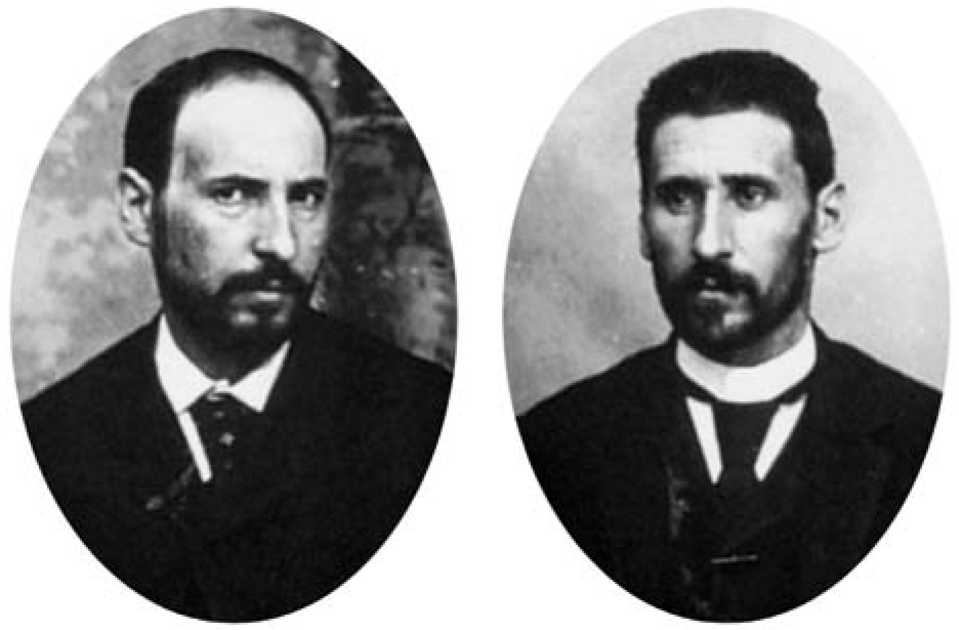
Figure 1.
Santiago Ramón y Cajal at 36 (left) and Pedro Ramón y Cajal at 34 years of age (right); detail from a group gelatin bromide photo of the four Ramón y Cajal brothers and sisters, shot in 1888, with the typical studio painted landscape in the background ([7], p. 84; [19], p. 57).
Introduction
Pedro Ramón was the younger brother of the illustrious neuroanatomist Santiago Ramón y Cajal [1]. Despite several general and technical articles centring on the life and distinguished medical and academic career of ‘the second’ Ramón y Cajal published in Spanish over the past four decades [2,3,4,5,6,7], and even a whole doctoral thesis devoted to him [8], doctor P. Ramón appears to have remained little known even in Spain [6]. Recognition has caught up in his homeland with the inclusion of his biography in the Grand Aragonese Encyclopaedia [9] and the decision in February 2002 by the City Council of Ayerbe to name a new street after don Pedro Ramón y Cajal [6]. Following the occasion of the 150th birthday anniversaries of the two fraternal investigators (Figure 1), the work of P. Ramón, particularly in histology, has attracted the scientific interest of neurobiologists outside Spain [10,11].
The aim of the present article is to present a concise outline of Pedro Ramón’s life and work, placing special emphasis on his ground-breaking discoveries on the structure of the nervous system. It is by now clear that those discoveries provided his elder brother with crucial clues needed to formulate both the neuron theory and the law of dynamic neuronal polarisation [10,11,12], the two main conceptions that Santiago Ramón y Cajal has bequeathed to modern neuroscience.
It is characteristic that in his Nobel Prize acceptance lecture on December 12, 1906, in Stockholm, Cajal [13] refers to the work of his brother (whom he quotes not by name but as “mon frère”) no less than four times,stressing the latter’s critical contributions to the neuron doctrine and the law of dynamic polarisation of neurons, and making special reference to his “very interesting observations on the lateral geniculate body of mammals and the optic lobe of birds, reptiles and fish” and his observations on the intrinsic and extrinsic connections of the cerebellar cortex.
For ease to the reader of this article, but also following a historical tradition, we will refer to Santiago Ramón y Cajal as ‘Cajal’, and to Pedro Ramón y Cajal as ‘Ramón’, a trend consistently practised by the celebrated neuroanatomist in his writings [1,14,15,16,17], but also adopted by modern writers [18].
Life
The second son of Justo Ramón and Antonia Cajal, Pedro was born on October 23, 1854 in Larrés, a small town at an altitude of 3000 feet in the Aurín Valley, located 60 km from Huesca, 30 months after the birth of his brother Santiago [2,3,4,5,6,7,8,9,19].
The restless nature of the two brothers was evident early in their passion for ballistics and warlike games. Cajal recounts experimenting as an 11-year-old boy, along with his 9-year-old brother, with the construction of a cannon out of a bronze pipe, and their miraculous escaping with minor injuries after its accidental explosion “into a thousand pieces”.A metal particle did enter Cajal’s eye, leaving a permanent scar in his iris ([1], p. 73).
In 1866 Pedro accompanied Santiago to Huesca to attend secondary school. Cajal describes his younger brother as “a boy who was as docile and attentive as he was diligent and punctilious, possessing no doubt artistic inclinations” ([1], p. 106–7). Their father, don Justo, had high hopes on Pedro’s seriousness and obedience; fearing the contagion of Santiago’s rebelliousness, he lodged Pedro in a separate boarding house.
Figure 2.
Portrait of 56-year-old General Timoteo Aparicio (left) from Diario El Siglo,issue of June 1871, i.e. the year that Pedro Ramón landed at Uruguay. Aparicio had fought next to President Manuel Oribe (1792–1857) during the ‘Great Siege’ of Montevideo in 1843–1851, and in 1870 had initiated the ‘Revolution of the Lances’ against President Lorenzo Batlle (1810–1887) at the heights of Gualeguaychú. He died poor after a painful illness [20]. Portrait of Pedro Ramón (right) at 27 years [5].
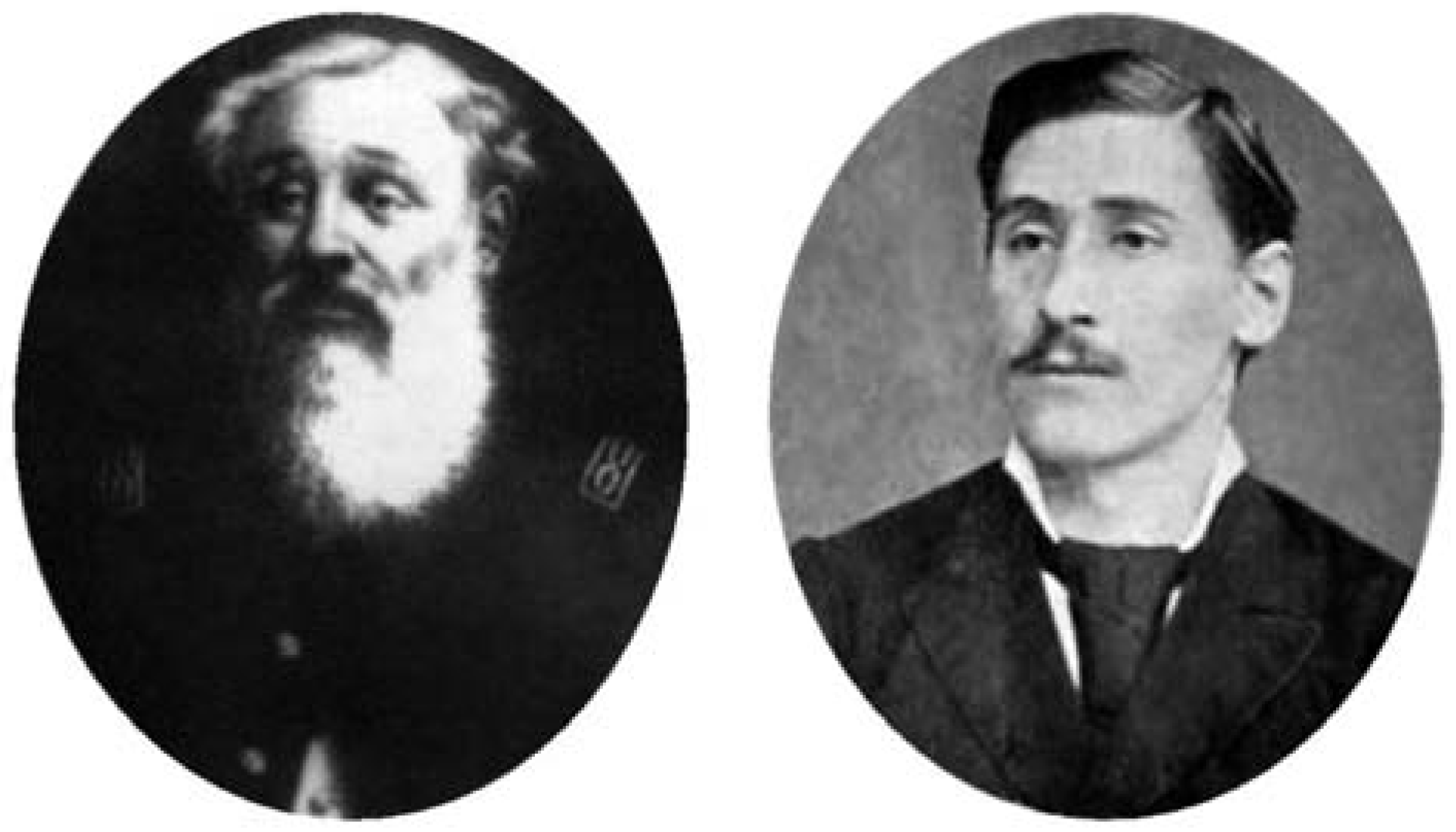
Contrary to such hopes, the “proper, impeccable, submissive and obedient son surpassed at one bound all the boasted wildness of his first born”; at the age of 17, Pedro embarked on a truly epic adventure: having been suspended in one of his last high school courses and not daring to appear before his father, he discarded his student’s gown and fled his home in the company of a “certain Italian seductive adventurer”, leaving Santiago humbled by his “failure to do anything so great” ([1], p. 204). Reaching Bordeaux, Pedro embarked as a cabin boy (grumete) without any money on the boat Queen, landing in Argentina and Uruguay three months later after an episodic journey [5]. In Latin America he enlisted in the revolutionary rows as a soldier and became a hero of the pampas, engaging in civil fights and wounded in several skirmishes. Pedro served as private secretary for General Timoteo Aparicio (1814– 1882), a native Uruguayan chief (‘caudillo’) and revolutionary of the National Party (Figure 2), after whom the 64th School in the Uruguayan city of Florida has been named by the National Administration of Public Education and Chamber of Representatives [20]. When Pedro was caught taking the horse and pistol of General Aparicio, he was sentenced to death, but was saved just before execution through intervention from the Italian and Spanish Consulates.
In 1878 he returned to Spain, landing at the port city of Santander in Cantabria and reaching Zaragoza by train via Bilbao [4]. During the following three years he attended medical school and obtained his MD degree with honours on October 17, 1881, coincidentally, the same year as Freud, whose MD is dated 200 days earlier ([21], p. 57).
Between 1881 and 1888 Ramón practised medicine in the rural areas of La Almolda and Fuendejalón [4,9]. On November 24, 1890 he obtained his PhD degree with high marks [5,6]. From 1890 to 1894 he worked as Director of Anatomical Works in Zaragoza.From 1894 to 1899 he was Professor and Chairman (Catedrático) of Normal Histology and Histochemistry and Pathological Anatomy in the Faculty of Medicine of the University of Cádiz. In 1899 he occupied the Chair of Gynaecology in the Faculty of Medicine of his Alma Mater, the University of Zaragoza, a position he held through his retirement in October 1924 [4,6]. After that, he continued to practise medicine.
As a Professor of Histology at Cádiz, Ramón published between 1896 and 1900 nine extensive studies [22,23,24,25,26,27,28,29,30] on the comparative neurology of fish, amphibians, reptiles and birds in the newly founded journal by Cajal in Madrid ([1], p. 461–2), the ‘Quarterly Micrographic Review’ (Revista Trimestral Micrográfica, which continued from January 1901 on as the renowned Trabajos del Laboratorio de Investigaciones Biológicas de la Universidad de Madrid). Some of these publications are listed in volumes 1–21 of Index Medicus, Series I (1879–1899) and volumes I–III of Bibliographia Medica (1900–1902).
On January 25, 1902 Ramón shared the Martínez y Molina Prize of the College of Physicians of Madrid, carrying a monetary tag of 4000 Pesetas, with his brother Santiago for a jointly written work ‘On the cerebral sensory centres in man and animals’ ([1], p. 626). In the same year P. Ramón founded and directed, together with doctors Ricardo Lozano and Royo Villanova, the journal La Clínica Moderna—Revista de Medicine y Cirugía (‘Modern Clinic, a Review of Medicine and Surgery’) where, until 1917, he published some three dozen histopathological and gynaecological works, as well as the obituary for Rudolf Virchow (1821–1902),the founder of cellular pathology [31]. Later, on the occasion of his retirement at 70, P. Ramón compiled an exposé on his scientific works [32].
Neuroanatomical discoveries
Pedro Ramón is considered one of the fin-dusiècle pioneers who carefully documented the character and distribution of neurons in the telencephalon and established morphological differences associated with the transition from amphibians to reptiles [33], one of the major adaptive radiations in vertebrate evolution [34].
Early in his professional life, beginning in 1888, Pedro Ramón occupied himself with comparative vertebrate histology and the neurohistology of birds, fish, amphibians and reptiles (Lacerta agilis), making valuable contributions to the anatomy of the optic lobe. His 1890 doctoral thesis, a comprehensive monograph in comparative neuromorphology, is entitled Investigations of the comparative histology of the visual centers in diverse vertebrates [35]. In that work he also examined in detail the cell layers of the superior colliculus and noted the descending visual (reflex) pathway associated with it ([17], p. 156).
In his classical Histology of the nervous system, Cajal borrows at least 15 figures from histological sketches drawn by his brother Pedro (for example, cf. figures 28, 36, 37, 133, 138–141, 143–146, 330, 391 and 392 in [17]). Cajal credits his brother for confirming in avian embryos his own observations on the course of the dorsal roots in the spinal cord ([14],p.395),for demonstrating that the disposition of cerebellar granule cells and their fibres, as well as that of Purkinje cells and their axons in the white matter, are essentially identical in all the vertebrates ([14], p. 416), and with confirming the discovery of climbing and mossy fibres in the cerebellum ([14], p. 420–1) and the establishment on Purkinje dendrites of axodendritic connections by the former ([15], p. 69), as well as the discovery of the growth cones of primitive axons using the silver chromate method of tissue impregnation ([14], p. 461). Pedro Ramón suggested that neuroglia,besides sustaining nerve cells and nerve fibres, may also act specifically as an isolating medium for nerve conduction ([14], p. 455).
Using the Golgi method, Pedro Ramón [35,36,37] discovered the free termination of the optic fibre arborisations in the lateral geniculate body, which is characteristic of animals with binocular vision, such as the cat, the rabbit and the dog; that finding was subsequently confirmed by Kölliker, Edinger, van Gehuchten, Tello and Cajal ([16], p. 23; [17], p. 316). In particular, Ramón’s [35] description of the terminal ramifications of the visual fibres in the optic lobe of fish and reptiles was judged by his elder brother as “masterful” ([15], p. 90–1).
Studies on the amphibian brain
Ramón [33,38] provided detailed descriptions of the cytologic structure of the olfactory bulb in amphibians. He described the scattered periglomerular cells in the glomerular layer, which are comparable to the external granule cells of many animals ([39], p. 1293). He also demonstrated that mitral cells receive afferent input from two or more glomeruli and speculated that such a more complicated organisation of the amphibian glomerular layer as well as the complex dendritic branching pattern of mitral cells would imply a finer analysis of odour stimuli [33,40,41]. In that context, it is worth noting that, while in gyrencephalic mammals (and humans) mitral cells elaborate one ‘primordial’ dendrite, in fish, reptiles and birds they can sometimes have four, six or more dendrites per cell ([17], p. 540).
Ramón provided some of the most careful descriptions of the structure of the cerebral cortex of batrachians ([18], p. 449; [30,33,37,42]). In particular, he showed that many of the axons of the neurons of the granule cell layer (which is equivalent to the pyramidal layer of reptiles and mammals) form in the ventrolateral region of the anterior cerebral vesicle the ‘lateral fasciculus’, which arrives at the midbrain and joins with a peduncular pathway originating in the basal or primordial ganglion ([18], p. 450). In the plexiform layer, he showed the presence of displaced pyramidal cells whose axons first descend and later ascend, as well as of dwarf stellate cells ([17], p. 692; [18], p. 451).
Pedro Ramón described the two decussating bundles of the anterior commissure which constitute the inferior arcuate fasciculus and which are found dorsally and caudally to the dorsal fascicles of the median forebrain bundle ([33,38]; [39], p. 1304). He also described the structure of the amphibian habenula ([17], p. 342; [37]) and confirmed the dorsal root bifurcation of spinal ganglion cells ([17], p. 402; [43]).
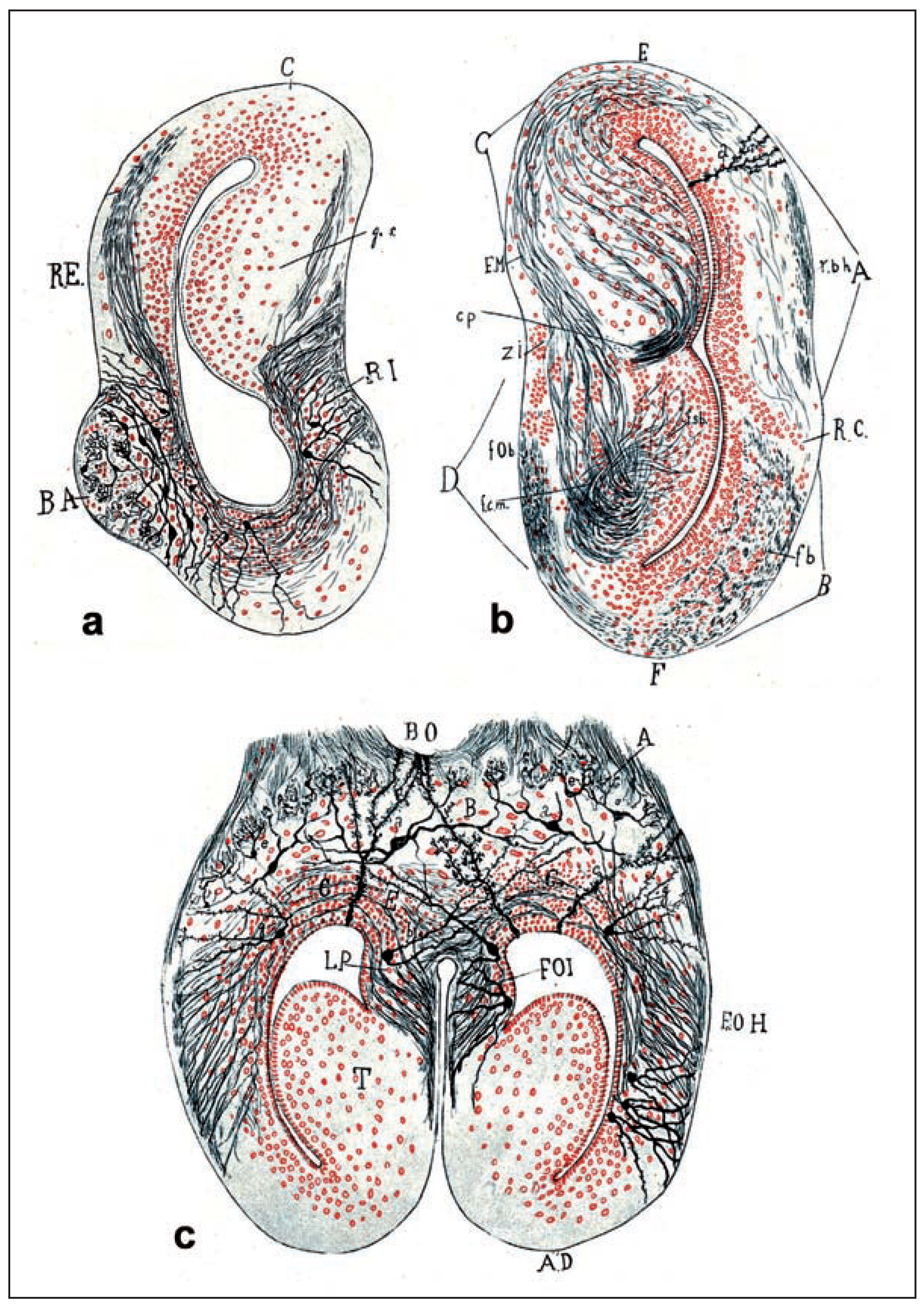
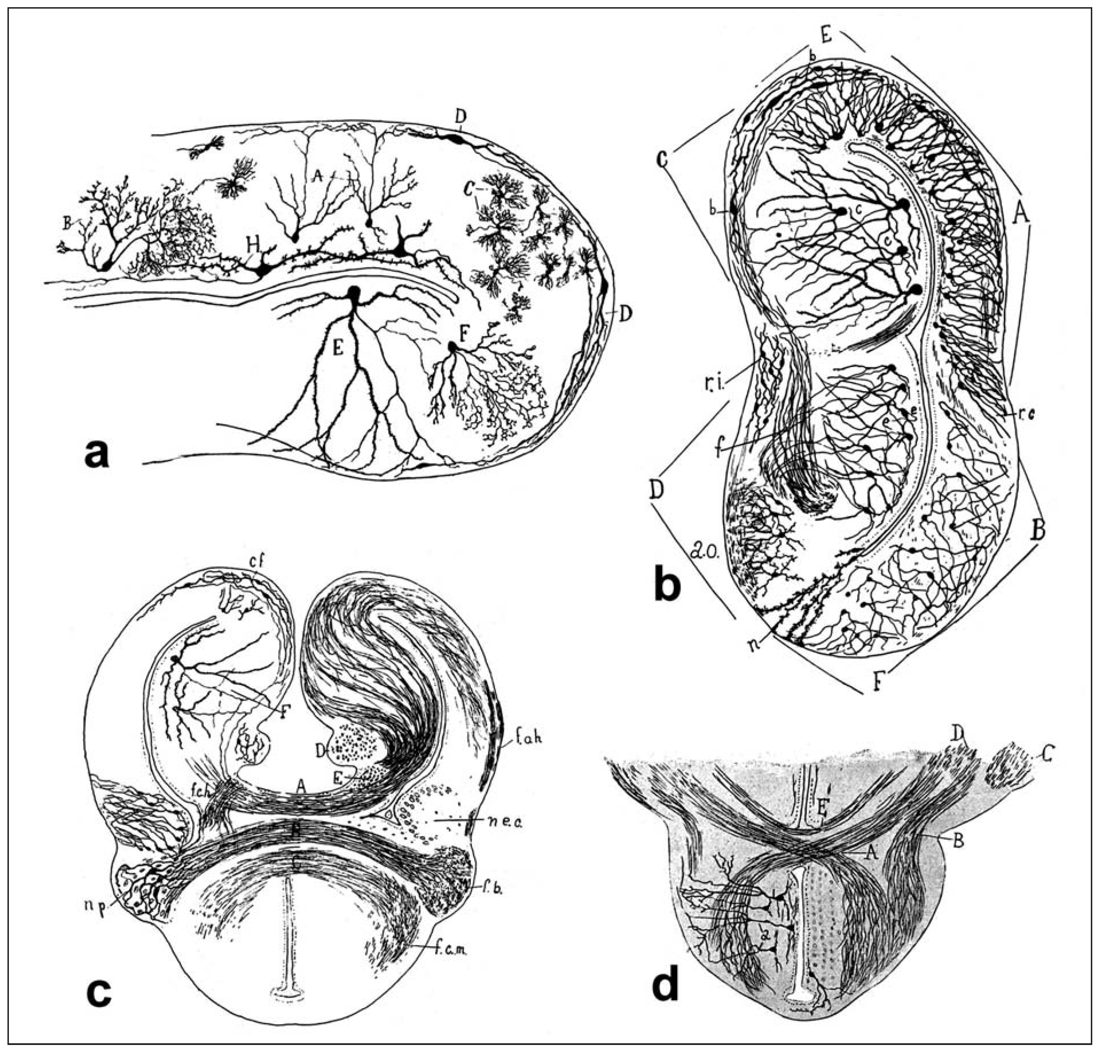
In the special two-volume edition by students and admirers honouring Santiago Ramón y Cajal on the occasion of his 70th birthday, Ramón [38] contributed an extensive overview of his findings on the anatomy and histology of the frog nervous system (Figure 3 and Figure 4). His last neuroanatomical publication [44] was also centred on the frog brain.
Studies on the brain of reptiles
In the chameleon Pedro Ramón showed that the reflex that involves the pigmented cells of connective tissue and their related nerve terminals (its sensory or afferent pathway being represented by the optic nerve), which enables the animal to imitate the colour of its surroundings, is interrupted when the skin is anaesthetised with chloroform, when the peripheral motor tracts are cut, or when the spinal cord is experimentally destroyed ([14], p. 171; [45]).
In the phylogenetic passing from amphibians to reptiles, the archipallium becomes markedly differentiated into a cortex-like structure containing a dorsal and a dorsomedial hippocampal part. In the telencephalon of Varanus and Lacerta, Ramón [46,47,48] recognised a hippocampal region. In the portion that corresponds to the dorsomedial part of the hippocampus, he demonstrated a transition in cell type from true granule cells in the outermost portion to true Ammon’s pyramidal cells in the innermost portion; in certain forms the dorsomedial hippocampal part is clearly separated into a lateral pyramidal and a medial granular layer ([37]; [39], p. 1334, 1478).
Figure 3.
Histological drawings of the frog brain by Pedro Ramón, from his 1922 paper in the honourary volume for his brother Cajal [38]. Note the ‘family’ traces of resemblance between the artistic talent of the younger Ramón and the classical Cajal ‘paintings’. a) Section of a cerebral vesicle. Abbreviations:B. A.= accessory olfactory bulb; R. E. =external olfactory root and bulbo-cortico-habenular fasciculus; R. I. = internal root or basal bulbo-olfactory fasciculus; C = cortex; g. s. = superior ganglion of the septum. b) Frontal section of the anterior vesicle. Abbreviations:A= cerebral cortex or external superior region of the cerebrum; B = inferior region or basal ganglion; C = superior region or dorsal ganglion of the septum; D = inferior region of the internal cerebral segment; f. b. = basal fasciculus; R. C. = curved region of the cortex; z. i.= zona interna. c) The principal olfactory bulbs. Abbreviations:A = zone of the glomeruli; B = zone of the large mitral cells; C = zone of the granules; E = zone of the deep olfactory fibres; F. O. H. = habenular olfactory fasciculus; F. O. I. = internal olfactory fasciculus; L. P. = minor postolfactory lobule; T. = cerebral septum; c.= intraglomerular cells; a = large mitral cells; g = granules.
In the reptilian cortex Ramón described in the second (or pyramidal cell) layer three types of axon collaterals, ‘recurrent’ (to the external plexiform layer), ‘horizontal or tangential’ (arborised in the inner plexiform layer) and ‘association’ (arising in the white matter) ([17], p. 687; [18], p. 446; [27]). Ramón was the first to indicate that neuroglia in the fourth layer (or layer of the white matter) are represented by radial epithelial cells that start at the ventricle and terminate with a tuft of spiny filaments at the cerebral surface ([18], p. 77; [22,36]); moreover, in the same layer, he distinguished four pathways, namely, an ipsilateral association pathway, a longitudinal association pathway (the ‘sagittal bundle of P. Ramón’), a contralateral association pathway or corpus callosum, and a direct and crossed projection pathway ([18], p. 446–7).
Among the fibre connections of the reptilian telencephalon, Ramón observed the lateral forebrain bundle; he also traced the axons of thalamic neurons into the bundles and indicated that a substantial portion of the thalamostriate fibres are ascending ([39], p. 1353–4; [45]). Ramón also considered the optico-commissural fasciculus—a bundle similar to the tractus tectothalamicus cruciatus—as composed in part of optic and in part of supraoptic fibres ([22]; [39], p. 1003). In the supraoptic system of the chameleon [22], he noted the tractus opticus commissuralis, an apparent homologue of what is also termed the tectothalamic or tectotectal tract ([39], p. 1007).
Ramón [25] discovered both descending (derived from the posterior commissure, from the homologue of the nucleus of Darkschewitsch and from the thalamus) and ascending fibres in the medial longitudinal fasciculus of the reptilian brain; the great complexity of that structure in reptiles may be related to the fact that it contains fibre groups which in the mammalian brain are found outside of it ([17], p. 219).
P. Ramón demonstrated the existence of the so-called ‘shepherd’s crook cell’—an instance where an axon issues from the distal end of a long dendrite—in the optic lobe of reptiles, including the chameleon, and of fish and amphibians ([17], p. 104–5, 120–1; [22,35,36]). In the reptilian optic tectum Ramón [22] described an elaborate architectonic arrangement of cells in 14 layers, including the ependyma; his nomenclature was later adopted by Ariëns Kappers ([39], p. 972–8).A similar cytoarchitectonic arrangement was seen in the optic tectum of the frog [33], while in the tectal centres of teleosts (Barbus fluviatilis) 10 layers are mentioned [29,35], beginning with the ventricular surface and counting towards the outer tectal surface ([39], p. 904). In the reptilian diencephalon Ramón described the various anatomical subcompartments of the lateral geniculate nucleus ([22]; [39], p. 996–7).
Studies on the avian brain
In the optic lobe of birds the valuable contributions of Ramón [28,35] strengthened Cajal’s view that, in sensory areas, afferent impulses are propagated by contact from the retinal centripetal fibres to the dendrites and somata of the central neurons ([1], p. 350–1). In the avian optic tectum Ramón [26,49] provided a most detailed cytoarchitectonic description, by identifying 15 layers of cells, where other investigators had been able to only see 3–9 layers ([39], p. 1015).
Figure 4.
Histological drawings of the frog brain by Pedro Ramón, from his 1922 paper in the honourary volume for his brother Cajal [38]. a) Corpuscle of Golgi or short axis cylinder, showing great resemblance to similar elements in the cortex of higher vertebrates.Abbreviations: A = cells of Martinotti; B = Golgi cell with short axon; C = dwarf cell with short axon; D = tangential fusiform cell; E = long axon cell of the septal dorsal ganglion; F = short axon cell of the septum. b) Frontal section of a cerebral vesicle. Abbreviations: A = external cortex; B = basal ganglion or corpus striatum; C = lamina of the fornix or superior ganglion of the septum; E = dorsal angle of the cerebrum; F = ventral angle; D = inferior ganglion of the septum; a. o. = olfactory area; r. i. = intermediate region of the septum; f = corticomedial fasciculus; b = fusiform cortical cells;r.c. = curved region; c = cells of the superior ganglion of the septum; e = cells of the inferior ganglion; n = ependymal cell. c) Frontal and posterior section of the cerebral vesicles of the frog. Abbreviations: A = commissure of the pallium; B = anterior commissure; C = ventral commissure or decussation of the corticomedial fasciculi; D = supracommissural nuclei; E = juxtacommissural nuclei; F = superior ganglion of the internal cerebral segment or septum; f. o. h.= habenular olfactory fasciculus; n.e. c. = lateral spherical nucleus; f. b. = basal fasciculus; f. c. m. = corticomedial fasciculus; n. p. = nucleus of the peduncle and the anterior commissure; f. c. h. = corticohabenular fasciculus; c. f. = fusiform cells of the dorsal angle of the cortex. d) Frontal section of the frog hypothalamus. Abbreviations: A= crossed-over fornix; B = fasciculus terminalis of the corticomedial bundle; E = Haller’s commissure; C = basal fasciculus; D = corticomedial fasciculus; a = cells of the infundibulum.
Regarding the fibre connections in the telencephalon of birds, Ramón showed that the reduction of the central paths in the olfactory nerve is greater than the peripheral reduction, since multiple olfactory neurons terminate into a single mitral cell, producing a summation of incoming signals; this is the opposite of what is seen in fish, amphibians and reptiles, where one mitral cell comes into contact with several olfactory glomeruli through its dendrites ([39], p. 1381–2).
In the basal ganglia of lower vertebrates Ramón noted the presence of dwarf (or ‘neurogliaform’) neurons ([17], p. 418). Fish and amphibians have mostly ovoid basal ganglion cells; in reptiles these neurons reach enormous size; and in birds one sees both morphological types found in the mammalian brain, i.e. giant and medium-sized ([17], p. 425).
Midbrain of lower vertebrates
In the midbrain Ramón showed that the anterior part of the mesencephalic nucleus in lower vertebrates occupies a different position than it does in mammals: while in the latter it is seen in lateral parts of the periaqueductal grey, in amphibians, reptiles and birds, studied with the reduced silver nitrate method, cells extend horizontally throughout the central grey, sometimes reaching the raphe nuclei ([17], p. 723–4; [39], p. 427–9; [50]). In newly hatched pigeons Ramón showed an inconsistent presence of dendrites in neurons of the dorsal part of the commissure between the optic lobes ([17], p. 724–5; [50]).
Cerebellar cortex
Pedro Ramón made several important discoveries regarding the comparative histology of the cerebellar cortex. In the molecular layer he observed the axosomatic contacts established by Purkinje axons on stellate cell bodies ([15], p. 16; [22]). He expressed the view that the protoplasmic expansions of cerebellar granule cells only arise after their migratory descent through the molecular layer to the internal granule cell layer; concomitantly, granule cells from bipolar become unipolar ([36]; [51], p. 253). With regard to mossy fibres, Ramón [23] reported centripetal fibres, arborised among granule cells and displaying large varicosities that apparently correspond to terminal rosettes ([17], p. 63–4).
Ramón provided the first description of the rostrocaudal flattening of the Purkinje dendritic trees in fish, amphibians and reptiles, as well as a respective increase in dendritic tree size and complexity in this order ([17], p. 60; [36,37,52]). On the other hand, Purkinje axon collaterals, which in mammals can number up to three or four, are reduced to two in birds, to one in reptiles and to one or sometimes none in amphibians and fish. The basket cell axons are shorter in reptiles, very quickly giving rise in the reptilian cerebellum to pericellular arborisations ([17],p.61;[52]). Ramón described the consistent properties of granule cell axons across vertebrate species, their upward course and T-branching in the molecular layer into two parallel fibres, which in reptiles may give off a few very short collaterals.
Envoy
In the official session of the Iberian Society of Natural Sciences in Zaragoza on October 8, 1938, Pedro Ramón presented his thoughts on the doctrine of the organic evolution of cerebral pyramidal cells [53], thus renewing a theme discoursed upon three dozen years earlier [54].
His early observations were decisive for Cajal’s establishment of the law of dynamic polarisation of neurons [11]. It is by now clear that through the demonstration in his doctoral dissertation [35] of an abundance of axon terminals in areas of the optic tectum—which Cajal had thought contained only dendrites—he helped his brother solve one of the major problems and thus opened the way to the formulation of the ‘law of functional polarisation’ [11,12].
His histological studies on lower vertebrates have been cited by some of the most notable neuroanatomists, such as Arthur van Gehuchten (1861– 1914), Albert von Kölliker (1817–1905), Wilhelm von Waldeyer (1836–1921), Wilhelm His (1831– 1904), Ludwig Edinger (1855–1918), Cornelius Ariëns Kappers (1877–1946) and James Papez (1883–1958). Ramón confessed [32] that he did not comprehend why his modest contributions would draw the attention of researchers of such international calibre.
Legacy
Later on, Pedro Ramón distinguished himself particularly with his work in gynaecologic pathology; he was a pioneer in the use of pre- and intraoperative biopsies, as well as in the histopatho-logical study of the effects of radiation on malignant tumours [5].
Figure 5.
A mature Graffian follicle in the dog, drawn by Pedro Ramón; Golgi method. Abbreviations:A = stratum granulosum; B = proligerous disc. Used as figure 502 in Histology([14], p. 584).
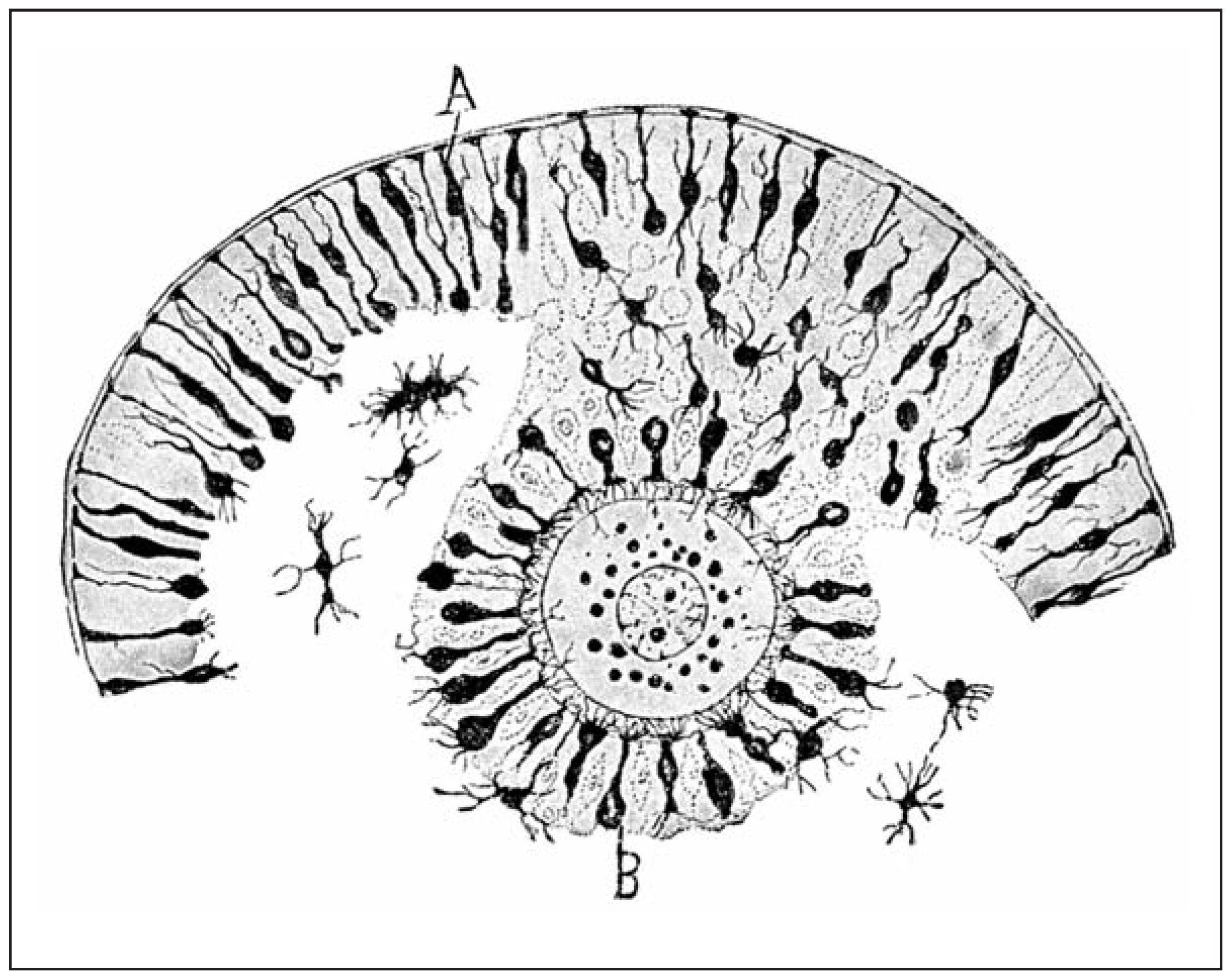
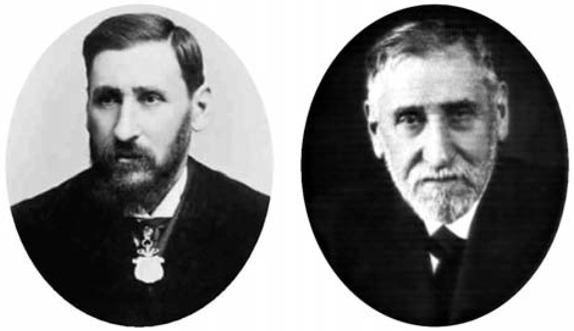
Figure 6.
Pedro Ramón [5,6] as a young professor at the prime of his life (left) and as an older gentleman in his seventies (right).
With his histological studies on the reproductive system (Figure 5), Pedro Ramón first confirmed the idea of Flemming that, during the phase of the embryonic vesicle, the protoplasmic expansions,
which place in communication the protoplasm of the ovum with the immediate epithelial cells, pass through fine radiating ducts; and second, that the cells of the stratum granulosum and the proligerous disc of the Graffian follicle are of a connective type, taking a spindle shape with polar expansions,which commonly terminate with brooches of ramifications ([14], p. 584).
The continued and present interest in the contributions of Pedro Ramón [10,11] is a clear indication that the life and work of ‘Cajal the younger’ are finally,and deservedly,emerging form the shadow of his celebrated brother.
A highly respected gentleman, doctor Pedro Ramón y Cajal (Figure 6), or ‘don Pedro’ to friends and students, died on December 10, 1950 in his house in Zaragoza at the age of 96, having outlived his brother Santiago by 16 years. He was laid to rest in a private ceremony the following day at the Cemetery of Torrero [5].
Being a rounded physician ‘of the old school’, he had practised histology, anatomic pathology, internal medicine, surgery, gynaecology, radiotherapy, and even hypnotism for neuroses [4,7]. Professor Ramón left—besides his fundamental discoveries in comparative neurohistology—a legacy ingrained with exemplary humility, generosity, serenity, tolerance and a profound religiosity (a noted difference from his brother) that were characteristic of his personality [4].
His honours include election to Member of the Royal Academy of Medicine of Zaragoza (1893), President of the Spanish Society of Natural History (1907), Merit Fellow of the Academy of Sciences of Bologna, Italy (1907), of the Imperial Academy of Anthropology of Moscow (1901) and the Imperial Academy of Saint Petersburg, Russia (1913), Corresponding Member of the Royal National Academy of Medicine of Spain and the bestowal of the Gold Medal by the City of Zaragoza [4,6].
The real honours and the way Pedro Ramón viewed his own work are reflected in the following sentences:“ I am an original and romantic investigator who without any pretence, and solely for an inner satisfaction, has consecrated more than thirty years to the contemplation of the mysterious nervous plot, preferentially the brain of inferior vertebrates […].The honours I obtained have been great: to be Spanish, to be Aragonese, to be a medical doctor, and to be Santiago’s brother […]. One of my greatest satisfactions was to be the unique friend and confidant of my brother Santiago” [6].
References
- Ramón y Cajal, S. Recollections of My Life [originally published as ‘Recuerdos de mi vida’ (1901–1907); translated by E. H. Craigie and J. Cano]. In The Classics of Neurology and Neurosurgery Library; Gryphon Editions: Birmingham (AL), 1988. [Google Scholar]
- Galán Bergua, P. Pedro Ramón y Cajal. In Biografías Aragonesas; Institución ‘Fernando el Católico’: Zaragoza, 1967; pp. 195–200. [Google Scholar]
- Horno Liria, R. Pedro Ramón y Cajal. In Semblanzas de ginecólogos Aragoneses; Real Academia de Medicina: Zaragoza, 1975; pp. 59–65. [Google Scholar]
- Solsona, F. Pedro Ramón y Cajal (1854–1950). Serrablo Rev Trimest (Sabiñánigo). 2000, 30, pp. 1–4. Available online: http://www.serrablo.org/boletin/s117/s117a6.html?q=revista/s117/s117a6.html.
- de Carlos Segovia, JA; Ramón y Cajal Agüeras, S. Pedro Ramón y Cajal: médico, histólogo y docente. Rev Españ Patol. 2002, 35, 465–74. [Google Scholar]
- Ramón y Cajal Abelló, P. Pedro Ramón y Cajal. Comarca (Ayerbe). 2002, 33, 2–4. [Google Scholar]
- Ramón y Cajal Abelló, P. Los hermanos Ramón y Cajal.Sus relaciones familiares y profesionales. In Visiones: Santiago Ramón y Cajal 150 aniversario; Grasa Adé, I, Romero Santamaría, A, Eds.; Diputación de Zaragoza – Félix Arilla: Zaragoza, 2002; pp. 83–91. [Google Scholar]
- Rodríguez Martín, B. Vida y obra de D. Pedro Ramón y Cajal. Tesis doctoral, Facultad de Medicina de la Universidad de la Laguna, 1985. [Google Scholar]
- Gran Enciclopedia Aragonesa OnLine. Biographical entry ‘Ramón y Cajal, Pedro’. Available online: http://www.enciclopediaaragonesa.com/voz.asp?voz_id=10639 (accessed on 26 October 2006).
- Triarhou, LC; del Cerro, M. Professor Pedro Ramón (1854–1950), Cajal’s brother. Hellenic Soc Neurosci Abstr (Athens) 2003, 18, 176–9. [Google Scholar]
- Kuljis, RO; Karten, HJ. Pedro Ramón y Cajal’s Role Launching the Comparative Analysis of the Brain and Rescuing Cajal’s Theory of Dynamic Neuronal Polarization. In Program No. 24.12.2007 Neuroscience Meeting Planner; Online; Society for Neuroscience: San Diego (CA), 2007. [Google Scholar]
- Yuste, R; Tank, DW. Dendritic integration in mammalian neurons a century after Cajal. Neuron 1996, 16, 701–16. [Google Scholar] [CrossRef]
- Ramón y Cajal, S. Structure et connexions des neurones. Conférence Nobel faite à Stockholm le 12 décembre 1906. In Les Prix Nobel en 1906; Hasselberg, KB, Pettersson, SO, Mörner, KAH, af Wirsén, CD, Santesson, MCG, Eds.; Kungl. Boktryckeriet P. A. Norstedt & Söner: Stockholm, 1908 [1907]; pp. 1–27. [Google Scholar]
- Ramón y Cajal, S; Tello-Muñoz, JF. Histology; Fernán-Núñez, M., Translator; William Wood & Company: Baltimore (MD), 1933. [Google Scholar]
- Ramón y Cajal, S. Neuron Theory or Reticular Theory? Objective evidence of the anatomical unity of nerve cells; Purkiss, M. Ubeda; Fox, C. A., Translators; Consejo Superior de Investigaciones Científicas – S. Aguirre Torre: Madrid, 1954. [Google Scholar]
- Ramón y Cajal, S. Studies on the Diencephalon [translated by E. Ramón-Moliner]; Charles C. Thomas Publisher: Springfield (IL), 1966. [Google Scholar]
- Ramón y Cajal, S. Histology of the Nervous System; Swanson, N.; Swanson, L. W., Translators; Oxford University Press: New York, Oxford, 1995. [Google Scholar]
- DeFelipe, J; Jones, EG. Cajal on the Cerebral Cortex: an Annotated Translation of the Complete Writings; Oxford University Press: Oxford, New York, 1988. [Google Scholar]
- Romero, A. Fotografía Aragonesa/1: Ramón y Cajal; Diputación Provincial de Zaragoza: Zaragoza, 1984. [Google Scholar]
- Christiansen, CE. General Timoteo Aparicio (XLVIa Legislatura, Carpeta N° 263, Repartido N° 298); Cámara de Representantes, Comisión de Educación y Cultura: Montevideo, República Oriental del Uruguay, 2005; pp. 1–3. [Google Scholar]
- Jones, E. The Life and Work of Sigmund Freud. In the Formative Years and the Great Discoveries, 1856– 1900; Basic Books: New York, 1953; Volume 1. [Google Scholar]
- Ramón y Cajal, P. Estructura del encéfalo del camaleón. Rev Trimest Microgr (Madrid) 1896, 1, 131–67. [Google Scholar]
- Ramón y Cajal, P. Las células estrelladas de la capa molecular del cerebelo de los reptiles. Rev Trimest Microgr (Madrid) 1896, 1, 221–2. [Google Scholar]
- Ramón, P. Los corpúsculos nerviosos de axón corto o células sensitivas de Golgi en los vertebrados inferiores. Rev Trimest Migrogr (Madrid) 1897, 2, 29–32. [Google Scholar]
- Ramón y Cajal, P. El fascículo longitudinal posterior en los reptiles. Rev Trimest Microgr (Madrid) 1897, 2, 153–62. [Google Scholar]
- Ramón y Cajal, P. Centros ópticos de las aves. Rev Trimest Microgr (Madrid) 1898, 3, 141–97. [Google Scholar]
- Ramón y Cajal, P. La célula piramidal del cerebro en los reptiles. Rev Trimest Microgr (Madrid) 1899, 4, 73–6. [Google Scholar]
- Ramón y Cajal, P. Adiciones a nuestros trabajos sobre los centros ópticos de las aves. Rev Trimest Microgr (Madrid) 1899, 4, 77–85. [Google Scholar]
- Ramón y Cajal, P. El lóbulo óptico de los peces teleósteos. Rev Trimest Microgr (Madrid) 1899, 4, 87–108. [Google Scholar]
- Ramón y Cajal, P. Ganglio basal de los batracios y fascículo basal. Rev Trimest Microgr (Madrid) 1900, 5, 23–36. [Google Scholar]
- Ramón P. Virchow (necrología). Clín Moderna (Zaragoza) 1902, 1, 312–4.
- Ramón y Cajal, P. Breves datos referentes a mi labor científica y docente. Clín Lab (Zaragoza) 1924, 4, 301–30. [Google Scholar]
- Ramón y Cajal, P. L’encéphale des amphibiens. Bibliogr Anatomique (Paris) 1896, 4, 233–52. [Google Scholar]
- Northcutt, RG; Royce, GJ. Olfactory bulb projections in the bullfrog Rana catesbeiana. J Morphol. 1975, 145, 251–68. [Google Scholar] [CrossRef]
- Ramón y Cajal, P. Investigaciones de histología comparada en los centros ópticos de distintos vertebrados. Tesis del doctorado, Facultad de Medicina de la Universidad Central de Zaragoza, 1890. [Google Scholar]
- Ramón y Cajal, P. El encéfalo de los reptiles; Trabajos del Laboratorio de Histología de la Facultad de Medicina: Zaragoza, 1891; pp. 1–33. [Google Scholar]
- Ramón y Cajal, P. Investigaciones micrográficas en el encéfalo de los batracios y reptiles, cuerpos geniculados y tubérculos cuadrigéminos de los mamíferos; Trabajos Anatómicos de la Facultad de Medicina-La Derecha: Zaragoza, 1894; pp. 1–88. [Google Scholar]
- Ramón y Cajal, P. El cerebro de los batracios. In Libro en honor de D. S. Ramón y Cajal: Trabajos originales de sus admiradores y discípulos, extranjeros y nacionales; Jiménez y Molina Impresores: Madrid, 1922; Volume Tomo I, pp. 13–59. [Google Scholar]
- Ariëns Kappers, CU; Huber, GC; Crosby, EC. The Comparative Anatomy of the Nervous System of Vertebrates, Including Man, 3 Volumes; Hafner Publishing Co.: New York, 1965. [Google Scholar]
- Nezlin, LP; Schild, D. Structure of the olfactory bulb in tadpoles of Xenopus laevis. Cell Tissue Res. 2000, 302, 21–9. [Google Scholar] [CrossRef]
- Nezlin, LP; Schild, D. Individual olfactory sensory neurons project into more than one glomerulus in Xenopus laevis tadpole olfactory bulb. J Comp Neurol. 2005, 481, 233–9. [Google Scholar] [CrossRef]
- Ramón y Cajal, P. El encéfalo de los batracios. Primera parte. Mem Real Soc Españ Hist Nat (Madrid) 1905, 3, 165–88. [Google Scholar]
- Ramón y Cajal, P. Las fibras colaterales de la substancia blanca en la médula de las larvas de los batracios. Gac Sanit Barcel 1890, 2, 78–89. [Google Scholar]
- Ramón y Cajal, P. El cerebro de los batracios. Trab Inst Cajal (Madrid) 1946, 38, 41–111. [Google Scholar]
- Ramón y Cajal, P. Experiencias sobre los cambios de color de la piel del Camaleón; Asociación Española para el Progreso de las Ciencias: Madrid, 1908; pp. 1–9. [Google Scholar]
- Ramón y Cajal, P. Nuevo estudio del encéfalo de los reptiles. Trab Lab Invest Biol Univ Madrid 1917, 15, 83–100. [Google Scholar]
- Ramón y Cajal, P. Nuevo estudio del encéfalo de los reptiles. Trab Lab Invest Biol Univ Madrid 1918, 16, 309–33. [Google Scholar]
- Ramón y Cajal, P. Nuevo estudio del encéfalo de los reptiles; Imprenta de Nicolás Moya: Madrid, 1919; pp. 1–25. [Google Scholar]
- Ramón y Cajal, P. Lóbulos ópticos de las aves. Trab Inst Cajal (Madrid) 1943, 35, 3–20. [Google Scholar]
- Ramón y Cajal, P. Origen del nervio masticador en las aves, reptiles y batracios. Trab Lab Invest Biol Univ Madrid 1904, 3, 153–62. [Google Scholar]
- Ramón y Cajal, S. Etudes sur la neurogenèse de quelques vertébrés; Tipografía Artística: Madrid, 1929. [Google Scholar]
- Ramón y Cajal, P. Notas preventivas sobre la estructura de los centros nerviosos. Gac Sanit Barcel 1890, 3, 10–8. [Google Scholar]
- Ramón y Cajal, P. Algunas reflexiones sobre la doctrina de la evolución de los corpúsculos piramidales del cerebro en la escala de los vertebrados. Rev Soc Ibér Cienc Nat (Zaragoza) 1938, 36, 7–15. [Google Scholar]
- Ramón, P. Algunas reflexiones sobre la doctrina de la evolución orgánica de los corpúsculos piramidales del cerebro. Clín Moderna (Zaragoza) 1902, 1, 17–25. [Google Scholar]
This is an open-access article distributed under the terms of the Creative Commons Attribution Non-Commercial No Derivatives (CC BY-NC-ND) 4.0 License.