


Summary
Gérard Roth, a prominent Swiss neurologist and neurophysiologist, passed away on May 10, 2006, at the age of 83 years. He was a pioneer in the discovery of multifocal motor neuropathy (MMN) and the first to describe persistent conduction blocks in several other disorders. His death occurred a few days before a symposium was held to commemorate the 20th anniversary of the description of multifocal motor neuropathy. This symposium thus became the occasion to honour the role of this outstanding scientist, inspiring mentor and wonderful person.
Introduction
In this symposium dedicated to “Multifocal Motor Neuropathy—MMN”, my task was to give a talk on “Conduction Blocks—CB”. The unexpected death of Gérard Roth on May 10, 2006 (19 days before this symposium took place) led us to modify the topic of this lecture and to dedicate it to the keyrole he played in the discovery of multifocal motor neuropathy and in our understanding of conduction blocks. I have had the privilege to know and to collaborate with G. Roth during more than 25 years. This has given me the opportunity to witness the difficulties one may encounter to receive recognition of a pioneer work, as exemplified in the following lines.
Gérard Roth
Gérard Roth was born in Geneva on March 29, 1923. He died aged 83, a few months after his wife had passed away. He kept a perfect intellect until his last days. G. Roth spent his professional life in Geneva University Hospital, except for a 3-month visit in the laboratory of F. Buchthal in Copenhagen (DK). He was a remarkable neurologist and electromyographist. G. Roth was my mentor and excellent friend.
 Gérard Roth 1923–2006.
Conduction blocks
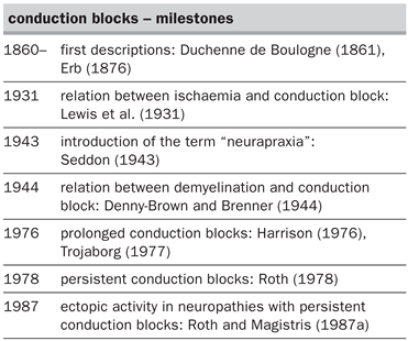
The history of multifocal motor neuropathy is closely linked to that of conduction blocks of the peripheral nerves. A conduction block consists in the non-propagation of the action potential passed a particular point along the nerve fibre.The phenomenon was first reported by Duchenne de Boulogne (1861) and Erb (1876). It can be detected and quantified by the electrophysiological measurement of a reduction of the size of the response evoked by proximal versus distal stimulation of a nerve.The clinical expression of a conduction block usually consists in a sensory-motor deficit that is reversible in less than to 2 months when it relates to a lesion of the myelin sheath. The phenomenon has been called “neurapraxia” (i.e., non-action of the nerve—Seddon, 1943). Rarely, a conduction block may be prolonged over a period of a few more weeks or months (Harrison, 1976; Trojaborg, 1977). Examples of frequent pathological conditions accompanied by conduction blocks of peripheral nerves are: trauma or compression of nerves (mononeuropathies), Guillain-Barré syndrome, chronic inflammatory demyelinating polyneuropathies (multineuropathies or polyneuropathies). The physiopathology of conduction block has been understood progressively (for a review see Kuntzer and Magistris, 1995, and Table 1).
Table 1.
Persistent conduction blocks
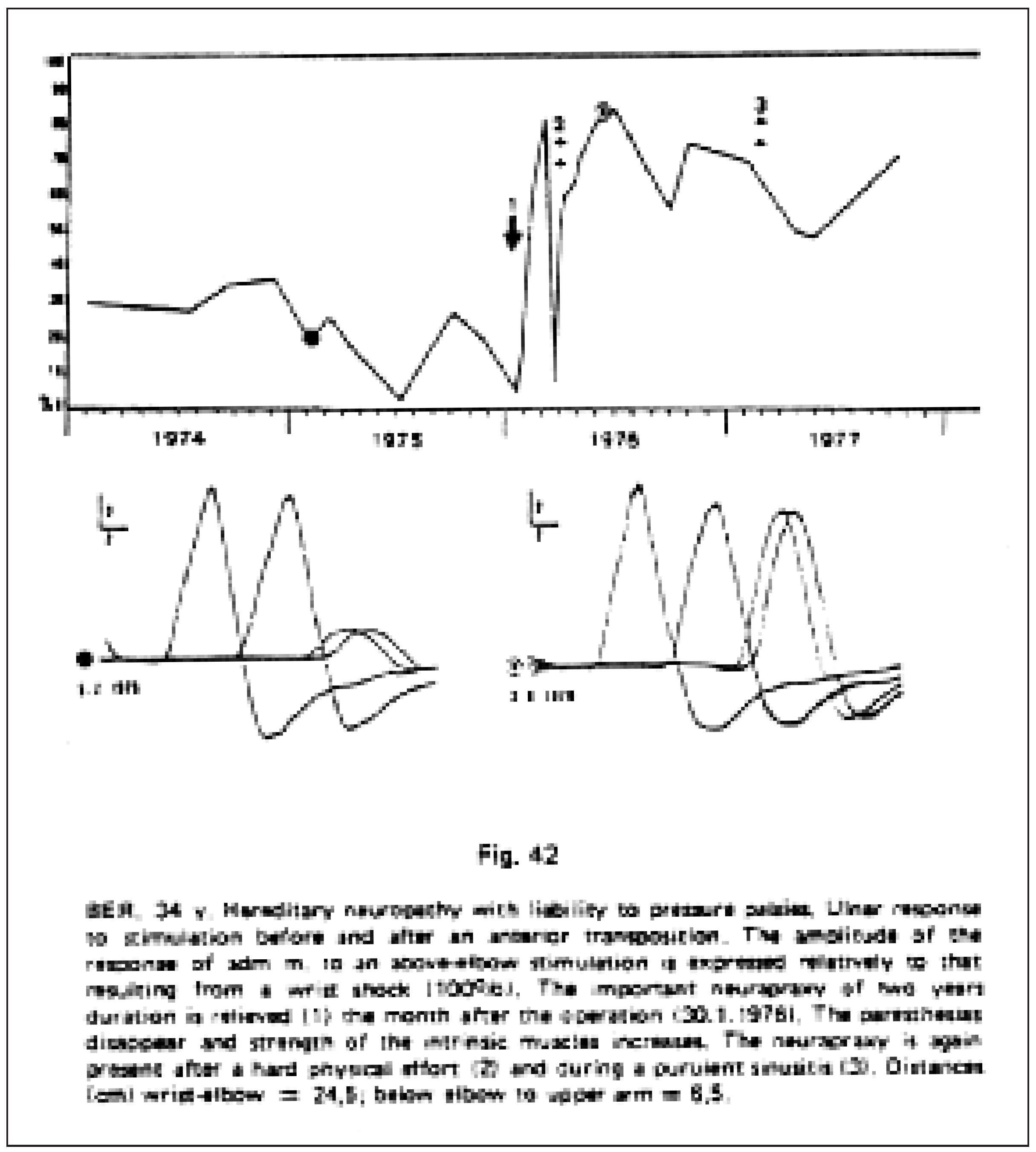
In 1978 G. Roth was the first to report the observation of “persistent CBs” in patients presenting with a hereditary disease of the peripheral myelin causing a susceptibility of peripheral nerves to compression: “in hereditary neuropathy with liability to pressure palsies, axonotmesis is most frequently associated with neurapraxia which can almost be permanent over years” (Roth, 1978, p. 329—cf. Figure 1).
Figure 1.
Persistent conduction block in hereditary neuropathy with liability to pressure palsies (from Roth, 1978).
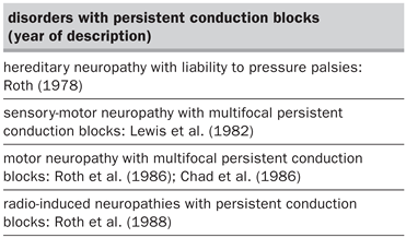
The description of persistent conduction blocks is a milestone in the history of peripheral neuropathies. Conduction blocks in hereditary neuropathy with liability to pressure palsies have further been discussed in the following publications by our group (Roth and Magistris, 1984; Magistris and Roth, 1985) and by others (Sellman and Mayer, 1987). Subsequently, three other disorders of the peripheral nerves have been described that are explained by persistent conduction blocks (Table 2). Two of these were recognised by G. Roth and colleagues, namely multifocal motor neuropathy with persistent conduction blocks (Roth et al., 1986) and radiation neuropathies (Roth et al., 1988).
Table 2.
Multifocal motor neuropathy with persistent conduction blocks
Since 1974 G. Roth and colleagues examined two patients presenting with a disorder resembling amyotrophic lateral sclerosis (ALS), but differing by the absence of conspicuous signs of upper motor neuron involvement and amyotrophy. In these patients focal proximal conduction blocks were repeatedly noted which surprisingly solely concerned the motor nerve fibres. These conduction blocks increased in number and severity during the follow-up. A manuscript describing these two patients was sent to an American Journal in 1984; it was rejected by the editor who believed that these patients had amyotrophic lateral sclerosis and that the authors were probably inexperienced. It must be recalled that, for neurologists of that time, a peripheral deficit restricted to motor symptoms could only relate to disorders of the anterior horn, the neuromuscular junction or the muscle. The deficit caused by the lesion of a peripheral nerve had to include sensory symptoms! These patients were presented and examined by two renowned professors of neurology from Paris and Lyon. They concluded that the patients probably suffered from amyotrophic lateral sclerosis or alternatively from a conversion syndrome since their weak muscles were not clearly amyotrophic (as usually observed in amyotrophic lateral sclerosis and peripheral axonopathies, but not in case of conduction blocks). Our colleague F. Ochsner presented these two patients in his doctoral thesis in 1986 (see his article in the present issue). The manuscript by G. Roth and colleagues, which was rejected in 1984, was modified to report only the most characteristic patient. This article was received by the journal European Neurology on June 5, 1985, and was eventually published in June 1986. Around that time, the world of neurophysiology was ready to discover multifocal motor neuropathy since similar patients were independently reported by two other groups, that of Parry and Clarke, in an abstract published in September 1985 (and subsequently in a full article in 1988), and that of Chad et al. (1986). Interestingly, whereas the condition of the patient described by Chad et al. (1986) resolved almost completely spontaneously after 3 years, that of our patient steadily worsened to lead to quadriplegia and eventually to death 21 years after the beginning of the disease (Magistris and Roth, 1992). These two cases remained for a long time the mildest and the worst example of multifocal motor neuropathy. Subsequently, several articles reported numerous patients presenting with this disorder (see other articles of this issue).
Ectopic muscle activity in neuropathies with persistent conduction blocks
In 1987 Roth and Magistris showed that fasciculations (the spontaneous contraction of the muscle fibres belonging to a motor unit) may be abundant and occur repetitively (grouped fasciculations or myokymic discharges), leading to clinical “myokymia” (shivering of the skin overlying the muscle affected by this involuntary activity) in the different neuropathies presenting with persistent conduction blocks (Roth and Magistris, 1987a). This ectopic activity, which is generated on the blocked axons—distally from or at the level of the conduction block –, demonstrates that the axonal membrane of blocked axons is hyperexcitable (Roth and Magistris, 1987a; Roth et al., 1988). This hyperexcitability and consecutive involuntary activity of the muscles paralysed by conduction blocks are of great interest. It is mainly the involuntary muscle contractions, occurring in case of a purely motor syndrome, which led to confuse multifocal motor neuropathy and amyotrophic lateral sclerosis in the past. The clinical observation of myokymia occurring in muscles innervated by a nerve trunk (rather than occurring diffusely as in amyotrophic lateral sclerosis) should lead the neurologist to suspect that a conduction block is involved and the neurophysiologist to search for the conduction block preferentially on such nerve supply. The absence of amyotrophy, in peripheral paralysis caused by persistent conduction blocks, may be mediated and explained by the ongoing involuntary activity. Eventually, the axonal hyperexcitability probably explains the increased response of the muscle to percussion performed at the motor point in case of conduction block (Magistris and Kohler, 1996).
Techniques to detect and quantify conduction blocks
G. Roth and his colleagues played a further role in the domain by reporting new techniques improving the clinical and electrophysiological detection and quantification of conduction blocks: namely the description of “percutaneous monopolar stimulation”, allowing the supra-maximal stimulation of proximal deeply located nerves (Roth et Magistris, 1987b) often required to detect the proximal conduction blocks observed in multifocal motor neuropathy; the triple stimulation technique, allowing for the detection and quantification of conduction block despite desynchronisation of the evoked response (Roth and Magistris, 1989); the description of the increased contraction response to direct muscle percussion in case of conduction block, a test allowing to suspect a conduction block and to distinguish it from an axonal lesion at bedside (Magistris and Kohler, 1996).
Conclusion
G. Roth has been a cautious scientist and author. His publications are not very numerous (altogether about 50 articles and one book—Roth, 2000), but they are all original and informative. I am not aware of one statement made by G. Roth in his articles that could be considered an error or unsupported speculation. G. Roth has been an outstanding scientist and a wonderful person. He will remain in the heart and minds of the several pupils he has inspired.
References
- Chad DA, Hammer K, Sargent J. Slow resolution of multifocal weakness and fasciculation: a reversible motor neuron syndrome. Neurology. 1986;36:1260–3. [CrossRef] [Scilit] [PubMed]
- Denny-Brown D, Brenner C. Paralysis of nerve induced by direct pressure and by tourniquet. Arch Neurol Psychiatry. 1944;51:1–26. [CrossRef] [Scilit]
- Duchenne de Boulogne GBA. De l’électrisation localisée et son application à la pathologie et à la thérapeutique. 2nd edition. Paris: J.-B. Baillière et fils; 1861.
- Erb WH. Diseases of the peripheral cerebro-spinal nerves. In: Von Ziemssen HW, editor. Encyclopedia of the Practice of Medicine. Vol. 2. London, New-York: William Wood and Co.; 1876.
- Harrison MJG. Pressure palsy of the ulnar nerve with prolonged conduction block. J Neurol Neurosurg Psychiatry. 1976;39:96–9. [CrossRef] [Scilit]
- Kuntzer T, Magistris MR. Blocs de conduction et neuropathies périphériques. Rev Neurol. 1995;151:368–82. [PubMed]
- Lewis T, Pickering GW, Rothschild P. Centripetal paralysis arising out of arrested blood flow to the limb, including notes on a form of tingling. Heart. 1931;16:1–32.
- Lewis RA, Sumner AJ, Brown MJ, Asbury AK. Multifocal demyelinating neuropathy with persistent conduction block. Neurology. 1982;32:958–64. [CrossRef] [Scilit] [PubMed]
- Magistris MR, Roth G. Persistent conduction block in hereditary neuropathy with liability to pressure palsies. Neurology. 1985;35:1639–41. [CrossRef] [Scilit] [PubMed]
- Magistris MR, Roth G. Motor neuropathy with multifocal persistent conduction blocks [letter]. Muscle Nerve. 1992;15:1056–7. [PubMed]
- Magistris MR, Kohler A. Contraction response to muscle percussion is increased in peripheral nerve conduction block. Neurology. 1996;47:1243–6. [CrossRef] [Scilit] [PubMed]
- Ochsner F. Neuropathie motrice avec blocs de conduction proximaux et persistants. Fasciculations et myokymies [Thèse de médecine, No 8080]. Genève; 1986.
- Parry GJ, Clarke S. Pure motor neuropathy with multifocal motor conduction blocks masquerading as motor neuron disease [abstract]. Muscle Nerve. 1985;8:617.
- Parry GJ, Clarke S. Multifocal acquired demyelinating neuropathy masquerading as motor neuron disease. Muscle Nerve. 1988;11:103–7. [CrossRef] [Scilit] [PubMed]
- Roth G. Intranervous regeneration of lower motor neuron. Electromyogr Clin Neurophysiol. 1978;18:(I)225–88, (II)311–50.
- Roth G, Magistris MR. Fragilité héréditaire des nerfs périphériques. Etude clinique et neurophysiologique de 23 cas. Revue de la littérature. Schweiz Arch Neurol Neurochir Psychiatry. 1984;135:243–64.
- Roth G, Rohr J, Magistris MR, Ochsner F. Motor neuropathy with proximal multifocal persistent conduction block, fasciculations and myokymia. Evolution to tetraplegia. Eur Neurol. 1986;25:416–23. [CrossRef] [Scilit]
- Roth G, Magistris MR. Neuropathies with prolonged conduction block, single and grouped fasciculations, localized limb myokymia. Electroencephalogr Clin Neurophysiol. 1987a;67:428–38. [CrossRef] [Scilit]
- Roth G, Magistris MR. Detection of conduction block by percutaneous stimulation of the brachial plexus. Electromyogr Clin Neurophysiol. 1987b;27:45–53.
- Roth G, Magistris MR, Le Fort D, Desjacques P, Della Santa D. Plexopathie brachiale post-radique. Blocs de conduction persistants. Décharges myokymiques et crampes. Rev Neurol. 1988;144:173–80. [PubMed]
- Roth G, Magistris MR. Identification of motor conduction block despite desynchronisation. A method. Electromyogr Clin Neurophysiol. 1989;29:305–13.
- Roth G. Clinical motor electroneurography: evoked responses beyond the M-wave, ectopic activity. Amsterdam: Elsevier; 2000.
- Seddon HJ. Three types of nerve injury. Brain. 1943;66:237–88. [CrossRef] [Scilit]
- Sellman MS, Mayer RF. Conduction block in hereditary neuropathy with susceptibility to pressure palsies. Muscle Nerve. 1987;10:621–5. [CrossRef] [Scilit] [PubMed]
- Trojaborg W. Prolonged conduction block with axonal degeneration. An electrophysiological study. J Neurol Neurosurg Psychiatry. 1977;40:50–7.
This is an open-access article distributed under the terms of the Creative Commons Attribution Non-Commercial No Derivatives (CC BY-NC-ND) 4.0 License.