


Summary
The author presents here the clinical history of 2 patients with chronic asymmetric motor neuropathy of the upper limbs. The affected muscles were weak but not wasted, and there were many fasciculations and myokymia. Electrophysiological examination recognised (i) proximal multifocal persistent conduction blocks located outside the usual entrapment sites, (ii) asynchronous and arrhythmic firing of a large number of fasciculation potentials recorded in the paretic muscles that were isolated or grouped, discharging distally on the blocked axons, and (iii) reduction of the number of motor unit potentials according to the severity of the conduction block during maximal voluntary contractions. During follow-up the weakness of these 2 patients slowly increased, and new conduction blocks could be seen at each examination, together with progressive denervation and amyotrophy. The patients died several years after onset, but no nerve biopsy was available. The patients were described in a medical thesis in 1986 and were the first two to be published and known to suffer from a peripheral neuropathy nowadays known as multifocal motor neuropathy. It is underlined that in 1986 the nosology of this neuropathy was unknown and there were neither biological markers nor treatment.
Introduction
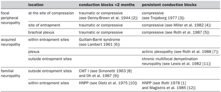
Until the beginning of the eighties, a focal neuropathy was the almost exclusive sign of an entrapment syndrome within a compartment of a relatively fixed size. Its cause was primarily a compression of external or internal origin, a repetitive trauma and some other aetiology, affection of a nerve on a focal segment. At that time, the persistent conduction blocks were recent observations [1] and the underlying mechanisms were unknown; as an example the classification of the causes of conduction blocks is presented in Table 1.
Table 1.
List of the type of conduction blocks that were known in the 1980s. What was relatively new at that time was the persistence of the conduction blocks, such as encountered in HNPP (hereditary neuropathy with liability to pressure palsy) and in postactinic brachial plexopathies.
Among the non-focal neuropathies Lewis and colleagues were able to isolate a “multifocal demyelinating neuropathy with persistent conduction blocks” from the other chronically acquired demyelinating neuropathies [11]. This neuropathy was characterised by asymmetric sensory motor deficits resembling mononeuritis multiplex, most pronounced in the upper extremities. The course of the disease could be either slowly or rapidly progressive over a long period with a subacute onset. The conduction blocks were not localised at entrapment sites.
In 1984, as I started to learn how to perform electrodiagnostic studies, Gérard Roth, my mentor at this time, asked me to describe in detail 2 unusual patients that he had encountered several months earlier and who had pure motor signs, no sensory involvement and persistent conduction blocks. The studies of the different charts and the nerve conduction studies performed in these 2 patients were the basis of my medical thesis [13], and we published one of the two cases the same year [14]. The aim of this review paper is to point out the difficulties which we encountered when we were studying these patients and the difficult time we have had to publish our article on this new disease.
Patients and methods
Patient 1
This 47-year-old man was first examined in February 1977, presented with a slowly progressive weakness of the right hand (since 1969) and the right leg (since 1974). When I examined him in July 1984, he had a symmetrical weakness of the upper extremities, which was more severe distally, and only slight voluntary contractions of the radial innerved muscles were possible. Fasciculations and myokymia were observed in the four limbs with slight amyotrophy in both thighs and hand muscles. Tendon reflexes were weak or absent, and there was no sensory deficit or symptom, but frequent painful cramps were reported.
During the last years palsy extended to involve neck, tongue muscle with fasciculations, resulting in difficulty of speech and mastication. He died at the age of 59 years as a result of respiratory distress, after an evolution of 21 years, as reported by Magistris and Roth [15].
Relatives were examined and had normal neurological and neurophysiological examinations. The patient underwent extensive laboratory studies which were all normal (in March 1990 anti-GM1antibodies were found to be elevated at a titre of 88 UI/L, normal <20). Histological examination of the sural nerve was compatible with a ‘very mild ischemic or toxic polyneuritis’.
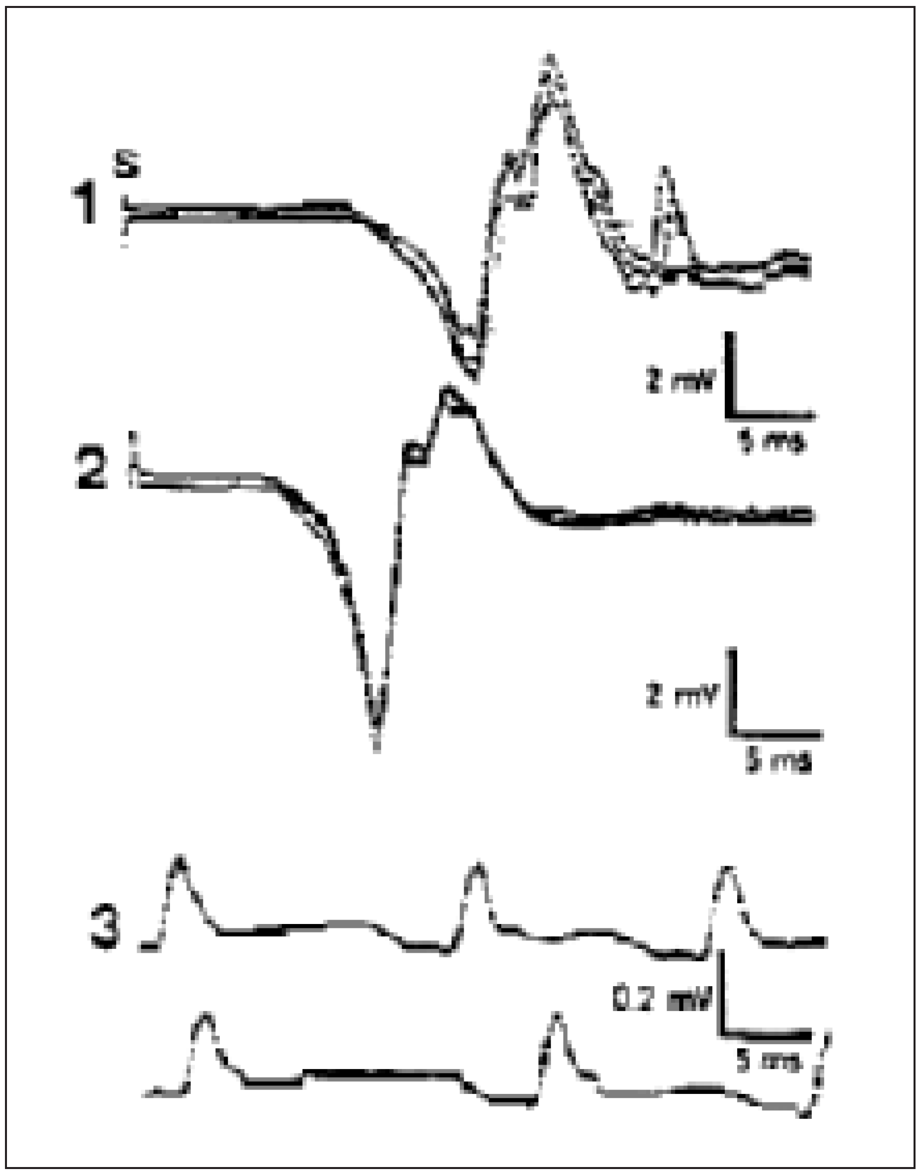
The hallmark of the examination was the observation of multifocal conduction blocks (Figure 1). The conduction blocks increased in severity and number at each examination (2 conduction blocks during the first exam in 1977, 12 conduction blocks during the last exam in 1984). Long latency motor axon reflexes with late motor unit potential (MUP) were evoked in the muscles with the partial conduction blocks. Continuous asynchronous and arrhythmic firing of a large number of fasciculation potentials were recorded in the weak muscles, causing clinical myokymia. The spontaneous activity appeared either as isolated fasciculations or occasionally as doublets, recurring irregularly over long periods of time or as sporadically occurring trains of grouped fasciculations (Figure 2) with interpotential intervals varying from 7 to 70 ms. The extent of spontaneous activity reflected the severity of the conduction block. The number of motor unit potentials was reduced during maximal effort in accordance with the severity of the conduction block. When stimulation could not be applied proximally, for example of the sciatic nerve, the existence of a suspected conduction block was confirmed when the number of motor unit potentials evoked by graded stimulation was much greater than could be obtained during maximal contraction and/or the total area of the evoked compound muscle action potential (CMAP) was much greater than the sum of the areas of the individual motor unit potentials (Figure 3).
Figure 1.
Patient 1: An almost complete conduction block of the right ulnar nerve at the brachial plexus could be demonstrated (surface electrodes over the abductor digiti minimi muscle), stimulation at the wrist (1), axilla (2) and Erb’s point (3).
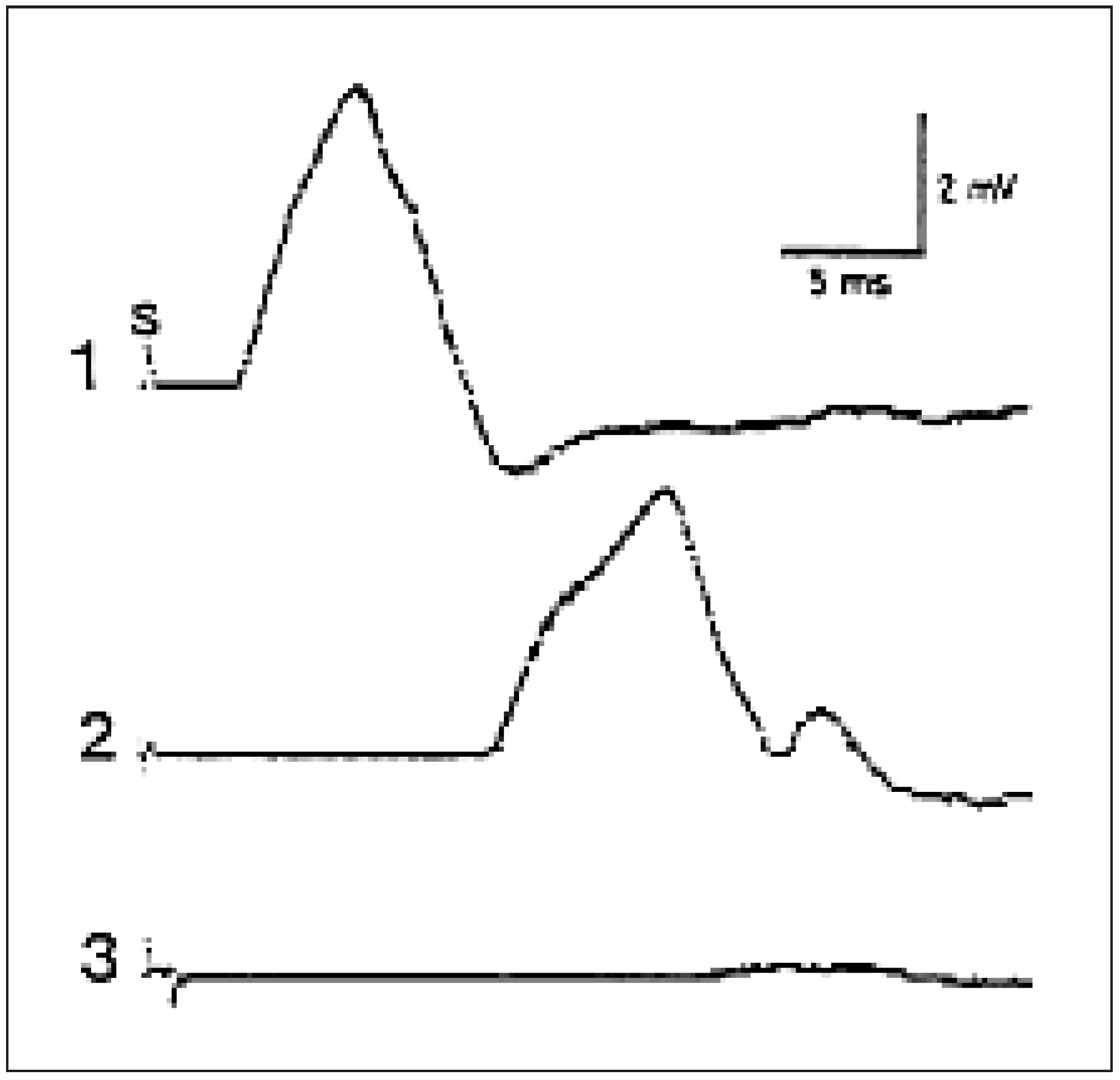
Figure 2.
Patient 1: Fasciculation potentials as seen with a needle electrode, from a thenar muscle. The appearance of the fasciculations was either stable or altered through successive isolated or grouped discharges.
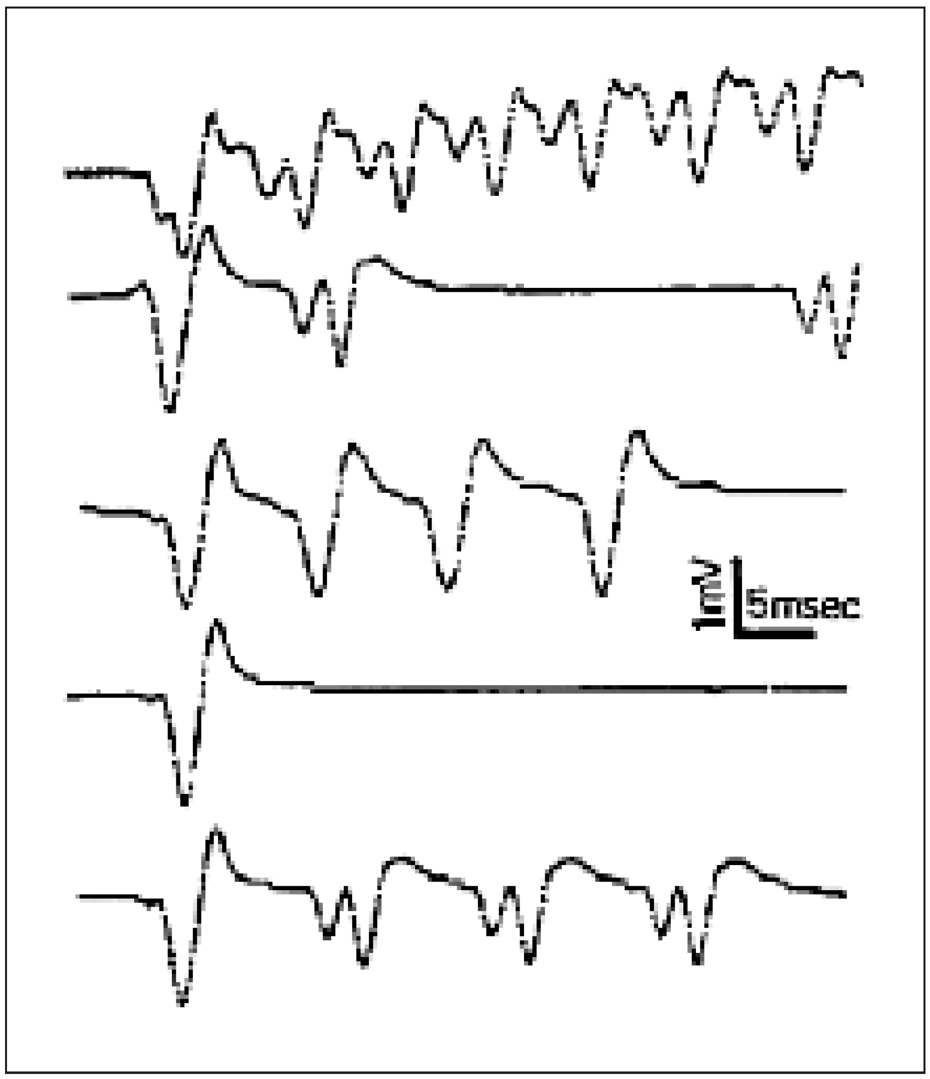
Figure 3.
Patient 1: Proximal conduction block of the right sciatic nerve. A single motor unit is obtained during maximal contraction, tibialis anterior muscle (3). Stimulation at the gluteal skin fold with needle electrode (1), at the fibular neck (2).
Patient 2
During the first examination in 1974, this 74-year-old man complained of a slowly progressive weakness of the right hand without pain or sensory symptom. Within 9 months he had developed a severe weakness of radial nerve innervated muscles, except triceps brachii and extensor carpi radialis, the right extensor and flexor foot muscles, with slight amyotrophy. Fasciculations and myokymia were evident and seen in the weak muscles, with absence of tendon reflexes but with no sensory deficit. The amyotrophy of the paretic muscles worsened gradually during successive examinations from 1976 to 1986 with a bilateral ulnar paresis of the hands, clearer on the left. In 1986 only the left leg remained normal, without motor weakness. The patient died quadriplegic, probably at the beginning of the 1990s.
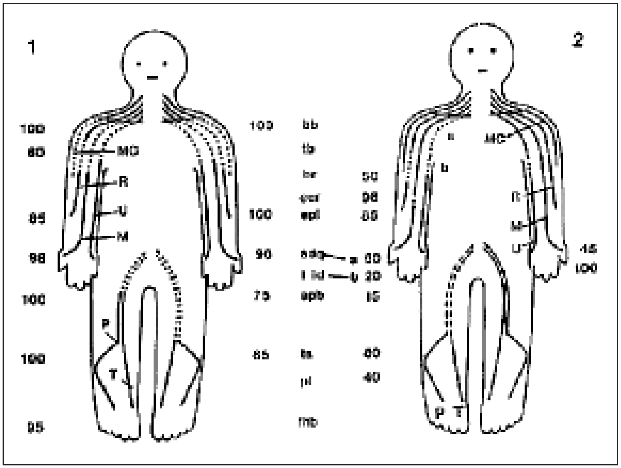
The conduction blocks of the involved nerves were located on the segment indicated by dotted lines. The importance of the block for the axons of individual muscles was expressed in per cent and depicted in Figure 4.
Figure 4.
Distribution of the conduction blocks in patient 1 and 2: each conduction block was located somewhere on a segment indicated by the dotted lines. The importance of the conduction block for the axon of individual muscles is expressed in per cent.
Discussion
These 2 patients were presented to J. Lapresle, professor of neurology in Kremlin-Bicêtre (F) and to B. Bady, professor of neurology in Lyon (F). These two experts favoured the diagnosis of amyotrophic lateral sclerosis (ALS), based on (i) ‘sharp reflexes’, actually an exaggeration of the contraction response to muscle percussion (as will be demonstrated later by Magistris and Kohler in 1996 [16]) and (ii) suspected questionable methodology, owing in particular to the difficulty in stimulating the proximal nerves.
The reality of the proximal multifocal persistent conduction blocks was based on the successive electrophysiological studies performed in the 2 patients that certified the persistence of the blocks, and also because Gérard Roth and his pupils were accustomed to diagnose conduction blocks in patients who had motor involvement of the upper limbs following radiation of the brachial plexus or in patients presenting with hereditary neuropathy with liability to pressure palsies. This purely motor syndrome associated with the chronic persistence of blocked nerves, progression of the weakness and incessant fasciculations without known cause was submitted to publication under the title ‘Motor neuropathy with proximal multifocal persistent conduction block, fasciculations and myokymia. Evolution to tetraplegia’. The paper was eventually accepted by the journal European Neurology after having previously been rejected by an American journal, arguing that conduction blocks were not a real problem in these patients, and allegedly resulted from the inadequate methodology and inexperience of the authors.
The discussion in the medical thesis, written in 1985 [13], underlines some clinical aspects: the insidious onset of the muscle weakness, its asymmetry, the abundant spontaneous activity seen at rest, the absence of sensory disturbances, all signs that raise questions about motor neuron diseases, such as ALS or a special type of progressive spinal amyotrophy.We were, however, convinced that our patients did not suffer from a motor neuron disease because of the presence of (i) persistent conduction blocks and (ii) evidence of numerous long latency motor axon reflexes suggesting presence of a neuropathy [17].
The cause of weakness was clearly established by electrophysiological evidence of proximal multifocal conduction blocks of individual nerves, not located within the usual entrapment sites. Certain characteristics of the patients’ illness were similar to the cases reported earlier by Lewis and Sumner [11], such as conduction blocks, but the following differences were sufficiently clear to ascertain that our 2 patients were suffering from another type of neuropathy: no numbness, no pain, no sensory disturbance were reported and furthermore, the conduction blocks were proximally located in our cases, with the presence of an abundant spontaneous activity seen at rest.
In 1986 the persistent conduction blocks had recently been described and the underlying mechanisms were unknown. Their electrophysiological demonstration was not easy, simply because not all EMG machines were able to measure the negative area of the CMAP and there was the dogma of a close link between presence of fasciculations and occurrence of ALS. The unsolved questions at that time were (i) the physiopathology of the disease reported in these 2 patients; (ii) the possible biological markers to be searched for in the serum and CSF as all results were unrevealing; (iii) the treatment to propose, as prednisone did not improve our patients. Some of those questions received an answer in the 1990s.
Acknowledgements
The author is very grateful to Thierry Kuntzer, MD (Lausanne), and Michel R. Magistris, MD (Geneva), for their time, help and comments on the manuscript.
References
- Roth G. Intranervous regeneration of lower motor neuron. Study of 1153 motor axon reflexes. Electromyogr Clin Neurophysiol. 1978;18:225–88, 211–351.
- Denny-Brown D, Brenner C. Paralysis of nerve induced by direct pressure and by tourniquet. Arch Neurol Psychiatry. 1944;51:1–26. [CrossRef] [Scilit]
- Trojaborg W. Prolonged conduction block with axonal degeneration. An electrophysiological study. J Neurol Neurosurg Psychiatry. 1977;40:50–7. [CrossRef] [Scilit] [PubMed]
- Miller RG, Olney RK. Persistent conduction block in compression neuropathy. Muscle Nerve. 1982;5:s154–6. [PubMed]
- Roth G, Magistris MR. Neuropathies with prolonged conduction block, single and grouped fasciculations, localized limb myokymia. Electroencephalogr Clin Neurophysiol. 1987;67:428–38. [CrossRef] [Scilit] [PubMed]
- Lambert EH. Neurophysiological techniques useful in the study of neuromuscular disorders. Res Publ Assoc Res Nerv Ment Dis. 1961;38:247–373.
- Roth G, Magistris MR, Le Fort D, Desjacques P, Della Santa D. Plexopathie brachiale post-radique. Blocs de conduction persistants. Décharges myokymiques et crampes. Rev Neurol. 1988;144:173–80. [PubMed]
- Simonetti C. Neuropatia periferica ereditaria. (Contributo clinico alle forme atipiche della malattia di Charcot-Marie). Rev Neurobiol. 1963;9:383–417.
- Oh SJ, Chang CW. Conduction block and dispersion in hereditary motor sensory neuropathy. Muscle Nerve. 1987;10:656–7.
- Dietz V, Schenck E. Familiäre Polyneuropathie mit Neigung zu Druckparesen. Klinische und neurographische Befunde einer Familie. Arch Psychiatr Nervenkr. 1975;220:67–78. [CrossRef] [Scilit]
- Lewis RA, Sumner AJ, Brown MJ, Asbury AK. Multifocal demyelinating neuropathy with persistent conduction block. Neurology. 1982;32:958–64. [CrossRef] [Scilit] [PubMed]
- Magistris MR, Roth G. Long-lasting conduction block in hereditary neuropathy with liability to pressure palsies. Neurology. 1985;35:1641. [CrossRef] [Scilit]
- Ochsner F. Neuropathie motrice avec blocs de conduction proximaux et persistants. Fasciculations et myokymies (contribution à l’étude des blocs de conduction). [Thèse de Médecine 8080.] Genève: Médecine & Hygiène; 1986.
- Roth G, Rohr J, Magistris MR, Ochsner F. Motor neuropathy with proximal multifocal persistent conduction block, fasciculations and myokymia. Evolution to tetraplegia. Eur Neurol. 1986;25:416–23. [CrossRef] [Scilit] [PubMed]
- Magistris MR, Roth G. Motor neuropathy with multifocal persistent conduction blocks. Muscle Nerve. 1992;15:1056. [CrossRef] [Scilit] [PubMed]
- Magistris MR, Kohler A. Contraction response to muscle percussion is increased in peripheral nerve conduction block. Neurology. 1996;47:1243–6. [CrossRef] [Scilit] [PubMed]
- Roth G. Intranervous regeneration: the study of motor axon reflexes. J Neurol Sci. 1979;41:139–48. [CrossRef] [Scilit] [PubMed]
This is an open-access article distributed under the terms of the Creative Commons Attribution Non-Commercial No Derivatives (CC BY-NC-ND) 4.0 License.