


The aim of this section is to prepare the neurologist-in-training for the FMH examination, to confront her or him with specific problems of everyday neurological practice and to give him or her updates on recent controversies in clinical neurology.
Neurological MCQ
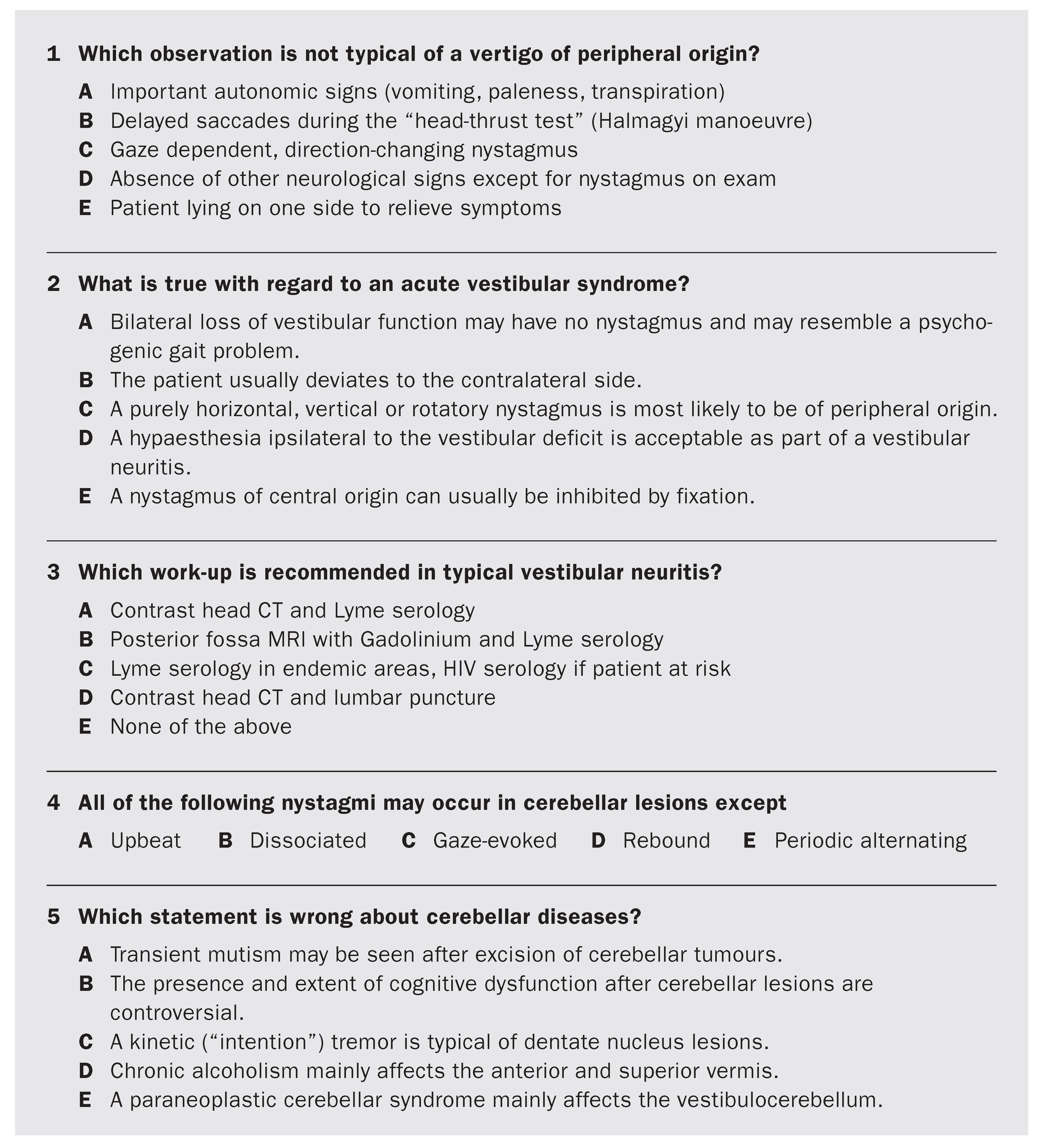
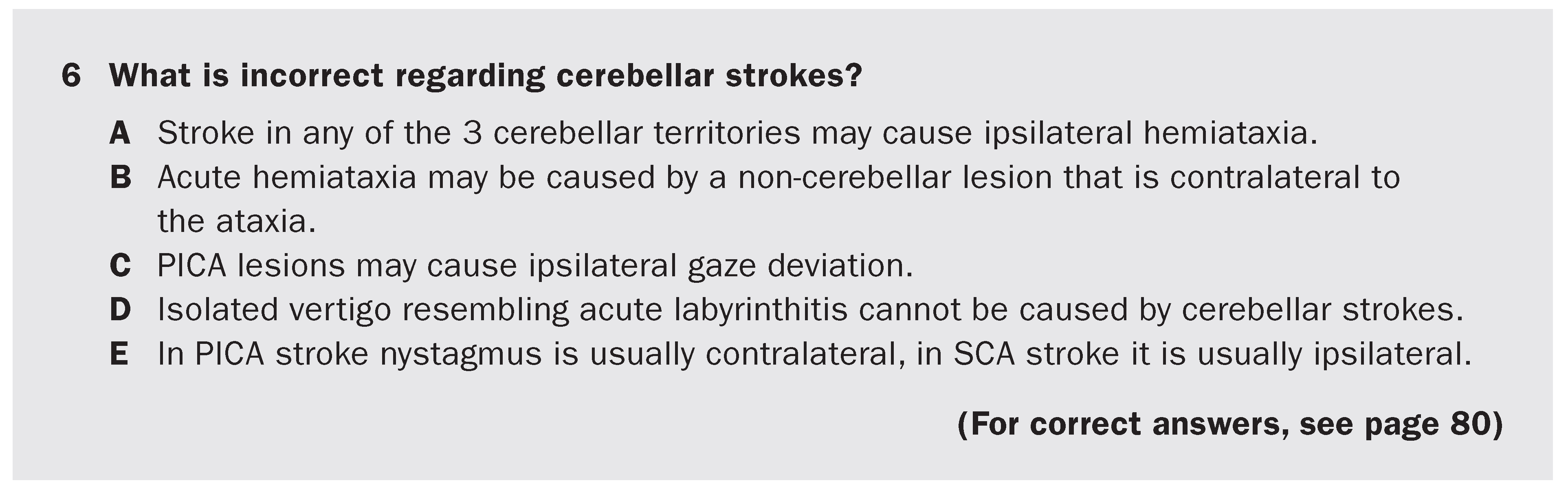
Select the one correct answer.
References
- Brazis PW, Masdeu JC, Biller J. Localisation in Clinical Neurology. 4th ed. Philadelphia: Lippincott, Williams & Wilkins; 2001.
- Baloh RW. Vestibular neuritis. N Engl J Med 2003;348:1027–32.
Neuroradiology, neuroanatomy and EEG
Read for you
Update on Creutzfeldt-Jakob disease (CJD)
Prion diseases are considered as a family of degenerative diseases [1] that can occur sporadically, genetically (autosomal dominant mutations within the short arm of chromosome 20) or by infection. The prion is a physiologically occurring protein (PrPc) which is a constituent of the neuronal cell membrane. Its precise physiological role is unknown.
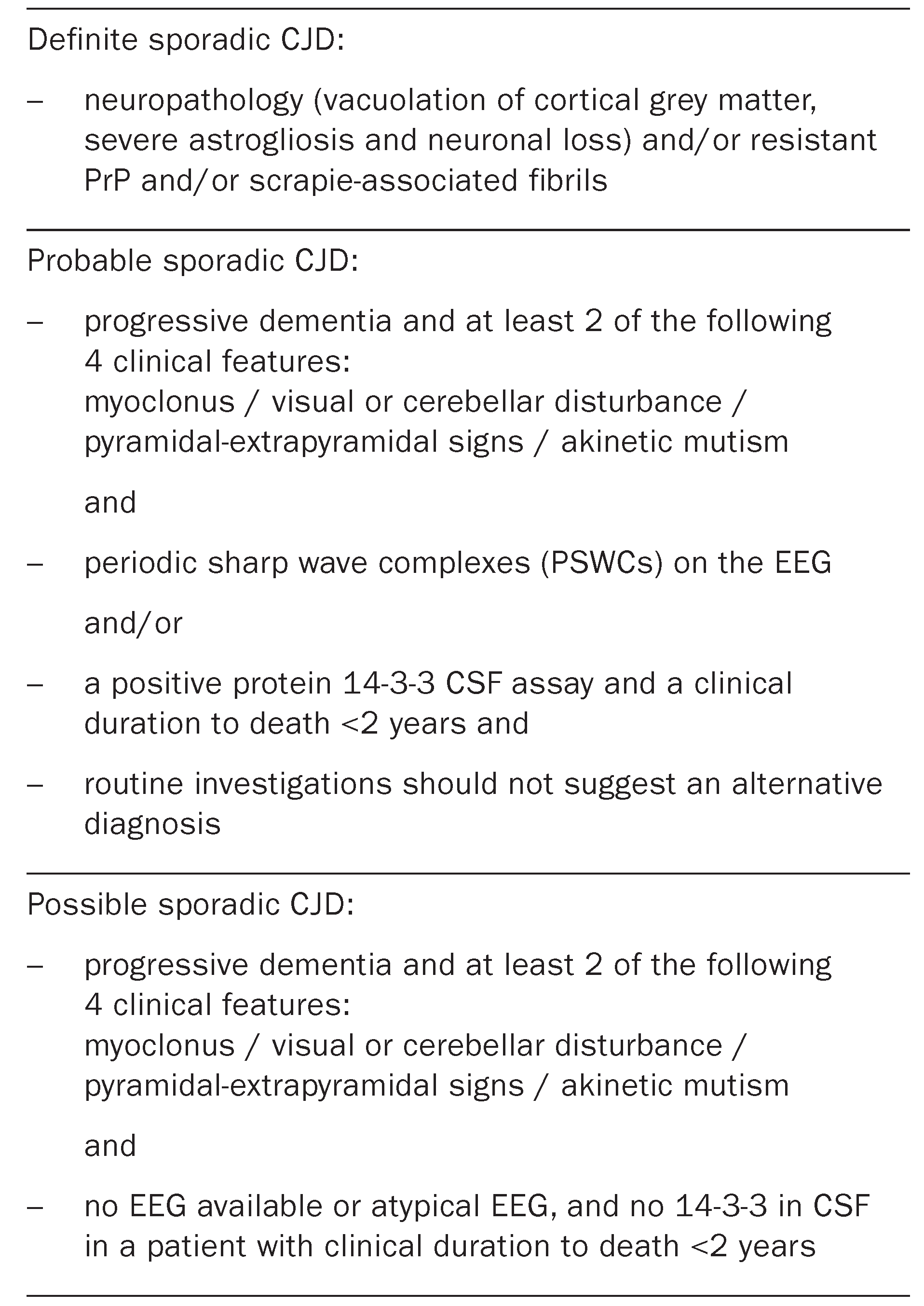
When PrPc comes into contact with the abnormal prion protein (PrPSc), the former changes its conformation into the pathological isoform and becomes proteinase resistant. It is hypothesised that this process may occur in continuitatem [2]. The abnormal prion protein aggregates to produce amyloid. There are five known major syndromes in humans: sporadic CJD (sCJD), Gerstmann-Sträussler-Scheinker syndrome (GSS), fatal familial insomnia (FFI), kuru and the new variant of CJD (vCJD). The main differential diagnoses of CJD are Alzheimer’s disease, dementia with Lewy bodies and cerebrovascular dementia. The typical clinical triad comprises cognitive disturbances, ataxia and myoclonus and is present in about 60 to 70% of sCJD patients. Since 1998, sCJD is diagnosed according to the “Revised World Health Organization Criteria” (Table 1) [3].
Table 1.
Revised World Health Organization Criteria of sporadic Creutzfeldt-Jakob disease (CJD).
Among the additional exams, EEG was the first to be integrated in the current diagnostic criteria. The characteristic pattern of the EEG is one of bilateral (initially unilateral) periodic discharges of 0.5 to 2 Hz and is present in 60 to 70% of cases at some time during the illness (sensitivity 66% and specificity 74%) [4]. Additionally, CSF 14-3-3 protein measures (sensitivity 94% and specificity 84%) [5] and CSF neuron-specific enolase can contribute to the probable diagnosis but one has to keep in mind that false-positive results are found in diseases such as herpes simplex encephalitis, stroke, Hashimoto encephalopathy and paraneoplastic disorders [6].
So far, MRI is not an official criterion for the diagnosis of CJD. However, MRI abnormalities have now been described for more than 10 years.
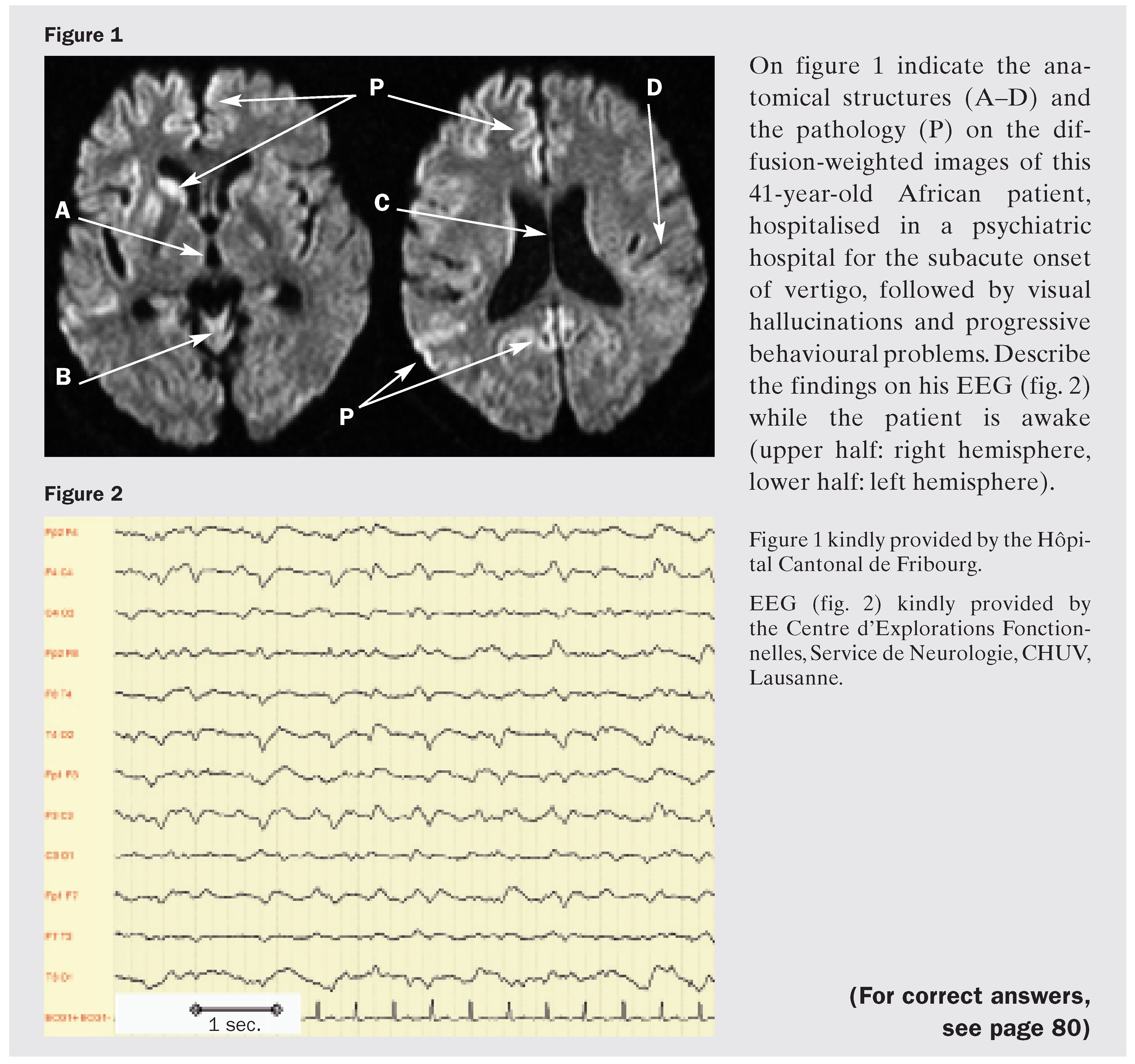
They include hyperintensities in the basal ganglia (mainly caudate nucleus and putamen), thalamus (including the pulvinar) and in a multifocal distribution along the cortical ribbon (cerebral or cerebellar) in up to 80% of cases [7,8]. Although T2-weighted and fluid-attenuated inversion recovery (FLAIR) sequences show many of these lesions, diffusion-weighted (DWI) sequence seems to be the most sensitive to show cortical abnormalities [9]. The thalamic and cortical signals in sCJD are usually less intense than those in the putamen and caudate nucleus. These radiological findings are not completely specific, however, as they can also be seen in other neurological conditions such as encephalitis, hypoxia, CO poisoning, hypoglycaemia, Wilson’s disease, mitochondrial diseases and Huntington’s disease. Patients with the new variant of CJD typically show bilateral DWI changes in the pulvinar thalami (sensitivity 78% and specificity 100%) [10].
There is hope of stopping the disease process in humans by using substances that limit PrPSc production in animals, such as derivatives of acridine and phenothiazine (for example chlorpromazin or quinacrine) [11] or pentosan polysulphate which is derived from beechwood [12]. Preliminary experience in humans with such substances against sCJD or vCJD has been rather disappointing, however.
References
- Prusiner SB. Shattuck lecture – neurodegenerative diseases and prions. N Engl J Med 2001;344:1516–26.
- Russmann H, Vingerhoets F, Miklossy J, Maeder P, Glatzel M, Aguzzi A, et al. Sporadic Creutzfeldt-Jakob disease: a comparison of pathological findings and diffusion-weighted imaging. J Neurol 2005;252:338–42.
- Report of the WHO consultation on the global surveillance, diagnosis and therapy of human transmissible spongiform encephalopathies. Geneva: WHO; 1998.
- Steinhoff BJ, Zerr I, Glatting M, Schulz-Schaeffer W, Poser S, Kretzschmar HA, et al. Diagnostic value of periodic complexes in Creutzfeldt-Jakob disease. Ann Neurol 2004;56:702–8.
- Zerr I, Pocchiari M, Collins S, Brandel JP, de Pedro CJ, Knight RS, et al. Analysis of EEG and CSF 14-3-3 proteins as aids to the diagnosis of Creutzfeldt-Jakob disease. Neurology 2000;55:811–5.
- Kenney K, Brechtel C, Takahashi H, Kurohara K, Anderson P, Gibbs CJ Jr. An enzyme-linked immunosorbent assay to quantify 14-3-3 proteins in the cerebrospinal fluid of suspected Creutzfeldt-Jakob disease patients. Ann Neurol 2000;48:395–8.
- Poser S, Mollenhauer B, Krauß A, Zerr I, Steinhoff BJ, Schroeter A, et al. How to improve the clinical diagnosis of Creutzfeldt-Jakob disease. Brain 1999;122:2345–51.
- Tschampa HJ, Kallenberg K, Urbach H, Meissner B, Nicolay C, Kretzschmar HA, et al. MRI in the diagnosis of sporadic Creutzfeldt-Jakob disease: a study on interobserver agreement. Brain 2005;128:2026–33.
- Shiga Y, Miyazawa K, Sato S, Fukushima R, Shibuya S, Sato Y, et al. Diffusion-weighted MRI abnormalities as an early diagnostic marker for Creutzfeldt-Jakob disease. Neurology 2004;63:443–9.
- Collie DA, Summers DM, Sellar RJ, Ironside JW, Copper S, Zeidler M, et al. Diagnosing variant Creutzfeldt-Jakob disease with the pulvinar sign: MR imaging findings in 86 neuropathologically confirmed cases. Am J Neuroradiol 2003;24:1560–9.
- May BC, Fafarman AT, Hong SB, Rogers M, Deady LW, Prusiner SB, et al. Potent inhibition of scrapie prion replication in cultured cells by bis-acridines. Proc Natl Acad Sci U S A 2003;100:3416–21.
- Doh-ura K, Ishikawa K, Murakami-Kubo I, Sasaki K, Mohri S, Race R, et al. Treatment of transmissible spongiform encephalopathy by intraventricular drug infusion in animal models. J Virol 2004;78:4999–5006.
Neurologist-in-training
Answers to MCQ

Answers to neuroradiology, neuroanatomy and EEG
Figure 1
- A
- Third ventricle
- B
- Vermis cerebelli
- C
- Septum pellucidum
- D
- Central sulcus (Rolandii)
- P
- Multifocal hyperintensities on DWI in the head of the caudate, anterior pallidum, putamen and in the cerebral cortex, as typically seen in sporadic Creutzfeldt-Jakob disease. The radiological differential diagnosis is discussed in the section “Read for you”. This patient died 6 months later and the diagnosis was confirmed histologically.
Figure 2
This EEG shows generalised slowing in the 4–6 Hz range (theta) with pseudoperiodic discharges of 0.5 to 2 Hz, compatible with but not specific for Creutzfeldt-Jakob disease.

This is an open-access article distributed under the terms of the Creative Commons Attribution Non-Commercial No Derivatives (CC BY-NC-ND) 4.0 License.