


Summary
Fournier PJ,Aguilar M, Stephanov S. Severe neurological complications in an adult achondroplastic dwarf with generalised spinal stenosis.Schweiz Arch Neurol Psychiatr 2004;155:64–6.
A case is reported with generalised spinal stenosis and severe neurological complications.Neurological and radiological findings and surgical procedures are discussed. The patient was admitted with profound spastic lower paraparesis and the upper limbs were paretic. After an emergency lumbar decompression, the patient improved and on the twelfth day he was able to take a short walk.Twenty-two days after the lumbar decompression a cervico-thoracic decompression was performed which proved fatal.
Introduction
Despite its rarity, achondroplasia is well documented in the literature [1,2,3,4,5,6,7,8,9,10,11,12]. Recognisable at birth, it is the most common form of congenital bony dysplasia [4,7].The abnormal formation of enchondral bone leads to stenosis of the craniospinal axis [1,5].At birth the head size is often large, the foramen magnum is stenotic and the musculature is hypotonic. The spinal X-rays demonstrate a narrowing of the interpedicular distances in the lumbar spine [7]. A small spinal canal is present from birth but signs and symptoms of spinal cord compression are noted in middle age or later [7]. Before 1980 most neurological complaints in adult achondroplasia were attributed to thoraco-lumbar stenosis [4].The use of total myelography followed by CT scan permitted a multilevel spinal stenosis visualisation. MRI is also used in achondroplasia.
Morgan and Young [1] divided their 17 cases with spinal stenotic syndrome into 3 groups, the third of which consisted of two patients with generalised spinal stenosis who fared less well.The current opinion is that a decompressive procedure of the spine should be done early, before the spinal cord is significantly damaged [1,3,4,5,6,7]. It is worth noting that the earliest report of neurological complications associated with achondroplasia was provided by Walter Dandy in 1921 [12].
We present a case of an adult achondroplastic dwarf with generalised spinal stenosis and severe neurological deficit.
Case report
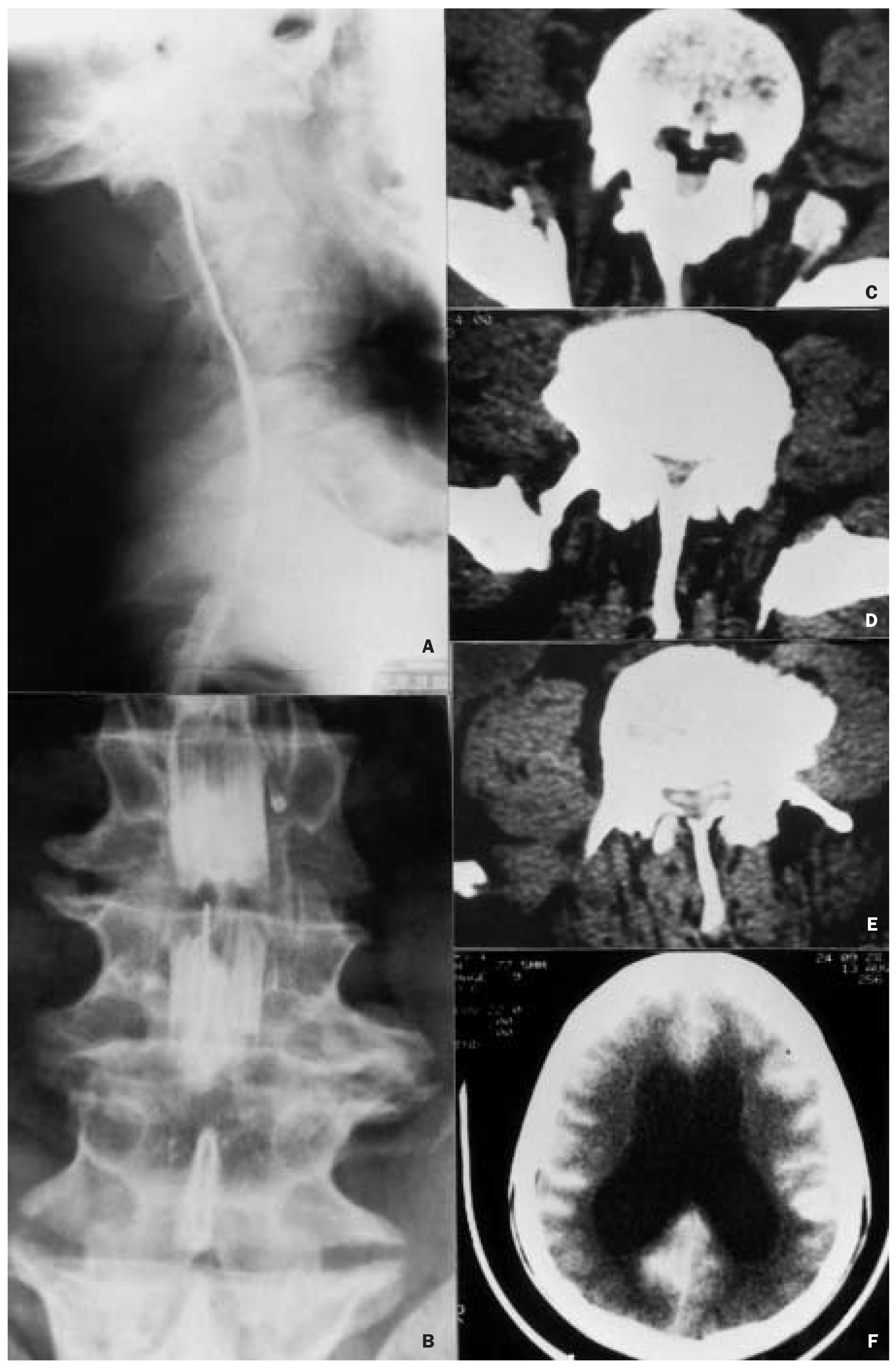
This 62-year-old man was admitted elsewhere for urinary retention and severe weakness of the lower limbs.He had been working for many years as a mason. The last 3 years, however, he was unable to do his job because of weakness of his legs and arms. The patient was seen in neurosurgical consultation. He presented a typical picture of achondroplastic dwarf, with a large head, short arms and legs. His abdomen was distended and a bladder catheter inserted. He exhibited a profound spastic paraparesis with Babinski signs and ankle clonus positive bilaterally; the distal parts of the upper limbs were paretic with mild hypotrophy of hands and hyperactive tendon reflexes. In view of this dramatic neurological picture an emergency total myelography, via lumbar puncture, followed by CT scan was performed, which showed stenosis of the entire spinal canal (fig. 1a–e). The subsequent head CT scan showed an internal hydrocephalus obviously arrested (fig. 1f).
Figure 1.
1a to 1e shows the results of a total myelography followed by CT scan: an extended stenosis of the spinal canal at all levels is presented; 1f demonstrates the arrested hydrocephalus.
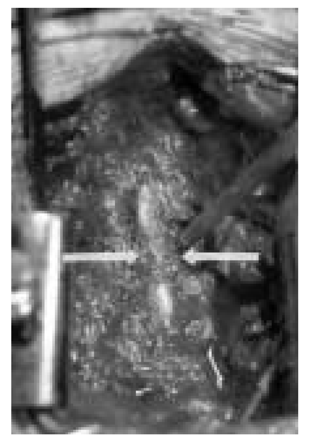
The patient was taken to the operating room. A laminectomy was performed from L1 through L5. The laminae were found very thick and broad and the ligamentum flavum hypertrophied. The dura was extremely thin. The decompression was difficult. Three days after the procedure, the patient started to move his legs. On the 12th postoperative day he was able to take a short walk with the aid of crutches. Twenty-two days after the lumbar decompression, we decided to decompress the low cervical and upper thoracic spine according to the myelographic features. The spinal processes were removed using a rongeur and the initial laminectomy was performed using a high-speed drill. A mini Kerrison punch was used to detach laterally the osseous cervical ring at T1 level. (fig. 2). The patient tolerated the operation well and he was placed on steroids. After the second decompression, however, the patient did not improve; his neurological status gradually deteriorated and he died from respiratory failure 3 weeks after the cervico-thoracic decompression.
Figure 2.
Peroperative view during cervico-thoracic laminectomy shows the osseous ring at T1 level (arrows).
Discussion
The experience of treating achondroplastic patients is limited [3]. Stenosis of the achondroplastic vertebral canal increases with time provoking neurological signs and symptoms as intermittent claudication, nerve root compression, paraparesis, paraplegia and quadriplegia [2,3,6]. The frequently seen hydrocephalus in this disease usually stops with time [10].
The achondroplasia presents a difficult management problem in spinal surgery [5‚ 9] and the preoperative duration of signs and symptoms and the age of the patient may equally determine the outcome of the operative treatment [3]. In the series of Morgan and Young of 17 patients with achondroplasia, 2 who had generalised spinal stenosis fared less well [1].These authors concluded that the prognosis for patients with this type of achondroplasia appears to be poor [1]. In extreme and selected cases with general spinal stenosis Uematsu et al. proposed a total craniospinal decompression along the entire neuroaxis in one or two stages [5]. In 1994 these authors reported their experience with 7 patients treated by this technique. The need for prophylactic decompressive laminectomy in achondroplasia has been raised by Verbiest [3]. According to this expert spine surgeon, if all midsagittal diameters, as judged by total myelography of the vertebral canal followed by CT scan, are 10 mm or less, decompressive laminectomy is needed at levels considered to be asymptomatic [3]. However, prospective studies will be necessary to document that prophylactic decompression is indicated in those patients before the onset of a neurological deficit [9].
Our unfortunate patient is an example how neglect of the complaints of an achondroplastic individual and his late referral to a neurosurgical unit can lead to an undesirable result.
References
- Morgan DF, Young RF. Spinal neurological complications of achondroplasia. Results of surgical treatment. J Neurosurg 1980;52:463–72.
- Alexander E Jr. Significance of the small lumbar spinal canal: cauda equina compresssion syndromes due to spondylosis. Part 5: Achondroplasia. J Neurosurg 1969;31:513–9.
- Verbiest H. Lumbar spine stenosis. In: Youmans JR, editor. Neurological Surgery. Vol. 4. 3rd ed. Philadelphia: W.B. Saunders Company; 1990. p. 2805–55.
- Winfield JA, Wang H. Achondroplasia: neuroradiological investigation and surgical management of craniocervical junction and pan-spinal stenosis. In: Long DM, editor. Current Therapy in Neurological Surgery 1985–1986. Toronto, Philadelphia: B.C. Deeker Inc; 1985. p. 144–6.
- Uematsu S, Wang H, Kopits SE, Hurko O. Total craniospinal decompression in achondroplastic stenosis. Neurosurgery 1994;35:250–8.
- Lutter LD, Langer LO. Neurological symptoms in achondroplastic dwarfs. Surgical treatment. J Bone Joint Surg 1977;59:A87–92.
- Tolo V. Spinal disorders associated with skeletal dysplasias and metabolic diseases. In: Rothman RH, Simeone FA, editors. The Spine. Vol. 1. 3rd ed. Philadelphia: W.B. Saunders Company; 1992. p. 349–64.
- Epstein BS, Epstein JA, Jones MD. Lumbar spinal stenosis. Radiol Clin North Am 1977;15:227–39.
- Weinstein PR. Achondroplasia: Comment. Neurosurgery 1994;35:257–8.
- Menezes AH. Congenital and acquired abnormalities of the craniovertebral junction. In: Youmans JR, editor. Neurological Surgery. Vol. 2. 4th ed. Philadelphia: W.B. Saunders Company; 1996. p. 1035–89.
- Uematsu S, Wang H, Hurko O. The subarachnoid fluid space in achondroplastic spinal stenosis. In: Nicoletti B, Kopits SE, Ascani E, McKusick VA, editors. Human Achondroplasia: A Multidisciplinary Approach. New York: Plenum Press; 1988. p. 275–81.
- Dandy WE. Hydrocephalus in achondroplasia. Bull Johns Hopkins Hospital 1921;32:5–10.
This is an open-access article distributed under the terms of the Creative Commons Attribution Non-Commercial No Derivatives (CC BY-NC-ND) 4.0 License.