


Summary
Restless Legs Syndrome (RLS) is a common disorder affecting, depending on the diagnostic criteria used, up to 5–10% of the general population and with an increasing in incidence with age. The clinical features include dysaesthesias and paraesthesias in the legs and sometimes in the arms, occurring at rest, worsening in the evening and night and alleviated by movements. Associated symptoms are insomnia, fatigue, sleepiness, mood and anxiety disorders. The diagnosis is mainly based on clinical features. Neurophysiological investigations, polysomnography and actimeter are used to objectively assess sensory-motor disorders and sleep disturbances.Apart from secondary cases related to neurological or medical diseases, the restless legs syndrome is a primary and idiopathic disorder showing a high rate of familiarity and implying a deficient dopaminergic transmission at spinal level and/or basal ganglia with a selective impairment of D2 receptors. The additional role of a dysfunction of the brain iron metabolism has been suggested but not defined in detail. Levodopa is an efficacious treatment, but adverse effects, i.e., rebound and augmentation, are frequently described. Dopamine agonists have largely replaced levodopa in moderate to severe restless legs syndrome, while slow-release levodopa is still used in mild cases. Treatment with opiates and gabapentin is proposed for cases not responding to dopamine agonists and/or when pain is the presenting symptom. Iron therapy is recommended only in patients with low level of ferritin.
Introduction
From the first extensive description by Ekbom in 1945 [1], progresses in sleep medicine have allowed a better understanding of the pathophysiological mechanisms and true clinical relevance of restless legs syndrome (RLS). The restless legs syndrome may represent a “primary sensory disorder” with a secondarily triggered motor response. Patients report dysaesthetic sensations described as “pins and needles”, “internal itch”, “crawling”, “creeping”, “burning”, “painful sensations” or a more general “sense of discomfort” inducing a need to move. Typically, symptoms occur at rest, are relieved by movements and present a typical circadian pattern with appearance or worsening in the evening and night. Most patients report difficulty in falling asleep or early wake-up in the middle of the night with dysaesthesias and paraesthesias forcing them to move and walk (“the nightwalkers”). Even though some patients report sleepiness, fatigue, tiredness and changes in mood are the most frequent diurnal symptoms. Restless legs syndrome often first appears in adult life as mild and transitory discomfort while at rest, but it slowly progresses with sleep disturbances appearing after 40 years of age. Early onset of restless legs syndrome, even in childhood, is possible particularly in familial cases. The diagnosis is primarily “clinical” and polysomnography is useful either to exclude other sleep disorders, to confirm the severity of insomnia and the presence of periodic leg movements during sleep (PLMS).
This review will highlight some of the recent clinical and pathophysiological aspects of restless legs syndrome and summarises the clinical approaches and therapeutic strategy in current use.
Definition
A recent consensus by the International Restless Legs Syndrome Study Group (IRLSSG) [2] on essential diagnostic criteria has stressed that the diagnosis of restless legs syndrome can be made in the presence of four “clinical” major criteria, namely: (1) a need to move the limbs usually associated with paraesthesias and dysaesthesias; (2) motor restlessness to relieve sensory symptoms; (3) symptoms that appear or are worse at rest with a partial relief by activity; and (4) symptoms that are worse in the evening and during the night. More recently the presence of PLMS and a good response to levodopa or dopamine agonists were suggested by the IRLSSG as important additional criteria to confirm restless legs syndrome. Other minor criteria are sleep disturbances, involuntary movements while awake at rest, periodic leg movements while awake (PLMW), an unremarkable neurological examination and a positive family history, criteria that, however, are not mandatory for diagnosis.
The first criterion for diagnosis is the presence of paraesthesias and dysaesthesias associated with the need or internal urge to move. The sensory symptoms are localised more frequently in the legs, especially the regions between the knees and the ankles, more frequently bilaterally. Paraesthesia or dysaesthesia is usually associated with an urge to move and patients typically walk around. Often rubbing the legs, tossing and turning in bed, stretching, crossing and flexing the legs, or pacing the floor may be tried as relief. The arms may be involved more rarely in 22–50% of patients [3,4,5,6] and particularly in severe cases [5,6].
The other two criteria are the onset at rest, for example in lying or sitting position, and the improvement with movements. If the patient is forced to lie still or stay at rest, the symptoms will continue to build up with a crescendo pattern in the urge to move inducing the appearance of periodic legs movements, i.e., periodic leg movements while awake, or involuntary jerks. Once movement ceases, however, the symptoms will return with a crescendo pattern.
An important clinical criterion, useful for differential diagnosis, is the circadian pattern, with symptoms predominating in the evening and at night and declining in the daytime. Studies analysing the over-time occurrence of sensory symptoms and PLMS [7,8,9] have found that the maximal score of sensory complaints could be detected at 11–12 pm, the lowest score at 9–11 am [7,10]. In a similar manner, PLMS progressively increased in night-time with a peak occurring in the falling phase of core temperature between midnight and 1 am and declining progressively between 9 am and 2 pm. The cause of this sensory and motor circadian pattern is unknown [7,8] but may reflect the circadian variation on dopamine metabolites [11,12] and/or in iron [13,14], a cofactor for tyrosine hydroxylase synthesis and, therefore, for dopamine synthesis.
Additional features for diagnosis are sleep disturbances, presence of PLMS, a normal neurological examination in idiopathic cases, a progressive clinical course and a positive family history in familial cases. More than 94% of patients have sleep disturbances, more frequently difficulties in falling asleep (58%) or maintenance insomnia with several awakenings related to occurrence of sensory symptoms [6,15]. Fatigue and tiredness are the most frequent diurnal consequences. Excessive daytime sleepiness is less common. An abnormal Epworth sleepiness score (>10) can be found in 24–34% of patients [4,6] and may be more common in patients with PLMS [15]. The relationship between sleepiness and PLMS is, however, debated, daytime sleepiness not being strongly related to PLMS index [16]. Symptoms of anxiety and depression are often found when a patient with restless legs syndrome is examined, and depressive symptoms at any time during life are reported by 33% of patients [6].
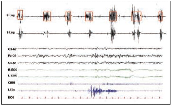
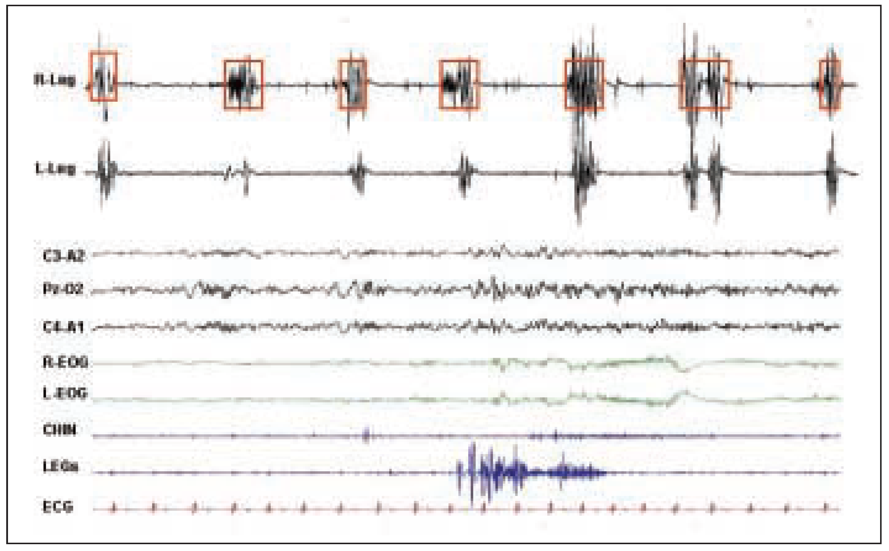
As about 80–90% of patients with restless legs syndrome have PLMS, these entities have traditionally been considered as necessarily associated. However, some patients with restless legs syndrome do not have PLMS, thus it seems reasonable to view the restless legs syndrome as a sensory disease and PLMS as a nonspecific motor response sharing a common underlying mechanism. PLMS are described as stereotyped flexion movements of the lower and sometimes upper extremities [17], lasting 0.5 to 5 s and occurring every 20 to 40 s throughout sleep (Figure 1 upper panel). They are associated with phasic heart rate and blood pressure rises [18] as well as with microarousal (Figure 1, lower panel), which can disturb sleep. Interestingly, recent studies have demonstrated that PLMS may occur in unstable sleep and without microarousals supporting the hypothesis that PLMS may be the expression of a dysfunction of the oscillatory neural networks regulating the cyclic arousability of the sleepy brain [19,20]. Periodic or aperiodic leg movements may also occur during wakefulness (PLMW), showing the same periodicity of PLMS but generally lasting more than 5 s and having a shorter intermovement interval [21]. Their occurrence during the “Suggested Immobilisation Test” (SIT) has been proposed as a diagnostic tool for definition of restless legs syndrome severity [22].
Figure 1.
The upper panel shows periodic limb movements during light sleep occurring every 20–40 seconds. The lower panel depicts a PLMS associated with microarousal and tachycardia.
Epidemiology
Prevalence studies conducted over the past decade have demonstrated that up to 5 [1] to 10–15% [23,24,25] of adults in Western countries are likely to have restless legs syndrome. Lower frequencies of restless legs syndrome are found when specific questionnaires are used in Asian populations and when patients are clinically assessed [26]. All the surveys examining the effect of age reported a strong increase in prevalence with age, more cases diagnosed between age 30 to 40 (18%) and between age 40 to 60 (33%) [3,6,25]. However, 38 to 45% of patients [4,26] experienced their first symptom before the age of 20, suggesting that restless legs syndrome is a progressive and chronic condition. Early onset of restless legs syndrome is common in familial forms (see below). A history of “growing pains” is not uncommon in patients with early onset restless legs syndrome. While no differences for sex were found by Lavigne et al. [23] and Phillips et al. [24], the prevalence for females was twice as high in the German sample [24].
Diagnosis
Clinical features
As previously described, diagnosis is essentially based on the patient’s symptoms and characterised by the four clinical features (see definition). These features allow clinicians to differentiate restless legs syndrome from lumbo-radicular or vascular pain syndromes, arthritis, leg cramps, painful legs and moving toes and drug-induced acathisia. Pain syndromes and arthritis do not have a circadian pattern, and movements do not rapidly relieve symptoms. Painful legs and moving toes do not have the focal urge to move, do not show the circadian pattern with a worsening at night and patients show repetitive semi-continuous movements of the toes. Drug-induced acathisia and leg cramps show some similarities with restless legs syndrome, i.e., circadian pattern, but they differ in other aspects. In acathisia there is a prior use of neuroleptic drugs or dopamine antagonists and more often the patients complain about an inner restlessness rather than leg discomfort, with a desire to move the whole body. Sensory symptoms are less frequent and complaints not worse when lying or sitting compared to the upright position. Patients with leg cramps complain about sudden attacks of sustained and painful contraction of the muscles without sensory symptoms which can be relieved by stretching the affected muscle.
The most difficult differential diagnosis is polyneuropathy with sensory symptoms not worsening at night, unrelieved by walking and in which electromyography is altered. This, however, does not exclude the association of restless legs syndrome with neuropathies and radiculopathies that have been described as possible secondary causes of restless legs syndrome [26] and with alteration of small fibres in mild idiopathic restless legs syndrome cases [27].
Interestingly, in some patients with anxiety or depressive symptoms restless legs syndrome-like symptoms may be present and in such patients the coexistence of complaints derived from psychiatric disease with those from restless legs syndrome makes differential diagnosis particularly difficult. These patients typically have no or only a few PLMS and do not respond to dopaminergic drugs.
Questionnaires
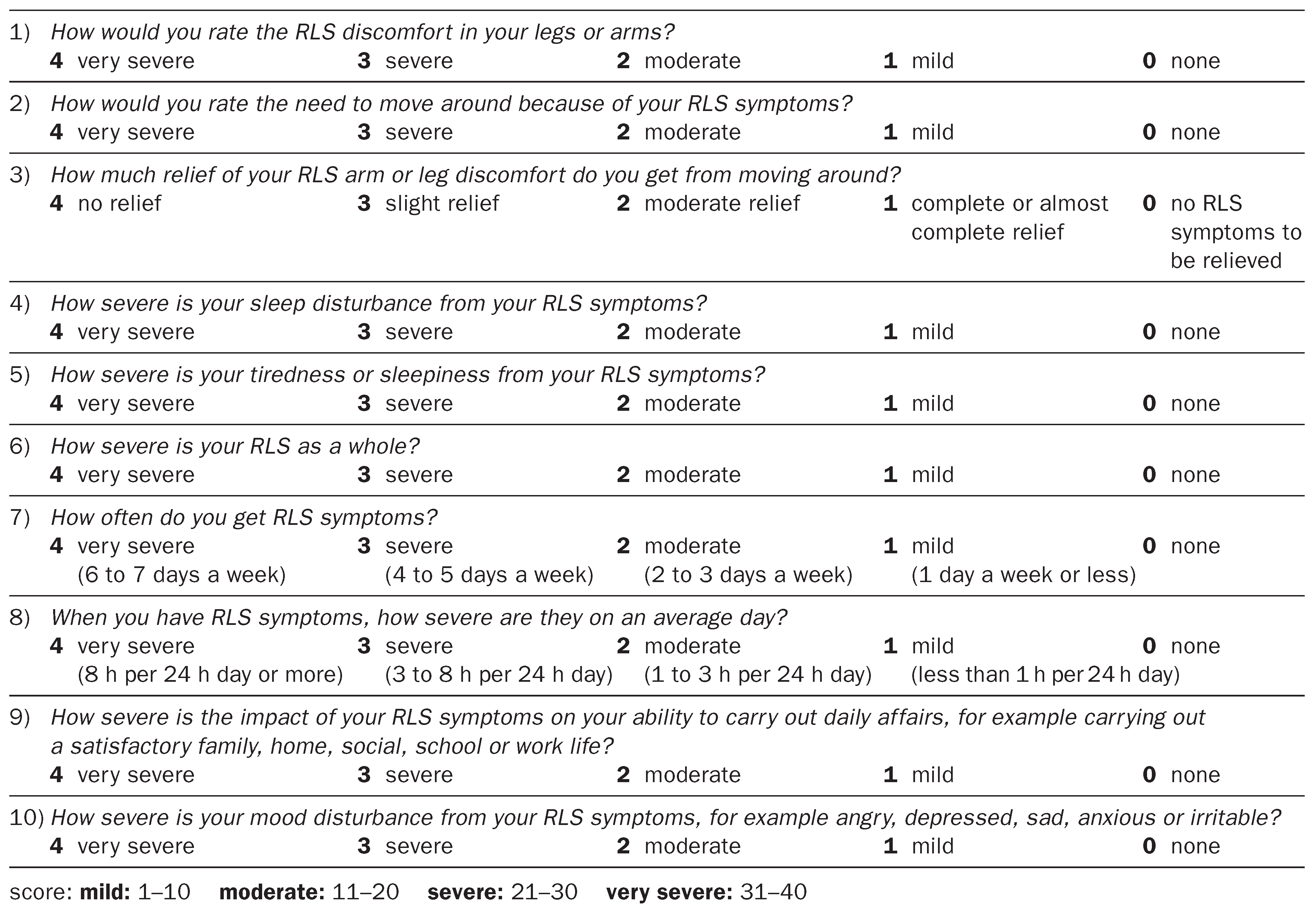
Two questionnaires have been developed to assess severity of the restless legs syndrome. The first is a 10-question scale (Table 1) developed by the IRLSSG filled in by patient or clinician at the visit considering frequency of the symptoms, diurnal consequences and nocturnal disturbances. According to the values obtained, a severity score could be obtained ranging from mild to severe. This scale has a good concordance with clinical rating and has previously been validated. The second questionnaire frequently used is the “Johns Hopkins Restless Legs Severity Scale” (JHRLSS) [28] providing a four-point scale from 0 (no symptoms) to 3 (severe cases) and evaluating the time of the day for usual occurrence of symptoms, severe cases having an onset in the afternoon.
Table 1.
The International Restless Legs Study Group RLS severity Scale.
Instrumental evaluation
Actimetry
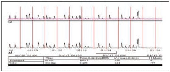
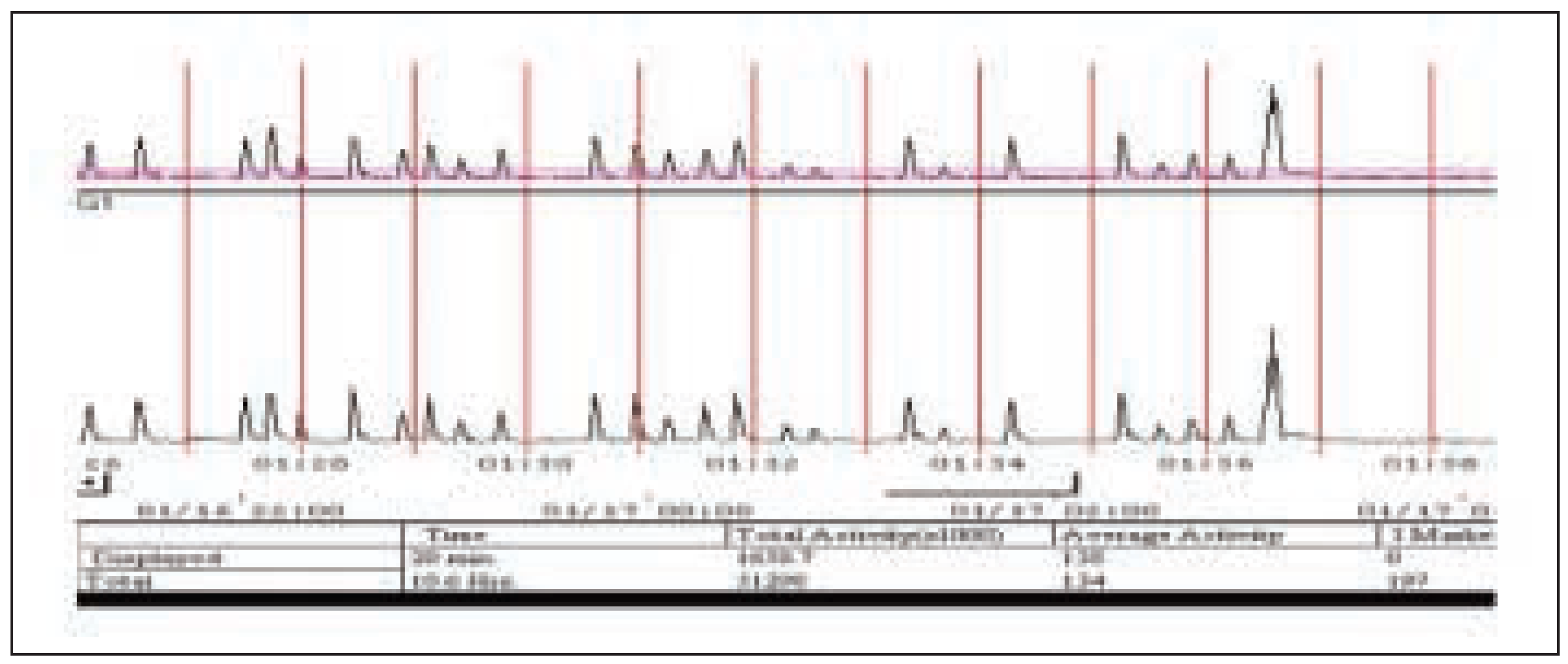
Ambulatory actigraphy devices (Figure 2) have been developed for diagnostic purposes and follow-up studies of treatment effect in order to replace the much more expensive polysomnography. Comparing actigraphy results with electromyographic activity, Kazenwadel et al. [29] found an agreement scoring of about 90%. After application of a modified algorithm and a manual correction, a lower sensitivity and specificity was found in a second study [30]. A more recent study conducted in a large sample of subjects without sleep complaints [31] demonstrated a modest and significant correlation between the number of PLM detected by actigraphy and subjectively reported restless legs syndrome severity. These results suggest that actimeter may be a useful tool for community studies and for follow-up in patients with a diagnosis of PLM and/or restless legs syndrome as suggested by clinical features and polysomnography.
Figure 2.
Actimeter recording in a patient with restless legs syndrome and periodic leg movements.
Polysomnographic recording
Even though not necessary for diagnosis, polysomnography is often performed to define the severity of restless legs syndrome, demonstrate the presence of PLMS and rule out other (or associated) sleep disorders, such as sleep apnoea syndrome. To diagnose restless legs syndrome and assess its severity Montplaisir et al. introduced a standardised provocation test, called the suggested immobilisation test (SIT) [22]. This is a 60-min test performed before the nocturnal polysomnography, in that the patient is asked to remain in bed, reclined at 45 degree with eyes open, legs stretched out and the instruction not to move and not to fall asleep [22]. Surface electromyography from right and left anterior tibialis muscles is used to quantify leg movements while leg discomfort is measured every 5 minutes with a electronic device using a scale from 0 (no discomfort) to 100 (extreme discomfort). The mean leg discomfort score (SIT MDS) represented the average value of the twelve measures while the PLMW calculated during the SIT period represented the number of PLM during the test. Both indices showed a good sensitivity and specificity and the combination allows diagnosis in 88% of the patients [32].
Nocturnal polysomnography is used to define the degree of sleep disturbances, assess the PLM index during wakefulness and sleep, and the PLM index associated with arousals. The PLM index during the nocturnal waking period is the best objective index to define the severity of the subjective complaints and sleep efficiency indicates the extent of sleep disturbance. PLM index and PLM index with arousal are assumed to describe sleep fragmentation, but the correlation with fatigue, tiredness and sleepiness during the day is moderate [16].
Laboratory and instrumental investigations
With the exception of symptomatic cases (see below) in which restless legs syndrome is associated with metabolic and neurological diseases, laboratory studies are always within physiological values and electromyography and motor nerve conduction measurements fail to reveal pathological signs. However, iron deficiency and iron excess should systematically be looked for in restless legs syndrome patients keeping in mind that serum iron is highly variable and not specific of iron metabolism. Transferrin, total iron binding capacity (TIBC) and ferritin are more sensitive markers of iron availability and storage respectively. Decreased iron availability is associated with reduced TIBC and increased transferrin levels while low ferritin (<50 µg/l) is always indicative of low iron stores.
Aetiology
Although in most cases the aetiology of restless legs syndrome is unknown (“idiopathic forms”), a large number of potential aetiologic factors have been reported (“secondary forms”). Conditions associated with a higher incidence of restless legs syndrome include pregnancy, iron deficiency, haemochromatosis, vascular insufficiency, chronic obstructive pulmonary disease, sleep-disordered breathing, polyneuropathies, rheumatoid arthritis, uraemia, diabetes, alcohol abuse, avitaminosis, amyloidosis, cryoglubulinaemia, lumbosacral plexus lesions, myelopathy, multiple sclerosis, Isaac’s syndrome, motor neuron disease, tumours, spinal anaesthesia, neuroataxia, syringomyelia, neurodegenerative diseases (e.g., Parkinson’s and Huntington’s disease) and medications (neuroleptics), tricyclic antidepressant, SSRI antidepressant (mirtazepine), antiepileptic drugs (zonisamide), beta-blockers and lithium. Acute-onset and transient restless legs syndrome can be observed in acute spinal cord lesions (stroke, myelitis, multiple sclerosis, etc.), as a consequence of severe bleeding and following operations or spinal anaesthesia [6,33]. Worsening has also been described by consuming coffee, nicotine and chocolate, while smoking marijuana may bring relieve in some patients. Improvement of restless legs syndromesymptoms and worsening of PLMS [34] are known to also occur with treatment of sleep-disordered breathing.
In the “primary” forms several studies have documented a familial occurrence in 43 to 64% of patients, and an autosomal dominant inheritance was proposed [35,36,37,38] with variable expressivity [38,39,40]. These studies suggest a low but notable degree of inheritance for restless legs syndrome that is much larger when symptoms appear early. Thus two phenotypes may be proposed based on the family history and time of disease onset: the “early-onset” and the “late-onset” cases. Patients with early onset (<45 years) more often have a positive family history, a slower progression of the disease, less frequently iron level abnormalities and the existence of a single major gene transmitted by an autosomal dominant mode of inheritance. Recent studies have described a susceptibility locus for restless legs syndrome on chromosome 12q [41] in a family with putative recessive inheritance and on chromosome 14 [42] in a 30-member Italian family. Patients with late-onset disease are more frequently secondary forms with pain as dominating symptom [6] most probably indicating a multifactorial pathogenesis.
Pathogenesis
The pathogenesis of restless legs syndrome remains uncertain but the therapeutic efficacy of dopaminergic agents crossing the blood-brain barrier stresses the role of a dopaminergic system dysfunction affecting the central nervous system. The localisation of restless legs syndrome symptoms in the extremities with sparing of the face, the acute appearance in association with spinal (peridural) anaesthesia and spinal disorders (see above), the association of restless legs syndrome with spino-cerebellar ataxias particularly SCA Type III [43] and the similarity between PLMS and flexor reflexes [44] favour the hypothesis of a primary dysfunction at spinal cord level probably affecting the polysynaptic reticulo-spinal tract (A11) [45].
On the other hand, an additional dysfunction of sensorimotor control at brain level has been suggested by neurophysiological and neuroimaging data. Studies using either single photon emission computer tomography (SPECT) [46,47] or positron emission tomography (PET) [48,49] have shown a post- and pre-synaptic D2 dysfunction at the level of basal ganglia, although the results of different studies are somewhat contradictory [50].
Since iron deficiency is associated with restless legs syndrome in some cases, iron seems to play an important role in secondary forms, such as in pregnancy and anaemia. Iron is necessary as a cofactor for the tyroxine hydroxylase, which is the rate-limiting enzyme in the conversion from levodopa to dopamine. Therefore, a decrease in iron may reduce dopamine synthesis and thus dopamine availability. However, some cases of restless legs syndrome improved significantly with iron therapy [51] and previous studies have demonstrated both a decrease in ferritin in cerebrospinal fluid [52] and a lower iron concentration in the substantia nigra in restless legs syndrome patients [53]. These findings have supported the hypothesis of a primary alteration of iron metabolisms in the pathogenesis of restless legs syndrome.
Therapy
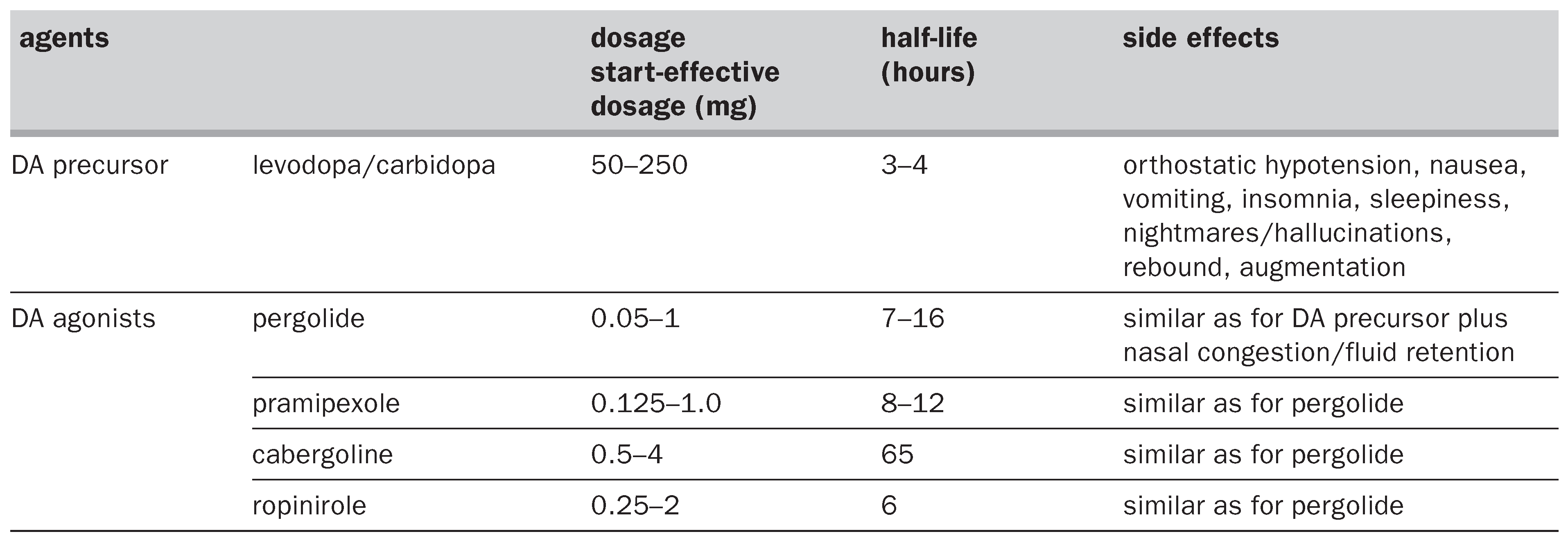
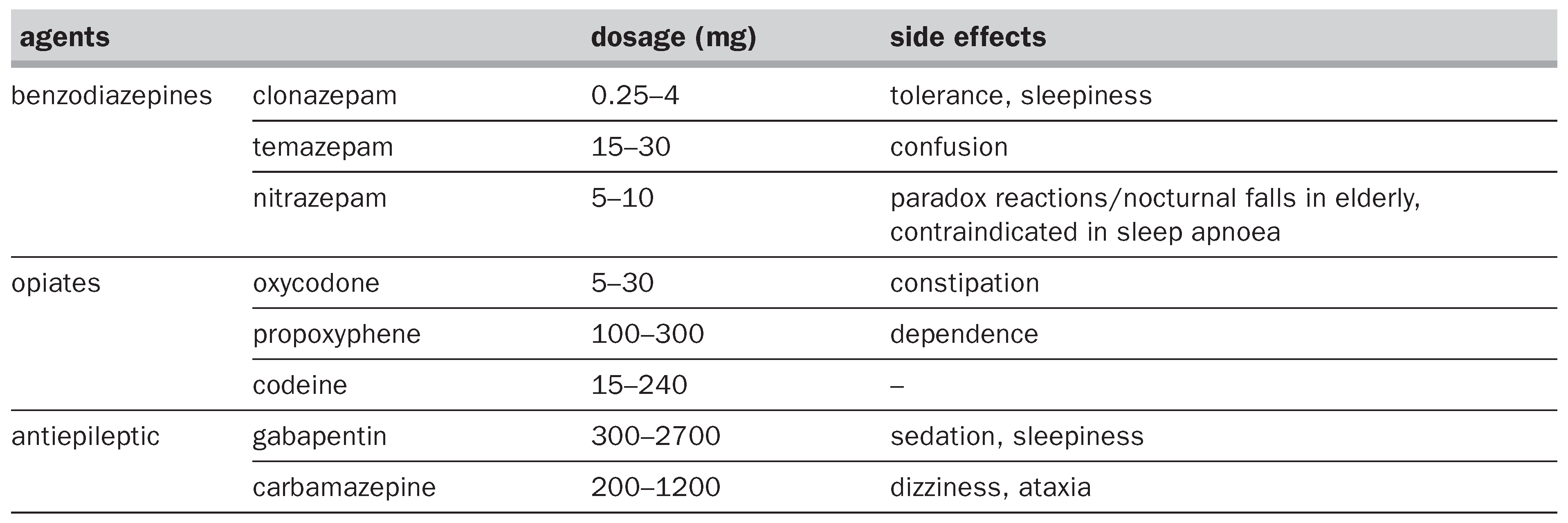
In some patients restless legs syndrome occurs sporadically with spontaneous remission and without persistent sleep complaints. In these cases the physician should use pharmacological therapy when appropriate. Chronic therapy is recommended if patients complain of restless legs syndrome occurring at least 3 nights per week and when symptoms induce diurnal consequences. The more common drugs used in the treatment of restless legs syndrome and the possible side effects are listed in Table 2a and Table 2b and summarised in some international guidelines recently published [54,55,56,57]. The treatment of restless legs syndrome now focuses on dopaminergic agonists considered the first choice therapy for patients who meet the specific clinical criteria and suffer from clinically relevant symptoms, either in primary or secondary forms. The use of benzodiazepines such as clonazepam may be limited to mild cases in which the therapeutic benefit is that to promote sleep.
Table 2a.
Dosage, half-life and adverse effects of dopamine agents.
Table 2b.
Dosage and adverse effects of other drugs.
In the early studies treatment with standard levodopa/benserazide induced an easy and rapid (after 1 h) improvement of the symptoms at a dosage of 100–400 mg levodopa at night. However, although symptoms were improved, a shifting of symptoms into the morning hours and into late afternoon was seen: the “rebound” and the “augmentation”, both side effects indicating a change in dopamine receptor sensitivity. The “rebound effect” seen more frequently with the short-acting form of the levodopa/benserazide is characterised by the appearance of symptoms 2 to 6 h after intake of the drug or at the morning awakening inducing the need for an additional dose. This effect was described in 46% of patients during night-time and 19% at the awakening. Sustainedrelease levodopa extended the benefit up to 7 h compared to the 3–4 h of relief of short-acting formulations and reducing, therefore, this adverse effect. The second and more interesting adverse effect is “augmentation”, i.e., increasing intensity of symptoms, earlier onset of the symptoms in the day, reduced time at rest until symptoms start and in some cases spreading of the symptoms to other body parts. Augmentation was more common than rebound effect and present in up to 82% of patients treated with standard levodopa at high doses (400 mg or more) and in about 30% of patients treated with sustained release levodopa. In this situation levodopa should be replaced by dopamine agonists.
Several recent studies have demonstrated, compared to placebo and levodopa, a greater efficacy of dopamine agonists [58,59] characterised by a prolonged duration of action, a direct action on D2 and D1 receptors and a reduced frequency of augmentation and rebound adverse effects. There are no studies that have demonstrated the superiority of one agonist compared to another and the choice depends on the personal clinical strategy. With dopamine agonists the common strategy is the start of treatment at a low single dosage before bedtime progressively increased, every 2–7 days, until clinical benefit is obtained. The slow increase of dosage is necessary to prevent the appearance of adverse effects such as hypotension and nausea. The latter can also be prevented by the coadministration of domperidone, particularly when doing this 3 days before starting the dopaminergic agent. Patients should also be advised about the potential increase in excessive daytime sleepiness and the risk of sleep attacks [58,60].
Opioid drugs are generally used as second choice for patients with augmentation or nonresponders to dopamine agonists. Antiepileptic drugs such as gabapentin should be considered for painful restless legs syndrome or restless legs syndrome secondary to neuropathy either as a single treatment or in combination with dopamine agonists. Iron therapy is recommended in patients with a low level of ferritin in a posology of 300–400 mg to increase the serum ferritin to level more than 50 µg/l. In cases where this substitution does not increase serum ferritin levels after 2 to 3 months an intravenous application can be tried [54].
References
- Ekbom, KA. Restless legs: a clinical study. Acta Med Scand 1945, 158, 1–123. [Google Scholar]
- The International Restless Legs Syndrome Study Group. Toward a better definition of the restless legs syndrome. Mov Disord 1995, 10, 634–42. [Google Scholar]
- Ondo, W; Jankovic, J. Restless legs syndrome: clinicoetiologic correlates. Neurology 1996, 47, 1435–41. [Google Scholar] [CrossRef] [Scilit] [PubMed]
- Montplaisir, J; Boucher, S; Poirier, G; Lavigne, G; Lapierre, O; Lesperance, P. Clinical, polysomnographic and genetic characteristics of restless legs syndrome: a study of 133 patients diagnosed with new standard criteria. Mov Disord 1997, 12, 61–5. [Google Scholar] [CrossRef] [Scilit]
- Michaud, M; Chabli, A; Lavigne, G; Montplaisir, J. Arm restlessness in patients with restless legs syndrome. Mov Disord 2000, 15, 289–93. [Google Scholar] [CrossRef] [Scilit]
- Bassetti, C; Mauerhofer, D; Gugger, M; Mathis, J; Hess, CW. Restless legs syndrome: a clinical study of 55 patients. Eur Neurol 2001, 45, 67–74. [Google Scholar]
- Hening, WA; Walters, AS; Wagner, M; Rosen, R; Chen, V; Kim, S; et al. Circadian rhythm of motor restlessness and sensory symptoms in the idiopathic restless legs syndrome. Sleep 1999, 22, 901–12. [Google Scholar] [CrossRef] [Scilit] [PubMed]
- Trenkwalder, C; Hening, WA; Walters, AS; Campbell, SS; Rahman, K; Chokroverty, S. Circadian rhythm of periodic limb movements and sensory symptoms of restless legs syndrome. Mov Disord 1999, 14, 102–10. [Google Scholar] [CrossRef]
- De La Llave, Y; Garcia-Borreguero, D; Barrio, S; Larrosa, O; Granizo, JJ; Allen, R. Severity of disease and circadian patterns of periodic leg movement activity in restless legs syndrome: preliminary report. Sleep 2001, 24, A101. [Google Scholar]
- Boecker, MR; Badia, P; Shaffer, JI. The relationship between core temperature and time-of-the night patterns in PLMS activity. Sleep Res 1995, 23, 235. [Google Scholar]
- Davila, R; Zummarraga, M; Andia, I; Friedhoff, AJ. Persistence of cyclicity of the plasma dopamine metabolite, homovanillic acid, in neuroleptic treated schizophrenic patients. Life Sci 1989, 44, 1117–21. [Google Scholar] [PubMed]
- Schade, R; Vick, K; Ott, T; Sohr, R; Pfister, C; Bellach, J; et al. Circadian rhythms of dopamine and cholecystokin in nucleus accumbens and striatum of rats – influence on dopaminergic stimulation. Chronobiol Int 1995, 12, 87–99. [Google Scholar]
- Scales, WE; Vander, AJ; Brown, MB; Kluger, MJ. Human circadian rhythms in temperature, trace metals and blood variables. J Appl Physiol 1988, 65, 1840–6. [Google Scholar] [PubMed]
- Tarquini, B. Iron metabolism: clinical chronobiological aspects. Chronobiologica 1978, 5, 315–36. [Google Scholar]
- Coleman, RM; Bliwise, DL; Sajben, N; Boomkamp, A; De Bruyn, LM; Dement, WC. Daytime sleepiness in patients with periodic movements in sleep. Sleep 1982, 5, S191–S202. [Google Scholar] [CrossRef] [Scilit]
- Mendelson, WB. Are periodic leg movements associated with clinical sleep disturbance. Sleep 1996, 19, 219–23. [Google Scholar] [CrossRef] [Scilit] [PubMed]
- Chabli, A; Michaud, M; Montplaisir, J. Periodic arm movements in patients with restless legs syndrome. Eur Neurol 2000, 44, 133–8. [Google Scholar]
- Sforza, E; Nicolas, A; Lavigne, G; Gosselin, A; Petit, D; Montplaisir, J. EEG and cardiac activation during periodic leg movements in sleep. Support for a hierarchy of arousal responses. Neurology 1999, 52, 786–91. [Google Scholar]
- Droste, DW; Krauss, JK; Hagedorn, G; Kaps, M. Periodic leg movements are part of the B-wave rhythm and the cyclic alternating pattern. Acta Neurol Scand 1996, 94, 347–52. [Google Scholar] [CrossRef] [Scilit]
- Karadeniz, D; Ondze, B; Besset, A; Billiard, M. EEG arousals and awakenings in relation with periodic leg movements during sleep. J Sleep Res 2000, 9, 273–7. [Google Scholar]
- Nicolas, A; Michaud, M; Lavigne, G; Montplaisir, J. The influence of sex, age and sleep/wake state on characteristics of periodic leg movements in restless legs syndrome. Clin Neurophysiol 1999, 110, 1168–74. [Google Scholar] [CrossRef] [Scilit] [PubMed]
- Montplaisir, J; Michaud, M; Nicolas, A; Lesperance, P; Gosselin, A; Rompré, P; et al. Immobilization test and periodic leg movements in sleep for diagnosis of restless legs syndrome. Mov Disord 1998, 13, 324–9. [Google Scholar] [CrossRef] [Scilit]
- Lavigne, GJ; Montplaisir, J. Restless legs syndrome and sleep bruxism: prevalence and association among Canadians. Sleep 1994, 17, 739–43. [Google Scholar] [CrossRef] [Scilit] [PubMed]
- Phillips, B; Young, T; Finn, L; Asher, K; Hening, WA; Purvis, C. Epidemiology of restless legs symptoms in adults. Arch Intern Med 2000, 160, 2137–41. [Google Scholar] [CrossRef] [Scilit]
- Walters, A; Hickey, K; Maltzman, J; Verrico, T; Hening, JW; Wilson, V; et al. A questionnaire study of 138 patients with restless legs syndrome: the “Night-Walkers” survey. Neurology 1996, 46, 92–5. [Google Scholar] [CrossRef] [Scilit]
- Hening, WA; Walters, AS; Chokroverty, S. Motor functions and dysfunction in sleep. In Sleep Disorders Medicine; Chokroverty, S, Ed.; Butterworth-Heinemann: Boston, 1994; pp. 255–93. [Google Scholar]
- Polydefkis, M; Allen, RP; Hauer, P; Earley, CJ; Griffin, JW; McArthur, JC. Subclinical sensory neuropathy in late-onset restless legs syndrome. Neurology 2000, 55, 1115–21. [Google Scholar] [CrossRef] [Scilit]
- Allen, RP; Earley, CJ. Validation of the Johns Hopkins Restless Legs Severity Scale. Sleep Med 2001, 3, 239–42. [Google Scholar] [CrossRef] [Scilit]
- Kazenwadel, J; Pollmacher, T; Trenkwalder, C; Oertel, WH; Kohnen, R; Kunzel, M; et al. New actigraphic assessment method for periodic leg movements (PLM). Sleep 1995, 18, 689–97. [Google Scholar] [CrossRef] [Scilit] [PubMed]
- Sforza, E; Zamagni, M; Petiav, C; Krieger, J. Actigraphy and leg movements during sleep: a validation study. J Clin Neurophysiol 1999, 16, 154–60. [Google Scholar] [CrossRef] [Scilit]
- Morrish, E; King, MA; Pilsworth, SN; Shneerson, JM; Smith, IE. Periodic limb movement in a community population detected by a new actigraphy technique. Sleep Med 2002, 3, 489–95. [Google Scholar] [CrossRef] [Scilit] [PubMed]
- Michaud, M; Paquet, J; Lavigne, G; Desautels, A; Montplaisir, J. Sleep laboratory diagnosis of restless legs syndrome. Eur Neurol 2002, 48, 108–13. [Google Scholar] [CrossRef] [Scilit]
- Högl, B; Frauscher, B; Seppi, K; Ulmer, H; Poewe, H. Transient restless legs syndrome after spinal anaesthesia. Neurology 2002, 59, 1705–7. [Google Scholar] [CrossRef] [Scilit] [PubMed]
- Briellmann, RS; Mathis, J; Bassetti, C; Gugger, M; Hess, CW. Patterns of muscle activity in legs in sleep apnea patients before and during nCPAP therapy. Eur Neurol 1997, 38, 113–8. [Google Scholar] [CrossRef] [Scilit] [PubMed]
- Montplaisir, J; Godbout, R; Boghen, D; DeChamplain, J; Young, SN; Lapierre, O. Familial restless legs with periodic leg movements in sleep: electrophysiologic, biochemical and pharmacologic study. Neurology 1985, 35, 130–4. [Google Scholar] [CrossRef] [Scilit] [PubMed]
- Trenkwalder, C; Seidel, VC; Gasser, T; Oertel, WH. Clinical symptoms and possible anticipation in a large kindred of familial restless legs syndrome. Mov Disord 1996, 11, 389–94. [Google Scholar] [CrossRef] [Scilit]
- Ondo, WG; Vuong, KD; Wang, Q. Restless legs syndrome in monozygotic twins: clinical correlates. Neurology 2000, 55, 1404–6. [Google Scholar] [CrossRef] [Scilit]
- Winkelmann, J; Wetter, TC; Collado-Seidel, V; Gasser, T; Dichgans, M; Yassouridis, A; et al. Clinical characteristics and frequency of the hereditary restless legs syndrome in a population of 300 patients. Sleep 2000, 23, 597–602. [Google Scholar] [CrossRef] [Scilit]
- Walters, AS; Picchietti, D; Hening, W; Lazzarini, A. Variable expressivity in familial restless legs syndrome. Arch Neurol 1990, 47, 1219–20. [Google Scholar] [CrossRef] [Scilit]
- Winkelmann, J; Muller-Myhsok, B; Wittchen, HU; Hock, B; Prager, M; Pfister, H; et al. Complex segregation analysis of restless legs syndrome provides evidence for an autosomal dominant mode of inheritance in early age at onset families. Ann Neurol 2002, 52, 297–302. [Google Scholar] [CrossRef] [Scilit]
- Desautels, A; Turecki, G; Montplaisir, J; Sequeira, A; Verner, A; Rouleau, GA. Identification of a major susceptibility locus for restless legs syndrome on chromosome 12q. Am J Hum Genet 2001, 69, 1266–70. [Google Scholar] [CrossRef] [Scilit]
- Bonati, MT; Ferini-Strambi, L; Aridon, P; Oldani, A; Zucconi, M; Casari, G. Autosomal dominant restless legs syndrome maps on chromosome 14 q. Brain 2003, 126, 1485–92. [Google Scholar] [CrossRef] [Scilit]
- Schols, L; Haan, J; Riess, O; Amoiridis, G; Przuntek, H. Sleep disturbances in patients with hereditary ataxias. Neurology 1998, 51, 1603–7. [Google Scholar]
- Bara-Jimenez, W; Aksu, M; Graham, B; Sato, S; Hallett, M. Periodic limb movements in sleep: state-dependent excitability of the spinal flexor reflex. Neurology 2000, 54, 1609–16. [Google Scholar] [CrossRef] [Scilit]
- Ondo, WG; He, Y; Rajasekaran, S; Le, WD. Clinical correlates of 6-Hydroxydopamine injections into A11 dopaminergic neurons in rats: a possible model for restless legs syndrome? Mov Disord 2000, 15, 154–8. [Google Scholar]
- Michaud, M; Soucy, JP; Chabli, A; Lavigne, G; Montplaisir, J. SPECT imaging of striatal preand postsynaptic dopaminergic status in restless legs syndrome with periodic leg movements in sleep. J Neurol 2002, 249, 164–70. [Google Scholar] [CrossRef] [Scilit]
- Turjanski, N; Lees, AJ; Brooks, DJ. Striatal dopaminergic function in restless legs syndrome. Neurology 1999, 52, 932–7. [Google Scholar] [CrossRef] [Scilit] [PubMed]
- Trenkwalder, C; Walters, AS; Hening, WA; Chokroverty, S; Antonini, A; Dhawan, V; et al. Positron emission tomographic studies in restless legs syndrome. Mov Disord 1999, 14, 141–5. [Google Scholar] [CrossRef]
- Ruottinen, HM; Partinen, M; Hublin, C; Bergman, J; Haaparanta, M; Solin, O; et al. An FDOPA PET study in patients with periodic limb movement disorder and restless legs syndrome. Neurology 2000, 54, 502–4. [Google Scholar] [CrossRef] [Scilit] [PubMed]
- Wetter, TC; Linke, R; Eisensehr, I; Noachtar, S; Tatsch, K; Trenkwalder, C. Iodine-123-IPT SPECT imaging in idiopathic restless legs syndrome: preliminary findings. Sleep 2000, 23, A130. [Google Scholar]
- Earley, CJ; Heckler, D; Allen, RP. IV iron treatment for RLS. Sleep 2000, 24, 359. [Google Scholar]
- Allen, RP; Earley, CJ. Restless legs syndrome. A review of clinical and pathophysiologic features. J Clin Neurophysiol 2001, 18, 128–47. [Google Scholar] [CrossRef] [Scilit]
- Allen, RP; Barker, PB; Wehrl, F; Song, HK; Early, CJ. MRI measurement of brain iron in patients with restless legs syndrome. Neurology 2001, 56, 263–5. [Google Scholar] [CrossRef] [Scilit]
- Hening, W; Allen, R; Earley, C; Kushida, C; Picchietti, D; Silber, M. The treatment of restless legs syndrome and periodic leg movements. Sleep 1999, 22, 970–99. [Google Scholar]
- Chesson, AL; Wise, M; Davila, D; Johnson, S; Littner, M; Anderson, MD; et al. Practice parameters for the treatment of restless legs syndrome and periodic limb movement disorder. Sleep 1999, 22, 961–8. [Google Scholar] [CrossRef] [Scilit]
- Comella, CL. Restless legs syndrome. Treatment with dopaminergic agents. Neurology 2002, 58, S87–S92. [Google Scholar] [CrossRef] [Scilit]
- Stasny, K; Oertel, WH; Trenkwalder, C. Clinical symptomatology and treatment of restless legs syndrome and periodic limb movement disorder. Sleep Med Rev 2002, 6, 253–65. [Google Scholar] [CrossRef] [Scilit] [PubMed]
- Frucht, S; Rogers, JD; Greene, PE; Gordon, MF; Fahn, S. Falling asleep at the wheel: motor vehicle mishaps in persons taking pramipexole and ropinirole. Neurology 1999, 52, 1908–10. [Google Scholar] [CrossRef] [Scilit] [PubMed]
- Bassetti, C; Clavadetscher, S; Gugger, M; Hess, CW. Pergolide-associated ‘sleep attacks’ in a patient with restless legs syndrome. Sleep Med 2002, 3, 275–7. [Google Scholar] [CrossRef] [Scilit] [PubMed]
- Earley, C. Restless legs syndrome. N Engl J Med 2003, 348, 2103–9. [Google Scholar] [CrossRef] [Scilit]

This is an open-access article distributed under the terms of the Creative Commons Attribution Non-Commercial No Derivatives (CC BY-NC-ND) 4.0 License.