


Summary
Narcolepsy is a long-life, usually sporadic (rarely familial) sleep-wake disorder characterised by an often disabling excessive daytime sleepiness (EDS) and so-called REM-sleep symptoms such as cataplexy (muscle tone loss triggered by emotions), sleep paralysis and hallucinations. Biological markers of narcolepsy are a specific HLA class II-subtype (DQB1*0602), the appearance of REM-sleep within 15–20′ after sleep onset (socalled sleep onset REM periods or SOREM) and the absence/reduction of the recently discovered peptide hypocretin-1 (also called orexin-A) in the cerebrospinal fluid. The neurobiology of narcolepsy has traditionally been attributed to a neurochemical (cholinergic-aminergic) dysbalance in the brainstem, based on genetic predisposition and environmental factors. The involvement of the hypocretin system in both human and animal forms of narcolepsy has led to the recognition of a central role of the hypothalamus in the pathophysiology of this disorder. Treatment of narcolepsy includes counselling, scheduled naps, stimulant as well as anticataplectic drugs. This article reviews clinical features, diagnostic criteria, pathophysiology and therapeutic strategies of human narcolepsy, also integrating the most recent data on the physiology and pathology of hypocretinergic neurotransmission.
Clinical symptomatology
Narcoleptic tetrad
The clinical features of narcolepsy are excessive daytime sleepiness in combination with intrusions of REM (rapid-eye-movement)-sleep features during wakefulness presenting as cataplexy, sleepparalysis and hallucinations. The classical narcoleptic “tetrad” of excessive daytime sleepiness (EDS), cataplexy, sleep paralysis and hypgnagogic/hypopompic hallucinations is only present in about 10–15% of narcoleptics [1]. Most patients suffer from excessive daytime sleepiness (90%) and cataplexy (70%), hallucinations and sleep paralysis are less frequent (50–75%). Only cataplexy is specific for narcolepsy, whereas hallucinations and sleep paralysis are also common in normal population.
Excessive daytime sleepiness is often the first and the most disabling symptom. Excessive daytime sleepiness refers to both a subjective feeling of sleepiness and to irresistible short naps (“sleep attacks”). The subjective feeling of sleepiness exhibits high abnormal (>14/24) scores when measured by the standardised Epworth Sleepiness Score (ESS) [2] and it is comparable to sleepiness after 32-h sleep deprivation in normal subjects [3]. Sleep attacks are short (<20 min) and even occur in active situations like talking, eating, standing, walking or sexual intercourse. Though labelled as “attacks” sleep onset is rarely abrupt but is usually preceded by prodromi of drowsiness like blurred vision, ptosis or nodding. Naps are typically short and refreshing but sleepiness will return after a few hours.
Cataplexy consists of a sudden bilateral loss of postural muscle tone in association with intense emotions, whereas sleep paralysis corresponds to a transient inability to perform voluntary movements either at sleep onset (hypnagogic or predormital form) or upon awakening (hypnopompic or postdormital form). Both are considered to be an intrusion of physiologic REM-sleep atonia into wakefulness. Cataplexy shares many clinical features of sleep paralysis although muscle atonia in sleep paralysis is usually complete and lasts longer (but usually less than 10 minutes). Cataplectic attacks are short, usually less than 1 minute and exceed 2 minutes in only 10–20% of cases [4]. The loss of motor tone in cataplexy is usually partial and bilateral, may be unnoticeable to others and most commonly involves the lower extremities but often also the upper limbs or face/neck muscles. Patients may report buckling or unlocking of the knees, zig-zag walking, closing of the eyelids, dropping of the head and slurred speech. Only one third of the patients experience falls. Muscle atonia is rarely complete but rapidly alternates with short phasic muscle activity presenting as muscle twitches [5]. Prolonged episodes >30 minutes (“status cataplecticus”) can occur after abrupt discontinuation of anticataplectic drugs. Although patients seem to be unresponsive due to muscle atonia, consciousness is typically preserved unless a transition to sleep or hypnagogic hallucinations occurs. Hypnagogic or hypnopompic hallucinations are dreamlike experiences occurring when the patient is awake. The intrusion of dreams would explain the not uncommon absurd/bizarre characteristics. Patients often feel a person or an animal nearby, standing over the bed or lying underneath or experience out-of-the-body dreams in a fantastic, mysterious or religious context. Also assaults by intruders, animals or monsters can be reported. Hypnopompic hallucinations tend to be more suggestive of narcolepsy than hypnagogic hallucinations.
Associated features
Other sleep disorders like sleep apnoea syndrome (10–20%), periodic limb movements in sleep (40–60%) or REM-sleep behavioural disorder (RBD) (12%) can be found in narcoleptic patients. REM-sleep behaviour disorder is a bizarre, often harmful behaviour during REM-sleep due to loss of physiological REM-atonia that allows patients to act out their dreams. For the clinician it is noteworthy that muscle tone dysregulation in narcolepsy may present in both ways, in a loss of muscle tone (during cataplexy and sleep paralysis) and/or in a loss of REM-atonia during REM-sleep behaviour disorder. Sleep-maintenance insomnia is often regarded as the fifth symptom of the narcoleptic tetrad. These sleep disorders need correct identification and specific therapies as they may contribute to daytime sleepiness. Sleepiness may account for the subjective complaints of memory deficits in narcoleptics. Depression is often associated with narcolepsy. Narcoleptics are occasionally obese, particularly in childhood, and on average, their body mass index is 10–20% higher than that of the normal population [6].
Epidemiology
Narcolepsy is not a rare but an underdiagnosed disorder. Recent epidemiological studies suggest the prevalence of narcolepsy in European countries around 26–47/100 000 [7,8,9], comparable to the prevalence of multiple sclerosis. Narcolepsy usually starts in the second or third decade and affects both sexes equally. In 5–15% narcolepsy starts in childhood [10]. Although late onset after the age of 40 is rare, patients may receive treatment only in adulthood due to long delay in diagnosis and therapy. Patients with early-onset narcolepsy may have a positive family history and are associated with a severe form of the disease.
Diagnosis and differential diagnosis
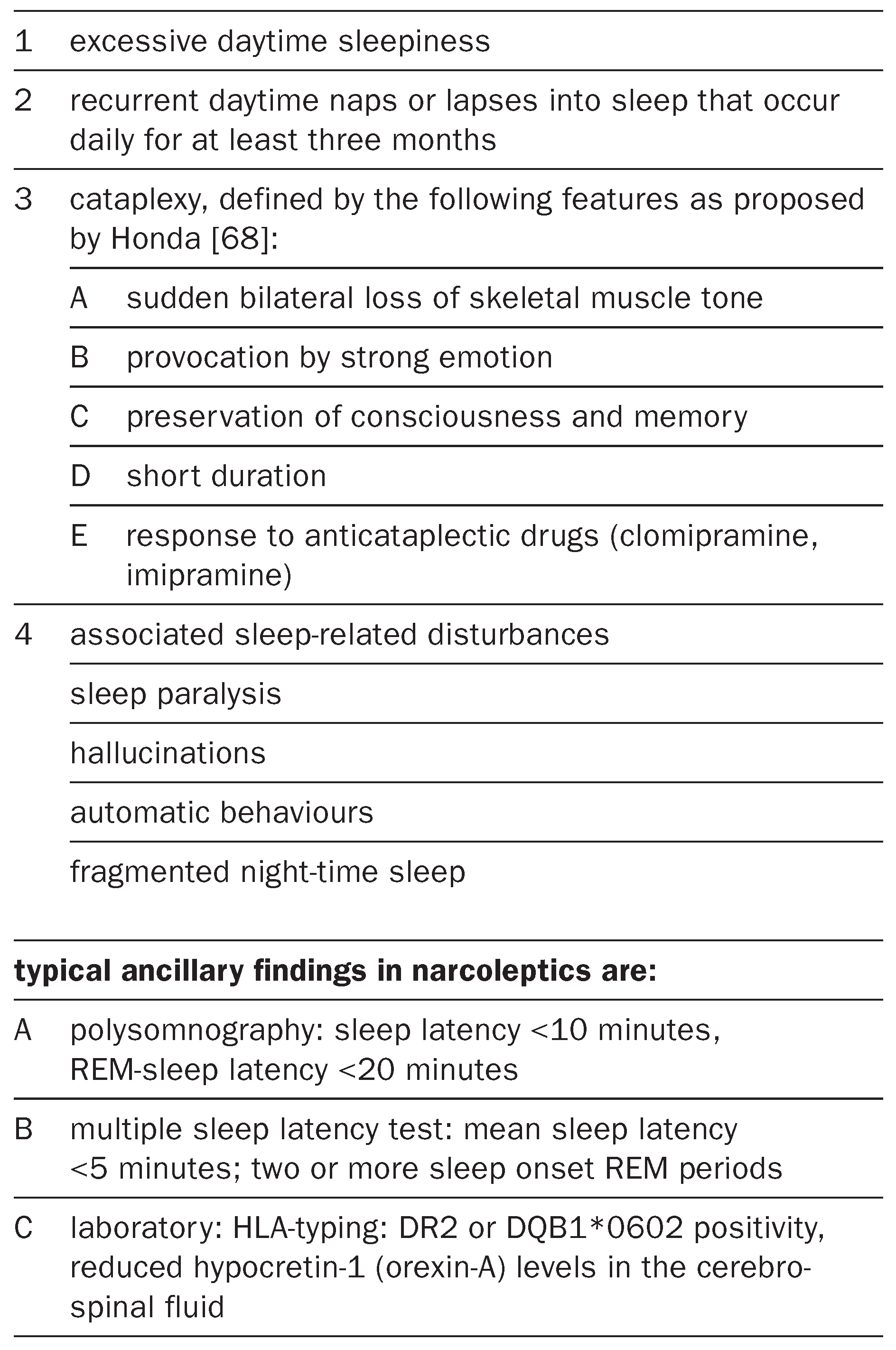
Table 1.
Diagnostic criteria for narcolepsy (modifed after the International Classification of Sleep Disorders).
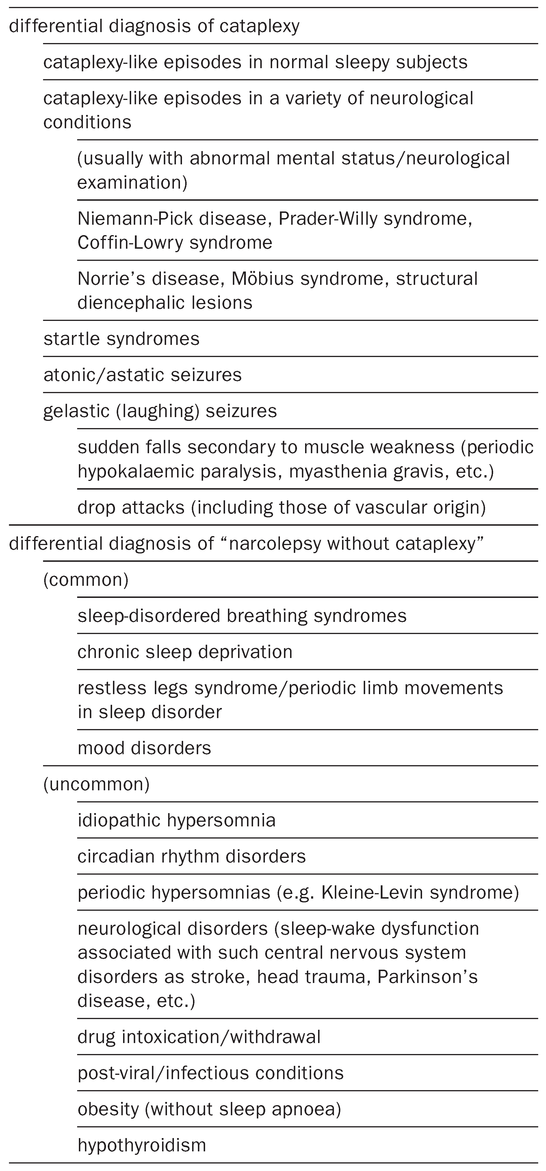
Table 2.
Differential diagnosis of narcolepsy.
Figure 1.
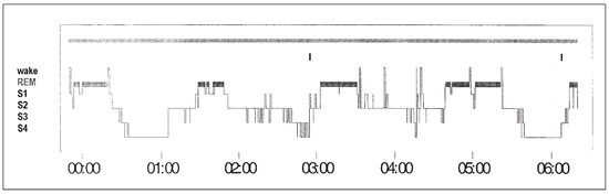
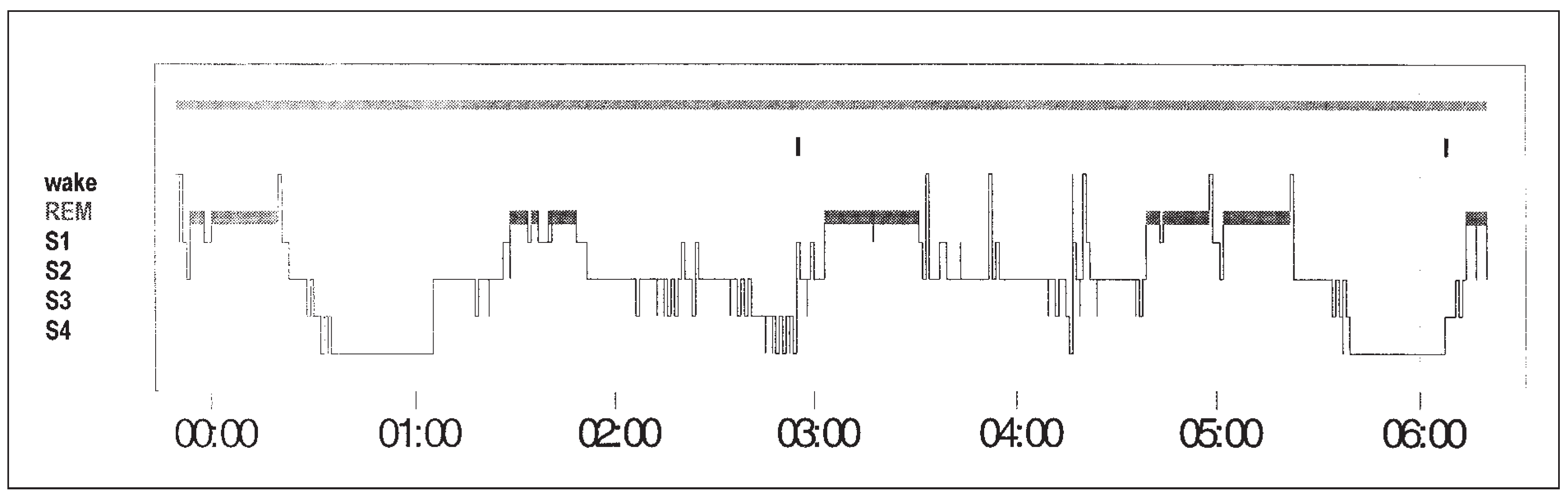
Diagnostic work-up in a patient with narcolepsy-cataplexy. A 34-year-old man referred to our institution because of severe excessive daytime sleepiness (ESS 16/24) and short episodes of muscle weakness tone triggered by laughter, but no sleep paralysis or concomitant hallucinations. Polysomnography: characteristic short sleep latency (3.5 min to NREM2), SOREM (1 min) and fragmented sleep are shown. There are no other results explaining excessive daytime sleepiness (no sleep apnoea or periodic limb movements in sleep).
The diagnosis of narcolepsy is basically a clinical one and usually straightforward in patients with typical and frequent (“clear-cut”) cataplexy. Approximately 15–30% of patients may remain undiagnosed, especially in early or late stages of disease when cataplexy is mild or absent. Cataplexy usually manifests at the same time or within a few years after excessive daytime sleepiness. The frequency of cataplexy tends to decline with age and disappears in one third of patients. Cataplexy rarely appears during medical consultation and its differentiation from other motor symptoms relies on the patients’ history. There is no diagnostic test that reliably evokes cataplexy but watching humorous videos may be a successful provocation. During the cataplectic attack areflexia [11] is an important clinical feature to differentiate other forms of transient muscle weakness. Cataplexy-like episodes following strong emotions in healthy subjects may be difficult to distinguish [12]. These episodes tend to be more common in subjects complaining of excessive daytime sleepiness, usually involve only the lower limbs and are more commonly triggered by negative emotions (e.g., stress, anxiety, sorrow) than “clear-cut” (true) cataplexy [Sturzenegger and Bassetti, The clinical spectrum of narcolepsy with cataplexy: a reappraisal, submitted].
Sleep paralysis is not specific for narcolepsy. It occurs once in a lifetime in 6.2% of the general population [7]. Up to 0.8% of the population suffers from severe sleep paralysis (weekly episodes). Sporadic hypnagogic/hypnopompic hallucinations are reported in 24% of healthy subjects. The association between sleep paralysis and anxiety was described in the 1940s by Rosenthal as “halluzinatorisch-kataplektisches Angstsyndrom” [13].
Hypnagogic/hypnopompic hallucinations may be seen with a variety of conditions including schizophrenia, substance-induced hallucinations, metabolic encephalopathies or parkinsonism [14].
The International Classification of Sleep Disorders (ICSD) defined clinical criteria and recommended multiple sleep latency test (MSLT) and laboratory tests when diagnosis is equivocal [15]. MSLT measures sleep propensity during daytime when the patient is allowed to sleep in a two-hour interval. Short sleep latencies (<5 min) in combination with at least two SOREMs in MSLT suggest narcolepsy [4]. Aldrich found a sensitivity of 70% and a specificity of 97% for the combination of ≥2 SOREMs and a mean sleep latency <5 min in a large series of narcoleptic patients [16]. More than 3 SOREMs in combination with short sleep latency (<5 min) increased specificity up to 99.2% but decreased sensitivity (46%). The maintenance of wakefulness test (MWT) is often applied in narcoleptic patients to measure the ability to stay awake. Similar to MSLT, MWT consists of polysomnographic recordings conducted 4–5 times during daytime in a two-hour interval. Patients sit and are instructed to resist sleep for 20 min. Untreated narcoleptic patients have mean sleep latencies of 6 min whereas normal healthy subjects stay awake the entire 20 min [17]. To assess driving capacity MWT may be more appropriate than MSLT. Occasionally patients with narcolepsy may have short latencies on MSLT but a normal MWT (see case report, Figure 1).
Two laboratory markers are diagnostic for narcolepsy with cataplexy: a specific HLA-II subtype in the DQ-region (DQB1*0602) and reduced CSF hypocretin levels. Although some markers in the DR-region of HLA-system have an even tighter association of up to 100% in selected populations, recent studies found the DQB1*0602 marker in up to 90% across all ethnic groups compared to a 12–38% positivity in the normal population [8,18]. Low hypocretin levels in the CSF may become the most sensitive and specific diagnostic criterion for narcolepsy with cataplexy. In a recent study a cut-off level of 110 pg/mL showed a sensitivity of 87% and a specificity of 99% for narcolepsy with cataplexy [19]. Sensitivity and specificity decline in narcoleptic patients without or with atypical cataplexy [20]. In addition, hypocretin may be normal in familial cases and – according to our own observation – even in monozygotic twins concordant for narcolepsy. These preliminary data suggest that hypocretin may be differently involved in the pathophysiology of narcolepsies with a strong genetic component.
Pathophysiology
The cause of excessive daytime sleepiness remains unknown. Disturbed night sleep is common in narcoleptics and may contribute to sleepiness, but even patients with normal night sleep suffer from excessive daytime sleepiness. Hence sleepiness may not represent a deficiency of sleep regulation but result from a deficit in the wake-maintaining systems. Night sleep of narcoleptic patients typically starts with REM-sleep within 20 min (= sleep onset REM, SOREM), whereas in healthy controls REM sleep occurs after 90 min [16]. Sleep onset REM is a polysomnographic hallmark for narcolepsy and indicates a more profound sleepwake dysfunction in mechanisms that regulate the transitions of wakefulness, REM-sleep and NREM-sleep.
Cataplexy is traditionally explained as REMsleep atonia occurring during wakefulness. Evidence came from clinical observation and subsequent electrophysiological data showing that REM-sleep and cataplexy are accompanied by areflexia and H-reflex attenuation [4].These results have led to the hypothesis that atonia during cataplexy and REM-sleep are generated by a similar neurophysiological mechanism: an inhibition of spinal alpha-motoneurons due to the activation of neurons in the ponto-medullary midbrain. The validity of this concept has recently been challenged for several reasons. Firstly, the equation cataplexy = REM-atonia does not explain why cataplexy is triggered by emotions [21]. Secondly, laughter and forced expiration in healthy subjects led to a similar diminution of H-reflexes as seen during cataplexy and REM-sleep [21,22,23]. Thirdly, a pharmacological study in narcoleptic dogs demonstrated effective suppression of cataplexy by D2/D3-antagonist whereas REM-sleep remained unchanged [24]. An alternative hypothesis considers cataplexy as a form of “tonic immobility” (TI) [21]. Tonic immobility refers to a reversible motor inhibition in response to a prey/predator confrontation and is found in a wide range of species. Besides several neuroanatomical aspects and pharmacological characterisations tonic immobility and cataplexy share the strong emotional trigger. In conclusion, atonia during cataplexy and REM-sleep may be generated by different mechanisms that eventually recruit a common descending ponto-medullary-spinal pathway.
Aetiology
Most cases of narcolepsy are idiopathic, rare symptomatic cases are usually associated with brainstem or diencephalic lesions. Although 99% of the cases are sporadic, genetic factors play an important role as the risk for a first-degree relative is 30–40 times higher than in the general population [8]. The only well-established genetic risk factors are the above-mentioned HLA class II-subtypes. The tight association of narcolepsy to the HLA system raises the question whether autoimmune mechanisms are involved in the aetiology of narcolepsy. However, to date, no proofs of autoimmune process have been identified. Considering that one third of narcoleptics in families with multiple affected individuals are in fact negative for HLADQB1*0602 and studies on monozygotic twins resulted in a low concordance rate of 25–30%, additional (unknown) environmental factors have to contribute the genetic susceptibility [8].
Neurobiology of narcolepsy
Until recently the neurobiology of narcolepsy has been linked to a dysbalance of neurotransmitters in the brainstem [5]. The discovery that the neuropeptides hypocretins (also called orexins) are involved in human narcolepsy has pointed to a key role of the hypothalamus in this disorder.The hypothalamus is known as an integrative centre for many basal functions like temperature, energy regulation and the homeostatic control of sleeping and waking. Hypocretins are two neuropeptides (hcrt1/2 or orexin A/B) found in a few ten thousands neurons exclusively located in the perifornical area of the latero-posterior hypothalamus [25,26]. These few neurons project to all relevant sleep/waking areas. Two hcrt-/orexin-receptors, Hcrt1r (= OX1R) and Hcrt2r (OX2R), have been identified. The role of hypocretins in the sleep/wake control has been discovered in hypocretin knockout mice [27]. Besides feeding abnormalities (orexin derives from the Greek word  = appetite) these animals surprisingly displayed a narcoleptic phenotype with sudden cataplexy-like arrests and rapid REMsleep-wake transitions. At the same time an OX2R mutation resulting in a non-functional hypocretin receptor has been identified to cause canine narcolepsy [28]. In narcoleptic patients Nishino et al. found reduced levels of cerebrospinal fluid hypocretin-1 (<100 pg/mL) [29]. Low or undetectable CSF hypocretin-1 levels in narcolepsy with cataplexy were confirmed by others [19,30,31]. In subsequent histopathological studies the absence of hypocretin was observed in the hypothalamus, suggesting a loss of hypocretin/orexin neurons [26,32]. Whether the loss is neurodegenerative in nature or based on autoimmune destruction as hypothesised earlier remains unknown. It is noteworthy that hypocretin/orexin deficiency described in human and in animal narcolepsy is distinct in nature: in humans it is an acquired loss of hypothalamic hypocretin/orexin neurons, in animals narcolepsy corresponds to congenital mutations in the hypocretin gene or hypocretin receptor 2 gene. There is one case of a mutation in the precursor hypocretin/orexin gene documented in a young boy suffering from a severe form of familiar narcolepsy [32]. All other 21 polymorphisms of hcrt1/2- and hcrtr1/2-genes currently published are not associated with narcolepsy [32,33,34]. New animal models (e.g., the orexin/ataxin 3 model that resembles human narcolepsy) may provide better insight into the chronic loss of hypocretin/orexin neurons [35].
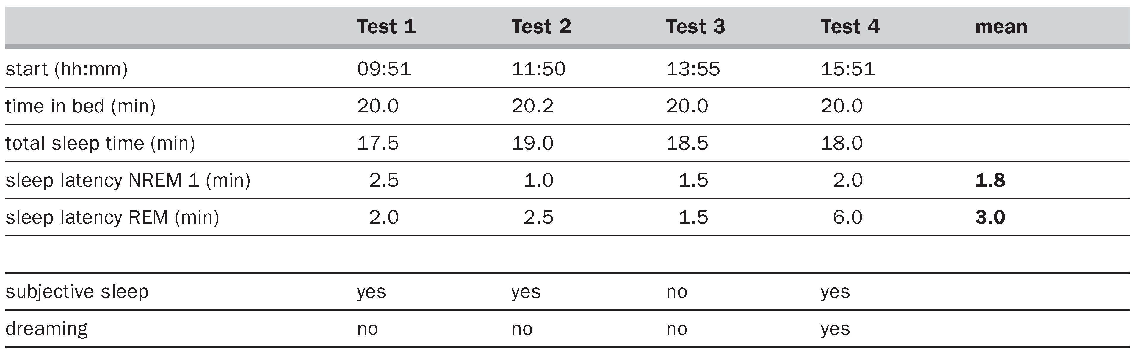
Results of multiple sleep latency test reflect high sleep propensity (pathological mean sleep latency 1.8 min) and high REM pressure (3 of 4 SOREM). Noteworthy is the patient’s misperception of sleep in one of the 4 naps.
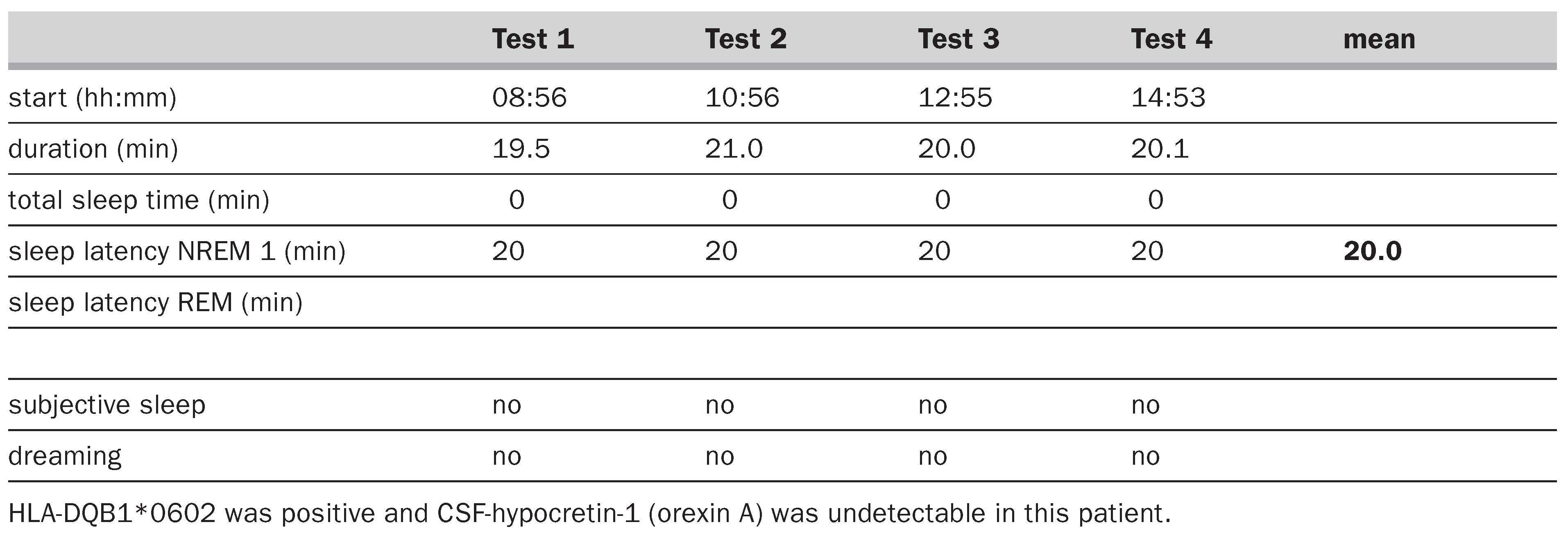
Interestingly maintenance of wakefulness test documents the patient’s ability to stay awake for 20 min when tested in a non-stimulant surrounding in a 2-hour interval 4 times a day.
Hypocretins (orexins) and sleep-wake cycle
Considering that any disruption of hypocretin signaling causes narcolepsy in humans and animals, it has been hypothesised that these neuropeptides are essential for the regulation of wakefulness and REM-sleep. Indeed, in vivo recordings in animals show that most of neurons located in the periforneal area of the hypothalamus are active during wakefulness and REM-sleep, but silent during NREM-sleep [36]. Intraventricular injection of hypocretin-1 increases wakefulness and natural hypocretin release in CSF correlates to the sleepwake cycle. It peaks in the active period and shows a nadir during sleep [36,37,38,39]. This cyclic variation is directly related to circadian rhythm and homeostatic sleep regulation and does not depend on confounding factors like locomotor activity or stress during wakefulness [40].The mode of action, i.e., how hypocretins promote wakefulness, is not yet clear. Wakefulness is mediated by a complex interaction of aminergic and cholinergic neurons. Hypocretins may mediate wakefulness by enhancing the neuronal activity on aminergic “wake active” areas (such as noradrenergic locus coeruleus [41,42,43], serotoninergic nucleus raphe [44,45], histaminergic tuberomammillary nucleus [46,47,48] or dopaminergic system) or by activation of cholinergic neurons in the basal forebrain or the laterodorsal/pedunculo-pontine tegmental area [49,50,51,52]. Interestingly pharmacological manipulation with modafinil, used for the treatment of sleepiness in narcolepsy, increases the level of hypocretins and induces c-fos expression [49]. Alternatively hypocretins may maintain wakefulness by sleep-inhibitory properties. Gabaergic neurons in the ventrolateral preoptic area (VLPO) promote NREM-sleep by inhibiting monoaminergic and cholinergic cells. These cells reciprocally inhibit VLPO neuron cells during wakefulness [53]. The presence of OX1R in lateral preoptic area led to the assumption that hypocretins promote wakefulness by inhibition of VLPO-neurons [54] but these data need to be confirmed.
The REM/cataplexy regulatory effects of hypocretins are more controversial. REM-sleep is normally initiated by specific neurons located in the pons. These neurons generate a highly active EEG pattern via ascending thalamo-cortical transmission and mediate spinal motor atonia via descending pathways. During wakefulness these REM-active neurons are inhibited by aminergic and serotoninergic neurons. A low aminergic drive due to absent or dysfunctional hypocretin transmission would enhance REM by simple disinhibitory effects. Consequently, activity of hypocretin neurons is expected to be low during REM-sleep. However, most studies found high hypocretins during REM-sleep [36,37]. In clinical studies CSF hypocretin-1 is only found to be low or undetectable when cataplexy is present [25], suggesting a central role of orexins in cataplexy. A complex muscle tone regulatory effect of orexins has been shown in animal studies. Depending on the site of application hypocretins may increase or decrease muscle tone [55,56]. However, in patients with low cerebrospinal fluid hypocretin-1 levels related to neurological disorders other than narcolepsy and hypersomnia, but no cataplexy has been reported [31].
Treatment of narcolepsy
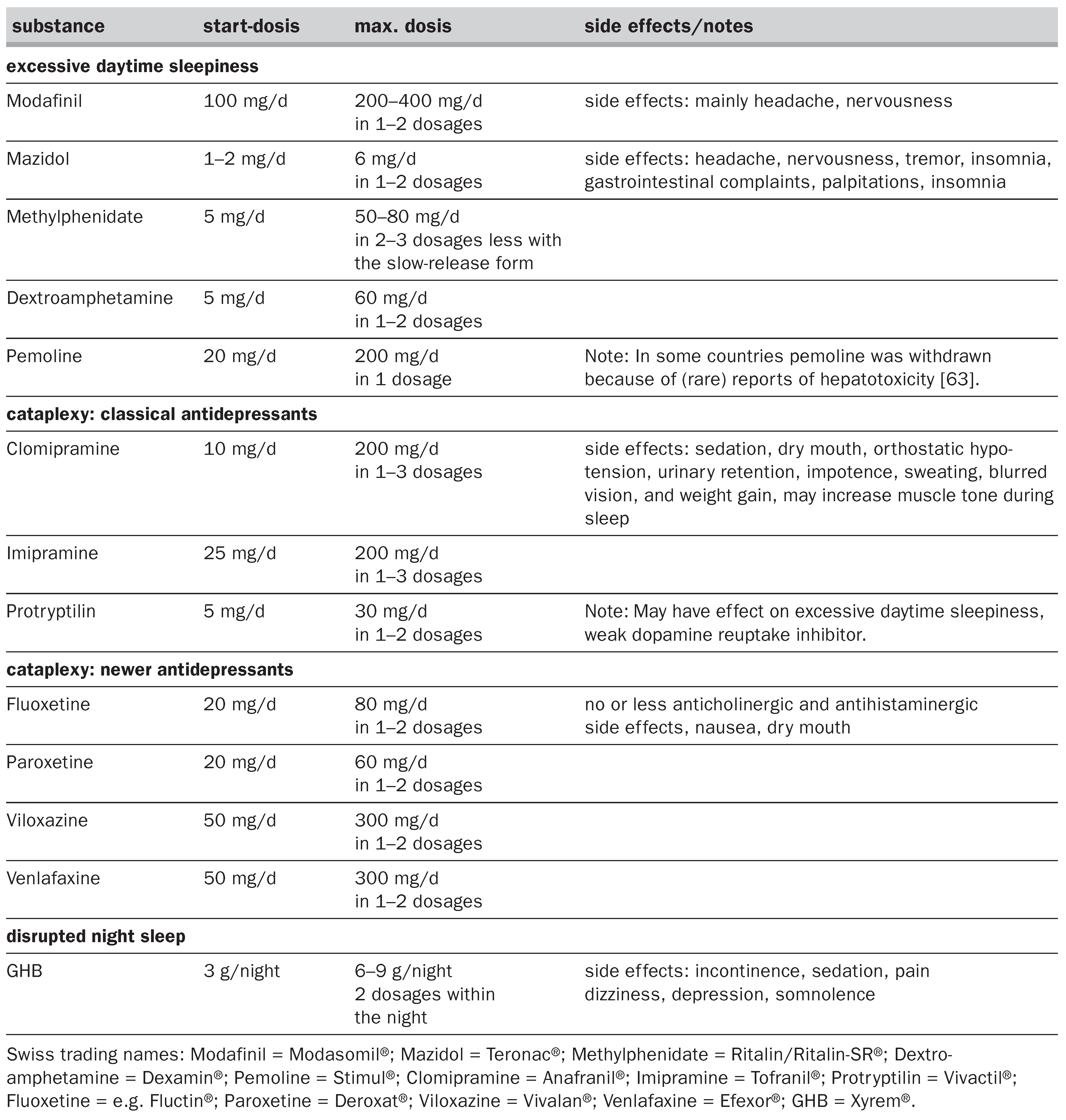
Current treatment of narcolepsy is symptomatic and includes non-pharmacological and pharmacological strategies (see Table 3). Patients should be informed about the chronic course of the disease and the value of counselling to address issues such as sleep hygiene, driving licence, professional choices, pregnancy. The most important nonpharmacological treatment are scheduled daytime naps even in late stages of disease. In milder forms short daytime nappings of 10–60 min may control excessive daytime sleepiness sufficiently but even in severely affected patients this strategy helps to reduce the dosage of stimulants. Most narcoleptic patients need pharmacological medication, which is effective in two thirds with tolerable side effects. Excessive daytime sleepiness and REM-sleep symptoms usually require distinct approaches although treating excessive daytime sleepiness positively influences cataplexy. There are only a few agents including sodium oxybate (gammahydroxybutyrate = GHB) that consolidate disturbed night sleep.
Table 3.
Pharmacological treatment of narcolepsy.
Excessive daytime sleepiness
The rationale of treatment with stimulants is to compensate the relative aminergic dysbalance by enhancing dopaminergic or noradrenergic activity. Drug of first choice for moderate and severe excessive daytime sleepiness is modafinil achieving a good effect in most patients. The effect of modafinil on excessive daytime sleepiness is similar to that of methylphenidate and probably inferior to that of amphetamines, but patients benefit from its lower side-effect profile, absent effect on nocturnal sleep and lower abuse potential [57]. Headache and nervousness are the most frequently reported side effects (up to 50% of patients). Tolerance has only rarely been observed. Alternatively mazidol or methylphenidate should be tried, in non-responsive patients metamphetamine and dextroamphetamine can be used [58,59,60,61]. Third-line drugs in the treatment of excessive daytime sleepiness include selegilin, sodium oxybate (GHB) and reboxetin (a selective noradrenalin reuptake inhibitor) [62]. Sodium oxybate markedly reduces the frequency of cataplectic attacks. The substance has been approved by the FDA in the US for the treatment of cataplexy but recent studies have also shown its efficacy against excessive daytime sleepiness [63]. GHB is probably the only available drug that consolidates night-time sleep in narcolepsy. Problematic is its short half-life and caution has to be paid to its sedating effects and abuse potential. Severe complications such as coma and death have been reported in non-narcoleptic drug abuses when taken together with alcohol or other drugs. The effect of propranolol and levodopa (250–500 mg/d) seem to be either limited to single patients or shortlived [61]. Some patients experience an improvement of mood with stimulants. Tolerance occurs in up to 30% of patients, more commonly at high dosages [41,61]. Drug holidays of 1–2 days/week (lower dosages or no medication) may be of benefit. Hypertension, myocardial ischaemia, ischaemic stroke and cerebral haemorrhage are rare in patients without associated cardiovascular risk factors. A neuronal toxicity has been described for amphetamines only at very high dosages of 500–3000 mg/d [64]. Addiction and psychosis are rare (<1–3% of cases) [65]. The use of low-dose stimulants seems to be safe in children, whereas it should be avoided when possible in pregnancy. Pemoline, mazidol and the selective serotonin reuptake inhibitors are probably the safest stimulants during pregnancy [61].
Cataplexy
In patients with frequent/severe cataplexy treatment should be started with tricyclic agents. The most effective drugs are clomipramine and imipramine [41,61,66]. Tolerance to these drugs may develop, and their abrupt discontinuation may cause a “status cataplecticus” [65]. Contraindications and side effects of these compounds are due to their anticholinergic effect or concomitant treatment with MAO-inhibitors (see Table 3). New and more selective antidepressants such as protryptilin, fluoxetine, paroxetine, viloxazine, venlafaxine or femoxetine can be tried in patients with untolerable anticholinergic side effects of the tricyclic agents [41]. These agents may also have a mild effect on excessive daytime sleepiness. Carbamazepine, clonidin or anticholinergic drugs can be tried in patients without response to other anticataplectic drugs. Prazosine should be avoided in narcoleptics because of its well-known aggravating effect on cataplexy [67]. Development of hypocretin/orexin-agonists may become a future therapeutic option for both excessive daytime sleepiness and cataplexy.
References
- Parkes, JD; Baraitser, M; Marsden, CD; Asselman, P. Natural history, symptoms and treatment of the narcoleptic syndrome. Acta Neurol Scand 1975, 52, 337–53. [Google Scholar] [CrossRef] [Scilit]
- Johns, MW. Sensitivity and specificity of the multiple sleep latency test (MSLT), the maintenance of wakefulness test and the Epworth sleepiness scale: failure of the MSLT as a gold standard. J Sleep Res 2000, 9, 5–11. [Google Scholar] [CrossRef] [Scilit] [PubMed]
- Hood, B; Bruck, D. A comparison of sleep deprivation and narcolepsy in terms of complex cognitive performance and subjective sleepiness. Sleep Med 2002, 3, 259–66. [Google Scholar] [CrossRef] [Scilit] [PubMed]
- Guilleminault, C. Cataplexy in narcolepsy. In Proceedings of the First International Symposium on Narcolepsy; Guilleminault, C, Dement, WC, Passouant, P, Eds.; Spectrum Publications, Inc.: Montpellier, 1976; pp. 125–43. [Google Scholar]
- Rubboli, G; d’Orsi, G; Zaniboni, A; Gardella, E; Zamagni, M; Rizzi, R; et al. A video-polygraphic analysis of the cataplectic attack. Clin Neurophysiol 2000, 11, S120–8. [Google Scholar] [CrossRef] [Scilit] [PubMed]
- Schuld, A; Hebebrand, J; Geller, F; Pollmacher, T. Increased body mass index in patients with narcolepsy. Lancet 2000, 355, 39–40. [Google Scholar] [CrossRef] [Scilit] [PubMed]
- Ohayon, MM; Priest, RG; Zulley, J; Smirne, S; Paiva, T. Prevalence of narcolepsy symptomatology and diagnosis in the European general population. Neurology 2002, 58, 1826–33. [Google Scholar] [CrossRef] [Scilit]
- Mignot, E. Genetic and familial aspects of narcolepsy. Neurology 1998, 50, 16–22. [Google Scholar] [CrossRef] [Scilit]
- Hublin, C; Partinen, M; Kaprio, J; Koskenvuo, M; Guilleminault, C. Epidemiology of narcolepsy. Sleep 1994, 17, 7–12. [Google Scholar] [CrossRef] [Scilit]
- Guilleminault, C; Pelayo, R. Narcolepsy in prepubertal children. Ann Neurol 1998, 43, 135–42. [Google Scholar] [CrossRef] [Scilit] [PubMed]
- Krahn, LE; Boeve, BF; Olson, EJ; Herold, DL; Silber, MH. A standardized test for cataplexy. Sleep Med 2000, 1, 125–30. [Google Scholar] [CrossRef] [Scilit] [PubMed]
- Paskind, HA. Effect of laughter on muscle tone. Arch Neurol Psychiatry 1932, 28, 623–8. [Google Scholar] [CrossRef] [Scilit]
- Rosenthal, C. Ueber das Auftreten von halluzinatorischkataplektischen Angstsyndromen, Wachanfällen und ähnlichen Störungen bei Schizophrenen. Monatsschrift für Psychiatrie und Neurologie 1939, 102, 11–38. [Google Scholar] [CrossRef] [Scilit]
- Bassetti, C; Aldrich, MS. Narcolepsy. Neurol Clin 1996, 14, 545–71. [Google Scholar] [CrossRef] [Scilit]
- American Sleep Disorders. A. International Classification of Sleep Disorders: diagnostic and coding manual, revised. Rochester, MN. 1997.
- Aldrich, MS; Chervin, RD; Malow, BA. Value of the multiple sleep latency test (MSLT) for the diagnosis of narcolepsy. Sleep 1997, 20, 620–9. [Google Scholar] [CrossRef] [Scilit] [PubMed]
- Mitler, MM; Walsleben, J; Sangal, RB; Hirshkowitz, M. Sleep latency on the maintenance of wakefulness test (MWT) for 530 patients with narcolepsy while free of psychoactive drugs. Electroencephalogr Clin Neurophysiol 1998, 107, 33–8. [Google Scholar] [CrossRef] [Scilit] [PubMed]
- Mignot, E; Lin, L; Rogers, W; Honda, Y; Qiu, X; Lin, X; et al. Complex HLA-DR and -DQ interactions confer risk of narcolepsy-cataplexy in three ethnic groups. Am J Hum Genet 2001, 68, 686–99. [Google Scholar] [CrossRef] [Scilit]
- Mignot, E; Lammers, GJ; Ripley, B; Okun, M; Nevsimalova, S; Overeem, S; et al. The role of cerebrospinal fluid hypocretin measurement in the diagnosis of narcolepsy and other hypersomnias. Arch Neurol 2002, 59, 1553–62. [Google Scholar] [CrossRef] [Scilit] [PubMed]
- Bassetti, C; Gugger, M; Bischof, M; Mathis, J; Sturzenegger, C; Werth, E; et al. The narcoleptic borderland: a multimodal diagnostic approach including cerebrospinal fluid levels of hypocretin-1 (orexin A). Sleep Med 2003, 4, 7–12. [Google Scholar] [CrossRef] [Scilit] [PubMed]
- Overeem, S; Lammers, GJ; van Dijk, JG. Cataplexy: tonic immobility rather than REM-sleep atonia? Sleep Med 2002, 3, 471–7. [Google Scholar] [CrossRef] [Scilit]
- Overeem, S; Lammers, GJ; van Dijk, JG. Weak with laughter. Lancet 1999, 354, 838. [Google Scholar] [CrossRef] [Scilit] [PubMed]
- Gandevia, SC; Butler, JE; Taylor, JL; Anand, A; Paintal, A. No laughing matter. Lancet 1999, 354, 2086. [Google Scholar] [CrossRef] [Scilit] [PubMed]
- Okura, M; Riehl, J; Mignot, E; Nishino, S. Sulpiride, a D2/D3 blocker reduces cataplexy but not REM sleep in canine narcolepsy. Neuropsychopharmacology 2000, 23, 528–38. [Google Scholar] [CrossRef] [Scilit] [PubMed]
- Kilduff, TS; Peyron, C. The hypocretin/orexin ligandreceptor system: implications for sleep and sleep disorders. TINS 2000, 23, 359–65. [Google Scholar] [PubMed]
- Thannickal, TC; Moore, RY; Nienhuis, R; Ramanathan, L; Gulyani, S; Aldrich, M; et al. Reduced number of hypocretin neurons in human narcolepsy. Neuron 2000, 27, 469–74. [Google Scholar] [CrossRef] [Scilit]
- Chemelli, RM; Willie, JT; Sinton, CM; Elmquist, JK; Scammell, T; Lee, C; et al. Narcolepsy in orexin knockout mice: molecular genetics of sleep regulation. Cell 1999, 98, 437–51. [Google Scholar] [CrossRef] [Scilit]
- Lin, L; Faraco, J; Li, R; Kadotani, H; Rogers, W; Lin, X; et al. The sleep disorder of canine narcolepsy is caused by a mutation in the hypocretin (orexin) receptor 2 gene. Cell 1999, 98, 365–76. [Google Scholar] [CrossRef] [Scilit] [PubMed]
- Nishino, S; Ripley, B; Overeem, S; Lammers, GJ; Mignot, E. Hypocretin (orexin) deficiency in human narcolepsy. Lancet 2000, 355, 39–40. [Google Scholar] [CrossRef] [Scilit] [PubMed]
- Krahn, LE; Pankratz, VS; Oliver, L; Boeve, BF; Silber, MH. Hypocretin (orexin) levels in cerebrospinal fluid of patients with narcolepsy: relationship to cataplexy and HLA DQB1*0602 status. Sleep 2002, 25, 733–6. [Google Scholar] [CrossRef] [Scilit]
- Ripley, B; Overeem, S; Fujiki, N; Nevsimalova, S; Uchino, M; Yesavage, J; et al. CSF hypocretin/orexin levels in narcolepsy and other neurological conditions. Neurology 2001, 57, 2253–8. [Google Scholar] [CrossRef] [Scilit]
- Peyron, C; Faraco, J; Rogers, W; Ripley, B; Overeem, S; Charnay, Y; et al. A mutation in a case of early onset narcolepsy and a generalized absence of hypocretin peptides in human narcoleptic brains. Nature Med 2000, 6, 991–7. [Google Scholar] [CrossRef] [Scilit]
- Olafsdottir, BR; Rye, DB; Scammell, TE; Matheson, JK; Stefansson, K; Gulcher, JR. Polymorphisms in hypocretin/ orexin pathway genes and narcolepsy. Neurology 2001, 57, 1896–9. [Google Scholar] [CrossRef] [Scilit]
- Gencik, MJ; Dahmen, N; Wieczorek, S; Kasten, M; Bierbrauer, J; Anghelescu, I; et al. A prepro-orexin gene polymorphism is associated with narcolepsy. Neurology 2001, 56, 115–7. [Google Scholar] [CrossRef] [Scilit]
- Hara, J; Beuckmann, CT; Nambu, T; Willie, JT; Chemelli, RM; Sinton, CM; et al. Genetic ablation of orexin neurons in mice results in narcolepsy, hypophagia, and obesity. Neuron 2001, 30, 345–54. [Google Scholar] [CrossRef] [Scilit]
- Alam, MN; Gong, H; Alam, T; Jaganath, R; McGinty, D; Szymusiak, R. Sleep-waking discharge patterns of neurons recorded in the rat perifornical lateral hypothalamic area. J Physiol 2002, 538, 619–31. [Google Scholar] [CrossRef] [Scilit] [PubMed]
- Kiyashchenko, LI; Mileykovskiy, BY; Maidment, N; Lam, HA; Wu, MF; John, J; et al. Release of hypocretin (orexin) during waking and sleep states. J Neurosci 2002, 22, 5282–6. [Google Scholar] [CrossRef] [Scilit] [PubMed]
- Estabrooke, IV; McCarthy, MT; Ko, E; Chou, TC; Chemelli, RM; Yanagisawa, M; et al. Fos expression in orexin neurons varies with behavioral state. J Neurosci 2001, 21, 1656–62. [Google Scholar] [CrossRef] [Scilit] [PubMed]
- Yoshida, Y; Fujiki, N; Nakajima, T; Ripley, B; Matsumura, H; Yoneda, H; et al. Fluctuation of extracellular hypocretin-1 (orexin A) levels in the rat in relation to the light-dark cycle and sleep-wake activities. Eur J Neurosci 2001, 14, 1075–81. [Google Scholar] [CrossRef] [Scilit]
- Zeitzer, JM; Buckmaster, CL; Parker, KJ; Hauck, CM; Lyons, DM; Mignot, E. Circadian and homeostatic regulation of hypocretin in a primate model: implications for the consolidation of wakefulness. J Neurosci 2003, 23, 3555–60. [Google Scholar] [CrossRef] [Scilit]
- Nishino, S; Mignot, E. Pharmacological aspects of human and canine narcolepsy. Prog Neurobiol 1997, 52, 27–78. [Google Scholar] [CrossRef] [Scilit]
- Hagan, JJ; Leslie, RA; Patel, S; Evans, ML; Wattam, TA; Holmes, S; et al. Orexin A activates locus coeruleus cell firing and increases arousal in the rat. Proc Natl Acad Sci 1999, 96, 10911–6. [Google Scholar] [CrossRef] [Scilit]
- Bourgin, P; Huitron-Resendiz, S; Spier, AD; Fabre, V; Morte, B; Criado, JR; et al. Hypocretin-1 modulates rapid eye movement sleep through activation of locus coeruleus neurons. J Neurosci 2000, 20, 7760–5. [Google Scholar] [CrossRef] [Scilit]
- Horvath, TL; Peyron, C; Diano, S; Ivanov, A; Aston-Jones, G; Kilduff, TS; et al. Hypocretin (orexin) activation and synaptic innervation of the locus coeruleus noradrenergic system. J Comp Neurol 1999, 415, 145–59. [Google Scholar] [CrossRef]
- Brown, RE; Sergeeva, O; Eriksson, KS; Haas, HL. Orexin A excites serotonergic neurons in the dorsal raphe nucleus of the rat. Neuropharmacology 2001, 40, 457–9. [Google Scholar] [CrossRef] [Scilit]
- Huang, ZL; Qu, WM; Li, WD; Mochizuki, T; Eguchi, N; Watanabe, T; et al. Arousal effect of orexin A depends on activation of the histamine system. Proc Natl Acad Sci 2001, 98, 9965–70. [Google Scholar] [CrossRef] [Scilit]
- Bayer, L; Eggermann, E; Serafin, M; Saint-Mleux, B; Machard, D; Jones, B; et al. Orexins (hypocretins) directly excite tuberomammillary neurons. Eur J Neurosci 2001, 14, 1571–5. [Google Scholar] [PubMed]
- Eriksson, KS; Sergeeva, O; Brown, RE; Haas, HL. Orexin/ hypocretin excites the histaminergic neurons of the tuberomammillary nucleus. J Neurosci 2001, 21, 9273–9. [Google Scholar] [CrossRef] [Scilit] [PubMed]
- Scammell, T; Estabrooke, IV; McCarthy, MT; Chemelli, RM; Yanagisawa, M; Miller, MS; et al. Hypothalamic arousal regions are activated during modafinil-induced wakefulness. J Neurosci 2001, 20, 8620–8. [Google Scholar] [CrossRef] [Scilit] [PubMed]
- Eggermann, E; Serafin, M; Bayer, L; Machard, D; Saint-Mleux, B; Jones, BE; et al. Orexin/hypocretins excite basal forebrain cholinergic neurons. Neuroscience 2001, 108, 177–81. [Google Scholar] [CrossRef] [Scilit] [PubMed]
- Methippara, MM; Alam, MN; Szymusiak, R; McGinty, D. Effects of lateral preoptic area application of orexin-A on sleep-wakefulness. Neuroreport 2000, 11, 3423–6. [Google Scholar] [CrossRef] [Scilit]
- Espana, RA; Baldo, BA; Kelley, AE; Berridge, CW. Wakepromoting and sleep-suppressing actions of hypocretin (orexin): basal forebrain sites of action. Neuroscience 2001, 106, 699–715. [Google Scholar] [CrossRef] [Scilit]
- Saper, CB; Chou, TC; Scammell, TE. The sleep switch: hypothalamic control of sleep and wakefulness. Trends Neurosci 2001, 24, 726–31. [Google Scholar] [CrossRef] [Scilit]
- Willie, JT; Chemelli, RM; Sinton, CM; Yanagisawa, M. To eat or to sleep? Orexin in the regulation of feeding and wakefulness. Ann Rev Neurosci 2001, 24, 429–58. [Google Scholar] [CrossRef] [Scilit] [PubMed]
- Kiyashchenko, LI; Mileykovskiy, BY; Lai, YY; Siegel, JM. Increased and decreased muscle tone with orexin (hypocretin) microinjections in the locus coeruleus and pontine inhibitory area. J Neurophysiol 2001, 85, 2008–16. [Google Scholar] [CrossRef] [Scilit]
- Xi, MC; Fung, SJ; Yamuy, J; Morales, FR; Chase, MH. Induction of active (REM) sleep and motor inhibition by hypocretin in the nucleus pontis oralis of the cat. J Neurophysiol 2002, 87, 2880–8. [Google Scholar] [CrossRef] [Scilit]
- U.S. Modafinil in Narcolepsy Study, Group. Randomized trial of modafinil for the treatment of pathological somnolence in narcolepsy. Ann Neurol 1998, 43, 88–97. [CrossRef] [Scilit]
- Mitler, M; Hajdukovic, R. Relative efficacy of drugs for treatment of sleepiness in narcolepsy. Sleep 1991, 14, 218–20. [Google Scholar] [CrossRef] [Scilit] [PubMed]
- ASDA. Practice parameters for the use of stimulants in the treatment of narcolepsy. Sleep 1994, 17, 348–51. [Google Scholar] [CrossRef] [Scilit]
- Fry, JM. Treatment modalities for narcolepsy. Neurology 1998, 50, 43–8. [Google Scholar] [CrossRef] [Scilit] [PubMed]
- Bassetti, C. Narcolepsy. Curr Treat Opt Neurol 1999, 1, 291–7. [Google Scholar] [CrossRef] [Scilit]
- Larrosa, O; de la Llave, Y; Bario, S; Granizo, JJ; GarciaBorreguero, D. Stimulant and anticatapletic effects of reboxetine in patients with narcolepsy: a pilot study. Sleep 2001, 24, 282–5. [Google Scholar] [CrossRef] [Scilit] [PubMed]
- Xyrem multicenter study group. A 12-month open label, multicenter extension trial of orally administered sodium oxybate for the treatment of narcolepsy. Sleep 2003, 26, 31–5. [Google Scholar]
- Ernst, T; Chang, L; Leonido-Yee, M; Speck, O. Evidence for long-term neurotoxicity associated with methamphetamine abuse: a 1H MRS study. Neurology 2000, 54, 1344–9. [Google Scholar] [CrossRef] [Scilit] [PubMed]
- Parkes, D; Dahlitz, M. Amphetamine prescriptions. Sleep 1993, 16, 201–3. [Google Scholar] [CrossRef] [Scilit] [PubMed]
- Passouant, P; Baldy-Moulinier, M; Aussilloux, C. Cataplexie associé avec la maladie de Gélinau: effect de la clomipramine. Rev Neurol 1970, 123, 56–60. [Google Scholar]
- Aldrich, M; Rogers, AE. Exacerbation of human cataplexy by prazosin. Sleep 1989, 12, 254–6. [Google Scholar] [CrossRef] [Scilit] [PubMed]
- Honda, Y. Clinical features of narcolepsy: Japanese experiences. In HLA in Narcolepsy; Honda, Y, Juji, T, Eds.; Springer: Berlin, 1988; pp. 24–57. [Google Scholar]

This is an open-access article distributed under the terms of the Creative Commons Attribution Non-Commercial No Derivatives (CC BY-NC-ND) 4.0 License.