


Summary
Hereditary spastic paraplegia (HSP) is a group of disorders characterised by continuously progressive paraplegia occurring at a wide variety of ages and sometimes accompanied by additional symptoms. Recent progress in hereditary spastic paraplegia molecular genetics has lead to the discovery of several new genes and loci, allowing first insights into the pathophysiology of this disorder. It is therefore timely to review the data about hereditary spastic paraplegia.
Zusammenfassung
Die spastische Spinalparalyse kann als reine oder komplexe Form mit Beteiligung verschiedener ZNS- und PNS-Regionen oder sogar anderer Systeme auftreten. Einige Loci wurden in den letzten Jahren beschrieben und einzelne Genmutationen gefunden.
Die häufigste der reinen Formen wird durch Mutationen des Spastin-Gens, lokalisiert auf dem langen Arm des Chromosoms 2, verursacht. Mutationen im Paraplegin-Gen werden auf autosomal-rezessive Art vererbt und können von einer reinen oder komplexen Form der Krankheit gefolgt werden. Zwei Gene auf dem X-Chromosom wurden beschrieben. Mutationen des Zelladhesionsmoleküls, L1-CAM, führen zu einem Syndrom mit Skelettdeformitäten und mentaler Retardation und Proteolipid-Protein-Mutationen, die allelisch zu der Pelizeus-Merzbacher-Krankheit sind, zu einem intermediären progressiven Syndrom.
Es ist zu erwarten, dass in den nächsten Jahren weitere Gene gefunden werden, die andere Syndrome verursachen. Diese Kenntnisse erlauben eine bessere Klassifikation der Krankheit und eine bessere genetische Beratung und allgemeine Betreuung der Patienten und ihrer Familien. Sie erlauben auch Einsicht in die Pathogenese der Krankheit, was hoffentlich zu besseren, rationaleren Therapieansätzen führen sollte. Wie auch bei anderen seltenen neurogenetischen Krankheitsbildern werden solche weiterführenden Erkenntnisse eine grosse Bedeutung für andere spinale Krankheiten haben.
Schlüsselwörter: pastische Spinalparalyse; Spastin; Paraplegin; Rückenmark; Erbkrankheiten
Hereditary spastic paraplegia is an old disorder occurring in pure and complex forms
The first description of inherited spastic paraplegia has been made by Strümpell in two brothers [1,2,3]. Later, after numerous case reports had been published, hereditary spastic paraplegia was classified according to age and to the presence of additional symptoms. However, newer genetic studies have now demonstrated that age might vary greatly even in families with the same mutation. A detailed evaluation of 22 families gave a thorough picture of what could be found in the area prior to molecular genetic findings [4]. Hereditary spastic paraplegia might occur as a pure form with steadily progressive paraplegia, starting at any age, but mainly between 20 and 40 leading to a gait disorder. Sometimes a urinary sphincter disturbance may be present as well. At examination a gait spasticity with brisk reflexes in the legs and positive Babinski sign are found. A slight decrease in vibration sense may be disclosed also. Pure hereditary spastic paraplegia is the most common form of the disorder, with a prevalence of 9.6 per 100 000 found in a carefully conducted study in Spain [5]. Autosomal dominant and recessive as well as X-chromosomal pure hereditary spastic paraplegia have been known for a while [6].
Table 1.
Accompanying symptoms found in complicated hereditary spastic paraplegia (HSP).
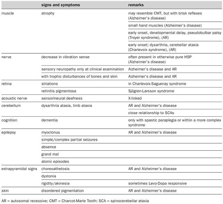
Disturbances of many other systems occur in the complicated form, pointing at central and peripheral nervous system involvement [6]. There are numerous descriptions available in the literature of families with hereditary syndromes in which spastic paraparesis is found. Almost all central and peripheral neurological regions may be involved. Furthermore, disorders of muscle, skin and retina may be included in some families (table 1).Ataxia and spastic paraplegia may coexist in the same syndrome, either in one individual or within families. Separation of hereditary spastic paraplegia from spinocerebellar ataxia on clinical grounds should therefore not be seen as definitive as it has previously been the case, since these syndromes overlap. This also leads to the suggestion that hereditary spastic paraplegia and spinocerebellar ataxia might share some pathogenetic features due to allelic mutations. Choreoatethosis, dystonia and rigidity have similarly been described in some families. Skin disorders and cardiac defects have been associated with hereditary spastic paraplegia. In addition, a whole series of rare syndromes has been described, in which spastic paraplegia is present as well. The list is far from being exhaustive and clarification among these hereditary syndromes awaits further molecular genetic advances.
Differential diagnosis of hereditary spastic paraplegia is wide
The great variety of symptoms found in complicated hereditary spastic paraplegia opens differential diagnostic reasoning widely. One of the most often contemplated diagnoses is multiple sclerosis, which may also on occasion occur within families, either due to the high prevalence of the disorder, or to a mendelian multiple sclerosis inheritance. Among viral disorders, HTLVI infection is the major cause to be considered, especially in pure hereditary spastic paraplegia. Furthermore, a variety of metabolic disorders may lead to symptoms similar to those found in hereditary spastic paraplegia, like xanthomatous cerebrotenidosis, xeroderma pigmentosum, metachromatic leukodystrophy, adrenomyeloneuropathy, and Lesch-Nyhan syndrome, among others. Clear neurological reasoning will help the physician to identify the right disorder. The most important will be to search for causes amendable to treatment. On the other hand, clear findings in the family history in an otherwise typical case should allow a diagnosis without any further investigation of the presenting case. In doubt, careful clinical examination of the family will be of great help. Furthermore, molecular genetic investigations may be performed in selected cases.
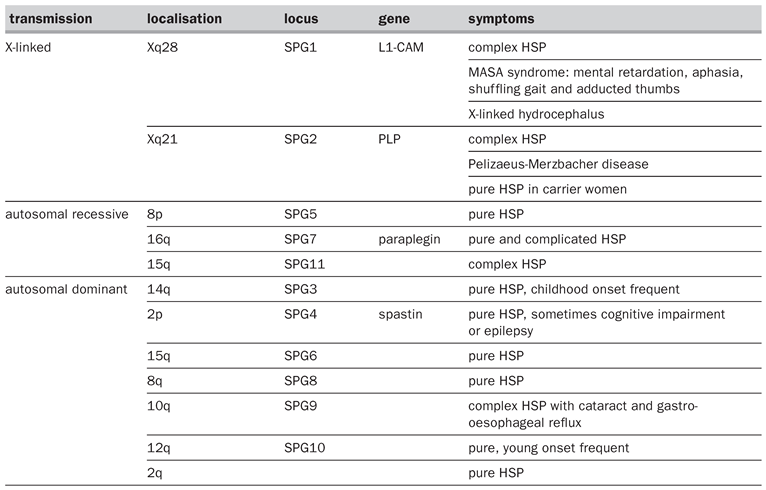
Table 2.
Molecular hereditary spastic paraplegia (HSP) classification.
Mutations of many genes may lead to hereditary spastic paraplegia
As has been the case in other groups of hereditary neurologic disorders, molecular classification is more useful than clinical. At present, one gene for a dominant form, spastin, one for a recessive form, paraplegin, and two for X-chromosomal forms, phospholipoprotein and L1-CAM, have been described (table 2). The discovery of these genes has pointed at many shortcomings of the above classification schemes. Further loci are known, and it is hoped that in the near future a whole set of new genes will be discovered. Furthermore, mutations of other sets of genes, like mitochondrial ones, may lead to syndromes including spastic paraplegia. The molecular pathophysiology of hereditary spastic paraplegia due to known mutation will now be reviewed. It is to be expected that in the near future many more genes will be discovered, allowing further insight into the molecular function of the spinal cord.
Spastin
Studies disclosing the SPG4 locus on chromosome 2p [7] were followed by the discovery of the gene itself, named spastin [8]. SPG4 is the most frequent locus found in 40% of the families with dominant hereditary spastic paraplegia [9] and most of those described so far have a pure form of the disorder. Large families were described in central Switzerland [10] and reexamined recently [11]. In three of them, linked to the SPG4 locus [12], a novel spastin mutation has been found, leading to skipping of exon 16 [13]. Spastin, located on chromosome 2, has 17 exons over a region of 90 kb [8]. Spastin belongs to a group of proteins named AAA for ATPases associated with diverse cellular activities [14]. These include cell cycle regulation, protein degradation, organelle biogenesis, and vesiclemediated protein functions. AAA proteins probably act as chaperones, allowing assembly and function of protein complexes also named proteasomes. They use energy from ATP to modulate tridimensional conformation of other proteins, for example molecular motors. AAA proteins are grouped into a family according to the gene sequence homology they have at the ATPase site. However, the remaining of the sequence may be very different among them, pointing at the numerous different uses of this core enzymatic activity in cell processes. The fact that spastin changes found in hereditary spastic paraplegia include missense, non-sense and splice-site mutations and that they are dominant suggest a loss of function. Some sequence homology with AAA proteins involved in nuclear function, more precisely in meiosis, would suggest that a disordered cell division may play a role. Later during life, other molecular events might then occur to induce cell death. Differences in these postulated events might explain the huge variety of symptom severity and age at onset even among members of the same family with the same mutation. At this time, however, the exact mechanisms of selective cell loss found in spastin deficiency have not yet been elucidated.
Proteolipid protein
The proteolipid protein (PLP) is one of the major CNS myelin protein components. Mutations of the proteolipid protein gene on the X chromosome may lead to a variety of syndromes. The most severe phenotype is the infantile form of PelizaeusMerzbacher disease. The disease starts in early childhood or infancy with nystagmus, hypotonia and cognitive impairment. Later, progressive spasticity and ataxia develop, and affected persons often die in childhood. Other mutations have been found to lead to a relatively benign spastic paraplegia in the adult, often with a normal life span. The SPG2 locus had been mapped in families with pure hereditary spastic paraplegia to the proximal long arm of the X chromosome by linkage to DXS17. After narrowing the interval in a larger pedigree, proteolipid protein was found to bear a point mutation segregating with the disorder [15]. Some other mutations lead to intermediate phenotypes and female carriers may show mild to moderate signs of the disease [16]. Proteolipid protein mutation induced disorder can be suggested when clinical signs with a steady progression are accompanied by the typical MRI picture with abnormal myelination.
L1CAM
SPG1 denotes a complex, X-chromosomal inherited phenotype which includes, beside spastic paraplegia, mental retardation, aphasia and diverse musculoskeletal abnormalities. SPG1 is mapped to the telomeric region of the long arm of the X chromosome [17]. The protein encoded by SPG1 is a cell adhesion molecule, L1CAM and a mutation in exon 26 has been found in one family [18]. SPG1 is allelic to the MASA syndrome and to X-linked hydrocephalus and corpus callosum agenesis. The group of diseases is also named CRASH syndrome (corpus callosum hypoplasia, retardation, adducted thumbs, spastic paraplegia and hydrocephalus) [19]. Cell adhesion molecules play important roles in the development of the central nervous system. L1CAM is a transmembrane glycoprotein expressed in neurons and Schwann cells involved in nerve growth and pathfinding [20].
Paraplegin
The locus of a relatively pure autosomal recessive form of hereditary spastic paraplegia, SPG7, had been mapped to 16q24.3 in a large informative family [21]. All examined affected members of this family had a 9.5 kb deletion at the SPG7 gene, named paraplegin [22]. Furthermore, frameshift mutations were seen in other families with pure and complex hereditary spastic paraplegia. In a study of 30 hereditary spastic paraplegia families of northeast England an additional paraplegin gene mutation leading to exon 11 deletion was found resulting in complex hereditary spastic paraplegia [23]. The paraplegin gene consists of 17 exons between 78 and 242 base pairs [24]. The sequence predicts two transmembrane domains compatible with a membrane localisation. The gene is expressed in a variety of tissues, with the highest expression found in the central nervous system, especially in amygdala, caudate, thalamus, subthalamic nucleus and spinal cord [24]. In the central nervous system a developmental regulation of SPG7 expression has been found, which was not the case in liver or kidney, suggesting a role for paraplegin in central nervous system development. Paraplegin is a mitochondrial metalloprotease with aminoacid sequence homology to yeast proteins Rca1p and Afg3p [25]. These proteins form another subclass within the AAA proteins family. In the mitochondria, paraplegin plays a role as a chaperone in the generation and assembling of proteins, in particular those involved in respiratory chain function. Paraplegin mutations in hereditary spastic paraplegia is one example of a nuclear encoded gene leading to mitochondrial dysfunction. This points at similar pathogenetic mechanisms in paraplegia found in this disorder and in mitochondrial disturbances. Other examples of mutations in nuclear encoded mitochondrial proteins genes include frataxin in Friedreich’s ataxia [26]. Furthermore, mutations in mitochondrial encoded genes may also lead to spastic paraplegia in the frame of complex syndromatic disorders.
Other examples
Charlevoix-Sagueny syndrome, as a complex form of hereditary spastic paraplegia with ataxia, slurred speech and distal amyotrophy have been described in Quebec [27]. SACS, the gene involved in this syndrome, is located on chromosome 13q11. The open reading frame encompasses only one exon, which is the largest found so far in vertebrates [28]. Sacsin probably functions as a chaperone, mediating protein folding in complex cellular processes.
Familial Alzheimer’s disease (AD) is found in about 5 to 10% of all patients diagnosed with Alzheimer’s disease. Familial Alzheimer’s disease is most often autosomal dominant and may be due to mutations in the genes encoding amyloid precursor protein, presenilin 1 and presenilin 2 [29]. Families with Alzheimer’s disease and additional paraparesis had been described and recently another mutation of the presenilin 1 gene has been found in one such family [30]. Presenilin 1 is a transmembrane protein with complex biological activities which are not known in detail [31]. The reason why some mutations of this gene lead to additional hereditary spastic paraplegia is not clear. These results lead to the suggestion that presenilin 1 mutations could be searched for in Alzheimer’s disease patients with paraparesis.
Conclusion
Hereditary spastic paraplegia is a good example of the discovery paradigm in neurogenetic disorders. Starting from clinical phenotypic classification, positional genetics lead to the cloning of several genes. Some of them were already known, with mutations leading to other phenotypes including spastic paraplegia, demonstrating genetic variation probably depending on their type. Others were new, further adding to the molecular complexity of the spinal cord and regions projecting there. The discovery of these genes now allows to pursue the search for an understanding of the pathophysiology of these disorders. Furthermore, this knowledge should lead to more rational therapeutic strategies in the future. It is hoped that accrued knowledge in the pathophysiology and therapy of hereditary spastic paraplegia should also help to better understand what happens in other spastic disorders. This illustrates one of the reasons why it is important to continue research about rare neurological disorders.
References
- Strümpell, A. Beiträge zur Pathologie des Rückenmarks. Arch Psychiatr Nervenkrankheiten 1880, 10, 676–717. [Google Scholar] [CrossRef] [Scilit]
- Strümpell, A. Ueber die hereditäre spastische Spinalparalyse. Dtsch Ztsch Nervenheil 1893, 4, 173–188. [Google Scholar] [CrossRef] [Scilit]
- Strümpell, A. Ueber eine bestimmte Form der primären combinierten Systemerkrankungen des Rückenmarkes. Arch Psychiatr 1886, 217–238. [Google Scholar]
- Harding, A.E. Early onset cerebellar ataxia with retained tendon reflexes: a clinical and genetic study of a disorder distinct from Friedreich’s ataxia. J Neurol Neurosurg Psychiatr 1981, 44, 871–883. [Google Scholar] [CrossRef] [Scilit]
- Polo, J.M.; Calleja, J.; Combarros, O.; Berciano, J. Hereditary ataxias and paraplegias in Cantabria, Spain. An epidemiological and clinical study. Brain 1991, 114, 855–866. [Google Scholar]
- McDermott, C.; White, K.; Bushby, K.; Shaw, P. Hereditary spastic paraparesis: a review of new developments. J Neurol Neurosurg Psychiatry 2000, 69, 150–160. [Google Scholar] [CrossRef] [Scilit]
- Hazan, J.; Fontaine, B.; Bruyn, R.P.M.; Lamy, C.; van Deutekom, J.C.T.; Rime, C.S.; et al. Linkage of a new locus for autosomal dominant familial spastic paraplegia to chromosome 2p. Hum Mol Genet 1994, 3, 1569–1573. [Google Scholar]
- Hazan, J.; Fonknechten, N.; Mavel, D.; Paternotte, C.; Samson, D.; Artiguenave, F.; et al. Spastin, a new AAA protein, is altered in the most frequent form of autosomal dominant spastic paraplegia. Nat Genet 1999, 23, 296–303. [Google Scholar] [PubMed]
- Reid, E.; Grayson, C.; Rogers, M.T.; Rubinsztein, D.C. Locusphenotype correlations in autosomal dominant pure hereditary spastic paraplegia. A clinical and molecular genetic study of 28 United Kingdom families. Brain 1999, 122, 1741–1755. [Google Scholar] [CrossRef] [Scilit] [PubMed]
- Taylor Carte, E. Beitrag zur Kenntnis der dominant vererbten spastischen Spinalparalyse; Orell Füssli: Zürich, Switzerland, 1963. [Google Scholar]
- von Fellenberg, J.; Paternotte, C.; Prud’homme, J.F.; Weissenbach, J.; Hazan, J.; Burgunder, J.M. Clinical and molecular genetic analysis of 4 Swiss families with the pure form of hereditary spastic spinal paralysis. Schweiz Med Wochenschr 1998, 128, 1043–1050. [Google Scholar] [PubMed]
- Dürr, A.; Davine, C.-S.; Paternotte, C.; von Fellenberg, C.J.; Cogilnicean, S.; Coutnino, P.; et al. Phenotype and genotype of autosomal dominant spastic paraplegia linked to chromosome 2. Brain 1996, 119, 1487–1496. [Google Scholar]
- Fonknechten, N.; Mavel, D.; Byrne, P.; Davoine, C.S.; Cruaud, C.; Boentsch, D.; et al. Spectrum of SPG4 mutations in autosomal dominant spastic paraplegia. Hum Mol Genet 2000, 9, 637–644. [Google Scholar] [CrossRef] [Scilit]
- Vale, R.D. AAA proteins. Lords of the ring. J Cell Biol 2000, 150, F13–F20. [Google Scholar] [CrossRef] [Scilit]
- Saugier, V.P.; Munnich, A.; Bonneau, D.; Rozet, J.M.; Le, M.M.; Gil, R.; et al. X-linked spastic paraplegia and PelizaeusMerzbacher disease are allelic disorders at the proteolipid protein locus. Nat Genet 1994, 6, 257–262. [Google Scholar] [CrossRef] [Scilit]
- Hodes, M.E.; Zimmerman, A.W.; Aydanian, A.; Naidu, S.; Miller, N.R.; Garcia Oller, J.L.; et al. Different mutations in the same codon of the proteolipid protein gene, PLP, may help in correlating genotype with phenotype in Pelizaeus-Merzbacher disease/X-linked spastic paraplegia (PMD/SPG2). Am J Med Genet 1999, 82, 132–139. [Google Scholar] [CrossRef] [Scilit]
- Kenwrick, S.; Ionasescu, V.; Ionasescu, G.; Searby, C.; King, A.; Dubowitz, M.; et al. Linkage studies of X-linked recessive spastic paraplegia using DNA probes. Hum Genet 1986, 73, 264–266. [Google Scholar] [CrossRef] [Scilit] [PubMed]
- Jouet, M.; Rosenthal, A.; Armstrong, G.; MacFarlane, J.; Stevenson, R.; Paterson, J.; et al. X-linked spastic paraplegia (SPG1), MASA syndrome and X-linked hydrocephalus result from mutations in the L1 gene. Nat Genet 1994, 7, 402–407. [Google Scholar] [PubMed]
- Fransen, E.; Lemmon, V.; Van Camp, G.; Vits, L.; Coucke, P.; Willems, P.J. CRASH syndrome: clinical spectrum of corpus callosum hypoplasia, retardation, adducted thumbs, spastic paraparesis and hydrocephalus due to mutations in one single gene, L1. Eur J Hum Genet 1995, 3, 273–284. [Google Scholar] [CrossRef] [Scilit]
- Doherty, P.; Williams, G.; Williams, E.J. CAMs and axonal growth: a critical evaluation of the role of calcium and the MAPK cascade. Mol Cell Neurosci 2000, 16, 283–295. [Google Scholar] [CrossRef] [Scilit]
- De Michele, G.; De Fusco, M.; Cavalcanti, F.; Filla, A.; Marconi, R.; Volpe, G.; et al. A new locus for autosomal recessive hereditary spastic paraplegia maps to chromosome 16q24.3. Am J Hum Genet 1998, 63, 135–139. [Google Scholar] [CrossRef] [Scilit]
- Casari, G.; De, F.M.; Ciarmatori, S.; Zeviani, M.; Mora, M.; Fernandez, P.; et al. A. Spastic paraplegia and OXPHOS impairment caused by mutations in paraplegin, a nuclearencoded mitochondrial metalloprotease. Cell 1998, 93, 973–983. [Google Scholar] [CrossRef] [Scilit]
- McDermott, C.J.; Dayaratne, R.K.; Tomkins, J.; Lusher, M.E.; Lindsey, J.C.; Johnson, M.A.; et al. Paraplegin gene analysis in hereditary spastic paraparesis (HSP) pedigrees in northeast England. Neurology 2001, 56, 467–471. [Google Scholar] [CrossRef] [Scilit]
- Settasatian, C.; Whitmore, S.A.; Crawford, J.; Bilton, R.L.; Cleton-Jansen, A.M.; Sutherland, G.R.; et al. Genomic structure and expression analysis of the spastic paraplegia gene, SPG7. Hum Genet 1999, 105, 139–144. [Google Scholar] [CrossRef] [Scilit]
- Patel, S.; Latterich, M. The AAA team: related ATPases with diverse functions. Trends Cell Biol 1998, 8, 65–71. [Google Scholar] [CrossRef] [Scilit] [PubMed]
- Becker, E.; Richardson, D.R. Frataxin: its role in iron metabolism and the pathogenesis of Friedreich’s ataxia. Int J Biochem Cell Biol 2001, 33, 1–10. [Google Scholar] [CrossRef] [Scilit] [PubMed]
- Bouchard, J.P.; Richter, A.; Mathieu, J.; Brunet, D.; Hudson, T.J.; Morgan, K.; et al. Autosomal recessive spastic ataxia of Charlevoix-Saguenay. Neuromuscul Disord 1998, 8, 474–479. [Google Scholar] [CrossRef] [Scilit]
- Engert, J.C.; Berube, P.; Mercier, J.; Dore, C.; Lepage, P.; Ge, B.; et al. ARSACS, a spastic ataxia common in northeastern Quebec, is caused by mutations in a new gene encoding an 11.5-kb ORF. Nat Genet 2000, 24, 120–125. [Google Scholar] [CrossRef] [Scilit]
- Cruts, M.; van Duijn, C.M.; Backhovens, H.; Van den Broeck, M.; Wehnert, A.; Serneels, S.; et al. Estimation of the genetic contribution of presenilin-1 and -2 mutations in a population-based study of presenile Alzheimer’s disease. Hum Mol Genet 1998, 7, 43–51. [Google Scholar] [CrossRef] [Scilit]
- Verkkoniemi, A.; Somer, M.; Rinne, J.O.; Myllykangas, L.; Crook, R.; Hardy, J.; et al. Variant Alzheimer’s disease with spastic paraparesis: clinical characterization. Neurology 2000, 54, 1103–1109. [Google Scholar] [CrossRef] [Scilit] [PubMed]
- Checler, F. The multiple paradoxes of presenilins. J Neurochem 2001, 76, 1621–1627. [Google Scholar] [CrossRef] [Scilit]
This is an open-access article distributed under the terms of the Creative Commons Attribution Non-Commercial No Derivatives (CC BY-NC-ND) 4.0 License.