


Summary
Some important steps forward were achieved in recent years in migraine research. A number of mutations were described in the monogenic subtype of migraine with aura, familial hemiplegic migraine, partly with some clinical overlap to the allelic disorders, episodic ataxia type 2 and spinocerebellar ataxia type 6. Migraine is now considered a neuro-vascular headache disorder and genetic studies point to-wards migraine in part as a central channelopathy, but also as a complex genetic disorder where genetic and environmental factors are interrelated. There is evidence that certain neurophysiological abnormalities, like deficient cortical information processing and diminished mitochondrial energy reserve, may be interictal endophenotypic markers of the genetic vulnerability to migraine; it remains to be proven that they also are of pathophysiological importance. Other neurophysiological abnormalities, such as subclinical impairment of cerebellar function and of neuromuscular transmission, are unlikely to play a causative role in migraine. If they are due, as suspected, to dysfunctioning neuronal ion channels, they could be useful for the selection of patients for genetic analyses by better defining the phenotype of subgroups of migraineurs. In future studies correlations between neurophysiological and genetic data need to be emphasised.
Zusammenfassung
In den letzten Jahren machte die Erforschung der Migräne wichtige Fortschritte. Mehrere Mutationen wurden für die familiäre hemiplegische Migräne (FHM) beschrieben. Die familiäre hemiplegische Migräne ist ein monogener Subtyp der Migräne mit Aura, der sich klinisch teilweise mit den allelischen Erkrankungen, episodischer Ataxie Typ 2 und spinozerebellärer Ataxie Typ 6, überschneidet.
Migräne gilt mittlerweile als neurovaskuläreres Kopfschmerzsyndrom. Genetische Studien legen eine Einstufung der Migräne einerseits als Ionenkanalerkrankung, anderseits aber auch als komplexe genetische Erkrankung nahe, bei der genetische und Umweltfaktoren eine Rolle spielen.
Es gibt Hinweise dafür, dass bestimmte neurophysiologische Abnormitäten, wie z.B. eine gestörte kortikale Informationsverarbeitung sowie eine verminderte mitochondriale Energiereserve, endophänotypische Marker einer genetisch bedingten Anfälligkeit für Migräne sein könnten; es ist jedoch noch unklar, inwieweit ihnen eine pathophysiologische Bedeutung zukommt. Es ist unwahrscheinlich, dass die subklinischen Störungen zerebellärer Funktion und neuromuskulärer Übertragung eine kausale Rolle bei der Migäne spielen. Falls die oben genannten Störungen, wie vermutet, auf eine Fehlfunktion neuronaler Ionenkanäle zurückzuführen sind, könnten sie nützlich sein, um Patienten für genetische Analysen auszuwählen, da man mit ihrer Hilfe den Phänotyp einiger Untergruppen besser definieren könnte.
Schlüsselwörter: Migräne; Genetik; Neurophysiologie; CACAN1A; familiäre hemiplegische Migräne
Introduction
Migraine is an episodic neurological disorder, affecting up to 12% of males and 24% of females in the general population. Two main types are distinguished: migraine without aura, typically characterised by attacks of severe unilateral pulsating headache accompanied by nausea or vomiting, photo- and phonophobia, and migraine with aura, in which the headache is preceded by transient focal neurological (usually visual) symptoms. Attacks of migraine without aura are found in about 70% of migraine patients and attacks with aura in about 30% [1]. Usually one type of attacks prevails, however, both types may coexist in the same patient.
Table 1.
Mutations, polymorphisms and linkage in migraine.
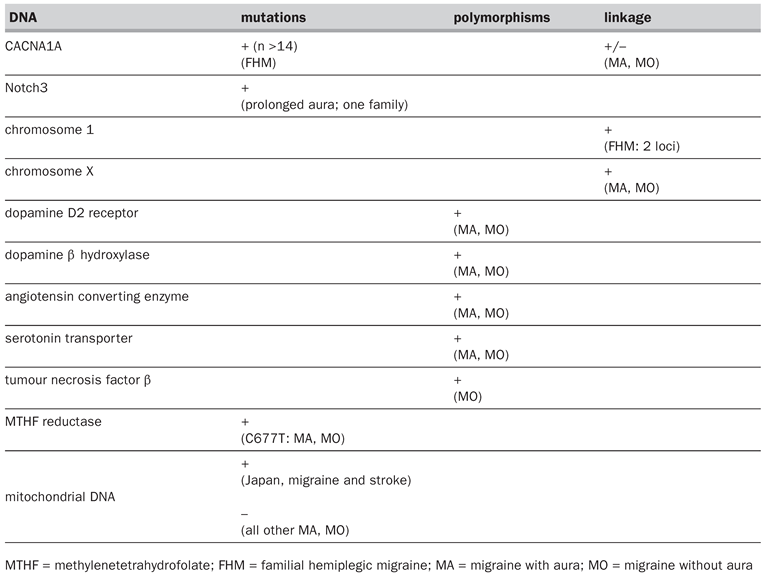
Migraine frequently runs in families, but family and segregation studies have produced conflicting results with respect to the mode of inheritance [2,3,4,5]. Up to now, the only known monogenic subtype of migraine is familial hemiplegic migraine (FHM). The more common migraine phenotypes appear to be complex genetic disorders, where additive genetic effects (susceptibility genes) and environmental factors are interrelated [6]. The weight of genetic factors seems to be more pronounced in migraine with aura than in migraine without aura [7]. Some studies suggest different liability loci for migraine headache and aura [7]. For an overview on mutations, polymorphisms and linkage, which are discussed below, see table 1.
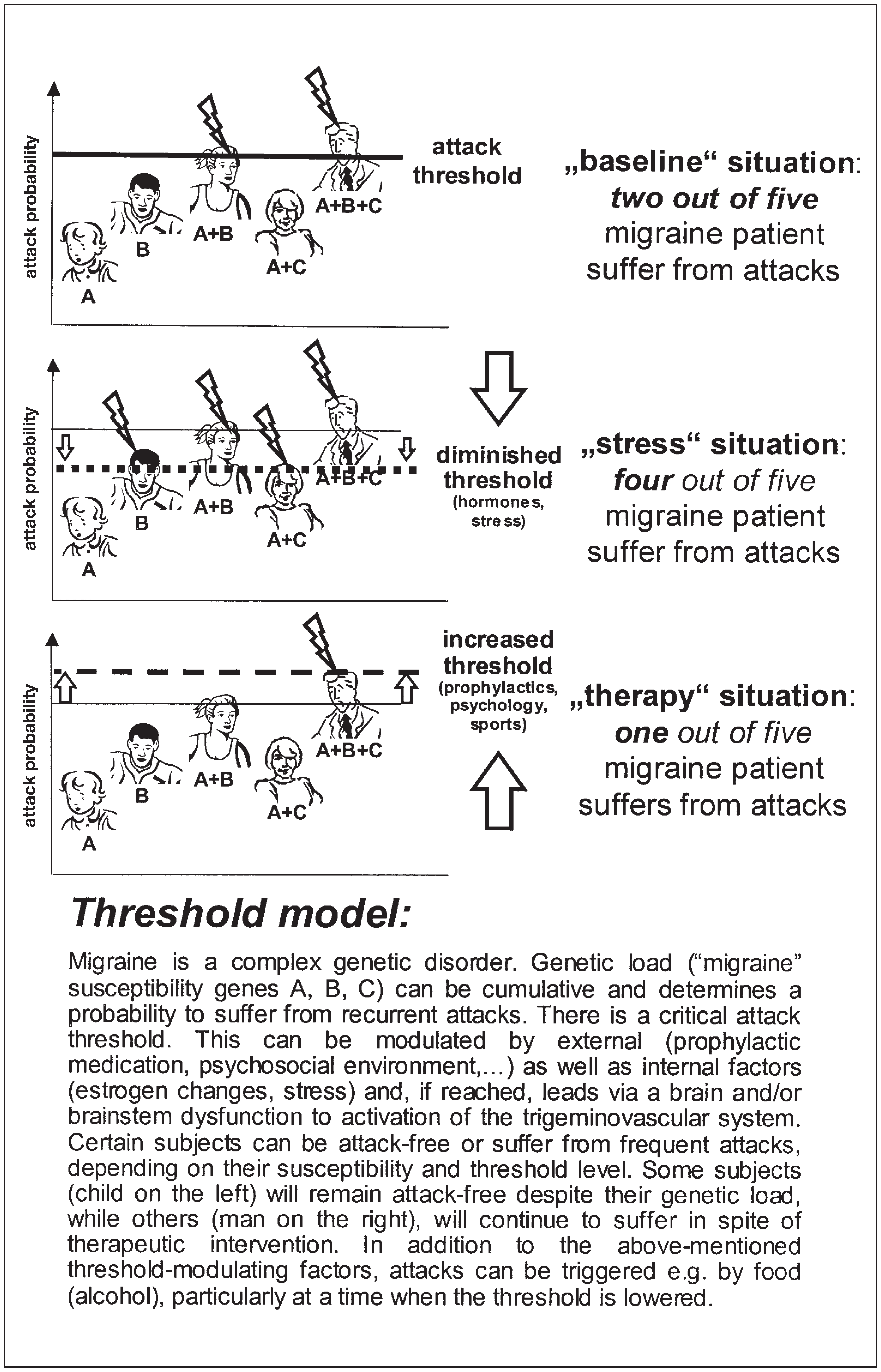
Migraine is characterised by recurrent attacks. Genetic load can be seen as determining on the one hand a critical migraine threshold that is modulated by external as well as internal factors and, if reached, leads via a brain and/or brainstem dysfunction to an attack (see fig. 1). On the other hand, genetic load may be responsible for interictal nervous system dysfunction that can produce subtle subclinical signs.
Familial hemiplegic migraine – a monogenic subtype of migraine with aura and allelic channelopathies
Phenotype
Familial hemiplegic migraine (FHM) is a rare autosomal dominant subtype of migraine with aura (IHS Headache Classification 1988). FHM patients have attacks of migraine which are associated with hemiparesis. Typically, the hemiparetic aura persists up to several days, much longer than the 20 to 30 minutes expected for the more common aura forms. In addition, some FHM families are associated with ictal and/or progressive (interictal) cerebellar ataxia. Patients with familial hemiplegic migraine and their family members may also have attacks of “non-hemiplegic” migraine. On this background it is plausible that familial hemiplegic migraine is most likely part of the migraine spectrum, and that genes involved in familial hemiplegic migraine are candidate genes for “nonhemiplegic” migraine, especially migraine with prolonged aura (i.e. longer than 60 min) and migraine associated with vestibulo-cerebellar symptoms [8].
Figure 1.
A threshold model of migraine.
FHM mutations in the gene CACNA1A
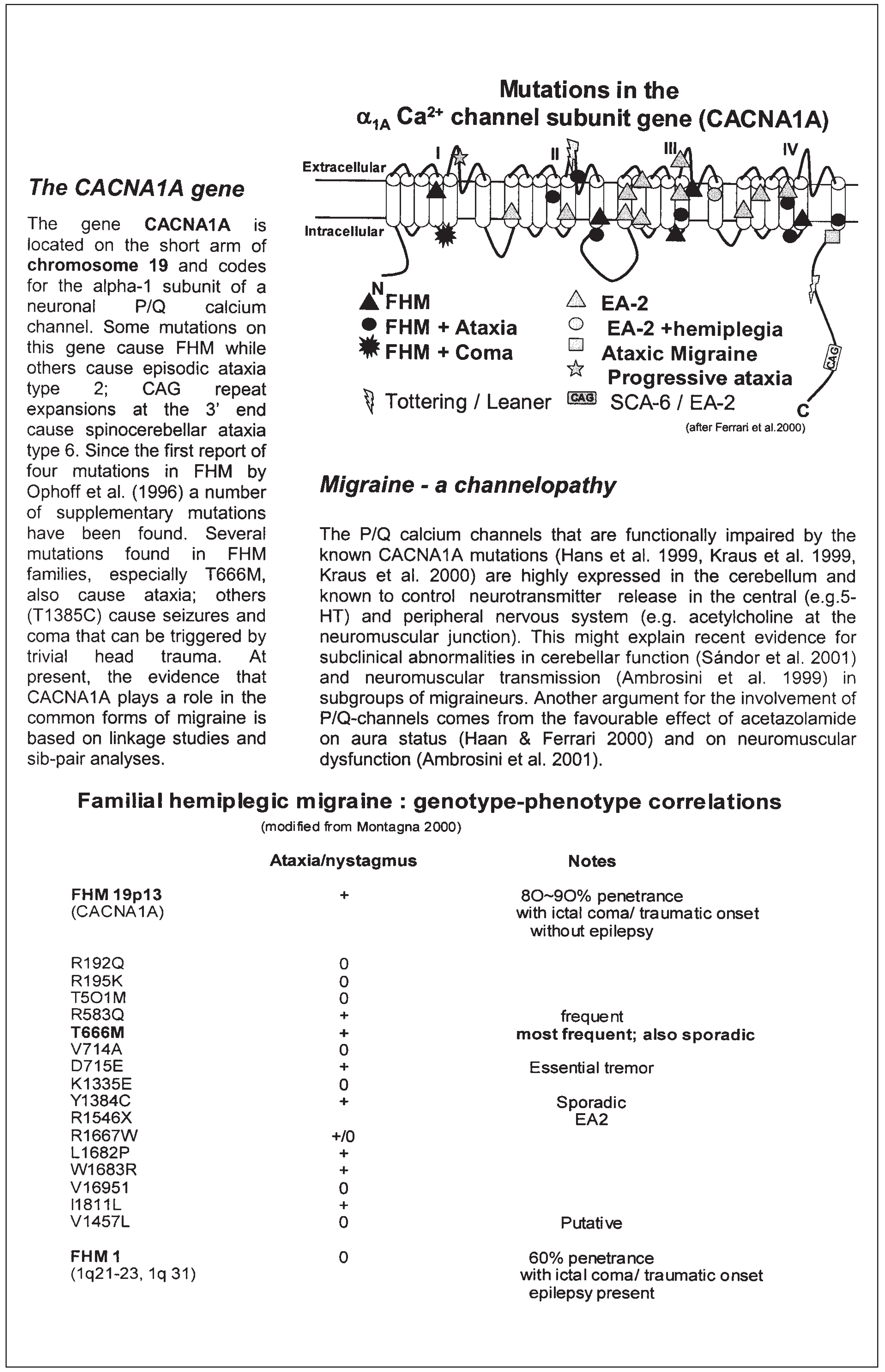
The calcium channel gene CACNA1A is located on the short arm of chromosome 19 and codes for the alpha-1 subunit of a neuronal P/Q Ca2+ channel. Some mutations on this gene cause familial hemiplegic migraine while others cause episodic ataxia type 2 (EA-2); CAG repeat expansions at the 3’ end cause spinocerebellar ataxia type 6. Since the first report of 4 mutations in familial hemiplegic migraine [7] at least 13 supplementary mutations have been found (cf. fig. 2; e.g. [9,10,11]). Several of those mutations, especially T666M, also cause ataxia [12]; others (T1385C) cause seizures and coma that can be triggered by trivial head trauma [13]. At present, the evidence that CACNA1A plays a role in the common forms of migraine is based on linkage studies and sib-pair analyses [14,15].
Functional consequences of these mutations
Six functional subclasses of Ca2+ channels have been defined by electrophysiological and pharmacological criteria. They fall into two major categories: low-voltage activated (T type) and high-voltage activated channels (L, N, P, Q, R type) [16]. Ca2+ channels are multiple-subunit complexes composed of a major transmembrane α1 unit and smaller auxiliary polypeptides which include a disulphide-linked α2 subunit and the β subunit. The α1A subunit, which is encoded by the CACNA1A gene, is the most important component of P and Q type channels; it acts as a voltage sensor and forms the ion-conducting pore [17].
In general, familial hemiplegic migraine is caused by missense mutations and episodic ataxia type 2 by truncating mutations in CACNA1A, but in some families these clinical phenotypes may overlap (e.g. [10], for review [18]).
The functional consequences of several of the FHM mutations were characterised in vitro. They were found to be rather complex: some mutations produced a gain of function, others a loss of functions and again others functional instability [19,20,21]. It therefore appears that mutations producing different channel dysfunctions may nonetheless cause similar clinical phenotypes.
In the cerebellum P/Q Ca2+ channels are highly expressed [22,23] and known to control neurotransmitter release in the central (e.g. serotonin, [24]) and peripheral nervous system (e.g. acetylcholine at the neuromuscular junction [25,26]).
Possible involvement of CACNA1A in non-hemiplegic migraine
Linkage studies
Linkage and sib-pair analyses suggest that the CACNA1A gene region 19p13 is also involved in the common forms of migraine with and without aura [15,27] although significant linkage was not found in one study [28].
Interictal nervous system dysfunction in migraineurs
Subclinical dysfunction of the cerebellum and of neuromuscular transmission
In addition to the direct evidence from studies of genetic epidemiology there is indirect neurophysiological evidence suggesting an involvement of Ca2+-channel genes in the common types of migraine. Mutations in the CACNA1A gene can cause familial hemiplegic migraine and/or cere-bellar ataxia. As P/Q Ca2+ channels are highly expressed in the cerebellum, we used a pointing paradigm and an infra-red opto-electronic tracking system to search for an impairment of motor control in migraineurs and found subclinical hypermetria as well as other subtle cerebellar signs in the common forms of migraine [29]. These cerebellar signs were more pronounced in migraine with than without aura and could reflect subtle dysfunctioning of genetically abnormal Ca2+ chan-nels in a CNS location which at first sight is not involved per se in migraine pathogenesis. It is of interest, however, that during and between attacks, vertigo, dizziness and episodes of dysequilibrium are frequent in migraineurs, especially in those suffering from migraine with aura [8,30].That the abnormalities are more pronounced in migraine with than in migraine without aura is compatible with the stronger genetic influence in the former migraine type [31,32].
Figure 2.
The CACNA1A gene, familial hemiplegic migraine and migraine as a channelopathy.
An example for a subclinical alteration in the peripheral nervous system of migraineurs seems to be the neuromuscular transmission abnormality recently found in a subgroup of migraine with aura patients [33]. A similar abnormality was described also in tottering mice, a CACNA1A mutant ([34], vide infra). P/Q Ca2+ channels are known indeed to control stimulation-induced acetylcholine release at the motor axon terminal [26,27].
No mutations have been identified up to now in the common forms of migraine but the Ca2++ channels could be functionally impaired because of alterations in channel kinetics due to more subtle genetic changes such as gene polymorphisms.A correlation between a CACNA1A single nucleotide polymorphism and neuromuscular transmission assessed by single fibre EMG was recently described in migraine with aura patients [32,35]. Another argument for the involvement of P/Q channels comes from the favourable effect of acetazolamide on neuromuscular dysfunction.
Possible involvement of the cortex
During the headache-free interval, an abnormal functioning of the migrainous brain can be demon-strated by psychophysical, neurophysiological and metabolic studies. Sensitivity to environmental stimuli is enhanced interictally in migraineurs [36]. On psychophysical tests of visual functions responses differ from normal in migraine patients who are particularly intolerant to certain visual patterns (see review in [37]).
Neurophysiological methods show that cortical information processing in migraineurs is characterised by a deficient habituation during repetition of the stimulation [38]. This has been demonstrated for event-related [39,40,41] and visual evoked potentials [42,43,44]. Moreover, intensity dependence of auditory evoked cortical potentials is increased in migraine patients compared to normal controls [45,46]. During prolonged visual stimulation we found less habituation-like behaviour of the BOLD signal in migraine with aura patients, confirming with the method of functional MRI a difference in cortical information processing (Sándor unpublished observations).
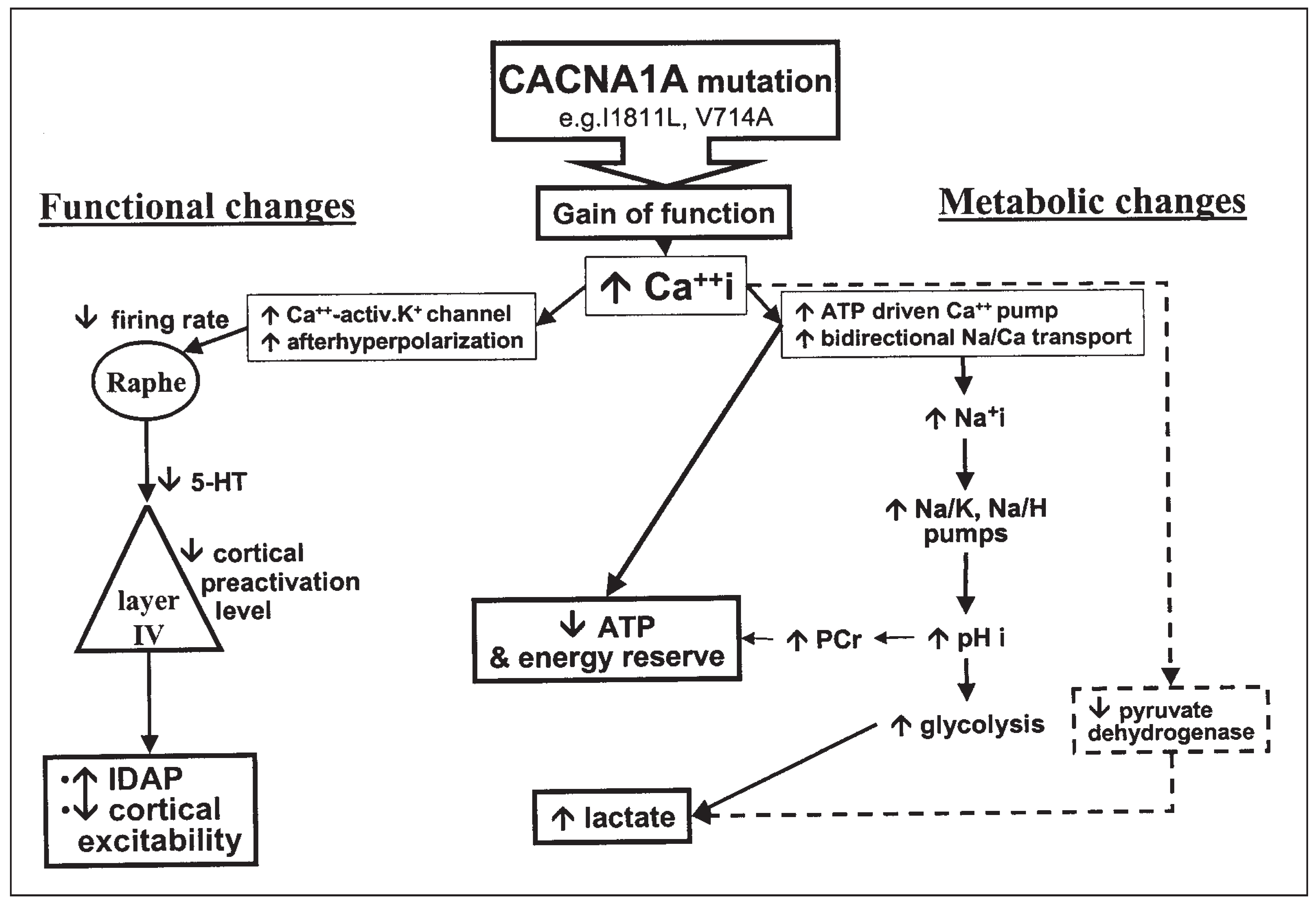
The precise cause for this cortical dysfunction is not known. However, it is likely to be genetically determined as shown by two independent studies [47,48] and may represent an “endophenotypic vulnerability marker”. It is likely on the one hand that an abnormal function of P/Q Ca2+ channels can change neuronal excitability and firing rate via changes in intracellular Ca2+ levels. Since the excitability of sensory cortices is known to be strongly modulated by subcortical aminergic neurons, a decreased firing rate of the latter might be responsible for the observed electrophysiological abnormalities (see fig. 3). On the other hand, a less obvious consequence of dysfunctioning Ca2+ channels has recently been demonstrated with NMR spectroscopy in episodic ataxia type 2: an increase in intracellular lactate levels and pH suggesting a disturbed mitochondrial metabolism (fig. 3) [49]. During prolonged visual stimulation in patients suffering from migraine with aura, we have found with functional magnetic resonance spectroscopy (MRS) an abnormal increase in lactate levels in the visual cortices which may be the metabolic consequence of the electrophysiological dishabituation or of a decreased mitochondrial energy reserve, or of both.
Mouse models of mutations in the FHM gene CACNA1A
There are three main mouse mutants that can serve as a model for human diseases with CACNA1A mutations: tottering, leaner and rolling Nagoya [50,51,52].The recessive tottering mice that have been studied extensively as models for human epilepsy [53] carry a missense mutation that is very similar to one of the FHM mutations, and most likely affects the pore function of the P/Q-type Ca2+ channel. It results in intermittent convulsions similar to human absence epilepsy, motor seizures, and mild ataxia. The more severely affected leaner mouse is associated with a mutation producing an aberrant intracellular terminus and resembling the mutations found in two EA-2 families. The leaner mouse suffers from absence-like (but not motor) seizures, is severely ataxic and typically dies early in life. Purkinje- and granule cell loss throughout the anterior cerebellum and reduced cerebellar size are found in this mutant. For the third mouse strain, the rolling Nagoya, which presents an intermediate phenotype, no mutation has yet been identified. Motor seizures do not occur, the ataxia is more severe than in the tottering mutant, and these mice have a normal life span [50].
It was shown that tottering mice have a modified threshold for cortical spreading depression [54], which is a phenomenon most likely involved in the pathophysiology of migraine aura. In the tottering mouse a cortical proliferation of noradrenergic axons from the locus coeruleus is considered to be involved in the generation of seizures [53]. Interestingly, positron emission tomography studies suggested brain stem structures in the region of the locus coeruleus and the dorsal raphe nucleus to be of pathophysiological importance in migraine attacks [55]. As mentioned above, tottering mice have clear abnormalities of acetylcholine release at the motor axon terminal [34]. In addition to serving as model for epilepsy and ataxia, the tottering mouse may therefore serve also as a channelopathy model for migraine.
Figure 3.
Putative functional and metabolic consequences of mutations in the α1A Ca2+ channel subunit gene (CACNA1A).
Other loci for familial hemiplegic migraine
Chromosomes 1 and X
While approximately 50% of FHM families have been assigned to chromosome 19p13 [56,57], about one third of families are not linked to chromosome 19, but to a yet to be identified gene on chromosome 1 [12,58,59] or to other undetermined genetic loci. Further analysis has to disclose whether chromosome 1q harbours one or two FHM genes. Some FHM families cannot be linked to chromosomes 19 or 1. Therefore, at least a third gene must be involved [58]. The X chromosome might be a candidate as in a study of familial typical migraine significant linkage to chromosome Xq24-28 was found [60].
Polymorphisms
The prevalence of various gene polymorphisms may be higher in migraineurs than in controls. This was reported for the dopamine D2 receptor [61], the angiotensin converting enzyme [62], the serotonin transporter [63], the dopamine b hydroxylase [64] and the TNFb genes [65]. The latter may be related to the decreased frequency of HLA Class II DR2 antigen reported in migraine without aura [66]. The role played by these various polymorphisms remains to be determined; some of them may not be specific to migraine, but they could increase susceptibility to the disorder and induce endophenotypic vulnerability markers (see above). A recent study in Japanese migraineurs showed a higher incidence of the homozygous C677T mutation of the methylenetetrahydrofolate reductase gene, which is associated with increased homocysteine levels and thrombosis. No evidence was found for an allelic association with migraine of a polymorphism in the inducible nitric oxide synthase gene [64], which may be relevant for the putative role of NO in migraine pathogenesis.
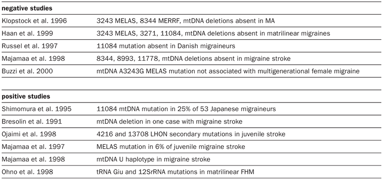
Table 2.
mtDNA studies in migraine (modified from Montagna P. Molecular genetics of migraine headaches: a review. Cephalalgia 2000, 20, 3–14).
Other conditions including migraine as a syndrome
CADASIL
Cerebral autosomal dominant arteriopathy with subcortical infarcts and leukoencephalopathy (CADASIL) is a disease with a multifaceted symptomatology, including recurrent subcortical ischaemic strokes, progressive vascular dementia, and mood disorders [67]. Remarkably, migraine with aura occurs in many patients with CADASIL [67,68,69]. Typical FHM attacks have been described in one CADASIL family [70] and migraine with white matter lesions resembling typical CADASIL on MRI in another family linked to the CADASIL locus [67]. A Notch3 gene mutation was identified to be cause of CADASIL [71]. It is not clear whether in CADASIL migraine attacks are directly related to the genetic abnormality or secondary to the vascular changes. Up to now, no further genetic data are available on the families, in which CADASIL occurs together with FHM-like migraine attacks. Interestingly, a mutation in the Notch3 gene was recently found in an Italian family affected by migraine with prolonged aura but no other neurological deficits [72].
Migraine and mitochondrial function
Metabolic studies using MRS have shown a low mitochondrial phosphorylation potential in the brain and muscles of migraineurs (fig. 3) [73,74,75]. Although mitochondrial DNA mutations were reported in some migraineurs with stroke-like epi-sodes [76,77,78,79], none of the known mitochondrial mutations was found up to now in the common forms of migraine [80,81,82]. Mitochondrial energy metabolism can, however, be impaired by Ca2+-channel dysfunction (see above and fig. 3) [83] and by decreased magnesium levels both of which may be involved in migraine [84,85,86]. It has been hypothesised that the conjunction of a decreased mitochondrial energy reserve and a deficit in habituation of cortical information processing, known to protect against overstimulation and lactate accumulation, might lead to activation of the major pain-signalling system of the head, the trigeminovascular system [41].
Preliminary results using functional MRS during prolonged visual stimulation (vide supra) suggest that the habituation deficit might suffice to produce an abnormal increase in cortical lactate levels. It is still to be determined whether the mitochondrial abnormality is an independent pathophysiological component or rather a consequence of other functional deficits.
Acknowledgments
We thank Mrs Ulli Sänger for excellent technical assistance.
References
- Russell, M.B.; Rasmussen, B.K.; Thorvaldsen, P.; Olesen, J. Prevalence and sex-ratio of the subtypes of migraine. Int J Epidemiol 1995, 24, 612–618. [Google Scholar] [CrossRef] [Scilit] [PubMed]
- Mochi, M.; et al. Testing models for genetic determination in migraine. Cephalalgia 1993, 13, 389–394. [Google Scholar] [CrossRef] [Scilit]
- Russell, M.B.; Olesen, J. The genetics of migraine without aura and migraine with aura. Cephalalgia 1993, 13, 245–248. [Google Scholar] [CrossRef] [Scilit]
- Stewart, W.F.; Staffa, J.; Lipton, R.B.; Ottman, R. Familial risk of migraine: a population-based study. Ann Neurol 1997, 41, 166–172. [Google Scholar] [CrossRef] [Scilit]
- Haan, J.; Terwindt, G.M.; Ferrari, M.D. Genetics of migraine. Neurol Clin 1997, 15, 43–60. [Google Scholar] [CrossRef] [Scilit]
- Svenson, L.W.; Cwik, V.A.; Martin, W.R. The prevalence of motor neurone disease in the Province of Alberta. Can J Neurol Sci 1999, 26, 119–122. [Google Scholar]
- Cicerone, K.D.; et al. Neuropsychological rehabilitation of mild traumatic brain injury. Brain Inj 1996, 10, 277–286. [Google Scholar] [CrossRef] [Scilit]
- Neuhauser, H.; Leopold, M.; von Brevern, M.; Arnold, G.; Lempert, T. The interrelations of migraine, vertigo, and migrainous vertigo. Neurology 2001, 56, 436–441. [Google Scholar] [CrossRef] [Scilit] [PubMed]
- Friend, K.L.; et al. Detection of a novel missense mutation and second recurrent mutation in the CACNA1A gene in individuals with EA-2 and FHM. Hum Genet 1999, 105, 261–265. [Google Scholar] [CrossRef] [Scilit] [PubMed]
- Jen, J.; et al. A novel nonsense mutation in CACNA1A causes episodic ataxia and hemiplegia. Neurology 1999, 53, 34–37. [Google Scholar] [CrossRef] [Scilit]
- Battistini, S.; et al. A new CACNA1A gene mutation in acetazolamide-responsive familial hemiplegic migraine and ataxia. Neurology 1999, 53, 38–43. [Google Scholar] [CrossRef] [Scilit]
- Echenne, B.; et al. Recurrent episodes of coma: an unusual phenotype of familial hemiplegic migraine with linkage to chromosome 1. Neuropediatrics 1999, 30, 214–217. [Google Scholar] [CrossRef] [Scilit] [PubMed]
- Vahedi, K.; et al. CACNA1A gene de novo mutation causing hemiplegic migraine, coma, and cerebellar atrophy. Neurology 2000, 55, 1040–1042. [Google Scholar] [CrossRef] [Scilit] [PubMed]
- Bleiberg, J.; Halpern, E.L.; Reeves, D.; Daniel, J.C. Future directions for the neuropsychological assessment of sports concussion. J Head Trauma Rehabil 1998, 13, 36–44. [Google Scholar] [CrossRef] [Scilit]
- Nyholt, D.R.; et al. Evidence for an X-linked genetic component in familial typical migraine. Hum Mol Genet 1998, 7, 459–463. [Google Scholar] [CrossRef] [Scilit]
- Catterall, W.A. Structure and function of voltage-gated ion channels. Annu Rev Biochem 1995, 64, 493–531. [Google Scholar] [CrossRef] [PubMed]
- Dunlap, K.; Luebke, J.I.; Turner, T.J. Exocytotic Ca2+ channels in mammalian central neurons. Trends Neurosci 1995, 18, 89–98. [Google Scholar] [CrossRef] [Scilit] [PubMed]
- Montagna, P. Molecular genetics of migraine headaches: a review. Cephalalgia 2000, 20, 3–14. [Google Scholar] [CrossRef] [Scilit]
- Hans, M.; et al. Functional consequences of mutations in the human alpha1A calcium channel subunit linked to familial hemiplegic migraine. J Neurosci 1999, 19, 1610–1619. [Google Scholar] [CrossRef] [Scilit]
- Kraus, R.L.; Sinnegger, M.J.; Glossmann, H.; Hering, S.; Striessnig, J. Familial hemiplegic migraine mutations change alpha1A Ca2+ channel kinetics. J Biol Chem 1998, 273, 5586–5590. [Google Scholar] [CrossRef] [Scilit]
- Kraus, R.L.; et al. Three new familial hemiplegic migraine mutants affect P/Q-type Ca(2+) channel kinetics. J Biol Chem 2000, 275, 9239–9243. [Google Scholar] [CrossRef] [Scilit]
- Stea, A.; et al. Localization and functional properties of a rat brain alpha1A calcium channel reflect similarities to neuron. Proc Natl Acad Sci USA 1994, 91, 10576–10580. [Google Scholar] [CrossRef] [Scilit] [PubMed]
- Randall, A.; Tsien, R.W. Pharmacological dissection of multiple types of Ca2+ channel currents in rat cerebellar granule neurons. J Neurosci 1995, 15, 2995–3012. [Google Scholar] [CrossRef] [Scilit] [PubMed]
- Craig, P.J.; et al. Distribution of the voltage-dependent calcium channel alpha(1A) subunit throughout the mature rat brain and its relationship to neurotransmitter pathways. J Comp Neurol 1998, 397, 251–267. [Google Scholar] [CrossRef]
- Katz, E.; Protti, D.A.; Ferro, P.A.; Rosato, S.; Uchitel, O.D. Effects of Ca2+ channel blocker neurotoxins on transmitter release and presynaptic currents at the mouse neuromuscular junction. Br J Pharmacol 1997, 121, 1531–1540. [Google Scholar] [CrossRef] [Scilit] [PubMed]
- Westenbroek, R.E.; Hoskins, L.; Catterall, W.A. Localization of Ca2+ channel subtypes on rat spinal motor neurons, interneurons, and nerve terminals. J Neurosci 1998, 18, 6319–6330. [Google Scholar] [CrossRef] [Scilit]
- May, A.; et al. Familial hemiplegic migraine locus on 19p13 is involved in the common forms of migraine with and without aura. Hum Genet 1995, 96, 604–608. [Google Scholar] [CrossRef] [Scilit]
- Hovatta, I.; Kallela, M.; Farkkila, M.; Peltonen, L. Familial migraine: exclusion of the susceptibility gene from the reported locus of familial hemiplegic migraine on 19p. Genomics 1994, 23, 707–709. [Google Scholar] [CrossRef] [Scilit]
- 29a Sándor, P.S.; Mascia, A.; Seidel, L.; de Pasqua, V.; Schoenen, J. Subclinical cerebellar impairment in the common types of migraine: a three-dimensional analysis of reaching movements. Ann Neurol 2001, 49, 668–672. [Google Scholar] [CrossRef]
- Baloh, R.W. Neurotology of migraine. Headache 1997, 37, 615–621. [Google Scholar] [CrossRef] [Scilit]
- Gervil, M.; Ulrich, V.; Kaprio, J.; Olesen, J.; Russell, M.B. The relative role of genetic and environmental factors in migraine without aura. Neurology 1999, 53, 995–999. [Google Scholar] [CrossRef] [Scilit] [PubMed]
- Ulrich, V.; Gervil, M.; Kyvik, K.O.; Olesen, J.; Russell, M.B. Evidence of a genetic factor in migraine with aura: a population-based Danish twin study. Ann Neurol 1999, 45, 242–246. [Google Scholar] [CrossRef] [Scilit]
- Ambrosini, A.; de Noordhout, A.M.; Alagona, G.; Dalpozzo, F.; Schoenen, J. Impairment of neuromuscular transmission in a subgroup of migraine patients. Neurosci Lett 1999, 276, 201–203. [Google Scholar] [CrossRef] [Scilit]
- Plomp, J.J.; et al. Abnormal transmitter release at neuromuscular junctions of mice carrying the tottering alpha(1A) Ca(2+) channel mutation. Brain 2000, 123, 463–471. [Google Scholar] [CrossRef] [Scilit]
- Afra, J.; Ambrosini, A.; Genicot, R.; Albert, A.; Schoenen, J. Influence of colors on habituation of visual evoked poten-tials in patients with migraine with aura and in healthy volunteers. Headache 2000, 40, 36–40. [Google Scholar] [CrossRef] [Scilit]
- Hay, K.M.; Mortimer, M.J.; Barker, D.C.; Debney, L.M.; Good, P.A. 1044 women with migraine: the effect of environmental stimuli. Headache 1994, 34, 166–168. [Google Scholar] [CrossRef] [Scilit]
- Afra, J.; Mascia, A.; Gerard, P.; Maertens de Noordhout, A.; Schoenen, J. Interictal cortical excitability in migraine: a study using transcranial magnetic stimulation of motor and visual cortices. Ann Neurol 1998, 44, 209–215. [Google Scholar] [CrossRef] [Scilit]
- Bohnen, N.; Jolles, J.; Twijnstra, A. Neuropsychological deficits in patients with persistent symptoms six months after mild head injury. J Neurosurg 1992, 30, 692–695. [Google Scholar]
- Maertens de Noordhout, A.; Timsit-Berthier, M.; Timsit, M.; Schoenen, J. Contingent negative variation in headache. Ann Neurol 1986, 19, 78–80. [Google Scholar] [CrossRef] [Scilit] [PubMed]
- Kropp, P.; Gerber, W.D. Is increased amplitude of contingent negative variation in migraine due to cortical hyperactivity or to reduced habituation? Cephalalgia 1993, 13, 37–41. [Google Scholar] [CrossRef] [Scilit] [PubMed]
- Schwartz, R.B.; Komaroff, A.L.; Garada, B.M.; Gleit, M.; Doolittle, T.H.; Bates, D.W.; et al. SPECT imaging of the brain: comparison of findings in patients with chronic fatigue syndrome, AIDS dementia complex, and major unipolar depression. Am J Roentgenol 1994, 162, 943–951. [Google Scholar] [CrossRef] [Scilit] [PubMed]
- Schoenen, J. Cortical electrophysiology in migraine and possible pathogenetic implications. Clin Neurosci 1998, 5, 10–17. [Google Scholar] [PubMed]
- Bird, N.; MacGregor, E.A.; Wilkinson, M.I. Ice cream headache-site, duration, and relationship to migraine. Headache 1992, 32, 35–38. [Google Scholar] [CrossRef] [Scilit]
- Lahat, E.; Barr, J.; Barzilai, A.; Cohen, H.; Berkovitch, M. Visual evoked potentials in the diagnosis of headache before 5 years of age. Eur J Pediatr 1999, 158, 892–895. [Google Scholar] [CrossRef] [Scilit]
- Wang, W.; Timsit-Berthier, M.; Schoenen, J. Intensity dependence of auditory evoked potentials is pronounced in migraine: an indication of cortical potentiation and low serotonergic neurotransmission? Neurology 1996, 46, 1404–1409. [Google Scholar] [CrossRef] [Scilit]
- Siniatchkin, M.; Kropp, P.; Neumann, M.; Gerber, W.; Stephani, U. Intensity dependence of auditory evoked cortical potentials in migraine families. Pain 2000, 85, 247–254. [Google Scholar] [CrossRef] [Scilit]
- Sándor, P.S.; Afra, J.; Proietti-Cecchini, A.; Albert, A.; Schoenen, J. Familial influences on cortical evoked potentials in migraine. Neuroreport 1999, 10, 1235–1238. [Google Scholar] [CrossRef] [Scilit]
- Kropp, P.; et al. Slow cortical potentials in migraine: a comparison of adults and children. Cephalalgia 1999, 19, 60–64. [Google Scholar] [CrossRef] [Scilit] [PubMed]
- Sappey-Marinier, D.; Vighetto, A.; Peyron, R.; Broussolle, E.; Bonmartin, A. Phosphorus and proton magnetic resonance spectroscopy in episodic ataxia type 2. Ann Neurol 1999, 46, 256–259. [Google Scholar] [CrossRef] [Scilit]
- Fletcher, C.F.; et al. Absence epilepsy in tottering mutant mice is associated with calcium channel defects. Cell 1996, 87, 607–617. [Google Scholar] [CrossRef] [Scilit]
- Hess, E.J. Migraines in mice? Cell 1996, 87, 1149–1151. [Google Scholar] [CrossRef] [Scilit]
- Doyle, J.; Ren, X.; Lennon, G.; Stubbs, L. Mutations in the Cacnl1a4 calcium channel gene are associated with seizures, cerebellar degeneration, and ataxia in tottering and leaner mutant mice. Mamm Genome 1997, 8, 113–120. [Google Scholar] [CrossRef] [Scilit]
- Kostopoulos, G.K. The tottering mouse: a critical review of its usefulness in the study of the neuronal mechanisms underlying epilepsy. J Neural Transm 1992, 35, 21–36. [Google Scholar]
- Ayata, C.; Shimizu-Sasamata, M.; Lo, E.H.; Noebels, J.L.; Moskowitz, M.A. Impaired neurotransmitter release and elevated threshold for cortical spreading depression in mice with mutations in the alpha1A subunit of P/Q type calcium channels. Neuroscience 2000, 95, 639–645. [Google Scholar] [CrossRef] [Scilit] [PubMed]
- Weiller, C.; et al. Brain stem activation in spontaneous human migraine attacks. Nat Med 1995, 1, 658–660. [Google Scholar] [CrossRef] [Scilit] [PubMed]
- Joutel, A.; et al. Genetic heterogeneity of familial hemiplegic migraine. Am J Hum Genet 1994, 55, 1166–1172. [Google Scholar]
- Ophoff, R.A.; et al. Genetic heterogeneity of familial hemiplegic migraine. Genomics 1994, 22, 21–26. [Google Scholar] [CrossRef] [Scilit]
- Ducros, A.; et al. Mapping of a second locus for familial hemiplegic migraine to 1q21-q23 and evidence of further heterogeneity. Ann Neurol 1997, 42, 885–890. [Google Scholar] [CrossRef] [Scilit]
- Gardner, K.; Barmada, M.M.; Ptacek, L.J.; Hoffman, E.P. A new locus for hemiplegic migraine maps to chromosome 1q31. Neurology 1997, 49, 1231–1238. [Google Scholar] [CrossRef] [Scilit] [PubMed]
- Nyholt, D.R.; Curtain, R.P.; Griffiths, L.R. Familial typical migraine: significant linkage and localization of a gene to Xq24-28. Hum Genet 2000, 107, 18–23. [Google Scholar]
- Del Zompo, M.; et al. Association between dopamine receptor genes and migraine without aura in a Sardinian sample. Neurology 1998, 51, 781–786. [Google Scholar] [CrossRef] [Scilit] [PubMed]
- Paterna, S.; et al. Angiotensin-converting enzyme gene deletion polymorphism determines an increase in frequency of migraine attacks in patients suffering from migraine without aura. Eur Neurol 2000, 43, 133–136. [Google Scholar] [CrossRef] [Scilit] [PubMed]
- Ogilvie, A.D.; et al. Altered allelic distributions of the serotonin transporter gene in migraine without aura and migraine with aura. Cephalalgia 1998, 18, 23–26. [Google Scholar] [CrossRef] [Scilit]
- Lea, R.A.; et al. Evidence for allelic association of the dopamine beta-hydroxylase gene (DBH) with susceptibility to typical migraine. Neurogenetics 2000, 3, 35–40. [Google Scholar] [CrossRef] [Scilit]
- Martelletti, P. Proinflammatory pathways in cervicogenic headache. Clin Exp Rheumatol 2000, 18, S33–S38. [Google Scholar]
- Martelletti, P.; et al. Chromosome 6p-encoded HLA-DR2 determination discriminates migraine without aura from migraine with aura. Hum Immunol 1999, 60, 69–74. [Google Scholar] [CrossRef] [Scilit] [PubMed]
- Chabriat, H.; et al. Clinical spectrum of CADASIL: a study of 7 families. Cerebral autosomal dominant arteriopathy with subcortical infarcts and leukoencephalopathy. Lancet 1995, 346, 934–939. [Google Scholar] [CrossRef] [Scilit]
- Verin, M.; et al. New phenotype of the cerebral autosomal dominant arteriopathy mapped to chromosome 19: migraine as the prominent clinical feature. J Neurol Neurosurg Psychiatry 1995, 59, 579–585. [Google Scholar] [CrossRef] [Scilit]
- Jung, H.H.; et al. Cerebral autosomal dominant arteriopathy with subcortical infarcts and leukoencephalopathy: a clinicopathological and genetic study of a Swiss family. J Neurol Neurosurg Psychiatry 1995, 59, 138–143. [Google Scholar] [CrossRef] [Scilit]
- Hutchinson, M.; et al. Familial hemiplegic migraine and autosomal dominant arteriopathy with leukoencephalopathy (CADASIL). Ann Neurol 1995, 38, 817–824. [Google Scholar] [CrossRef] [Scilit] [PubMed]
- Joutel, A.; et al. Notch3 mutations in CADASIL, a hereditary adult-onset condition causing stroke and dementia. Nature 1996, 383, 707–710. [Google Scholar] [CrossRef] [Scilit]
- Ceroni, M.; et al. Migraine with aura and white matter abnormalities: Notch3 mutation. Neurology 2000, 54, 1869–1871. [Google Scholar] [CrossRef] [Scilit]
- Welch, K.M.; Levine, S.R.; D’Andrea, G.; Schultz, L.R.; Helpern, J.A. Preliminary observations on brain energy metabolism in migraine studied by in vivo phosphorus 31 NMR spectroscopy. Neurology 1989, 39, 538–541. [Google Scholar] [CrossRef] [Scilit]
- Barbiroli, B.; Funicello, R.; Ferlini, A.; Montagna, P.; Zaniol, P. Muscle energy metabolism in female DMD/BMD carriers: a 31P–MR spectroscopy study. Muscle Nerve 1992, 15, 344–348. [Google Scholar] [CrossRef] [Scilit]
- Montagna, P.; et al. 31P-magnetic resonance spectroscopy in migraine without aura. Neurology 1994, 44, 666–669. [Google Scholar] [CrossRef] [Scilit]
- Bresolin, N.; et al. Muscle mitochondrial DNA deletion and 31P-NMR spectroscopy alterations in a migraine patient. J Neurol Sci 1991, 104, 182–189. [Google Scholar] [CrossRef] [Scilit] [PubMed]
- Sano, M.; et al. The T-C(8356) mitochondrial DNA mutation in a Japanese family. J Neurol 1996, 243, 441–444. [Google Scholar] [CrossRef] [Scilit] [PubMed]
- Ojaimi, J.; Katsabanis, S.; Bower, S.; Quigley, A.; Byrne, E. Mitochondrial DNA in stroke and migraine with aura. Cerebrovasc Dis 1998, 8, 102–106. [Google Scholar] [CrossRef] [Scilit] [PubMed]
- Majamaa, K.; Finnila, S.; Turkka, J.; Hassinen, I.E. Mitochondrial DNA haplogroup U as a risk factor for occipital stroke in migraine. Lancet 1998, 352, 455–456. [Google Scholar] [CrossRef] [Scilit]
- Klopstock, T.; et al. Mitochondrial DNA in migraine with aura. Neurology 1996, 46, 1735–1738. [Google Scholar] [CrossRef] [Scilit]
- Haan, J.; et al. Search for mitochondrial DNA mutations in migraine subgroups. Cephalalgia 1999, 19, 20–22. [Google Scholar] [CrossRef] [Scilit]
- Buzzi, M.G.; et al. mtDNA A3243G MELAS mutation is not associated with multigenerational female migraine. Neurology 2000, 54, 1005–1007. [Google Scholar] [CrossRef] [Scilit]
- Sappey-Marinier, D.; Vighetto, A.; Peyron, R.; Broussolle, E.; Bonmartin, A. Phosphorus and proton magnetic resonance spectroscopy in episodic ataxia type 2. Ann Neurol 1999, 46, 256–259. [Google Scholar] [CrossRef] [Scilit] [PubMed]
- Ramadan, N.M.; et al. Low brain magnesium in migraine. Headache 1989, 29, 590–593. [Google Scholar] [CrossRef] [Scilit] [PubMed]
- Schoenen, J.; Sianard-Gainko, J.; Lenaerts, M. Blood magne-sium levels in migraine. Cephalalgia 1991, 11, 97–99. [Google Scholar] [CrossRef] [Scilit] [PubMed]
- Mazzotta, G.; Sarchielli, P.; Alberti, A.; Gallai, V. Intracellular Mg++ concentration and electromyographical ischemic test in juvenile headache. Cephalalgia 1999, 19, 802–809. [Google Scholar] [CrossRef] [Scilit]
This is an open-access article distributed under the terms of the Creative Commons Attribution Non-Commercial No Derivatives (CC BY-NC-ND) 4.0 License.