


Summary
Pathogenesis of amyotrophic lateral sclerosis (ALS), a neurodegenerative disease, is unknown and several hypotheses are still under investigation. In order to test the occurrence of an autoimmune phenomenon in ALS (and in acquired lower motor neuron disease (LMND)), we (i) tested ALS/LMND sera on mouse central nervous system tissue sections, and (ii) passively immunized mice with ALS/LMND immunoglobulins (Ig). In in vitro and passive transfer studies, ALS/ LMND Ig (but not control Ig) labelled both neurons and astrocytes in the brain, brainstem, spinal cord and cerebellum of mice. These results emphasize the potential contribution of ALS/ LMND Ig to neuronal death process, and the role that astrocytes, that have the capacity to take part actively to immune responses, may play in this disorder.
Introduction
Amyotrophic lateral sclerosis (ALS) is a neurodegenerative disorder involving primarily upper and lower motor neurons (the latter is selectively impaired in acquired lower motor neuron disease (LMND)), and leading progressively to death within five years of onset [1]. Its pathogenesis is unknown although some recent findings have emphasized a role for neurotoxic and autoimmune factors as potential mechanisms implying neuronal death [2,3].The occurence of an autoimmune process in ALS was supported by postmortem studies [3], that showed astrocytes capacity to express major histocompatibility complex (MHC) class I and II antigens [4,5] additionally to the dramatic loss of motor neurons in anterior horns and of pyramidal cells in the IIIrd and Vth layers of the motor cortex. Cortical neurons positive for MHC class II were only rarely observed [5]. Leukocyte infiltrates, including CD8+ and/or CD4+ T cells [4,6], were as well detected in ventral horns and near corticospinal tracts. In addition, (i) some ALS patients possess immunoglobulin G (IgG)-type autoantibodies to L-type skeletal muscle voltagegated calcium (Ca2+) channels (VGCCs) inhibiting L-type Ca2+ current [7,8], (ii) ALS IgG are able to enhance Ca2+ current in a motor neuron-neuroblastoma hybrid cell line (VSC 4.1) by activating neuronal N- or P-type VGCCs, leading to cell death [9,10] and suggesting that ALS IgG may recognize epitopes common to the different VGCCs, and (iii) a subgroup of ALS patients, predominantly patients with lower motor neuron signs, present with IgM and IgG anti-ganglioside antibodies (especially GM1) [11,12], although they are also found in motor neuropathy with conduction blocks and LMND [13] and anti-GM1 antibodies are suspected to be part of the normal human antibody repertoire [14] being thus expected to participate to a secondary immune reaction after exposure of damaged neural tissue in neurological diseases. These are all arguments for an autoimmune response triggering or at least being concomitant to neuronal degeneration in ALS. On the other hand, the neurotransmitter glutamate is believed to be involved in neuronal cell death in ALS [15,16], and abnormalities in the transport and metabolism of glutamate have been demonstrated in ALS postmortem material [2]. Glutamate and aspartate tissue levels are decreased in ALS brain and spinal cord, which is likely to be due to the impairment of glutamate reuptake from the extracellular space by astrocytes and/or axon terminals, and marked decrease in the high-affinity sodium-dependent glutamate transport was demonstrated using synaptosomes prepared from ALS spinal cord, motor cortex and somatosensory cortex [2]. These alterations were region-specific, and were not found in patients with Alzheimer’s or Huntington’s disease. Finally, a role of free radicals in motor neuron damage is suggested in some of the 8 to 10% of ALS patients presenting with familial ALS linked to mutations in the Cu/Zn superoxide dismutase (SOD1) gene on chromosome 21 [17]. Mutations in the SOD1 gene have also been discovered in sporadic ALS [18,19,20], underlining its potential contribution to neuronal death in sporadic ALS as well.
We searched for the presence of a potential autoimmune factor directed against (motor) neurons in ALS/LMND. Patients’ sera were tested on mouse central nervous system (CNS) tissue sections for their binding, and patients’ Ig fractions were injected into mice in the attempt to develop a mouse passive transfer model of ALS/LMND.
Patients and methods
Patients
Serum was obtained from 9 patients with typical clinical and electromyographic features of ALS (with either bulbar and peripheral or purely peripheral presentation), and from 1 case with acquired LMND. Pooled sera from patients presenting with herniated disc (n = 3) or transient ischemic attack (n = 4),with no underlying inflammation,were used as controls.Anti-GM1 antibody activity was tested in all sera by ELISA, as described by Adams et al. [21].Purified ganglioside GM1 was kindly provided by Dr. C. Chizzolini, Fidia Research Laboratories, Abano Terme, Italy.
Immunocytochemical studies
Unfixed tissue blocks (in Ames OCT) of brain and spinal cord from an adult female BALB/c mouse were snap-frozen in isopentane cooled in liquid nitrogen and stored at –80° C. Cryostat sections (6 μm) were air-dried and then fixed in acetone. Sections were stained using a double indirect immunofluorescence technique (IIF) as follows: sections were incubated at room temperature with (1) patients’ and control sera (dilution 1/104), (2) goat anti-human (Hu) IgG(H+L) and anti-Hu IgM(Fc) (Pierce, Rockford, IL) preabsorbed with 15% normal mouse serum to preclude cross-reactions, (3) rabbit anti-goat IgG(H+L) coupled to tetramethylrhodamine isothiocyanate (TRITC; Pierce), previously blocked with 15% normal mouse serum (4a) a mouse monoclonal antibody cocktail to non-phosphorylated neurofilaments (SMI 311, Affiniti, Nottingham, UK) or (4b) a mouse monoclonal anti-glial acidic protein (GFAP) antibody (Amersham, Buckinghamshire, UK), (5) horse anti-mouse IgG(H+L) conjugated to fluorescein isothiocyanate (FITC; preabsorbed with 15% normal goat and rabbit sera) (Pierce). Sections were washed after every step in phosphatebuffered saline (PBS; pH 7.3), containing 0.5% bovine serum albumine (PBS/BSA). ALS and LMND patients’ sera with anti-GM1 activity were absorbed with 10 μg of purified ganglioside GM1 before they were tested on mouse CNS sections; absorbed sera and native control sera were devoid of anti-GM1 activity (ELISA).
Passive transfer studies
Crude Ig fractions were prepared from sera by precipitation with 50% ammonium sulfate followed by extensive dialysis, and IgG content determined by laser nephelometry. Five-week-old female BALB/c mice were injected intraperitoneally (i.p.) with the equivalence of 10 mg IgG, after a prior i.p. injection of cyclophosphamide (300 mg/kg) to suppress the immune response to foreign Ig. Four animals were injected with LMND Ig for 8 to 12 days, 3 mice with ALS Ig (patient 1) for 12 days, and 3 mice with control Ig for 6 to 22 days. Two animals received no treatment. Mice were sacrificed by ether inhalation 24 to 48 hours after the last i.p. injection. Animals were bled on day 8 and on the day of sacrifice from the retro-orbital venous plexus for quantitative determination of Hu circulating IgG (radial immunodiffusion; LC-Partigen-IgG, Behringwerke, Marburg), and for anti-GM1 antibody activity (ELISA). Mice were directly dissected after sacrifice. Unfixed whole spinal cord, brainstem, cerebellum and brain were snap-frozen as described previously and stored at –80° C. Cryostat sections (6 μm) were air-dried, fixed in acetone, and stained by double IIF as described earlier (omission of step (1)). Some sections were also incubated with a lectin from Bandeiraea simplicifolia (BS)-FITC (Sigma, St Louis, MO) to identify blood vessels. Lumbar, dorsal, cervical spinal cord and one brain hemisphere were systematically examined using double IIF, and the adjacent sections stained with Toluidin Blue to identify neuronal cell types and/or anatomical structures, whereas brainstem and cerebellum were studied less extensively.
Results
Anti-GM1 activity
No anti-GM1 activity was detected in control sera used for immunocytochemical studies or for passive transfer experiments. ALS patient 1 had an anti-GM1 antibody titer of 590 (normal <20), ALS patients 2 to 9 were negative, and LMND patient had a low anti-GM1 antibody titer of 60 (likely to reflect a secondary immune process).
ALS/LMND Ig binding to normal mouse CNS sections
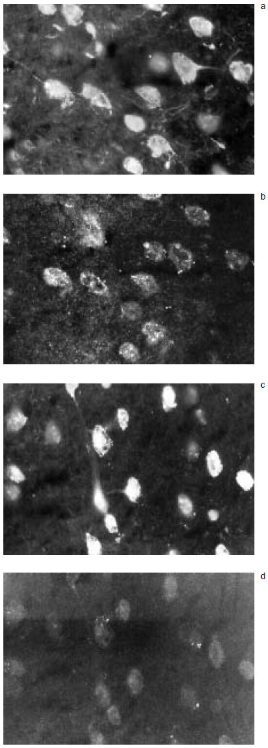
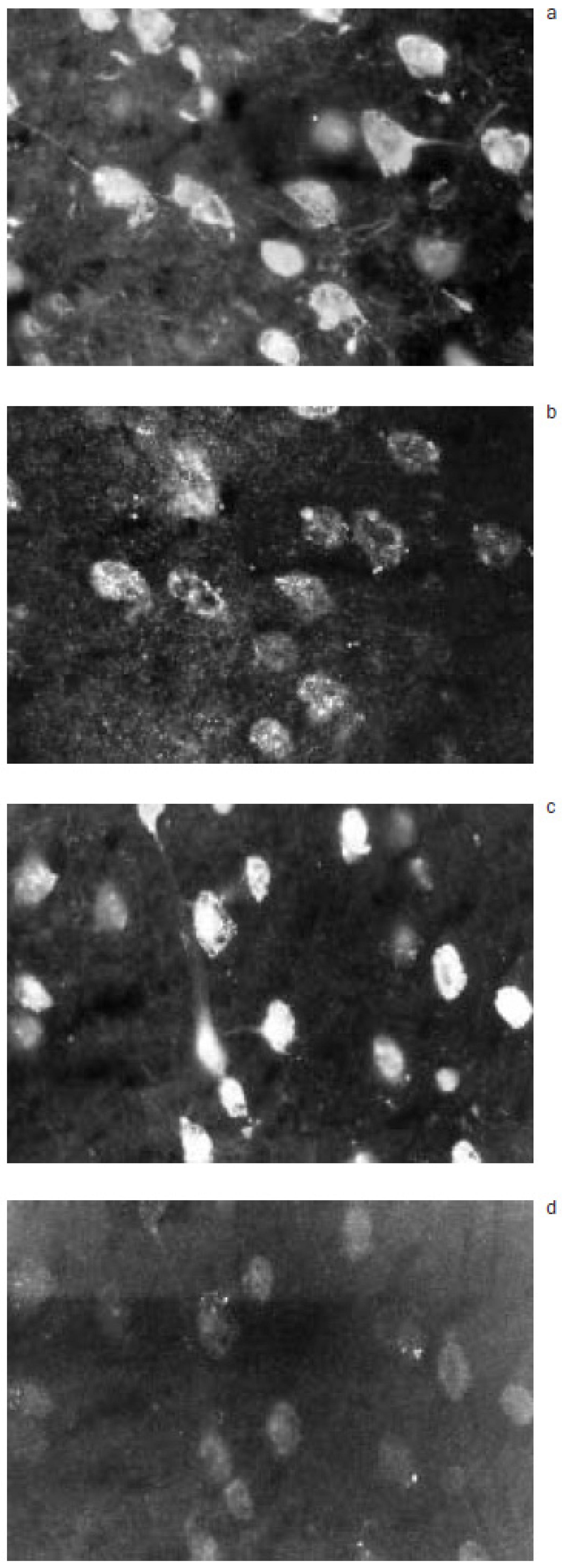
90 to 100% of neurons (SMI 311 positive cells, FITC) from normal mouse brain/spinal cord were stained for Hu Ig(G+M) (TRITC) when sections were incubated with serum from 5/9 ALS and LMND patients (serum from patient 1 and LMND patient was preabsorbed for anti-GM1 activity) (Figure 1a, b). Neuronal cell bodies were constantly positive for Hu Ig(G+M), but axons and dendrites were rarely labelled. A majority of astrocytes were also recognized by Ig(G+M) from the same 5/9 ALS and LMND patients (Figure 1c, d), however, the number and staining intensity of TRITC-positive astrocytes were inferior to those obtained for neurons. No neuron or astrocyte was TRITC positive when sections were tested with control sera (not shown).
Figure 1.
When normal mouse brain sections were incubated with serum from ALS patient 1, preabsorbed for its anti-GM1 antibody activity, most neurons (a; SMI 311-FITC) are labelled for Hu Ig(G+M) (b; TRITC), and numerous astrocytes (c; GFAPFITC) show diffuse positive staining for Hu Ig(G+M) (d; TRITC) (x 980).
Passive transfer studies
We did not detect any clinical deficit in mice injected i.p. with ALS/LMND Ig (ALS patient 1) or with control Ig. Particularly, they did not show weight loss or muscle weakness. The lack of clinical impairment may be explained by the short experimental duration. Circulating Hu IgG levels ranged from 3.8 to 7.4 mg/ml on day 8, and 3.0 to 8.5 mg/ml on day of sacrifice. No trace of Hu IgG with anti-GM1 activity was detected in mouse sera when tested at day 8 and on day of sacrifice. Control and ALS/LMND Ig(G+M) (TRITC) bound to all CNS blood vessels (endothelial cells, perivascular microglial cells) identified with BS-FITC, although Hu Ig-TRITC staining pattern was not as homogeneous as BS-FITC. Apart from this exception, CNS cells (whole spinal cord, brain, brainstem and cerebellum) from mice injected with control Ig showed no labelling for Hu Ig(G+M).
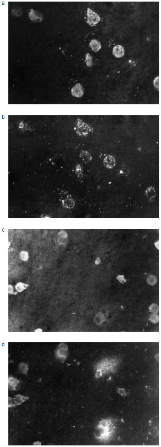
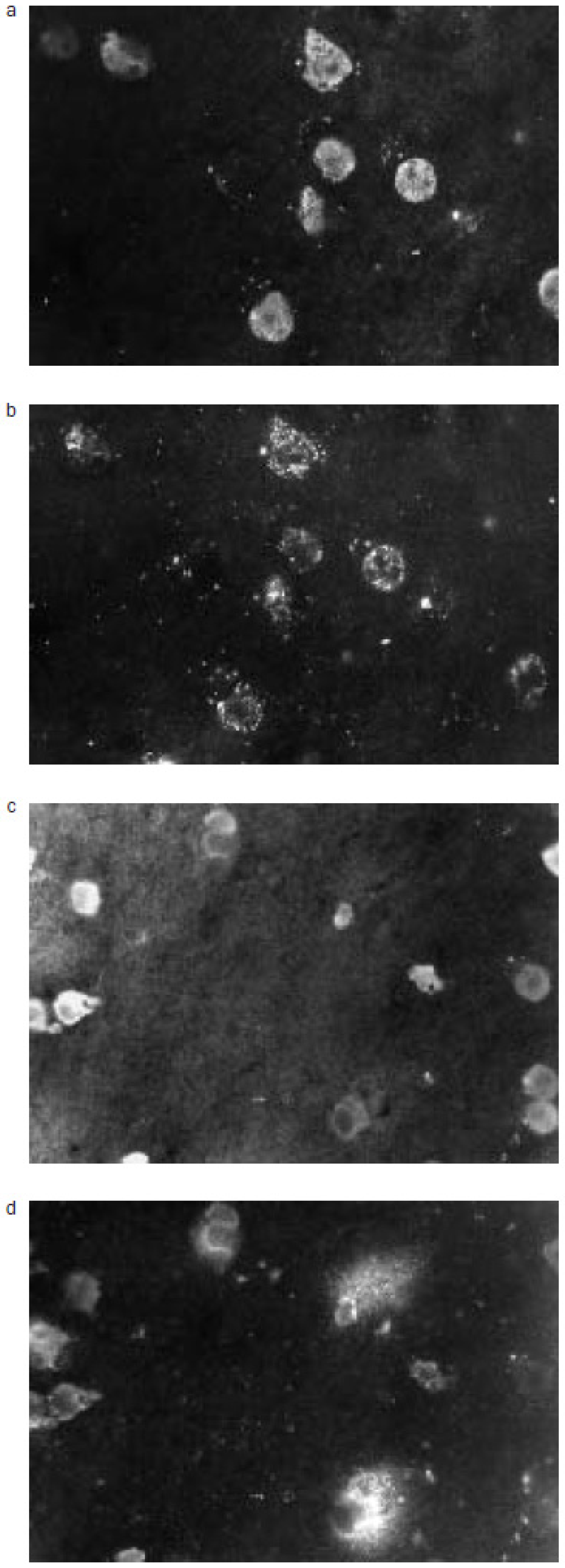
CNS sections from mice injected with ALS/ LMND Ig demonstrated neuronal cells positive for Hu Ig(G+M) in lumbar, dorsal, and cervical spinal cord, brainstem, brain and cerebellum. Only a restricted number of neurons were stained. In spinal cord, they were not specifically located in anterior horns and thus were not exclusively motor neurons. In motor cortical areas, pyramidal cells were more rarely labelled than other neurons; the latter were on the whole the neuronal types preferentially labelled, although some neurons in the frontal cortex, thalamus, caudate nucleus, putamen and pallidum were also positive for Hu Ig(G+M). TRITC-positive neurons were either isolated or in cluster, and their frequency per section was highly variable. In cerebellum, rare positive neurons were only seen in the molecular layer and in white matter; no Purkinje or granular cell bound ALS/LMND Ig. Neuronal TRITC staining pattern was variable: neurons were usually weakly stained, with either diffuse labelling of the neuronal body or with mainly TRITC staining of the cell membrane (Figure 2a, b). All TRITCpositive neurons showed consistently complete or partial staining of their cell body, whereas axons and dendrites were only extremely rarely stained. Astrocytes positive for Hu Ig(G+M) were only detected in mice injected with ALS/LMND Ig, and were located in the same CNS structures and levels as TRITC-positive neurons.Their distribution among the different CNS areas was the same as for TRITC-positive neurons, but their frequency was lower. Interestingly, large clusters of neurons stained for Hu Ig(G+M) were associated with the presence of numerous astrocytes positive for Hu Ig(G+M), identified in an adjacent section in the corresponding area.Groups of 2 to 4 TRITC-positive astrocytes were also observed in close contact with vessels. As for neurons, astrocyte cell bodies could be decorated with Hu Ig but their processes were usually TRITC negative (Figure 2c, d). We found no correlation between the number of days mice were injected and immunohistological observations, and detected no difference in the results obtained for mice injected with ALS Ig or LMND Ig. The delay of sacrifice (24 to 48 hours), motivated by the potential role of Ig retrograde transport from the presynaptic membrane to the neuronal cell body in the pathogenesis of ALS/LMND, did not influence significantly the amount of CNS cells labelling.
Figure 2.
Brain sections from a mouse injected with ALS Ig show some neurons (a; SMI 311-FITC) positive for Hu Ig(G+M) (b; TRITC), with TRITC staining sometimes limited only to the cytoplasmic membrane, and groups of astrocytes (c; GFAP-FITC) stained with Hu Ig(G+M) (d; TRITC) with scattered TRITC negative astrocytes (x 980).
Discussion
We have demonstrated that Ig from 5/9 ALS patients and 1 LMND patient (but not control Ig) (i) specifically bind to neurons and astrocytes from mouse CNS, independently of clinical presentation, degree of neurological handicap or length of disease, and that (ii) when passively transferred into mouse,ALS/LMND Ig are able to pass the blood-brain barrier (BBB) and recognize neuronal and astrocytic epitopes. This last observation may be partly explained by the fact that high levels of circulating Hu Ig(G) (control or ALS/LMND) should be able to some extent to cross the intact BBB, by analogy to human cerebrospinal fluid observations. Uniform decoration of CNS endothelial cells with control and ALS/ LMND Ig strengthens this hypothesis, with the suspicion, however, for a more specific capacity of ALS/LMND Ig to cross the BBB. As we have shown, only ALS/LMND Ig-labelled mouse neurons and astrocytes; this indicates strongly an autoimmune phenomenon occurring in ALS/ LMND, at least concomitantly to neuronal death and that does not involve anti-GM1 antibodies. However, the small number of patients included in our study can only suggest this kind of process and its potential targets in ALS/LMND, and further experiments including cell culture models and testing a larger number of ALS/LMND patients are warranted.
Motor neurons were not the only neuronal cells to be targeted by ALS/LMND Ig, as it might be expected. It is conceivable that, in case these autoantibodies are part of a secondary reaction to neuronal death, they can be generated against epitopes common to different types of neurons as well as against antigens specific to motor neurons. Furthermore, if these autoantibodies participate to the pathogenic process leading to neuronal death, the fact that they do not seem to be specifically restricted to neurons contributing to the corticospinal tract and to motor neurons in the anterior horns corroborates observations giving some evidence that other systems are impaired in ALS [22,23]. Pathological studies in ALS patients with long-lasting course of the disease may be of crucial importance for the understanding of ALS etiological mechanisms, since prolonged clinical course of ALS possibly represents a late step in the ALS degenerative process, affecting different types of neurons and leading to multisystemic degeneration. Widespread disturbances in ALS are also supported by the demonstration of a generalized glucose hypometabolism, using positron emission tomography [24]; the utilisation of glucose is decreased not only in motor areas but in particular also in parietal and occipital lobes, frontal cortex, caudate nucleus, putamen and thalamus. The occurrence of cognitive impairment [25] and somatosensory loss (evoked potentials studies [26]) was as well found in ALS patients.
These in vitro and passive transfer studies finally also emphasize the possible role of astrocytes in ALS/LMND pathogenesis. Astrocytes have the capacity to take part actively in immune responses [27,28], since they can express spontaneously in culture MHC class I antigens (increased after stimulation i.e. with interferon-γ), and MHC class II antigens under appropriate stimulation. Expression by astrocytes of both MHC class I and II antigens was inhibited when they were in contact with viable neurons in culture [27,28]. This observation may explain lack of expression of these antigens by astrocytes in vivo, in the vicinity of neurons [29]. It is then conceivable that when neuronal function is impaired or when there is an important loss of (motor) neurons, such as in ALS, astrocytes could recover their ability to express MHC antigens as it has been demonstrated in some postmortem studies in ALS patients [4,5]. We then postulate, since moreover CD4+ and/or CD8+ lymphocyte infiltrates have been demonstrated in ALS CNS [4,6], that some immune mechanisms may contribute to ALS pathogenesis and bring out the hypothesis that (i) CD8+ T cells may recognize antigenic epitopes presented by astrocytes in association with MHC class I molecules [30] and contribute to neuronal death, and that (ii) potentially primed CD4+ T cells would induce astrocytes to express MHC class II antigens, which can then restimulate CD4+ T cells and perpetuate the immune response [30].
Acknowledgments
The authors wish to thank Professor PJ Magistretti for helpful discussions and for reviewing the manuscript, and Mr E Razgallah for expert technical assistance.This work was supported by the Helmut Horten Foundation, the Puccini Foundation and Sandoz Pharma AG, Berne, Switzerland.
References
- Williams DB, Windebank AJ. Motor neuron disease (amyotrophic lateral sclerosis). Mayo Clin Proc 1991;66:54–82.
- Rothstein JD, et al. Decreased glutamate transport by the brain and spinal cord in amyotrophic lateral sclerosis. N Engl J Med 1992;326:1464–8.
- Engelhardt JI, Appel SH. IgG reactivity in the spinal cord and motor cortex in amyotrophic lateral sclerosis. Arch Neurol 1990;47:1210–6.
- Troost D, et al. Immunohistochemical characterization of the inflammatory infiltrate in amyotrophic lateral sclerosis. Neuropathol Appl Neurobiol 1990;16:401–10.
- Troost D, et al. Neuronophagia in the motor cortex in amyotrophic lateral sclerosis. Neuropathol Appl Neurobiol 1993;19:390–7.
- Engelhardt JI, et al. Lymphocytic infiltrates in the spinal cord in amyotrophic lateral sclerosis. Arch Neurol 1993;50:30–6.
- Smith RG, et al. Serum antibodies to L-type calcium channels in patients with amyotrophic lateral sclerosis. N Engl J Med 1992;327:1721–8.
- Kimura F, et al. Amyotrophic lateral sclerosis patient antibodies label Ca2+ channel a1 subunit. Ann Neurol 1994;35:164–71.
- Smith RG, et al. Cytotoxicity of immunoglobulins from amyotrophic lateral sclerosis patients on a hybrid motoneuron cell line. Proc Natl Acad Sci 1994;91:3393–7.
- Mosier DR, et al. Amyotrophic lateral sclerosis immunoglobulins increase Ca2+ currents in a motoneuron cell line. Ann Neurol 1995;37:102–9.
- Pestronk A, et al. Serum antibodies to GM1 ganglioside in amyotrophic lateral sclerosis. Neurology 1988;38:1457–61.
- Pestronk A, et al. Patterns of serum IgM antibodies to GM1 and GD1a gangliosides in amyotrophic lateral sclerosis. Ann Neurol 1989;25:98–102.
- Lamb NL, Patten BM. Clinical correlations of anti-GM1 antibodies in amyotrophic lateral sclerosis and neuropathies. Muscle Nerve 1991;14:1021–7.
- Shy ME, et al. Antibodies to GM1 and GD1b in patients with motor neuron disease without plasma cell discrasia. Ann Neurol 1989;25:511–3.
- Plaitakis A, et al. The neuroexcitotoxic amino acids glutamate and aspartate are altered in the spinal cord and brain in amyotrophic lateral sclerosis. Ann Neurol 1988;24:446–9.
- Rothstein JD, et al. Abnormal excitatory aminoacid metabolism in amyotrophic lateral sclerosis. Ann Neurol 1990;18:18–25.
- Rosen DR, et al. Mutations in Cu/Zn superoxide dismutase gene are associated with familial amyotrophic lateral sclerosis. Nature 1993;362:59–62.
- Jones CT, et al. Identification of a novel SOD1 mutation in an apparently sporadic amyotrophic lateral sclerosis patient and the detection of Ile113Thr in three others. Hum Mol Genet 1994;3:649–50.
- Jones CT, et al. Cu/Zn superoxide dismutase (SOD1) mutations and sporadic amyotrophic lateral sclerosis. Lancet 1993;342:1050–1.
- Bergeron C, et al. Copper/zinc superoxide dismutase mRNA levels are increased in sporadic amyotrophic lateral sclerosis motoneurons. Brain Res 1994;659:272–6.
- Adams D, et al. Predictive value of anti-GM1 ganglioside antibodies in neuromuscular diseases: A study of 180 sera. J Neuroimmunol 1991;32:223–30.
- Martin JE, Swash M. The pathology of motor neuron disease. In: Leigh PN, Swash M, eds. Motor neuron disease. Biology and management. London: Springer-Verlag; 1995. p. 93–118.
- Takahashi H, et al. Widespread multiple system degeneration in a patient with familial amyotrophic lateral sclerosis. J Neurol Sci 1993;120:15–21.
- Hatazawa J, et al. Cortical motor-sensory hypometabolism in amyotrophic lateral sclerosis: A PET study. J Comput Assist Tomogr 1988;12:630–6.
- Kew JJM, et al. The relationship between abnormalities of cognitive function and cerebral activation in amyotrophic lateral sclerosis. Brain 1993;116:1399–423.
- Georgesco M, et al. Les potentiels évoqués somesthésiques dans la sclérose latérale amyotrophique et la sclérose latérale primaire. Rev Neurol (Paris) 1994;150:292–8.
- Tontsch U, Rott O. Intercellular regulation of major histocompatibility complex class I expression in neural cells. Immunol 1993;80:507–9.
- Tontsch U, Rott O. Cortical neurons selectively inhibit MHC class II induction in astrocytes but not in microglial cells. Int Immunol 1993;5:249–54.
- Gehrmann J, et al. Microglia in the immune surveillance of the brain: Human microglia constitutively express HLA-DR molecules. J Neuroimmunol 1993;48:189–98.
- Sedgwick JD, et al. Major histocompatibility complexexpressing nonhematopoietic astroglial cells prime only CD8+ T lymphocytes: Astroglial cells as perturbators but not initiators of CD4+ T cell responses in the central nervous system. J Exp Med 1991;173:1235–46.
This is an open-access article distributed under the terms of the Creative Commons Attribution Non-Commercial No Derivatives (CC BY-NC-ND) 4.0 License.