Introduction
When one looks at the interactions between the nervous system and the immune system, there are a number of levels that should be considered (table 1). Both systems have much in common, first on a functional level, the nervous system receives inputs from sensory organs, the skin and on the output side controls skeletal muscles according to a reflex or a voluntary mode. The immune system is a system capable of identifying an infinite number of antigens and discriminate between self and non-self. At a macroscopical level, the nervous system is divided between a central and a peripheral nervous system. The immune system has also a centre and a periphery if one considers the thymus as opposed to the lymph nodes. At a cellular level, the brain has 30 x 109 neurons, the immune system is composed of about 1012 lymphocytes. At a molecular level, the brain relies on neurotransmitters, adhesion molecules and trophic factors for communication, while the immune system possesses a variety of cytokines, antibodies and antigen molecules to perform its function. Molecular dissection of these ligands and receptors has forced us to alter the long held concept that neurotransmitters, neuropeptides and neurotrophins only function in the brain and in neuronal networks and on the other hand cytokines and adhesion molecules are found only in the immune system. It has therefore been suggested that the brain and the immune system speak a common biological language (1).
Table 1.
A comparison between the nervous and the immune system.
Table 1.
A comparison between the nervous and the immune system.
When one considers diseases, there are sometimes situations when the brain is overwhelmed by infection or inflammation causing serious neurological dysfunction. There are other conditions where there is a subtle imbalance in the homeostasis between the brain and the immune system. Slight dysbalance in immune functions has been reported in psychiatric disorders and is the subject of a field called psychoneuroimmunology.
Communication between brain and immune system
Some argue that there is a real crosstalk between the brain and the immune system and that these interactions are formed by bidirectional circuits between the nervous system and the immune system. Evidence indicates that circuits exist between the brain and the immune system (2).While researchers once considered the body’s network of immune defences as a system unto itself, they have learned that it is intimately intertwined with the nervous and endocrine systems. There are direct physical links, for example neurons that innervate immune organs such as the spleen and lymph nodes, but more important we have been able to unravel the molecular links: these include the interleukins originally viewed only as regulators of immune cells, the neurotransmitters, which were once thought to act only between nerve cells and hormones, the endocrine messengers. As these connections come to light, they are helping to explain some previously mysterious correlations between mental and hormonal states and the immune system. Let us now briefly consider some of the mechanisms of auto-immunity in the brain.
Inflammation in the brain – the example of multiple sclerosis
When inflammatory cells are crossing the bloodbrain barrier, a number of adhesion molecules are playing a key role to regulate the entry of lymphocytes in the brain: there are complementary adhesion receptor ligand pairs on endothelial cells and lymphocytes that allow interaction of the lymphocytes to the endothelial cells. An important step is the interaction between LFA-l, the leukocyte function associated antigen on lymphocyte and the receptor ICAM-l, an adhesion molecule on the surface of endothelial cells. Several secondary signals also influence leukocyte extravation, these include low-molecular weight chemoattractants, referred to as chemokines as well as members of the interleukine family, such as IL-1. As a consequence of this process, the lymphocyte binds to the endothelium. The next step will be an opening of the blood-brain barrier, allowing the extravasation of the lymphocyte in the brain parenchyma (3).
Cytokines are a group of heterogenous polypeptides that modulate the immune response. Originally described as modulatory molecules in the peripheral immune system, several cytokines have been shown to play a key role in the brain (4). Their synthesis in the central nervous system is not only due to invading immune cells but cytokines can be produced by resident cells of the CNS, such as microglia, astrocytes and probably also neurons. The demonstration also of the expression by neurons of receptors specific for cytokines support a potentially crucial role for these molecules in brain function. We know that TNF-α and IL-l play an important role in nociception and cytokines are released after nerve injury.
In multiple sclerosis (MS) autoreactive T-cells recognizing myelin proteins (MBP, MOG or MAG) circulate in the blood of these patients. Upon activation, for example in the course of a viral infection, they penetrate the blood-brain barrier. Adhesion molecules are then critically involved in the homing and migration process. In the central nervous system these autoreactive T-cells undergo local reactivation and proliferation. CD-4 positive T-helper-1 lymphocytes secrete TNF-α and interferon-γ, activating macrophages and microglia. Autoantibodies directed to myelin antigens may cross the blood-brain barrier once it has been opened by activated T-cells or are locally produced by B-cells that have been activated by T-helper-2 T-lymphocytes. These autoantibodies activate the complement system. The destruction of myelin results from the concerted action of autoaggressive T-cells together with inflammatory mediators (5).
Multiple sclerosis – the enigma of fatigue
While the immunological changes during acute multiple sclerosis are increasingly better understood and have been used for the development of effective immunomodulating treatments, other aspects of multiple sclerosis remain poorly understood. For example fatigue is a very common and disabling symptom of multiple sclerosis. Patients suffering from fatigue complain of an overwhelming sense of tiredness, lack of energy or feeling of exhaustion during or following motor or mental activity. Fatigue is distinguished from symptoms of depression which include lack of self-esteem, despair or feeling of hopelessness. Fatigue is present in about 80% of MS patients, 70% experience it daily and 22% suffer disruption of their daily activities because of fatigue. In addition fatigue is a major cause of unemployment in MS patients (6). The pathophysiology of fatigue in MS is not known. Mechanisms like poor physical fitness, defects in neuromuscular transmission or increased cytokines level have been postulated but have not been universally accepted. We have hypothesised that brain dysfunction in MS patients with fatigue could result from impaired interaction of the cerebral cortex with functionally related subcortical regions. In a recent study we have found reduced glucose metabolism in the striatum and prefrontal cortex of MS patients with fatigue compared with MS patients without fatigue, using 18-F fluorodeoxyglucose and positron emission tomography (7). It thus appears that fatigue is associated with an impaired interaction between functionally related areas of the frontal cortex and basal ganglia. It is interesting to note that in depression changes in cerebral blood flow have been shown in different regions from those found in the MS fatigue patients, namely reduced glucose metabolism in the dorsolateral prefrontal cortex and the caudate nucleus on the left brain side or bilaterally (8, 9).
Tracing the connections between the brain and the immune system
a) The hypothalamic-pituitary-adrenal (HPA) axis
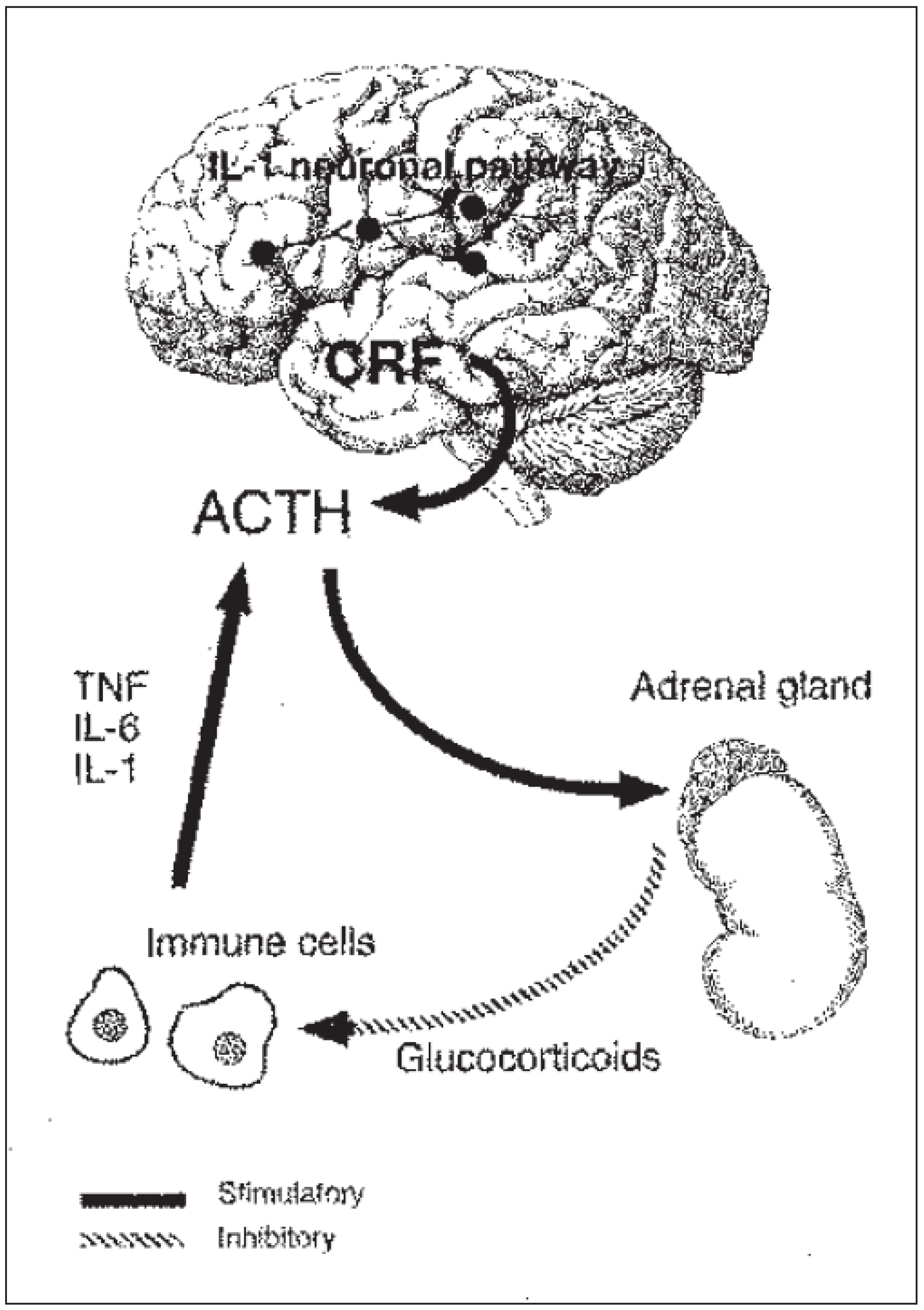
It is, however, the capacity of the brain to shape the immune response that has drawn most attention recently. As it has been suggested, mental states can influence the body’s resistance to disease. It has long been suspected that the brain influences immune function through hormones, particularly those in the hypothalamic-pituitary-adrenal axis. This axis is triggered when a stress stimulates the production of corticotropin releasing factor (CRF) by the hypothalamus of the brain (fig. 1). As an action, CRF causes the release of corticoids from the adrenal glands. These adrenal hormones have several actions, such as providing a burst of energy by raising blood sugar concentration that enables an individual to deal with a threatening or a stressful stimulus. In addition, however, corticosteroids inhibit IL-l production and thus reduce the inflammatory response, an effect that is the basis of their use as anti-inflammatory drugs.
Figure 1.
Brain-immune system connections. The brain influences immune functions and immune messengers in turn affect the brain.
Figure 1.
Brain-immune system connections. The brain influences immune functions and immune messengers in turn affect the brain.
Modification of the HPA axis can have good or bad consequences. The immune suppressing feedback can be helpful and may explain the fact that pregnancy may lessen the symptoms of multiple sclerosis. It has been shown that the fetus produces CRF that gets into the mother’s circulation and tends to make the HPA axis overly active. In addition the oestrogen increase during pregnancy may stimulate cortison secrection. Conversely there is mounting evidence that a depressed HPA axis resulting in two little corticosteroids can lead to a hyperactive immune system and increase the risk of developing autoimmune diseases. It has been shown for example that in two strains of rat which differ in their inflammatory response and their susceptibility to many experimental induced autoimmune diseases, such as allergic encephalomyelitis, a model for multiple sclerosis, the sensitive strain released much less CRF and less corticosteroids in response to stress than the resistant strain of rat. There are also many experiments in humans to suggest that damping down the immune system can lead to a decreased ability to fight infections. Influenza vaccinations are less likely to work in people caring for spouses with Alzheimer disease, a task known to cause a lot of stress, than in people of similar age and background not in those caretaker roles. The psychosocial consequences in children and adolescents of a parental chronic disease like multiple sclerosis is currently investigated by one of us. Further examples are that children suffering from atopic dermatitis or from asthma have a depressed HPA axis. These kinds of studies suggest an intimate link between the endocrine and the immune system on one hand and the brain on the other hand (10).
Stress while not being the cause of multiple sclerosis can affect the onset and subsequent disease activity. We know that the majority of MS patients report having experienced significant stress prior to the onset of their MS symptoms. Most patients believe that stress affects their symptoms. The majority (60% of MS patients) believe that stress affects their illness in general and 40% report that stress had brought on at least one exacerbation (11). MS patients report a great number of qualitatively extreme events particularly during the two to six months prior to symptom onset. However, the effects of stress on subsequent disease activity are less clear.
b) Emotion and the brain
The relationship between emotion and the brain is now being particularly well understood through the work of Le Doux (12) who has shown how the architecture of the brain gives the amygdala a privileged position as an emotional sentinel. His research has shown that sensory signals from the eye or the ear travel first in the brain to the thalamus and then across a single synapse, to the amygdala: a second signal from the thalamus is routed to the neocortex, the thinking brain. This branching allows the amygdala to begin to respond before the neocortex, thus demonstrating a neural pathway for feelings that bypass the neocortex. Anatomically the emotional system can act independently of the neocortex. Although the cortical pathway provides the amygdala with a more accurate representation than the direct pathway to the amygdala from the thalamus, it takes longer for the information to reach the amygdala by way of the cortex. In situations of danger, it is very useful to be able to respond quickly. This work has provided a cognitive model for examining the neural substrate of chronic stress or anxiety. The exposure to an aversive experience may lay down powerful memory traces that are difficult to extinguish and stored in a circuit in which the amygdala plays a central role. Because the amygdala has strong input to the hypothalamus, there are neuroendocrinological consequences: increased activity in the amygdala will trigger adrenocorticotropic hormone release in the hypothalamus leading in turn to corticosteroid release. In this way we will get an unusually high level of corticosteroids. In this context it should not escape to our attention that the sustained hyperactivity of the HPA axis has been associated with several psychiatric disorders, most notably major depression. Marked hypersecretion of ACTH and corticosteroids which is not appropriately suppressable by dexamethasone occurs in approximately 40 to 50% of patients with major depression (13). CRF levels may also be elevated in such patients. It is also of interest that a statistically significant percentage of patients with major depression have depressed immune function. Much current thinking supports the view that major depression represents a general stress response that has escaped it’s usual counterregulation. Negative emotional memories may represent a chronic “trait”, leading to depressive disorders. Their suppression or modulation (either by pharmacotherapy or psychotherapy) can bring remission of symptoms (14).
Conclusion
We have seen that some of the clinical enigmas of yesterdays are being replaced by the facts that are building the basis for a scientific approach to the understanding and treatment of neuroimmunological disorders. Thus in multiple sclerosis we know that oligodendrocytes and myelin are damaged or destroyed by an autoimmune process. New intriguing evidence is, however, mounting that in the chronic progressive form of multiple sclerosis the disease process seems to be based on continuous axonal loss, the exact mechanism, by which this occurs, remaining, however, unclear. Other aspects of multiple sclerosis, such as fatigue, depression or cognitive impairment remain poorly understood. The same is true for the vast literature dealing with stress disease links and describing such effects that stress can speed the metastases of cancer, increase the vulnerability to viral infection or exacerbate the formation of plaques in multiple sclerosis. Though the study of the brain and the immune system has allowed us to grasp some of the mechanisms at the molecular level, when we try to apply the same tools to study cognitive mechanisms or mental phenomena, many enigmas are remaining. This reflects the fact that transposing molecular mechanisms in mental phenomena remains a formidable task that will keep us busy well beyond the 20th century. Such an attempt is currently being carried by Damasio (15) who studies the connections between the brain and human experience, emotion and reason.