


Summary
Among the risk factors influencing the pathogenesis of Alzheimer’s disease (AD), genetic factors play a major role. The genes for early onset familial Alzheimer’s disease (FAD) include the ßamyloid protein precursor (APP) on chromosome 21, as well as the genes for presenilins 1 and 2 on chromosomes 14 and 1. On the other hand, the E4 allele of the apolipoprotein E gene is considered as a risk factor for late onset FAD and sporadic AD, and decreases onset age in FAD with an APP mutation. We are studying a Swiss family with an early onset AD, for which we have built up the pedigree based on 10 generations. We have identified numerous AD cases in 3 generations with a necropsy confirmation of the clinical diagnosis. The clinical features of the affected members are those of classical AD with a duration of the disease from 5 to 20 years. Several members presented epileptic characteristics, even before the onset of the disease. The brain pathology is almost similar to that of sporadic AD, but with a heavier degeneration of pyramidal neurons and fibres in the subiculum and hippocampal regions. The massive cortical degeneration involves primary sensory areas such as the visual cortex. Subcortical regions are affected as well. Partial genetic analysis has been performed on DNA from six necropsy cases and demonstrated the presence of the ApoE4 allele in all the six cases.The pathogenic mutations previously described in the APP and presenilin 2 genes were not detected. Screening for possible mutations in the presenilin 1 gene is now underway.
Résumé
Parmi les facteurs de risque décrits pour la maladie d’Alzheimer, les facteurs génétiques jouent un rôle primordial. Les gènes isolés à ce jour pour la forme familiale précoce comprennent le gène du précurseur de la protéine ß-amyloïde (APP) sur le chromosome 21, et les gènes des deux protéines présénilines 1 et 2 sur les chromosomes 14 et 1. Par ailleurs, l’allèle E4 de l’apolipoprotéine E est considéré comme un facteur de risque pour les formes familiales tardives et sporadiques et accroît la précocité de la maladie dans les formes avec mutation sur le gène APP. Nous étudions une famille suisse, avec une maladie d’Alzheimer à début précoce, pour laquelle nous avons reconstitué l’arbre généalogique sur 10 générations et identifié plusieurs cas avec confirmation neuropathologique du diagnostic clinique. Les caractéristiques cliniques sont similaires à celles des formes sporadiques ou familiales connues, mais comportent la présence d’une symptomatologie épileptique, parfois avant même le début de la maladie dont la durée varie entre 5 et 20 ans. La pathologie cérébrale est semblable à celle de la maladie d’Alzheimer classique, mais avec une dégénérescence plus importante dans les régions de l’hippocampe et du subiculum. Les régions corticales sont massivement touchées, y compris certaines aires sensorielles primaires comme le cortex visuel. La dégénérescence atteint aussi plusieurs régions sous-corticales. L’analyse génétique a été partiellement réalisée sur du matériel postmortem et a révélé la présence de l’allèle ApoE4 dans les 6 cas étudiés. Cependant, aucune mutation pathogène n’a pu être mise en évidence sur les gènes du précurseur de la protéine ß-amyloïde ou de la préséniline 2. L’examen des séquences sensibles du gène de la préséniline 1 est en cours.
Mots clés: maladie d’Alzheimer familiale, génétique, clinique, dégénérescence neuronale
Introductory review on genetic factors in Alzheimer’s disease
Alzheimer’s disease (AD) constitutes the first cause of dementia in the Western world and is now considered as an heterogeneous progressive neurodegenerative disorder including mainly sporadic and less common familial forms. Familial Alzheimer’s disease (FAD) is a large pathologic entity comprising early onset forms (EOFAD), beginning before the age of 65, and late onset forms (LOFAD), beginning after the age of 65 years. Among the risk factors influencing the pathogenesis of AD, three are clearly defined, age for the sporadic forms, genetic factors for the familial forms and the polymorphism of apolipoprotein E (ApoE) for both forms. Although EOFAD represents not more than 10% of all AD cases [1], the genes involved in its pathology are best characterized.
The first gene discovered was the one encoding the ß-amyloid protein precursor (APP) on chromosome 21. APP is a large transmembrane protein (695–770 amino-acids) normally wrapped in the neuronal as well as in other cellular membranes, highly conserved throughout evolution and playing a role in various biological functions among which cellular growth and maintenance of synaptic contacts. The ß-amyloid protein (ß-peptide) sequence itself is situated partially inside the membrane (ß29–42) and partially within the extracellular domain (ß1–28). Since 1991, several point mutations linked to autosomal dominant AD have been described at three different loci along the APP gene sequence. Three distinct mutations have been identified at codon 717 of the APP gene, 3 residues after the end of the ßpeptide sequence toward the C-terminal sequence. They produce the substitution of a valine into either isoleucine (London mutation), phenylalanine or glycine. Recently, a mutation at codon 716 has been described, replacing an isoleucine by a valine (Florida mutation). The third mutation site is known as the Swedish double mutation at codon 670–671 in the N-terminal sequence adjacent to that of the ß-peptide. The ß-amyloid protein is found in the brain, either as a diffuse and probably non toxic deposit, or as an aggregated form surrounded by glial cells and degenerating neurons in the senile plaque. The ß peptide has to be processed from its precursor in order to be deposited in the extracellular matrix. Three enzymes, the α, β, and γ secretases, are involved in the various cleavage reactions. The mutation sites are located very near to the cleavage sites of either the α secretase (Swedish mutation) or the γ secretase (mutations at codons716 and 717) and are supposed to interfere with the correct processing of APP. Indeed it has been shown that the total amount of ß peptide was increased in the Swedish mutation, while, in other mutations, the amount of its long and more amyloidogenic form (42–43 amino-acids) was increased compared to its short and less amyloidogenic form (40 amino-acids).This was shown in fibroblasts cultures of clinically affected and non affected carriers of the mutation as well as in transfected cells with a mutated APP. Thus a clear link seems to exist between mutations on the one hand and amyloid deposit and transmission of the disease on the other hand [review: 2–10].
As EOFAD with an APP mutation represents only a very small proportion of all EOFAD cases, linkage to other chromosomes have been searched for and in 1995, two other genes have been shown to be linked to the transmission of the disease in several families with an early onset. These two genes, located respectively on chromosomes 14 and 1, are encoding for proteins presenilin 1 (or S182) and presenilin 2 (or STM2). These two proteins share a great part of their sequence (about 450 amino-acids) and present, following the existing models, 8 transmembrane domains. Until now, more than 40 mutations have been isolated at about 30 sites in various families from different ethnic origins.While the mutations in presenilin 1 (chromosome 14) are considered to be responsible for most of all EOFAD cases, the mutations in presenilin 2 (chromosome 1) concern only 8 families, 7 Volga German families and one Italian family. The majority of the mutations are situated within two domains, the second transmembrane domain and the sixth hydrophilic loop, suggesting that these regions are important for the functions of the presenilins and that the mutated sites modify their normal metabolism. The latter is still largely unknown, but transfection of cells with mutated presenilins seems to induce a higher production of the long amyloidogenic form of the ß peptide, in a similar way as some APP mutations. Altogether, these data reinforce the hypothesis that the amyloid cascade represents a fundamental mechanism in the pathology of Alzheimer’s disease [review: 1, 6–11].
Besides the genetic loci on chromosomes 21, 14 and 1, a site linked to the transmission of familial Alzheimer’s disease with a late onset (LOFAD) was determined in 1991 on chromosome 19. Two years later, the gene was recognized as being the gene of apolipoprotein E, whose functions in the metabolism and transport of blood lipids and cholesterol were already well known. This gene is polymorphic and presents in the general population in three forms, E2, E3, E4, producing 6 different combinations (E2E2, E2E3, E2E4, E3E3, E3E4, E4E4). E3 is the most common allele (80–90% have at least one copy and 60% 2 copies), while E2 is very rare (10% have one copy and only 1–2% have 2 copies) and E4 has a general frequency of 15% in the Western countries, with some ethnic differences. Only 2% have an E4E4 genotype. The three resulting apolipoproteins (299 amino-acids) differ only by 2 aminoacids at the positions 112 and 158. Since 1993, a huge number of studies have demonstrated that the E4 allele is a risk factor for late onset FAD and sporadic AD, with the frequency of E4 varying from 10 to 20% in the control groups and from 30 to 60% in the AD groups. In addition, the presence of E4 was associated with a decreased onset age in FAD with an APP mutation.However, the E4 allele represents only a risk factor with a statistical value and not a factor allowing the prediction of AD. Only patients with a clinical diagnosis of AD and an E4E4 genotype present a strong probability that AD will be confirmed after death. The E4 carriers seem to have less total amount of ApoE protein in the brain and thus an impaired transport of lipids and cholesterol to the sites of lesion and reparation. The ApoE4 protein could play a role as a pathological chaperone, influencing the fibrillation of the ß peptide, while ApoE2 and ApoE3 might prevent the protein tau to undergo an excessive phosphorylation leading to neurofibrillary degeneration [review: 12–20].
As large families with an inherited form of Alzheimer’s disease offer a worthy opportunity to study the link between genetic defect, clinical pattern and pathological changes in the brain, we are investigating a Swiss family with early onset AD.
Data on the Swiss family
Pedigree (Figure 1)
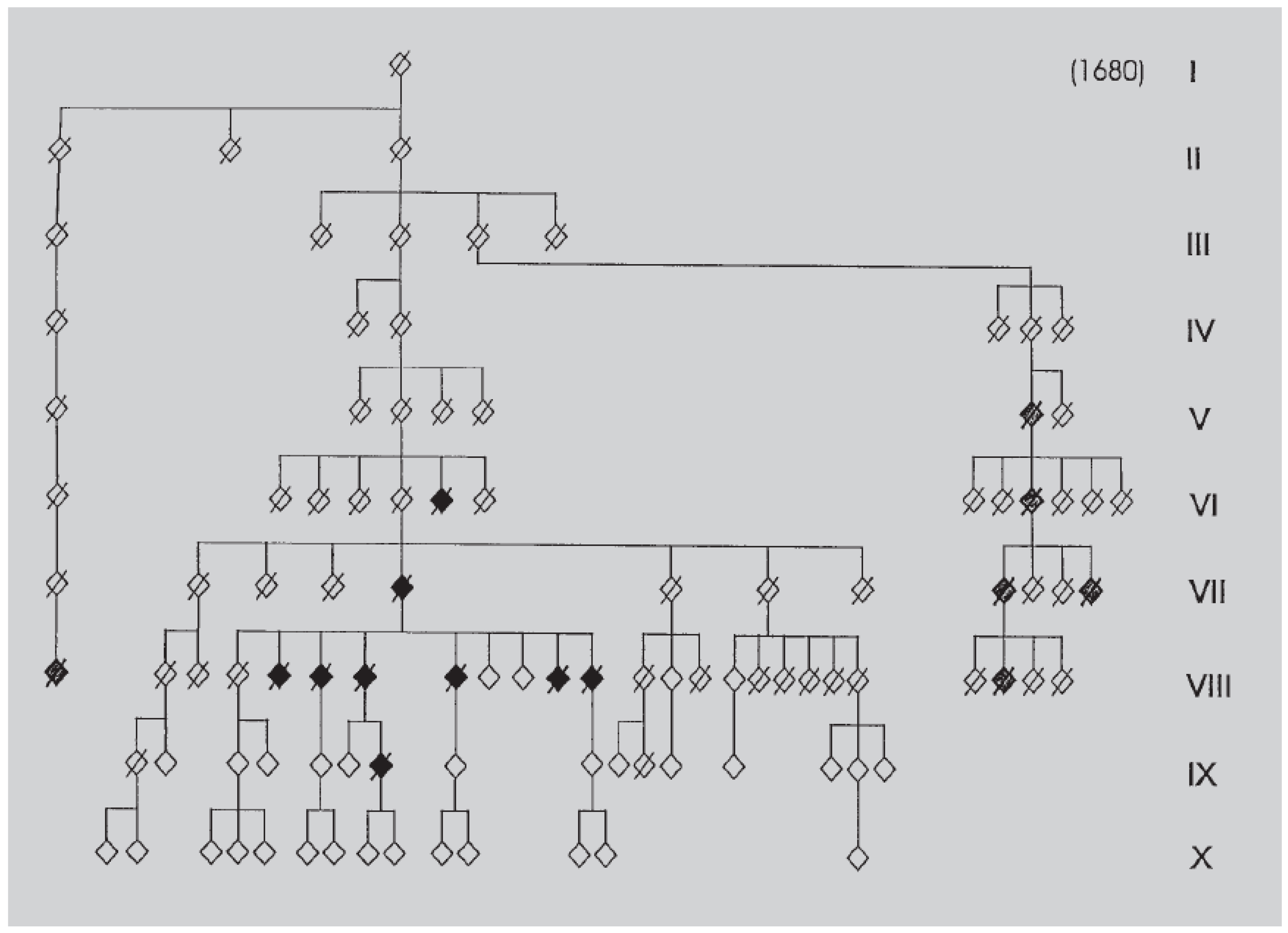
Information about the family was obtained from several sources, mainly medical files, interviews of family members and state archives. The pedigree of the Swiss family goes back to the seventeenth century, as we have discovered a common ancestor to several branches with psychiatric disorders, among which we found depression, suicide, alcoholic dementia and AD. In the main branch with AD, the first two documented cases are case 6 from generation VI and case 5 from generation VII, for which we have clinical data indicating senile dementia. In generation VIII, six of nine siblings are clinically well documented, and AD has been confirmed after death. The diagnosis of AD is doubtful for three other cases. One case is suspected for suicide and one member has committed suicide. Two members are unaffected. In generation IX, only one member until now was affected by a very early and severe AD, confirmed after death. One member died precociously after an epileptic attack, one committed suicide and one is depressed. Other are still asymptomatic. In generation X, most members are young and asymptomatic. In two other branches with the same ancestor, we have found several cases with alcoholic dementia. Interestingly in the main branch, three cases with a diagnosis of AD are also documented for alcoholism.
Figure 1.
Pedigree of the Swiss family with 10 generations. Solid symbols indicate family members with AD. Shaded symbols indicate family members with alcoholic dementia. Slashed figures indicate deceased members. The pedigree is disguised to prevent recognition of the family. See text.
Clinical data (Table 1 and Table 2)
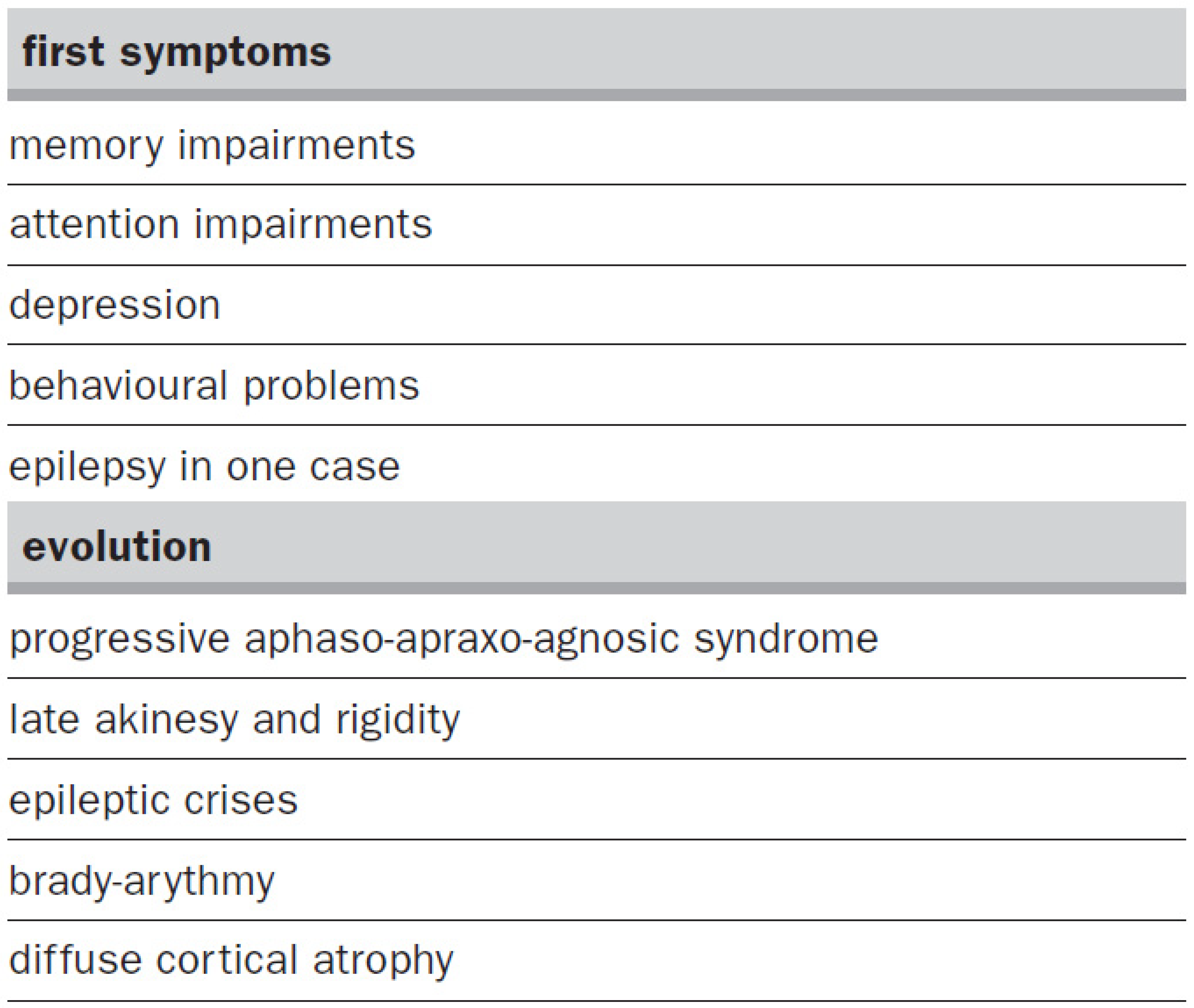
The diagnosis of AD was established on the basis of the DSM-IIIR criteria. The age of onset was between 43 and 64 years, and for all patients, first clinical signs were memory and attention impairments. Depression and behavioural problems were also present rather early in the course of the disease. In one case, epilepsy started long before the beginning of AD symptoms. On the whole, the clinical features do not differ from those of sporadic AD, except from additional epileptic features and/or chronic alcoholism in several members. In two other branches of the pedigree, several cases of alcoholic dementia were documented. The evolution of the AD affected members was similar to that of sporadic AD, with a progressive aphaso-apraxo-agnosic syndrome evaluated clinically, without the use of recognised deterioration scales. The duration of the disease was highly variable, between 5 and 21 years.
Table 1.
Description of the 8 members of the Swiss family with clinical Alzheimer’s disease. For all cases except the first one, the postmortem examination of the brain confirmed the diagnosis.
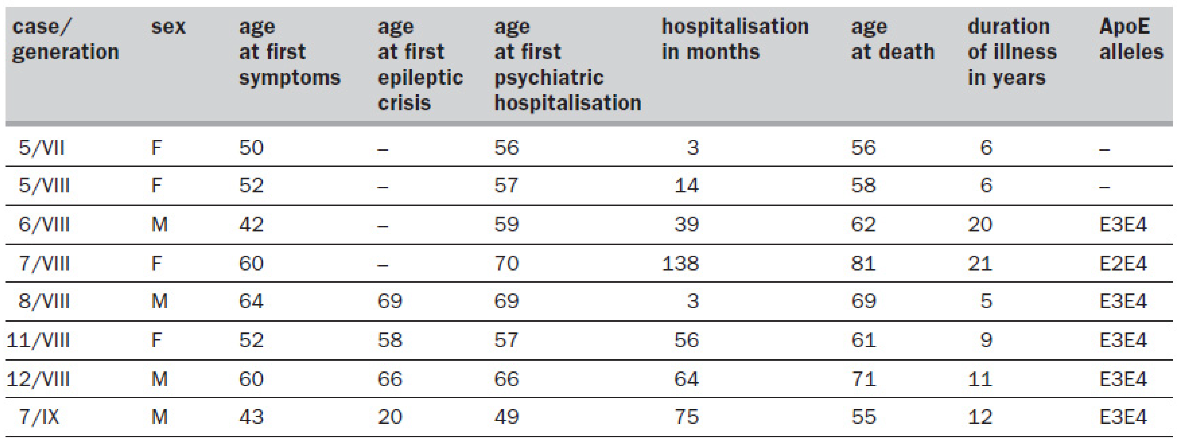
Table 2.
Clinical signs described for the first symptoms and evolution of AD in the family. See text.
Pathological data (Figure 2 )
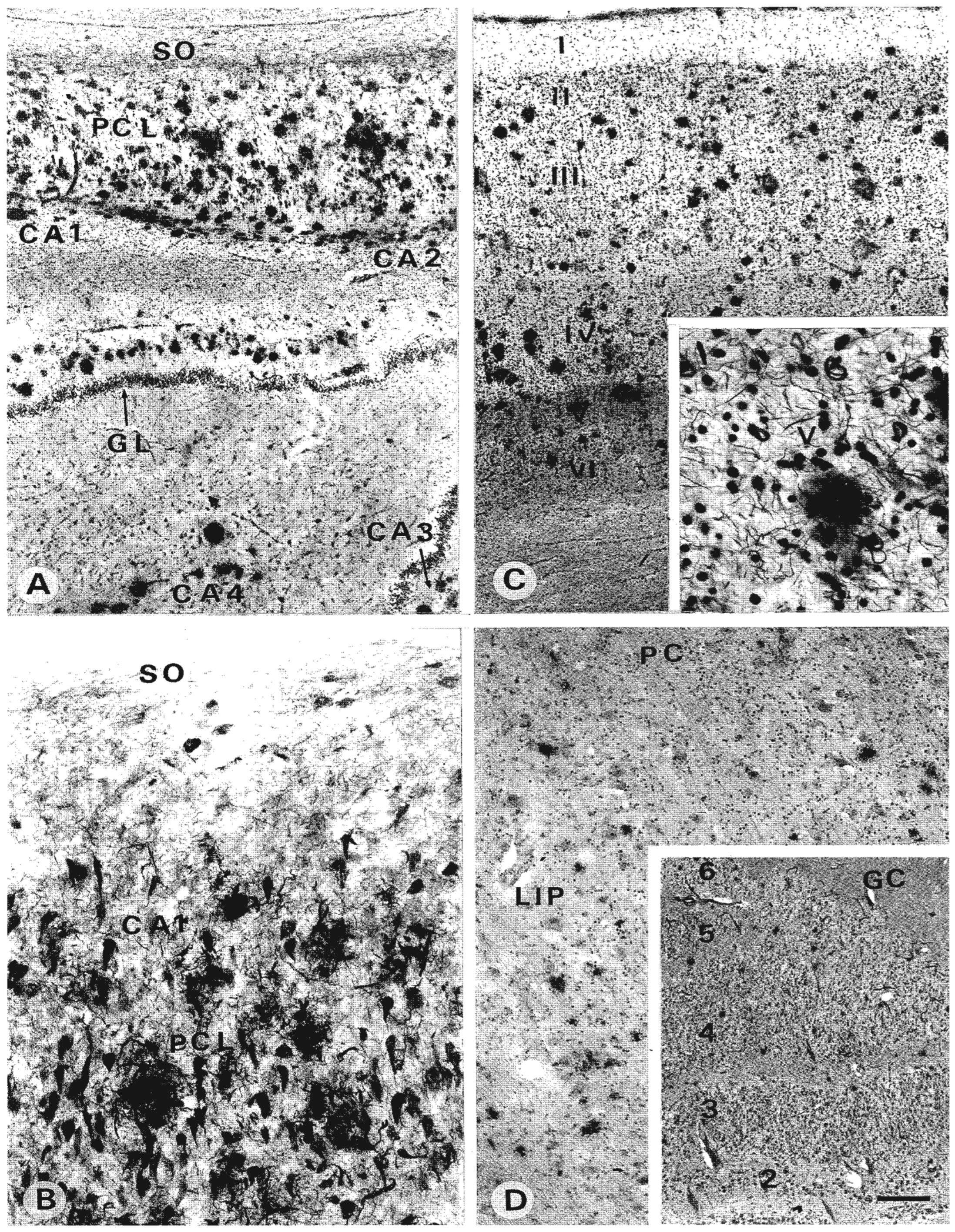
The brains were available in all cases from generations VIII and IX.The post-mortem AD diagnosis was confirmed by the qualitative examination of senile plaques (SP) and neurofibrillary tangles (NFT) in several cortical, including entorhinal and hippocampal, and subcortical regions. Any concomitant pathology was also excluded. We have studied in greater details the brains of three cases (cases 7 and 12 from generation VIII and case7 from generation IX). In all three cases, the hippocampus as well as the subiculum and entorhinal cortex were heavily affected. A ß-amyloid immunostaining was used to reveal ß-amyloid deposits on some sections, but the silver methenamin staining was more largely used to reveal all types of SP. The Gallyas staining was used to show neurofibrillary degeneration in the neuronal perikarya as well as in the neuropil and white matter (threads). A high number of ß-amyloid deposits and senile plaques were present in all sectors of the hippocampus. However, CA4 was less affected. SP were mainly in the pyramidal layer, but the white matter under the pyramidal layer and outside the granular layer of the dentate gyrus was not spared. In addition, a high amount of neurofibrillary degeneration was found in various types of neurons. Threads, representing dendritic and axonal degeneration, were noticed around the neurons as well as in the white matter. The adjacent subiculum and entorhinal cortex also showed massive degeneration. Most cortical areas showed high degrees of SP and NFT deposits, and even sensory areas such as the primary visual cortex exhibited similar features. In subcortical visual structures such as the lateral geniculate nucleus, the pulvinar and the superior colliculus, all cases showed signs of degeneration. The NFT degeneration was more prominent in the superior colliculus than in the pulvinar which showed mainly SP. On the whole, the most severely affected case appeared to be case 7 from generation IX, which was also the more precociously touched by the clinical symptoms.
Figure 2.
50-µm frozen sections stained with silver methenamin and Gallyas. (A) Hippocampus of case 12 from generation VIII: the pyramidal cell layer (PCL), here showing CA1–2 and partially CA3–4, is completely filled with senile plaques; GL = granular layer; SO = stratum oriens. (B) Higher magnification in CA1 of the same case: almost all pyramidal cells are degenerated and neuritic plaques and threads are present. (C) Primary visual cortex of case 7 from generation IX: a similar heavy deposit of senile plaques is observed in all cortical layers (I–VI). (Inset) Higher magnification of layer V, showing a high amount of degenerated fibres intermingled with cell nuclei and NFT bearing neurons. (D) Among subcortical visual structures, the pulvinar and lateral geniculate nucleus (inset) of case 7 from generation VIII exhibit also senile plaques deposit, although less important than in the cortical areas; PC = pulvinar capsule; LIP = lateral inferior pulvinar; GC = geniculate capsule. Scale: (A) (C) 0.4 mm; (inset of C) 50 µm; (B) 50 µm; (D) 175 µm; inset of (D) 0.3 mm.
Genetic data (Table 1)
As no living member is presently clinically affected by the disease, partial genetic analysis has been performed on DNA from six necropsy cases. DNA was first extracted from the brain specimen and then amplified by Polymerase Chain Reaction (PCR). ApoE alleles were determined by Restriction Fragment Length Polymorphism analysis (RFLP). All six cases were carriers of an E4 allele, together with an E3 allele for five of them and with an E2 allele for one case. Single Strand Conformation Polymorphism (SSCP) or sequencing analysis was performed with amplified exons 16 and 17 of the APP gene, as well as with exons 5 and 7 of the presenilin 2 gene, where all the described mutations are located. No mutated sequences were found, neither for APP gene (6 cases) nor for presenilin 2 gene (one case). The search for a mutation is now focusing on the presenilin 1 gene.
Discussion
Until now, a dozen families with a few pathological mutations in the amyloid protein precursor gene have been studied, while over 80 families are known to carry more than 40 different pathological mutations in the genes of the proteins presenilins 1 and 2 [1,3,9,10,11]. These mutations are responsible for autosomal dominant AD. Except for the age of onset, the clinical features of the APP mutated families do not differ essentially from those of sporadic AD.The mean age of onset is 55 years, but the onset may be as early as 43 years, and no healthy carrier of a pathological mutation has been observed after 67 years [3]. For the carriers of a pathological mutation in the presenilin 1, but not in presenilin 2, the clinical features seem more severe and include characteristics such as epilepsy, myoclonus and paratonia rather early in the course of the disease. In addition, the onset of clinical symptoms can be very precocious (30–50 years) and seems to depend on the location of the mutation inside the gene sequence, as families with a mutation in the same location have a clinical onset in a similar age range, different from that of families with a mutation in another location [1].
Our Swiss family presents some clinical characteristics similar to those of families with presenilin 1 mutation, such as the presence of epilepsy. Other features are different such as the variable age of onset between family members (43 – 64 years). As the known mutations in the genes of ß-amyloid precursor and presenilin 2 have been excluded, it seems reasonable to postulate a mutation in the presenilin 1 gene. However, in some families, the genetic defect has not been identified, implying that other unknown genes are involved in the transmission of the disease [9]. The presence of the E4 allele in the six postmortem cases with AD confirmation suggests that this could represent an additional risk factor. Another interesting clinical feature is the presence of chronic alcoholism in some cases, possibly related to the presence of alcoholic dementia in another branch of the family. In the latter, cases were discovered in the archives of the psychiatric hospital and the link with the AD family established thanks to the state archives (archives cantonales). However, the diagnosis of alcoholic dementia was used between 1850 and 1930, at a time when AD was either not described, before 1907 [21], or not used as a common clinical entity. It may have been confounded with a diagnosis of dementia linked to alcoholism. Indeed, depressive features are present rather early in all cases of the Swiss family and could favor an alcoholic behavior. To our knowledge, no clear association between AD and alcoholism has been described [22]. Finally, the rather high amount of suicides among members of the family speaks also in favor of a possible link between depression and dementia, as already suggested by other authors [23,24,25].
Brain studies have been performed for a few cases in some genetically described families, but were rather disappointing in the sense that no specific feature could be observed [26,27]. The pathology was that of classical AD with a heavy deposit of ß peptide, a high number of senile plaques and neurofibrillary tangles, mainly in the cortical and hippocampal regions. Our results in the Swiss family are in agreement with those of other authors and with the exception of a more massive degeneration in some cases, it would be almost impossible to recognize a family case on the basis of pathological data. Indeed, if the primary visual cortex, which is normally affected after other cortical areas in the course of the disease [28,29,30], is highly damaged in our cases, we have shown that this was also the case for sporadic AD [31]. The same is true for subcortical visual structures [32]. As mentioned in the introductory review, in vitro studies using cell cultures and transfected cells bring some more information, favoring the hypothesis that a common pathogenic mechanism should involve the accumulation of either the total amount of ß peptide or its long, more amyloidogenic form. However, aging brains without AD also show accumulation of ß amyloid and SP. On the other hand, no clear link has been demonstrated between ß-peptide accumulation and either neurofibrillary degeneration or neuronal and synaptic loss, the latter being best correlated with cognitive deterioration [33,34,35]. Thus if the presence of ß amyloid seems essential to induce AD degeneration, it may not be sufficient to produce the whole pathology and other molecular and cellular changes appear to be necessary. AD might have several etiologies but an unique phenotypical expression. Different genes, some of them being still unknown, could produce the same clinical and pathological symptoms, or the products of some genes could influence the expression of other genes.
In our family, the genetic defect is still unknown. The clinical portrait exhibits some specific features and the brain pathology is particularly severe, although not essentially different from that of sporadic AD. Further studies will bring more information on the link between these different components and may help to clarify the aetiology of Alzheimer’s disease. In addition, exclusion mapping of the various FAD genes is especially worthy as the involvement of yet unknown genes is still possible in early onset FAD.
Acknowledgement
This study has been approved by an ethical commission. It was supported by the Swiss National Science Foundation (3100-043573.95 and 3100-045960.95). We thank Prof. J. Wertheimer for his support and critical review of the clinical data, Prof. R. C. Janzer, Dr. J. Miklossy for providing autopsy material, F. Baeriswyl, C. Forsell, L. Lilius, C. Graviou for technical assistance.
References
- Van Broeckhoven C. Presenilins and Alzheimer’s disease. Nature Genet 1995;11:230–2.
- Ashall F, Goate AM. Role of the β-amyloid precursor protein in Alzheimer’s disease. Trends Biochem Sci 1994;19:42–6.
- Schellenberg GD. Genetic dissection of Alzheimer’s disease, a heterogeneous disorder. Proc Natl Acad Sci 1995;92:8552–9.
- Edelberg HK, Wei JY. The biology of Alzheimer’s disease. Mech Ageing Dev 1996;91:95–114.
- Hendriks L, Van Broeckhoven C. The β-A4 amyloid precursor protein gene and Alzheimer’s disease. Eur J Biochem 1996;237:6–15.
- Giannakopoulos P, Hof PR, Savioz A, Guimon J, Antonarakis SE, Bouras C. Early-onset dementias – clinical, neuropathological and genetic characteristics. Acta Neuropathol 1996;91:451–65.
- Levy-Lahad E, Bird TD. Genetic factors in Alzheimer’s disease: a review of recent advances. Ann Neurol 1996;40:829–40.
- Lannfelt L. Genetics of Alzheimer’s disease. Acta Neurol Scand 1994;168:25–7.
- HardyJ.Amyloid, the presenilins and Alzheimer’s disease. Trends Neurosci 1997;20:154–9.
- Selkoe DJ. Alzheimer’s disease: genotypes, phenotypes, and treatments. Science 1997;275:630–1.
- Rohan de Silva HA, Patel AJ. Presenilins and early-onset familial Alzheimer’s disease. Neuroreport 1997;8:i–xii.
- Uterman G. The apolipoprotein E connection. Current Biology 1994;4:362–5.
- Kamboh MI. Apolipoprotein E polymorphism and susceptibility to Alzheimer’s disease. Hum Biol 1995;67:195–215.
- Roses AD. Apolipoprotein E genotyping in the differential diagnosis, not prediction, of Alzheimer’s disease. Ann Neurol 1995;38:6–14.
- Poirier J, Minnich A, Davignon J. Apolipoprotein E, synaptic plasticity and Alzheimer’s disease. Ann Med 1995;27:663–70.
- Roses AD. Apolipoprotein E alleles as risk factors in Alzheimer’s disease. Ann Rev Med 1996;47:387–400.
- Strittmatter WJ, Roses AD. Apolipoprotein E and Alzheimer’s disease. Ann Rev Neurosci 1996;19:53–77.
- Mayeux R, Schupf N. Apolipoprotein E and Alzheimer’s disease: the implications of progress in molecular medicine. Am J Pub Health 1995;85:1280–4.
- Masliah E, Mallory M, Veinbergs I, Miller A, Samuel W. Alterations in apolipoprotein E expression during aging and neurodegeneration. Prog Neurobiol 1996;50:493–503.
- Higgins GA, Large CH, Rupniak HT, Barnes JC. Apolipoprotein E and Alzheimer’s disease: a review of recent studies. Pharm Biochem Behav 1997;56:675–85.
- Alzheimer A. Uber eine eigenartige Erkrankung der Hirnrinde. Allgemeine Zeitschrift für Psychiatrie und Physisch-Gerichtliche Medizin 1907;64:146–8.
- Fisman M, Ramsay D, Weiser M. Dementia in the elderly male alcoholic – a retrospective clinicopathological study. Int J Ger Psychiatry 1996;11:209–18.
- Fratiglioni L. Epidemiology of Alzheimer’s disease and current possibilities for prevention. Acta Neurol Scand 1996;93:33–40.
- Jost BC, Grossberg GT. The evolution of psychiatric symptoms in Alzheimer’s disease: a natural history study. J Am Geriatr Soc 1996;44:1078–81.
- Aksari P, Stoppe G. Risk factors in Alzheimer’s dementia. Fortschritte der Neurologie-Psychiatrie 1996;64:425–32.
- Lantos PL, Luthert PJ, Hanger D, Anderton BH, Mullan M, Rossor M. Familial Alzheimer’s disease with the amyloid precursor protein position 717 mutation and sporadic Alzheimer’s disease have the same cytoskeletal pathology. Neurosci Lett 1992;137:221–4.
- Cairns NJ, Chadwick A, Lantos PL, Levy R, Rossor MN. Beta-A4-protein deposition in familial Alzheimer’s disease with the mutation in codon 717 of the beta-A4-amyloid precursor protein gene and sporadic Alzheimer’s disease. Neurosci Lett 1993;149:137–40.
- Hof PR, Morrison JH. Quantitative analysis of a vulnerable subset of pyramidal neurons in Alzheimer’s disease: II. Primary and secondary visual cortex. J Comp Neurol 1990;301:55–64.
- Delacoste MC, White CL. The role of cortical connectivity in Alzheimer’s disease pathogenesis – a review and model system. Neurobiol Aging 1993;14:1–16.
- Rebeck GW, Hyman BT. Neuroanatomical connections and specific regional vulnerability in Alzheimer’s disease. Neurobiol Aging 1993;14:45–7.
- Leuba G, Kraftsik R. Visual cortex in Alzheimer’s disease: occurrence of neuronal death and glial proliferation, and correlation with pathological hallmarks. Neurobiol Aging 1994;15:29–43.
- Leuba G, Saini K. Pathology of subcortical visual centers in relation to cortical degeneration in Alzheimer’s disease. Neuropathol Appl Neurobiol 1995;21:410–22.
- Terry RD, Masliah E, Salmon DP, Butters N, De Teresa R, Hill R, et al. Physical basis of cognitive alterations in Alzheimer’s disease: Synapse loss is the major correlate of cognitive impairment. Ann Neurol 1991;30:572–80.
- Samuel W, Terry RD, De Teresa R, Butters N, Masliah E. Clinical correlates of cortical and nucleus basalis pathology in Alzheimer’s dementia. Arch Neurol 1994;51:772–8.
- Nagy Z, Jobst KA, Esiri MM, Morris JH, King EMF, Macdonald B, et al. Hippocampal pathology reflects memory deficit and brain imaging measurements in Alzheimer’s disease–clinicopathologic correlations using three sets of pathologic diagnostic criteria. Dementia 1996;7:76–81.
This is an open-access article distributed under the terms of the Creative Commons Attribution Non-Commercial No Derivatives (CC BY-NC-ND) 4.0 License.