


Abstract
Recent developments in molecular genetics have had a profound influence on the diagnosis and classification of inherited movement disorders. Parkinson’s disease occurs in familial aggregation and one gene has recently been mapped. Eight genes responsible for different inherited dystonia syndromes have been mapped, and two of them have been identified. Essential tremor also occurs in familial aggregation and a first gene has recently been mapped. Huntington’s disease is caused by the expansion of an unstable trinucleotide repeat sequence. Molecular diagnosis can easily be performed, and the study of the effects of the repeat expansion on the function of the encoded protein will help in understanding the pathogenesis of the disease. Wilson’s disease is caused by a large number of different mutations in a copper-binding ATPase gene. The genetic basis of Gilles de la Tourette’s syndrome is still obscure. The available data indicate that movement disorders are in most cases genetically heterogeneous. Molecular genetics will provide new classifications for these rather common disorders.
Introduction
Until few years ago, evidence on the genetic transmission of movement disorders was restricted to Huntington’s disease (HD), a model of autosomal dominant inheritance, and to Wilson’s disease (WD) an autosomal recessive disorder. Evidence for familial aggregation in Parkinson’s disease (PD) has been looked for since last century [1] and later affirmed or denied until recently.
Knowledge on the genetics of movement disorders has significantly been renewed in recent years. It is now recognised that disorders of the basal ganglia have a genetic basis and it is reck-oned that molecular genetic data will provide clues to the pathophysiology of normal and abnormal motor control.
Parkinson’s disease
A parkinsonian syndrome is defined by the occurrence of at least two of the following features: resting tremor, akinesia (with akinetic fatigue), rigidity, postural impairment, and parkinsonian gait (festination, freezing). PD is just one out of several parkinsonian syndromes which may resemble each other to a large extent.
The role of genetic factors in the aetiology of PD has been controversial for many years. Familial studies have been hampered by a lack of reliable diagnostic criteria and by the difficulty to appreciate the value of genetic transmission in this common disorder, when it occurred in more than one member of a family. There is now growing evidence that genetic factors play a significant role in the susceptibility to PD. A review of classical studies on PD in twins, using strict clinical criteria and an appropriate statistical methodology, came to the conclusion that the genetic hypothesis was neither proved or disproved [2]. A more recent PET study using ligands such as fluorodopa on 17 twins estimated a concordance of 45% in monozygotes and 29% in dizygotes, supporting the genetic hypothesis [3]. The accurate description of a number of familial aggregations of typical PD with a significant number of first degree affected relatives and case-control studies have brought other contributions to the genetic hypothesis. These studies raised a broad consensus that a positive family history is one of the factors most consistently associated to PD [4].
The discovery of a large Italian kindred with an autosomal dominant model of inheritance and autopsy-proven Lewy bodies has given a further support to the role of genetic factors in PD [5]. A linkage-based wide genome search on this Italian family has recently linked a PD susceptibility gene on the long arm of human chromosome 4 (4q21–23) [6]. Sequence analysis of the 4th exon of the α-synuclein gene, coding for a presynaptic protein, thought to be involved in neuronal plasticity and located in the 4q21–22 region, has revealed a single base pair change (G209A) in the Italian kindred and in three unrelated families of Greek origin with autosomal dominant inherited PD [7]. Nevertheless, linkage with 4q21–23 and the α-synuclein mutation have not been found in a large number of European PD kindreds with familial aggregation (Dr. Jenny Vaughan, personal communication). This data support the hypothesis of genetic heterogeneity in familial PD, and new PD susceptibility genes are likely to be found in the future.
Evidence for a familial occurrence has also been collected for other parkinsonian syndromes, such as progressive supranuclear palsy [8].
Primary torsion dystonia
Dystonia is a clinical sign presenting as involuntary and sustained muscle contractions, causing prolonged movements or abnormal postures. It may affect any body part, and the classification based upon topographical criteria comprises focal, segmental, multifocal, generalised or unilateral (hemidystonia) forms [9]. Dystonia is a clinical sign of different movement disorders (e.g. Parkinson’s disease, Wilson’s disease) and represents the clinical hallmark of a group of movement disorders simply called dystonia [10]. They can affect men and women alike, and may be primary (i.e. idiopathic) or secondary. Primary torsion dystonia (PTD) has been divided into early-onset dystonia, which usually starts in a limb and often generalises in a few years, and adult-onset dystonia, which frequently starts in the upper part of the body and remains localised. Within a family there may be marked differences in distribution, age of presentation and severity. Variants of PTD are defined by additional features like DOPA-responsiveness, parkinsonism, myoclonus, paroxysmal occurrence [10]. Generalised PTD occurs at higher frequency among Ashkenazi Jews (prevalence: 68 cases per million) than in other ethnic groups (34 per million) [11]. At variance, adult-onset focal dystonia has been estimated to have a 10 times higher prevalence of 330 per million [12]. No epidemiological data on dystonia are available for Switzerland and sparse information is available from elsewhere. Based on data collected in other countries [12,13,14] it can be estimated that as many as 4000 individuals in Switzerland are affected by PTD.
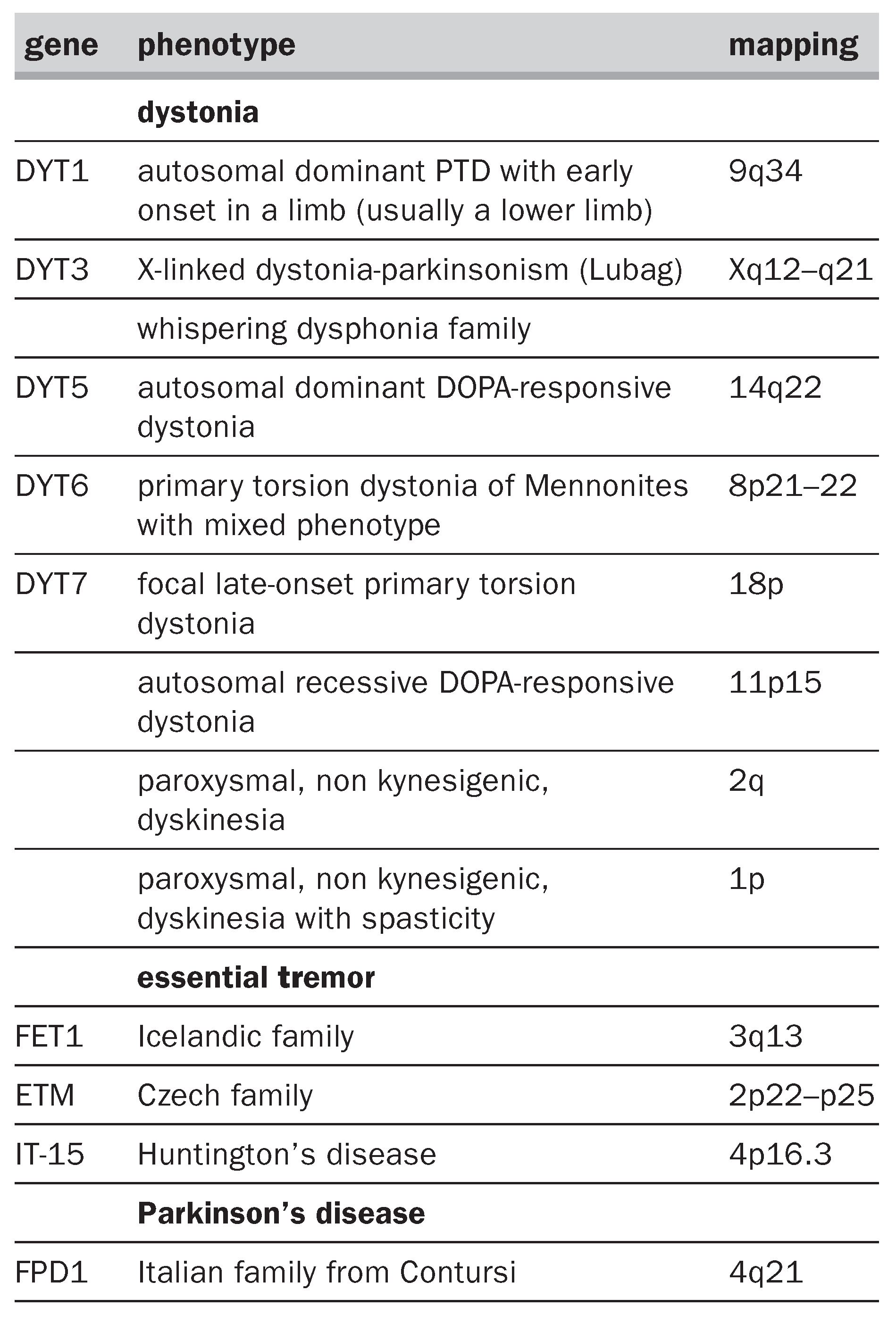
Most cases of PTD are inherited in an autosomal dominant pattern with reduced penetrance (30–40%) [15,16]. To date, 8 genes for well characterised forms of PTD have been identified (Table 1). Early-onset generalised PTD represents the most severe form of hereditary dystonia. The clinical spectrum is similar in all ethnic groups, with highest prevalence among Ashkenazi Jews due to a founder mutation [17]. The responsible gene (DYT1) has been linked to chromosome 9q34 [18,19,20] and this has recently been identified and sequenced. A 3-bp deletion in the coding sequence of the DYT1 gene was found in all affected and obligate carrier individuals with 9q-linked ITD, regardless of ethnic background and surrounding haplotype. The deletion results in loss of a glutamic-acid residue in a conserved region of a novel ATP-binding protein, termed torsin A [21]. A form of adult-onset, purely focal PTD has been linked to chromosome 18p in a German family [22], while in a German Mennonite family showing a mixed phenotype (variable age at onset, distribution and severity) a novel PTD gene has been mapped on chromosome 8 [23]. However, genetic studies performed on several ITD families have excluded linkage to known chromosomal locations [24,25,26,27,28].
It is clear that PTD is a clinically and genetically heterogeneous group of movement disorders; neuropathology is usually negative and the pathogenic mechanism leading to dystonia is still unknown. Several lines of evidence suggest that altered dopaminergic input to the basal ganglia may be implicated in dystonia. In addition to torsin A, only two other dystonia genes have been identified: GTP cyclohydrolase I on 14q [29] and tyrosine hydroxylase on 11p [30], which both implicate decreased dopaminergic transmission. Understanding the function of torsin A will help to elucidate the neuronal mechanism underlying loss of movement control in the basal ganglia, but further studies are required in order to determine whether clinically and genetically different forms of dystonia share a common underlying pathogenic mechanism.
The genes linked to different forms of paroxysmal, non kynesigenic dyskinesias have also been identified (see Table 1).
Table 1.
Mapped movement disorders genes.
Essential tremor
Essential tremor (ET) is the movement disorder with the highest prevalence all over the world. It is unknown to what extent ET clusters within families, and the role of genetic susceptibility in aetiology of ET has not been adequately investigated at the population level. Tremor in at least one first-degree relative is reported by 17% to 70% of patients with ET [31,32,33,34]. The high variability is probably due to methodological differences among the studies. It has also been shown that ET may coexist with ITD [35], thus requiring a thorough clinical analysis of the phenotype in each family member. Some studies suggest that earlyonset ET may be familial although increased awareness and earlier recognition of symptoms is an issue [36]. The lack of solid clinical genetic data explains why the genes responsible for ET have been elusive so far. Recently, a genome-wide scan performed in 16 Icelandic families affected by familial essential tremor inherited as a dominant trait, linked the disease to the long arm of chromosome 3 (3q13) [37]. A second gene responsible for “typical” ET has been found in a Czech family [38]. The responsible genes have not yet been identified.
Huntington’s disease
The cause of Huntington’s disease (HD) is an unstable expansion of a CAG trinucleotide repeat in the open reading frame of the IT15 gene on chromosome 4 [39]. The length of the CAG repeat in HD mutation does not appear to correlate with motor or psychiatric signs in presentation of the syndrome, or with the mode of progression after onset [40]. However, an inverse correlation between the number of CAG repeats and the age at onset has been demonstrated [41]. Confidence intervals for predicted age at onset, however, are quite broad and the number of CAG repeats accounts for only 50.4% variation in the age at onset [40]. Other genetic and environmental factors must influence age of onset and phenotypic expression in HD. In addition, it has been recently reported that the HD gene mutation has incomplete penetrance supporting the apparently healthy survival into old age of some individuals with 36–39 repeats [42]. The phenomenon of anticipation with paternal transmission of the gene is well recognised and large expansions in HD occur almost exclusively with paternal transmission [41,43].
The (CAG)n repeat (encoding for a polyglutamine tract) appeared to be located within the coding sequence of a predicted protein of about 348 kD (called huntingtin) that is widely expressed but unrelated to any known gene. Immunocytochemistry indicated that huntingtin is located in neurons throughout the brain, with the highest levels evident in larger neurones. In the human striatum, huntingtin was enriched in a patch-like distribution, potentially corresponding to the first areas affected in HD. The protein appears to be associated particularly with microtubules although some is associated with synaptic vesicles. Since a huntingtin-associated protein (HAP 1) is enriched in the brain, this may represent the possible basis for the selective brain pathology in HD [44]. The binding between the two proteins is enhanced by an expanded polyglutamate repeat. Also, it has been found that huntingtin is bound to a protein present in brain homogenates partially identical to the N terminus of glyceraldehyde-3-phosphatedehydrogenase (GAPD) which also bind to another protein with a polyglutamine tract, namely the DRPLA protein. Based on these findings, it has been postulated that diseases characterised by the presence of an expanded CAG repeat, which share a common mode of heritability, may also share a common metabolic pathogenesis involving GAPD as a functional component.
Several authors presented evidence that apoptosis is a mode of cell death in HD. Apopain, a human protease which plays a key role in proteolitic events leading to apoptosis, has been shown to cleave huntingtin, and the rate of cleavage increases with the length of the polyglutamine tract. These observations suggest that the disorder results from a gain-of-function mutation, in which anomalous protein–protein interactions may lead to inappropriate apoptosis [45,46].
Wilson’s disease
In Wilson’s disease (WD), the basal ganglia and liver undergo changes that express themselves in neurologic manifestations (such as tremor and dystonia) and signs of cirrhosis, respectively. A disturbance in copper metabolism is somehow involved in the mechanism. Typical features of the disease are high levels of copper in the liver and low levels of ceruloplasmin in the serum, presence of Kayser-Fleischer ring at the periphery of the cornea (due to copper deposits), hypercalciuria and nephrocalcinosis. The disease is treated successfully with copper chelant drugs, such as penicillamine and zinc salts. The disease is transmitted in an autosomal recessive manner. The world-wide prevalence of Wilson’s disease is estimated to be in the order of 30 per 1 million, with a gene frequency of 0.56% and a carrier frequency of 1 in 90. In 1985 the WD locus was assigned by linkage studies on the long arm of chromosome 13 and subsequently narrowed to the region 13q14.3. Physical mapping of this region identified a sequence similar to that coding for the proposed copper-binding regions of the putative ATPase gene defective in Menkes’ disease (an X-linked recessive disorder characterized by early retardation in growth, peculiar hair, and focal cerebral and cerebellar degeneration, and due to a defect in the intestinal absorption of copper) [47]. It has been shown that this sequence forms part of a P-type copper-transporting ATPase gene that is very similar to Menkes’ disease, with 6 putative metal-binding regions similar to those found in prokaryotic heavy metal transporters. The gene, expressed in liver and kidney, was found to lie within a 300-kb region likely to include the WD locus. Two WD patients were found to be homozygous for a 7-bp deletion within the coding region of the gene which was officially designated as ATP7B, the gene that is mutant in Menkes’ disease being ATP7A. More than 25 different mutations accounting for WD phenotype have been described so far.
Gilles de la Tourette’s syndrome
Gilles de la Tourette’s syndrome (TS) is a neurologic disorder manifested particularly by motor and vocal tics and associated with behavioral abnormalities such as obsessive-compulsive disorder (OCD). About three-fourths of patients are male, with onset occurring usually before 14 years. About 10% of patients have a family history of the same condition. In a large study on 338 biologic relatives of TS probands, 21 biologically unrelated relatives of adopted TS probands and 22 relatives of normal subjects, it has been shown that the rates of TS, chronic tics, and OCD in the total sample of biologic relatives of TS probands were significantly greater than in the relatives of controls [48]. In addition, the morbid risks of TS, OCD, and chronic tics were not significantly different in families of probands with OCD when compared to relatives of probands without OCD. These findings support the evidence that OCD is etiologically related to TS. Extended genetic studies suggested that the susceptibility to TS is conveyed by a major locus in combination with a multifactorial background. Other models of inheritance were definitely rejected, including strictly polygenic models, all single major locus models, and mixed models with dominant and recessive major loci. The frequency of the TS susceptibility allele was estimated to be 0.01. The major locus accounted for over half of the phenotypic variance for TS, whereas a multifactorial background accounted for approximately 40% of phenotypic variance.
Penetrance estimates suggested that all individuals homozygous for the susceptibility allele at the major locus are affected, whereas only 2.2% of males and 0.3% of females heterozygous at the major locus are affected. Of individuals affected by TS, approximately 62% are heterozygous and approximately 38% are homozygous at the major locus. While none of the families had 2 parents affected by TS, 19% of families had 2 parents affected with the broader TS phenotype which includes TS, chronic tic disorder, or OCD [49]. An autosomal dominant pattern of inheritance, with incomplete penetrance and variable expression, stands as the most widely accepted model of inheritance [50]. Assuming that there is a single genetic vulnerability factor identical in all families, about 80% of the genome could be excluded as the site for the TS gene by studies with over 600 DNA markers in an international collaborative effort. The frequency of bilineal (i.e. from both maternal and paternal sides) transmission of TS has been assessed in 39 families, in which 5 or more relatives were reported to be affected; bilineal transmission and homozygosity are common in TS and may play a role in severity of illness as well as account for difficulties in localizing the gene defect by linkage analysis [51].
References
- Gowers WR. A manual of diseases of the nervous system. v. 1. London: Churchill; 1886.
- Johnson WG, Hodge SE, Duvoisin RC. Twin studies and the genetics of Parkinson’s disease. A reappraisal. Mov Disord 1990;5:187–94.
- Burn DJ, Mark MH, Playford ED, Maraganore DM, Zimmerman TR, Duvoisin RC, et al. Parkinson’s disease in twins studied with 18F-DOPA and positron emission tomography. Neurology 1992;42:1894–900.
- Wood N. Genes and parkinsonism.J Neurol Neurosurg Psychiatry 1997;62:305–9.
- Golbe LI, Di Iorio G, Bonavita V, Miller DC, Duvoisin RC. A large kindred with autosomal dominant Parkinson’s disease. Ann Neurol 1990;27:276–82.
- Polymeropoulos MH, Higgins JJ, Golbe LI, Johnson WG, Ide SE, Di Iorio G, et al. Mapping a gene for Parkinson’s disease to chromosome 4q21–q23.Science 1996;274:1197–9.
- Polymeropoulos MH, Lavedan C, Leroy E, Ide SE, Dehejia A, Dutra A, et al. Mutation in the alpha-synuclein gene identified in families with Parkinson’s disease.Science 1997;276:2045–7.
- de Yébenes JG, Sarasa JL, Daniel SE, Lees AJ. Familial progressive supranuclear palsy. Description of a pedigree and review of the literature. Brain 1995;118:1095–103.
- Jankovic J, Fahn S. Dystonic disorders. In: Jankovic J, Tolosa E (editors). Parkinson’s disease and movement disorders. 2nd ed. Baltimore: Williams and Wilkins; 1993. p. 337–74.
- Albanese A. I gangli motori e i disturbi del movimento. Neurobiologia clinica. Padova: Piccin; 1991.
- Zilber N, Korkzyn AD, Kahana E, Fried K, Alter M. Inheritance of idiopathic torsion dystonia among Jews.
- Nutt JG, Muenter MD, Aronson A, Kurland LT, Melton LJ. Epidemiology of focal and generalyzed dystonia in Rochester, Minnesota. Mov Disord 1988;3:188–94.
- Defazio G, Livrea P, Guanti G, Lepore V, Ferrari E. Genetic contribution to idiopathic adult-onset blepharospasm and cranial-cervical dystonia. Eur Neurol 1993;33:345–50.
- Kandil MR, Tohamy SA, Fattah MA, Ahmed HN, Farwiez HM. Prevalence of chorea, dystonia and athetosis in Assiut, Egypt: a clinical and epidemiological study. Neuroepidemiology 1994;13:202–10.
- Fletcher NA, Harding AE, Marsden CD. A genetic study of idiopathic torsion dystonia in the United Kingdom.Brain 1990;113:379–95.
- Warner TT, Fletcher NA, Davis MB, Ahmad F, Conway D, Feve A, et al. Linkage analysis in British and French families with idiopathic torsion dystonia.Brain 1993;116:739–44.
- Bressman SB, Deleon D, Kramer PL, Ozelius LJ, Brin MF, Greene PE, et al. Dystonia in Ashkenazi Jews: clinical characterization of a founder mutation.Ann Neurol 1994;36:771–7.
- Kramer PL, de Leon D, Ozelius L, Risch N, Bressman SB, Brin MF, et al. Dystonia gene in Ashkenazi Jewish population is located on chromosome 9q32–34.Ann Neurol 1990;27:114–20.
- Kramer PL, Heiman GA, Gasser T, Ozelius LJ, de Leon D, Brin MF, et al. The DYT1 gene on 9q34 is responsible for most cases of early limb-onset idiopathic torsion dystonia in non-Jews. Am J Hum Genet 1994;55:468–75.
- Ozelius L, Kramer PL, Moskowitz CB, Kwiatkowski DJ, Brin MF, Bressman SB, et al. Human gene for torsion dystonia located on chromosome 9q32–q34.Neuron 1989;2:1427–34.
- Ozelius LJ, Hewett JW, Page CE, Bressman SB, Kramer PL, Shalish C, et al. The early-onset torsion dystonia gene (DYT1) encodes an ATP-binding protein.Nature Genet 1997;17:40–8.
- Leube B, Rudnicki D, Ratzlaff T, Kessler KR, Benecke R, Auburger G. Idiopathic torsion dystonia: assignment of a gene to chromosome 18p in a German family with adult onset, autosomal dominant inheritance and purely focal distribution. Hum Mol Genet 1996;5:1673–7.
- Almasy L, Bressman SB, Raymond D, Kramer PL, Greene PE, Heiman GA, et al. Idiopathic torsion dystonia linked to chromosome 8 in two Mennonite families.Ann Neurol 1997;42:670–3.
- Bentivoglio AR, Del Grosso N, Albanese A, Cassetta E, Tonali P, Frontali M. Non-DYT1 dystonia in a large Italian family. J Neurol Neurosurg Psychiatry 1997;62:357–60.
- Bressman SB, Hunt AL, Heiman GA, Brin MF, Burke RE, Fahn S, et al. Exclusion of the DYT1 locus in a non-Jewish family with early-onset dystonia.Mov Disord 1994;9:626–32.
- Bressman SB, Heiman GA, Nygaard TG, Ozelius LJ, Hunt AL, Brin MF, et al. A study of idiopathic torsion dystonia in a non-Jewish family: evidence for genetic heterogeneity. Neurology 1994;44:283–7.
- Bressman SB, Warner TT, Almasy L, Uitti RJ, Greene PE, Heiman GA, et al. Harding AE. Exclusion of the DYT1 locus in familial torticollis. Ann Neurol 1996;40:681–4.
- Holmgren G, Ozelius L, Forsgren L, Almay BG, Holmberg M, Kramer P, et al. Adult onset idiopathic torsion dystonia is excluded from the DYT1 region (9q34) in a Swedish family. J Neurol Neurosurg Psychiatry 1995;59:178–81.
- Furukawa Y, Shimadzu M, Rajput AH, Shimizu Y, Tagawa T, Mori H, et al. GTP-cyclohydrolase I gene mutations in hereditary progressive and dopa-responsive dystonia. Ann Neurol 1996;39:609–17.
- Ludecke B, Dworniczak B, Bartholome K. A point mutation in the tyrosine hydroxylase gene associated with Segawa’s syndrome. Hum Genet 1995;95:123–5.
- Hornabrook RW, Nagurney JT. Essential tremor in Papua, New Guinea. Brain 1976;99:659–72.
- Rajput AH, Offord KP, Beard CM, Kurland LT. Essential tremor in Rochester, Minnesota: a 45-year study. J Neurol Neurosurg Psychiatry 1984;47:466–70.
- Bain PG, Findley LJ, Thompson PD, Gresty MA, Rothwell JC, Harding AE, et al. A study of hereditary essential tremor. Brain 1994;117:805–24.
- Koller WC, Busenbark K, Miner K. The relationship of essential tremor to other movement disorders: report on 678 patients. Essential Tremor Study Group.Ann Neurol 1994;35:717–23.
- Jankovic J, Beach J, Pandolfo M, Patel PI. Familial essential tremor in 4 kindreds. Prospects for genetic mapping. Arch Neurol 1997;54:289–94.
- Louis ED, Ottman R. How familial is familial tremor? The genetic epidemiology of essential tremor. Neurology, 1996;46:1200–5.
- Gulcher JR, Jónsson P, Kong A, Kristjánsson K, Frigge ML, Kárason A, et al. Mapping of familial essential tremor gene, FET1, to chromosome 3q13.Nature Genet 1997;17:84–7.
- Higgins JJ, Pho LT, Nee LE. A gene (ETM) for essential tremor maps to chromosome 2p22–p25. Mov Disord 1997;12:859–64.
- The Huntington’s disease collaborative research group. A novel gene containing a trinucleotide repeat that is expanded and unstable on Huntington’s disease chromosomes. Cell 1993;72:971–83.
- Claes S, Van Zand K, Legius E, Dom R, Malfroid M, Baro F, et al. Correlations between triplet repeat expansion and clinical features in Huntington’s disease.Arch Neurol 1995;52:749–53.
- Ranen NG, Stine OC, Abbott MH, Sherr M, Codori AM, Franz ML, et al. Anticipation and instability of IT-15 (CAG)n repeats in parent-offspring pairs with Huntington’s disease. Am J Hum Genet 1995;57:593–602.
- Rubinsztein DC, Leggo J, Coles R, Almqvist E, Biancalana V, Cassiman JJ, et al. Phenotypic characterization of individuals with 30-40 CAG repeats in the Huntington’s disease (HD) gene reveals HD cases with 36 repeats and apparently normal elderly individuals with 36–39 repeats. Am J Hum Genet 1996;59:16–22.
- Norremolle A, Sorensen SA, Fenger K, Hasholt L. Correlation between magnitude of CAG repeat length alterations and length of the paternal repeat in paternally inherited Huntington’s disease. Clin Genet 1995;47:113–7.
- Li XJ, Li SH, Sharp AH, Nucifora FC Jr, Schilling G, Lanahan A, et al. A huntingtin-associated protein enriched in brain with implications for pathology.Nature 1995;378:398–402.
- Goldberg YP, Kalchman MA, Metzler M, Nasir J, Zeisler J, Graham R, et al. Absence of disease phenotype and intergenerational stability of the CAG repeat in transgenic mice expressing the human Huntington’s disease transcript. Hum Mol Genet 1996;5:177–85.
- Portera-Cailliau C, Hedreen JC, Price DL, Koliatsos VE. Evidence for apoptotic cell death in Huntington’s disease and excitotoxic animal models.J Neurosci 1995;15:3775–87.
- Bull PC, Thomas GR, Rommens JM, Forbes JR, Cox DW. The Wilson’s disease gene is a putative copper transporting p-type ATPase similar to the Menkes’ gene. Nature Genet 1993;5:327–37.
- Pauls DL, Raymond CL, Stevenson JM, Leckman JF. A family study of Gilles de la Tourette’s syndrome. Am J Hum Genet 1991;48:154–63.
- Walkup JT, La Buda MC, Singer HS, Brown J, Riddle MA, Hurko O. Family study and segregation analysis of Tourette’s syndrome: evidence for a mixed model of inheritance. Am J Hum Genet 1996;59:684–93.
- van de Wetering BJ, Heutink P. The genetics of the Gilles de la Tourette’s syndrome: a review. J Lab Clin Med 1993;121:638–45.
- Kurlan R, Eapen V, Stern J, McDermott MP, Robertson MM. Bilineal transmission in Tourette’s syndrome families. Neurology 1994;44:2336–42.
This is an open-access article distributed under the terms of the Creative Commons Attribution Non-Commercial No Derivatives (CC BY-NC-ND) 4.0 License.