


Simple Summary
Patients with immune deficiencies may develop both infections and inflammatory diseases, making it difficult to determine the cause of new or worsening symptoms. This diagnostic challenge becomes particularly important when symptoms resemble an existing autoimmune disease, as increasing immunosuppressive treatment may worsen an unrecognized infection. The present case illustrates this problem in a patient with common variable immunodeficiency and pre-existing psoriasis and psoriatic arthritis. It highlights the importance of considering infectious diseases when the clinical picture is atypical, involves several organ systems, or does not fit the expected course of the underlying disease. Careful clinical assessment and appropriate laboratory, pathological, and specialist investigations can help distinguish infection from inflammatory disease. Greater awareness of such diagnostic overlap may support earlier diagnosis and safer treatment decisions in patients with immune disorders.
Abstract
Introduction: Common variable immunodeficiency (CVID) is characterized by impaired antibody production and immune dysregulation, complicating the distinction between infection and autoimmunity. Aim: To present disseminated syphilis mimicking autoimmune disease in a patient with CVID. Case Presentation: A 26-year-old man with CVID, psoriasis, and psoriatic arthritis developed erythroderma, systemic inflammation, visual deterioration, and gastrointestinal symptoms suggestive of autoimmune exacerbation. Skin biopsy was inconclusive for psoriasis and raised suspicion of syphilis. Serology confirmed Treponema pallidum infection (RPR 1:128, TPHA positive). Cerebrospinal fluid analysis showed mild pleocytosis and elevated protein levels, with a negative CSF-VDRL. Ophthalmological examination revealed bilateral uveitis and optic nerve involvement. Immunological evaluation demonstrated persistent hypogammaglobulinemia and profound B-cell maturation defects with markedly reduced switched memory B cells. Ceftriaxone was administered because of a penicillin allergy, followed by a Jarisch–Herxheimer reaction and partial clinical improvement. Conclusions: This case highlights that disseminated syphilis may mimic autoimmune disease in a patient with CVID. Targeted infectious evaluation is essential to avoid inappropriate immunosuppression and enable timely antimicrobial treatment.
1. Introduction
Common variable immunodeficiency (CVID) comprises a heterogeneous group of inborn errors of immunity (IEI) characterized by impaired B-cell differentiation and defective immunoglobulin production, often accompanied by varying degrees of T-cell dysfunction [1]. Although recurrent infections are a hallmark of CVID, many patients also develop features of immune dysregulation, including autoimmune diseases, lymphoproliferative disorders, and an increased risk of malignancy [1,2]. In some cases, these manifestations may predominate and pose significant diagnostic challenges [2]. The coexistence of immunodeficiency and immune dysregulation makes it difficult to distinguish between infectious and autoimmune etiologies, particularly in patients presenting with systemic inflammatory features [2]. This distinction is especially important in the era of biological therapies, where inappropriate immunosuppression may worsen underlying infections [3].
Syphilis is a multisystem infection that may mimic a wide range of inflammatory and autoimmune diseases [4,5]. Its cutaneous manifestations may also resemble other dermatoses, including pityriasis rosea, infectious exanthems, and cutaneous drug eruptions [6,7]. Ocular and neurological involvement may further complicate the diagnosis, particularly in immunocompromised individuals [8].
Here, we present a case of disseminated syphilis in a patient with CVID, initially presenting with features suggestive of psoriatic arthritis (PsA) exacerbation. This case highlights the diagnostic complexity arising from the overlap between infection and immune-mediated disease.
2. Detailed Case Description
A 26-year-old man with CVID, initially treated with intravenous immunoglobulin (IVIG) since childhood and subsequently maintained on subcutaneous immunoglobulin (SCIG) replacement therapy (8 g weekly, 0.5 g/kg), was admitted for suspected syphilis with ocular manifestations and possible central nervous system involvement.
His medical history included chronic plaque psoriasis (since 2011), PsA, chronic sinusitis, bronchiectasis with fibrotic changes, and gastrointestinal inflammation.
At diagnosis in childhood, the patient had markedly reduced immunoglobulin levels (IgG 1.64 g/L; (reference range [RR]: 6.38–17.00 g/L), IgA 0.21 g/L; (RR: 0.7–4.0 g/L), IgM 0.27 g/L (RR: 0.4–2.3 g/L), with preserved T-cell proliferative responses to mitogens and anti-CD3 stimulation. Immunophenotyping revealed an increased proportion of CD19+ B cells (33%; 1158/µL; RR: 7.0–23.00%; 110–530/µL). Detailed B-cell subset analysis demonstrated a predominance of naïve B cells (IgD+CD27−, 95.8% of CD19+ cells; RR: 2.52–14.05%) and markedly reduced switched memory B cells (IgD−CD27+, 0.3%; RR: 0.95–4.28%), as well as non-switched memory B cells (IgD+CD27+, 2.3%; RR: 0.74–4.73%), consistent with a severe B-cell maturation defect typical of CVID. No genetic testing for monogenic causes of inborn errors of immunity has been performed, and no history of autoimmune cytopenias was documented.
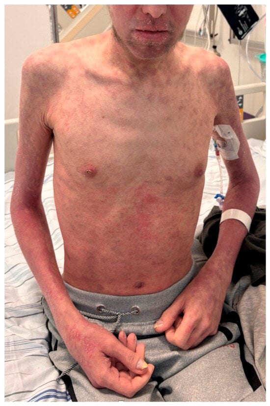
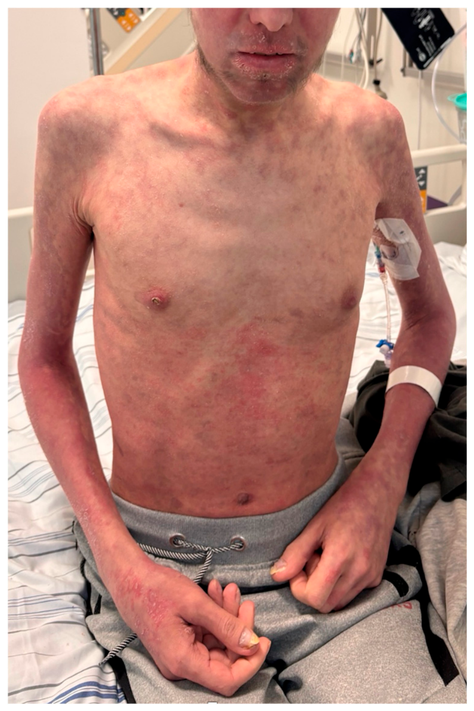
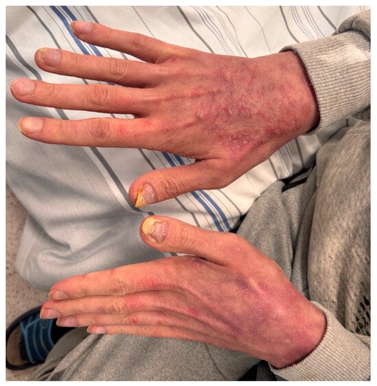
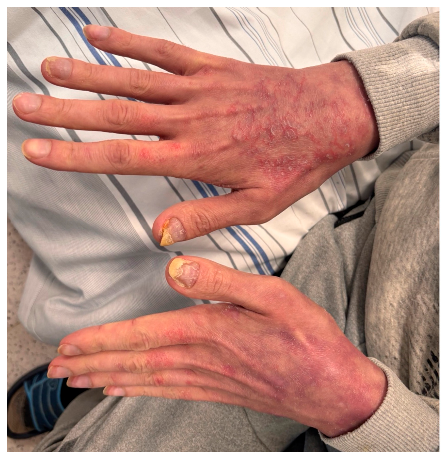
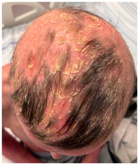
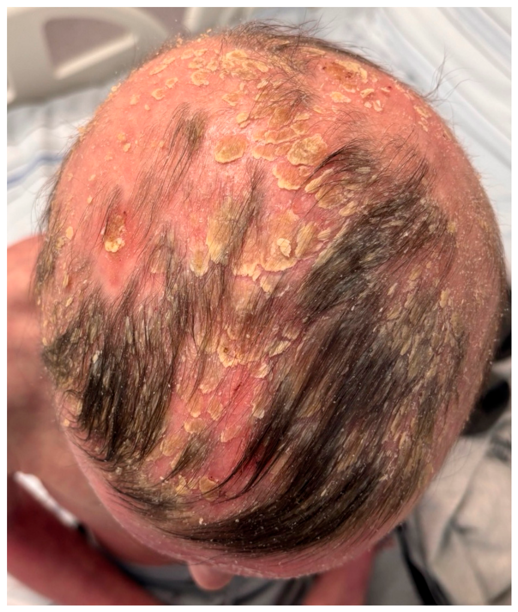
The patient was referred to the Department of Rheumatology and Immunology due to suspected exacerbation of PsA. Since August 2025, he has developed progressive erythroderma with generalized scaling and episodic joint pain and swelling. Methotrexate was discontinued after three doses due to adverse effects, including patchy hair loss involving the scalp, eyebrows, and eyelashes (Figure 1, Figure 2 and Figure 3).
Figure 1.
Erythematous and desquamative lesions on the face, trunk and limbs; hyperkeratotic lesions on the scalp.
Figure 2.
Erythematous and desquamative lesions localized on the dorsal surface of the palms, onycholysis and subungual hyperkeratosis.
Figure 3.
Hyperkeratotic lesions on the scalp, focal alopecia (“moth-eaten alopecia”).
From November 2025, the patient reported gradually progressive visual impairment characterized by blurred vision and increasing floaters in both eyes. Since December 2025, he has experienced chronic diarrhea, significant weight loss (10 kg), and intermittent fever up to 40 °C. Epidemiological history revealed high-risk sexual behavior (men who have sex with men, unprotected sexual contacts). Screening for blood-borne infections was performed. HIV-1/2 RNA, hepatitis B virus (HBV) DNA, and hepatitis C virus (HCV) RNA tests were negative. In light of the patient’s high-risk sexual behavior, extended microbiological screening was performed. Multiplex PCR testing of a urine sample yielded negative results for Chlamydia trachomatis (including LGV serovars), Neisseria gonorrhoeae, Mycoplasma genitalium, Mycoplasma hominis, Ureaplasma urealyticum, Ureaplasma parvum, Haemophilus ducreyi, Trichomonas vaginalis, and herpes simplex viruses (HSV-1 and HSV-2). Although Treponema pallidum was not detected by multiplex PCR, serological testing for syphilis was positive. Anorectal testing for Chlamydia trachomatis and Neisseria gonorrhoeae was not performed, and anal HPV infection status was not evaluated. HPV and HBV vaccinations were recommended.
On admission, the patient was in good general condition. Physical examination revealed tachycardia (100 bpm), generalized erythematous scaly lesions, areas of hair loss, psoriatic nail changes, bilateral ankle edema, and inguinal lymphadenopathy.
Laboratory findings showed elevated inflammatory markers (CRP 77 mg/L; RR: <5.0 mg/L), neutrophilic leukocytosis, lymphopenia, and mild microcytic anemia. Severe vitamin D deficiency was noted. Stool calprotectin was markedly elevated (5237 µg/g; RR: <50 µg/g). Follow-up immunological assessment confirmed persistent hypogammaglobulinemia despite replacement therapy (IgG as low as 4.8 g/L, IgA < 0.23 g/L, IgM 0.45 g/L). Extended immunophenotyping confirmed markedly reduced switched memory B cells (0.3% of CD19+; 0.04% of lymphocytes), reduced non-switched memory B cells, predominance of naïve B cells, and increased CD21^low B cells (15.4%). T-cell analysis showed lymphopenia with decreased CD8+ T cells.
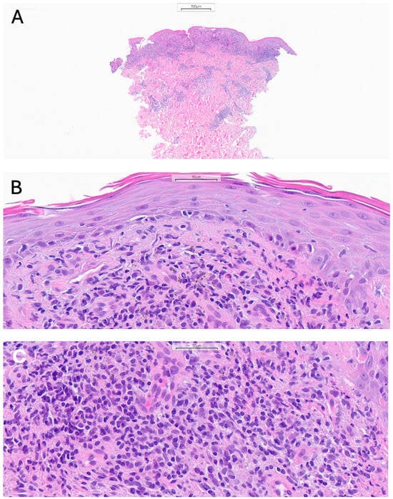
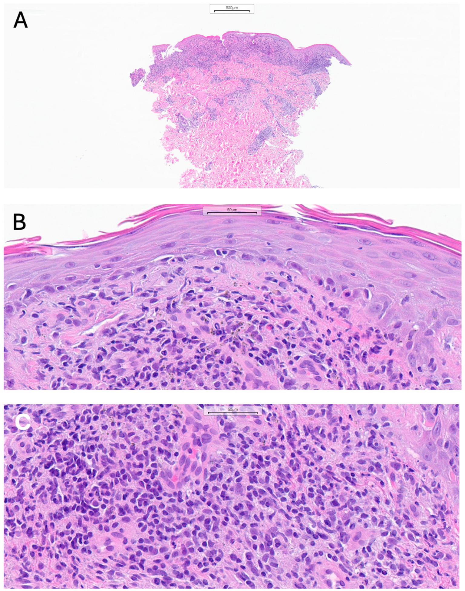
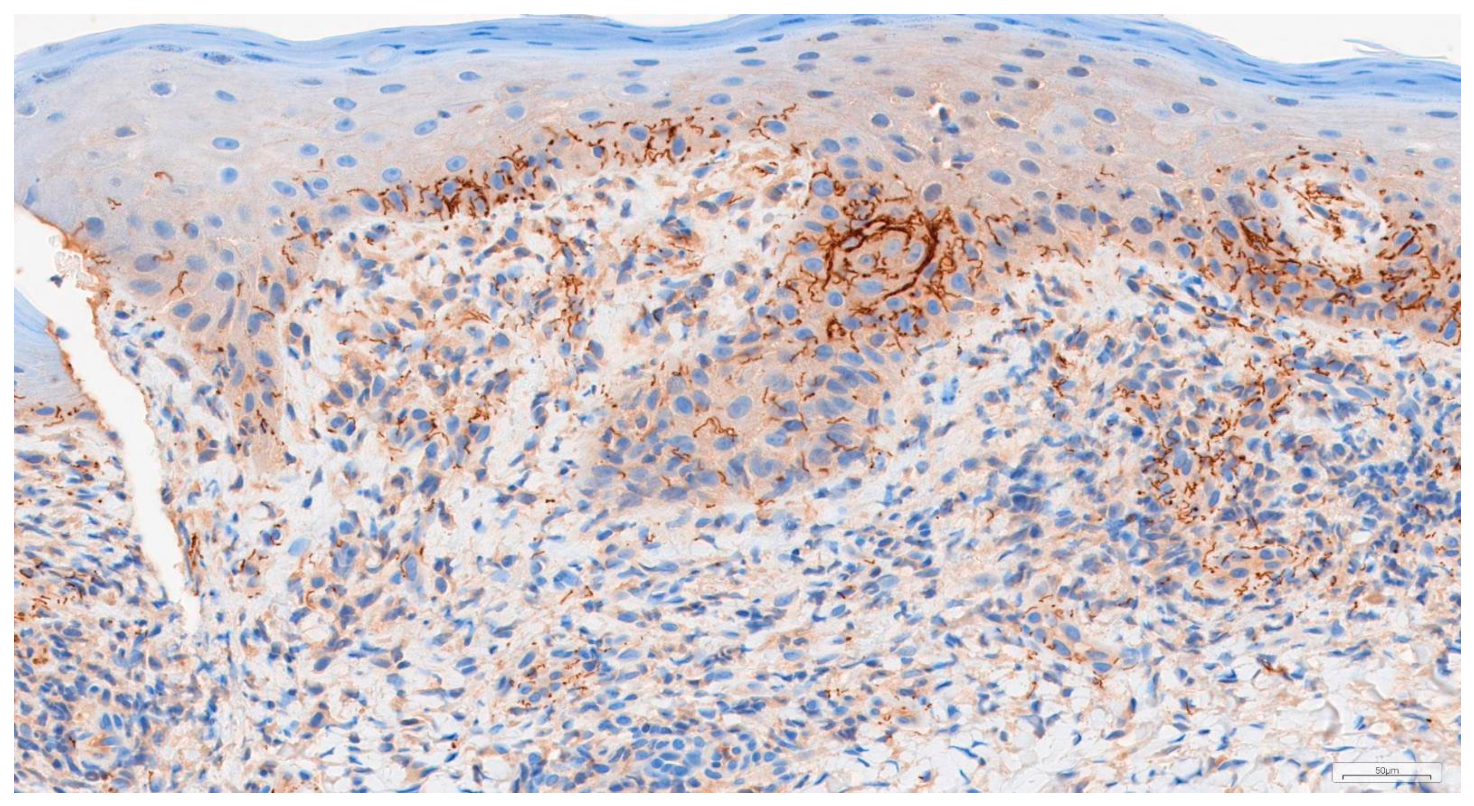
Histopathological examination of the skin demonstrated a partially atrophic epidermis with spongiosis and a dense lymphocytic infiltrate at the dermoepidermal junction with occasional eosinophils, accompanied by scattered colloid bodies and small periadnexal granulomas; no evidence of psoriasis or lymphoma was identified, while syphilis could not be excluded (Figure 4 and Figure 5). These findings raised suspicion of an underlying infectious process mimicking autoimmune disease. Serological testing revealed a positive VDRL/RPR (titer 1:128), and infection with Treponema pallidum was confirmed by TPHA. Cerebrospinal fluid (CSF) analysis showed mild pleocytosis and elevated protein levels, while CSF-VDRL was negative.
Figure 4.
Histopathological examination of the skin (hematoxylin and eosin staining). (A) Low-power overview of the biopsy specimen (scale bar: 500 µm). (B,C) Higher-magnification views showing focal interface dermatitis with scattered colloid bodies and a dense dermal inflammatory infiltrate composed predominantly of lymphocytes with some plasma cells. A small granuloma and pigment deposition are also present (scale bars: 50 µm).
Figure 5.
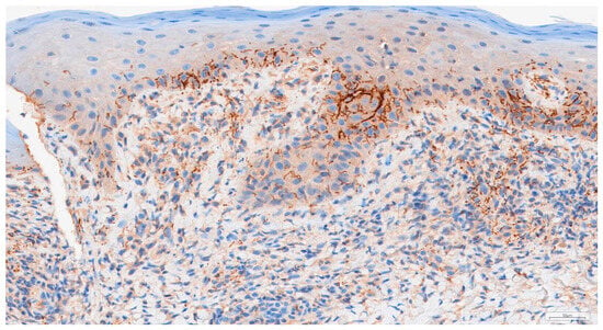
Numerous microorganisms can be seen in epidermis and in papillary dermis (Treponema pallidum immunostain, policlolnal antibody, BioSB, obj × 40, scale bar: 50 µm).
Magnetic resonance imaging (MRI) of the brain showed no parenchymal abnormalities, although extensive inflammatory changes were noted in the paranasal sinuses.
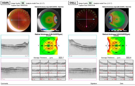
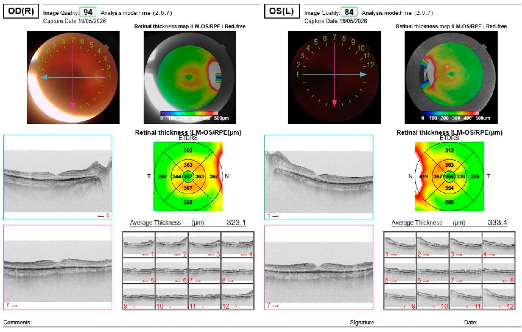
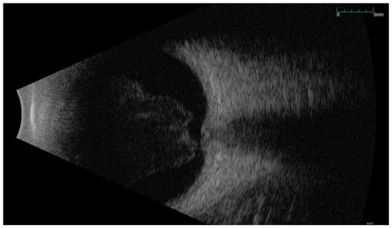
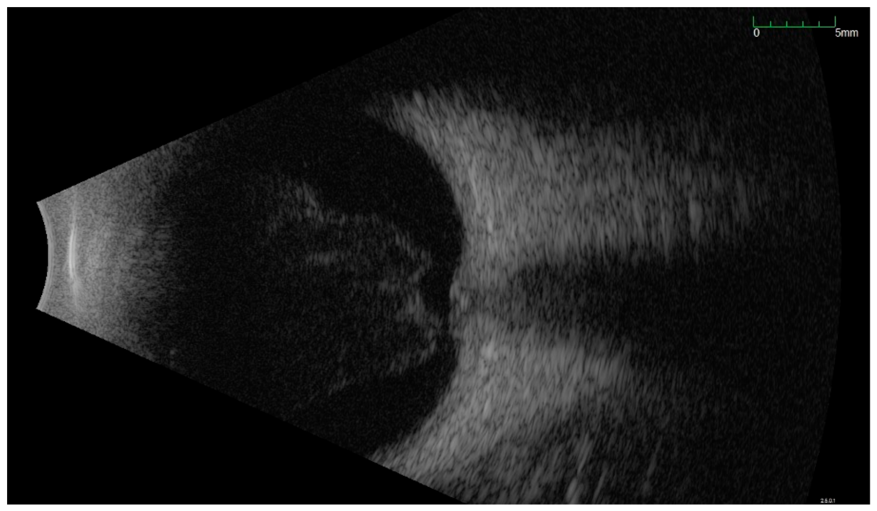
Further clinical evaluation revealed bilateral inflammatory eye disease characterized by interstitial keratitis, anterior and posterior uveitis, and optic nerve involvement. Fundus examination was partially obscured by dense vitreous inflammatory opacities. Fundus examination demonstrated a poorly delineated optic disc with features suggestive of mild-to-moderate optic disc swelling in both eyes. Optical coherence tomography (OCT) confirmed bilateral optic disc edema (Figure 6). In addition, OCT revealed hyperreflective vitreous opacities adjacent to the optic nerve head, consistent with inflammatory vitreous exudation, and intraretinal fluid (IRF) in the fovea of the left eye. B-scan ultrasonography demonstrated bilateral prominence of the optic nerve heads consistent with optic disc edema, together with hyperechogenic vitreous opacities suggestive of active inflammatory vitreous infiltration (Figure 7). Orbital MRI demonstrated bilateral optic nerve sheath enhancement, consistent with inflammatory optic nerve involvement. In view of the confirmed serological diagnosis of syphilis and compatible ophthalmological findings, ocular syphilis was suspected. Although mild CSF pleocytosis and elevated protein levels raised concern for central nervous system involvement, the negative CSF-VDRL and nonspecific CSF abnormalities did not allow definitive confirmation of neurosyphilis.
Figure 6.
Optical coherence tomography (OCT) of both eyes demonstrating bilateral optic disc edema with hyperreflective vitreous opacities adjacent to the optic nerve head. A small amount of intraretinal fluid is visible in the foveal region of the left eye.
Figure 7.
B-scan ultrasonography demonstrating prominent optic nerve heads consistent with optic disc edema and echogenic vitreous opacities consistent with vitreous inflammation.
Best-corrected visual acuity (BCVA), measured using Snellen charts, improved markedly after four months of treatment, from 0.1 to 1.0 in the right eye and from hand motion (HM) to 0.7 in the left eye. This functional improvement was accompanied by regression of optic disc edema and inflammatory vitreous changes.
Due to penicillin allergy, intravenous ceftriaxone was initiated. Shortly after the first dose, the patient developed fever, chills, hypotension, and transient worsening of skin lesions, consistent with a Jarisch–Herxheimer reaction. Following antimicrobial therapy, partial clinical improvement was observed, including reduction in skin lesions, improvement in visual function, and alleviation of gastrointestinal symptoms. This clinical response supported an infectious rather than autoimmune etiology.
3. Discussion
To the best of our knowledge, this is the first reported case of disseminated syphilis mimicking PsA in a patient with CVID in the context of a pronounced immune dysregulation phenotype. This case illustrates the complex interplay between infection and immune dysregulation, where clinical manifestations may closely resemble autoimmune disease and lead to potential diagnostic delay. This diagnostic complexity may be particularly relevant in patients with psoriatic arthritis, in whom multisystem comorbidities and overlapping inflammatory manifestations may further complicate the clinical assessment. Recent nationwide Polish data demonstrated distinct patterns of comorbidity burden and healthcare utilization among patients with newly diagnosed rheumatoid arthritis and psoriatic arthritis, emphasizing the complexity of the clinical context in which inflammatory manifestations are evaluated [9].
The interaction between syphilis and immunodeficiency has been most extensively characterized in the setting of HIV infection. Numerous studies have described a bidirectional relationship between these two conditions, suggesting that impaired host immunity may contribute to the dissemination and atypical presentation of syphilis, while syphilitic infection itself may facilitate viral transmission through mucosal disruption and local immune activation [10,11,12,13]. In HIV-positive individuals, syphilis has been associated with more aggressive and multisystem disease, including increased rates of neurological, ocular, and otological involvement [11,12,14,15]. These observations suggest that host immune status may be relevant to the clinical presentation of syphilis; however, they do not establish that other forms of immunodeficiency have similar effects. HIV infection and CVID differ fundamentally in their underlying immunopathological mechanisms, and findings from HIV-associated syphilis cannot be directly extrapolated to patients with CVID.
Although CVID differs fundamentally from HIV infection in its pathophysiology, it is characterized by defective B-cell differentiation, impaired immunoglobulin production, and varying degrees of immune dysregulation [1,2]. The immunological profile observed in our patient, including markedly reduced switched memory B cells, predominance of naïve B cells, and expansion of CD21low B cells, is consistent with profound abnormalities of humoral immunity and B-cell maturation previously described in CVID [16,17]. These immunological abnormalities have been associated with the heterogeneous clinical and immunological phenotype of CVID, including impaired humoral immune responses and immune dysregulation [16,17]. Although such abnormalities may theoretically influence host responses to infectious pathogens, the present findings do not demonstrate impaired clearance of Treponema pallidum or establish a specific effect of the patient’s B-cell phenotype on the severity or dissemination of syphilis.
In CVID, immunodeficiency and immune dysregulation frequently coexist and may contribute to the complexity of infectious and inflammatory manifestations [18]. These processes are not necessarily mutually exclusive and may complicate the interpretation of systemic clinical findings. Impaired antibody-mediated immunity may theoretically affect pathogen control and contribute to infection persistence, whereas dysregulated immune responses may contribute to excessive or prolonged inflammation and clinical manifestations resembling autoimmune disease [16,17,18]. However, the extent to which these mechanisms operate in syphilis infection in patients with CVID remains unknown. In our patient, the coexistence of pre-existing psoriasis and PsA with the multisystem manifestations of syphilis created considerable diagnostic uncertainty and initially suggested an exacerbation of the underlying inflammatory disease. Although the profound abnormalities in humoral immunity provide an important immunological context, the available clinical and laboratory findings do not demonstrate that CVID-related immune abnormalities caused the dissemination of infection or amplified its inflammatory manifestations. The potential contribution of CVID to the clinical course of syphilis in this patient therefore remains uncertain.
Syphilis can present with a wide range of dermatological and systemic manifestations, often resembling other diseases [10,12]. Secondary syphilis, in particular, may present with psoriasiform eruptions, generalized scaling, and even erythroderma, closely resembling psoriasis both clinically and histopathologically [19,20]. In addition, infections are known triggers of psoriasis exacerbations through activation of immune pathways, including T helper (Th)17-mediated inflammation, further blurring the distinction between infection and autoimmune flare [20]. In our patient, the coexistence of pre-existing psoriasis likely further obscured the clinical picture, as similar cases of concurrent psoriasis and syphilis have been reported [13].
The multisystem involvement observed in this case, including dermatological and ocular manifestations and possible central nervous system involvement, is compatible with disseminated syphilis. Ocular manifestations, such as uveitis, keratitis, and optic nerve inflammation, are well documented in syphilis [11,13]. Importantly, studies have shown that advanced diagnostic approaches, including Treponema pallidum-specific testing in ocular fluids, may be required in complex cases [14,21]. The presence of visual symptoms in our patient represented a key diagnostic clue and should prompt consideration of infectious etiologies in similar clinical scenarios. The present case does not allow us to determine whether CVID contributed to the extent or severity of ocular or other organ involvement.
Another important consideration is the potential influence of underlying immunodeficiency on the clinical presentation and diagnostic assessment of syphilis. Although atypical clinical manifestations and altered serological responses have been described in patients with HIV infection [12,21], the extent to which similar phenomena may occur in CVID remains largely unknown. Given the impaired antibody production and broader immune dysregulation characteristic of CVID, a potential influence on the host response to Treponema pallidum infection cannot be excluded. However, this remains a theoretical consideration, and the present case does not establish a direct causal relationship between CVID and the dissemination or atypical presentation of syphilis.
From a therapeutic perspective, this case highlights the importance of excluding infection before initiating or escalating immunosuppressive or biological therapy. In patients with suspected autoimmune disease and underlying immunodeficiency, an unrecognized infection may be mistakenly interpreted as inflammatory disease, potentially delaying appropriate antimicrobial treatment. In our patient, biological therapy was withheld and antimicrobial treatment was initiated, followed by clinical improvement. This response supports an infectious rather than autoimmune explanation for the new multisystem manifestations, although a single case cannot establish the effects that immunosuppressive treatment would have had on the course of infection. The Jarisch-Herxheimer reaction shortly after initiation of antimicrobial therapy further supported active spirochetal infection [22].
Epidemiologically, the patient’s history of high-risk sexual behavior is also of importance. Men who have sex with men represent a population with a disproportionately high burden of syphilis and HIV co-infection, highlighting the need for targeted screening and prevention strategies [10,13]. Although our patient was HIV-negative, similar risk factors remain relevant for syphilis acquisition and should be systematically assessed in clinical practice.
Overall, this case illustrates the diagnostic challenges associated with the coexistence of immunodeficiency, immune dysregulation, and disseminated infection. Although the coexistence of CVID and disseminated syphilis raises the question of whether impaired humoral immunity may influence disease presentation, a single case cannot establish such an association. The findings emphasize the importance of maintaining a high index of suspicion for infectious etiologies in patients with CVID presenting with new, atypical, or treatment-refractory systemic inflammatory manifestations.
4. Conclusions
This case highlights the importance of considering syphilis in the differential diagnosis of new or worsening manifestations resembling psoriatic arthritis in patients with CVID. Although the coexistence of CVID and disseminated syphilis raises questions about the potential influence of immunodeficiency on disease presentation, this single observation cannot establish a causal relationship. Further studies are needed to determine whether CVID influences the clinical course or presentation of syphilis.
Author Contributions
Conceptualization, G.R., J.K.-W. and M.S. (Magdalena Strach); methodology, J.K.-W.; resources, G.R., M.M.-R., A.P., M.S. (Magdalena Spałkowska), I.K.-B., G.D. and A.K.-N.; data curation, G.R. and J.K.-W.; writing—all authors, J.K.-W.; writing—review and editing, J.K.-W. and M.S. (Magdalena Strach); visualization, J.K.-W.; supervision, J.K.-W. and M.S. (Magdalena Strach); project administration, J.K.-W. All authors have read and agreed to the published version of the manuscript.
Funding
This research received no external funding.
Institutional Review Board Statement
Ethical review and approval of this study were not required by the Institutional Review Board of the Bioethical Committee of Jagiellonian University (Kraków, Poland), because case reports are not considered research. The patient’s information has been de-identified.
Informed Consent Statement
The informed consent for participation was waived by the Institutional Review Board of the Bioethical Committee of Jagiellonian University (Kraków, Poland), due to the retrospective nature of the study and its medical record review design.
Data Availability Statement
The data presented in this study are available on reasonable request from the corresponding author.
Acknowledgments
During the preparation of this manuscript, the author used Claude (Opus 5) and Codex (Sol) to assist with language editing and with reviewing the statistical analysis code. The author reviewed and edited all output and takes full responsibility for the content of this publication.
Conflicts of Interest
The authors declare no conflicts of interest.
Abbreviations
The following abbreviations are used in this manuscript:
| BCVA | Best-corrected visual acuity |
| CVID | Common variable immunodeficiency |
| HBV | Hepatitis type B virus |
| HCV | Hepatitis type C virus |
| HIV | Human immunodeficiency virus |
| HM | hand motion |
| Ig | Immunoglobulin |
| IRF | Intraretinal fluid |
| IVIG | Intravenous immunoglobulin |
| MRI | Magnetic resonance imaging |
| OCT | Optical coherence tomography |
| PsA | Psoriatic arthritis |
| RR | Reference range |
| SCIG | Subcutaneous immunoglobulin |
References
- Bylica, J.; Karpiński, M.; Korkosz, M.; Kosałka-Węgiel, J. NF-κB1 haploinsufficiency due to a novel c.104delT variant in two patients with common variable immunodeficiency. Clin. Immunol. 2026, 285, 110703. [Google Scholar] [CrossRef] [Scilit] [PubMed]
- Fernando, S.L.; Jang, H.S.; Li, J. The Immune Dysregulation of Common Variable Immunodeficiency Disorders. Immunol. Lett. 2021, 230, 21–26. [Google Scholar] [CrossRef] [Scilit] [PubMed]
- Sadi Aykan, F.; Çölkesen, F.; Evcen, R.; Kılınç, M.; Yıldız, E.; Arslan, Ş. Common variable immunodeficiency and autoimmune diseases: A 10-year single-center experience. Arch. Rheumatol. 2024, 39, 588–597. [Google Scholar] [CrossRef] [Scilit] [PubMed]
- Vasconcelos, P.; Borges-Costa, J.; Loreto, H.; Marques, S. Secondary syphilis mimicking inflammatory bowel disease. Sex. Transm. Infect. 2013, 89, 98–99. [Google Scholar] [CrossRef] [Scilit] [PubMed]
- Shatley, M.J.; Walker, B.L.; McMurray, R.W. Lues and lupus: Syphilis mimicking systemic lupus erythematosus (SLE). Lupus 2001, 10, 299–303. [Google Scholar] [CrossRef] [Scilit] [PubMed]
- Drago, F.; Ciccarese, G.; Rebora, A.; Parodi, A. Relapsing pityriasis rosea. Dermatology 2014, 229, 316–318. [Google Scholar] [CrossRef] [Scilit] [PubMed]
- Vrysis, C.; Katsarou, A.; Lytras, T.; Katsikas, K.; Falagas, M.E. Rash associated with antibiotic administration in patients with infectious mononucleosis: A systematic review and meta-analysis. Eur. J. Clin. Microbiol. Infect. Dis. 2025, 44, 2183–2196. [Google Scholar] [CrossRef] [Scilit] [PubMed]
- Fanaei, A.; Metivier, A.J.; Morreale, P. Ocular Neurosyphilis in an Immunocompetent Patient Without HIV: A Diagnostic Challenge. Cureus 2025, 17, e95895. [Google Scholar] [CrossRef] [Scilit] [PubMed]
- Zaręba, W.; Szeląg, M.; Batko, K.; Krawiec, P.; Stajszczyk, M.; Podwójcic, K.; Żuber, Z.; Krajewska-Włodarczyk, M.; Batko, B. Comorbidity profiles at diagnosis in rheumatoid arthritis versus psoriatic arthritis: A nationwide Polish claims-based study. J. Clin. Med. 2026, 15, 5249. [Google Scholar] [CrossRef] [Scilit] [PubMed]
- Puspawati, N.M.D.; Yasa, I.W.P.S.; Manuaba, I.B.P.; Wande, I.N.; Somia, K.A. Bidirectional relationship of HIV and syphilis coinfection: A pathophysiological and clinical review. Arch. Balk. Med. Union 2025, 60, 258–265. [Google Scholar] [CrossRef] [Scilit]
- Zhou, H.Y.; Ye, J.J. Research progress of ocular manifestations and treatments of human immunodeficiency virus and syphilis co-infection. Zhonghua Yan Ke Za Zhi 2021, 57, 865–870. [Google Scholar] [CrossRef] [Scilit] [PubMed]
- Weintrob, A.; Crum-Cianflone, N.; Michael, N. Syphilis and Human Immunodeficiency Virus Coinfection: More Than the Sum of Its Parts. Infect. Dis. Clin. Pract. 2006, 14, 197–203. [Google Scholar] [CrossRef] [Scilit]
- Refugio, O.N.; Klausner, J.D. Syphilis incidence in men who have sex with men with human immunodeficiency virus comorbidity and the importance of integrating sexually transmitted infection prevention into HIV care. Expert Rev. Anti Infect. Ther. 2018, 16, 321–331. [Google Scholar] [CrossRef] [Scilit] [PubMed]
- Nair, N.; Sudharshan, S.; Anand, A.R.; Biswas, J.; Therese, K.L. Utility of Treponemal Testing from Aqueous Fluid in the Diagnosis of Ocular Syphilis in Patients with HIV/AIDS. Ocul. Immunol. Inflamm. 2022, 30, 444–450. [Google Scholar] [CrossRef] [Scilit] [PubMed]
- Witt, L.S.; Wendy Fujita, A.; Ho, J.; Shin, Y.M.; Kobaidze, K.; Workowski, K. Otosyphilis. Open Forum Infect. Dis. 2022, 10, ofac685. [Google Scholar] [CrossRef] [Scilit] [PubMed]
- Warnatz, K.; Denz, A.; Dräger, R.; Braun, M.; Groth, C.; Wolff-Vorbeck, G.; Eibel, H.; Schlesier, M.; Peter, H.H. Severe deficiency of switched memory B cells (CD27(+)IgM(-)IgD(-)) in subgroups of patients with common variable immunodeficiency: A new approach to classify a heterogeneous disease. Blood 2002, 99, 1544–1551. [Google Scholar] [CrossRef] [Scilit] [PubMed]
- Wehr, C.; Kivioja, T.; Schmitt, C.; Ferry, B.; Witte, T.; Eren, E.; Vlkova, M.; Hernandez, M.; Detkova, D.; Bos, P.R.; et al. The EUROclass trial: Defining subgroups in common variable immunodeficiency. Blood 2008, 111, 77–85. [Google Scholar] [CrossRef] [Scilit] [PubMed]
- Ho, H.E.; Cunningham-Rundles, C. Non-infectious Complications of Common Variable Immunodeficiency: Updated Clinical Spectrum, Sequelae, and Insights to Pathogenesis. Front. Immunol. 2020, 11, 149. [Google Scholar] [CrossRef] [Scilit] [PubMed]
- Santana, A.W.A.; Campos, C.G.; Soares, Q.S.; Salgado, C.C.C.R.; Souza, M.A. Severe syphilis in patient infected with HIV. Med. Cutánea Ibero-Lat.-Am. 2009, 37, 98–101. [Google Scholar] [CrossRef] [Scilit] [PubMed]
- Melin, U.N.; Dharmasanti, P.A. Secondary Syphilis with Multiple Co-Infections of Human Immunodeficiency Virus (HIV), Tuberculosis (TB), and Oral Candidiasis in a 19-Year-Old Men Who Have Sex with Men (MSM): A Case Report. Berk. Ilmu Kesehat. Kulit Dan Kelamin 2024, 36, 225–231. [Google Scholar] [CrossRef] [Scilit]
- Rotty, J.; Anderson, D.; Garcia, M.; Diaz, J.; Van de Waarsenburg, S.; Howard, T.; Dennison, A.; Lewin, S.R.; Elliott, J.H.; Hoy, J. Preliminary assessment of Treponema pallidum-specific IgM antibody detection and a new rapid point-of-care assay for the diagnosis of syphilis in human immunodeficiency virus-1-infected patients. Int. J. STD AIDS 2010, 21, 758–764. [Google Scholar] [CrossRef] [Scilit] [PubMed]
- Wood, J.M.; Sbar, E. Jarisch-Herxheimer reaction [updated 2025 Jul 7]. In StatPearls [Internet]; StatPearls Publishing: Treasure Island, FL, USA, 2026. [Google Scholar]
Disclaimer/Publisher’s Note: The statements, opinions and data contained in all publications are solely those of the individual author(s) and contributor(s) and not of MDPI and/or the editor(s). MDPI and/or the editor(s) disclaim responsibility for any injury to people or property resulting from any ideas, methods, instructions or products referred to in the content. |
© 2026 by the authors. Published by MDPI on behalf of the European Society of Dermatopathology. Licensee MDPI, Basel, Switzerland. This article is an open access article distributed under the terms and conditions of the Creative Commons Attribution (CC BY) license.