


Simple Summary
Eosinophilic, polymorphic, and pruritic eruption associated with radiotherapy is a rare skin reaction that can occur during or after radiation treatment. It may cause a widespread itchy rash with different types of skin lesions, including blisters, and can resemble other inflammatory or autoimmune skin diseases. We report a 59-year-old man with prostate cancer who developed a rapidly spreading blistering eruption shortly after starting pelvic radiotherapy. A skin biopsy showed eosinophil-rich inflammation, supporting the diagnosis. The eruption improved with corticosteroid treatment, and radiotherapy was later resumed and completed. This case highlights the importance of linking new skin eruptions to recent radiotherapy and of close communication between clinicians and pathologists to ensure the correct diagnosis.
Abstract
Eosinophilic, polymorphic, and pruritic eruption associated with radiotherapy (EPPER) is a rare and probably underrecognized cutaneous reaction to ionizing radiation with marked clinicopathological variability. We report a 59-year-old patient with prostate adenocarcinoma who developed a rapidly progressive, generalized pruritic eruption with tense bullae approximately two weeks after initiation of pelvic radiotherapy. The eruption began in the gluteal region and spread to the trunk and extremities, accompanied by shallow buccal erosions and mild systemic inflammation. Diagnostic evaluation showed no evidence of an autoimmune blistering disorder. Histopathology revealed an eosinophil-rich spongiotic dermatitis with vesiculobullous changes, supporting EPPER in the appropriate clinical context. With continued systemic and topical corticosteroid therapy, the eruption gradually resolved. Radiotherapy was temporarily interrupted but subsequently resumed and completed under corticosteroid treatment, with only a mild self-limited flare and no recurrent bullae. This case highlights the vesiculobullous spectrum of EPPER and underscores the importance of the temporal relationship to radiotherapy and close clinicopathological correlation for accurate diagnosis.
1. Introduction
Eosinophilic, polymorphic, and pruritic eruption associated with radiotherapy (EPPER) is a rare and probably underrecognized cutaneous reaction to ionizing radiation, first described by Rueda et al. in 1999 [1]. It may develop during radiotherapy or after treatment completion, with delayed onset occurring several months after irradiation in some patients. Although lesions may initially be confined to the irradiated field, the eruption can extend well beyond the treated area and become widespread. EPPER is characterized by marked polymorphism and intense pruritus. Reported manifestations include erythematous macules and papules, plaques, pustules, vesicles, nodules, excoriations, and tense bullae [1,2,3,4,5,6,7,8]. This broad clinical spectrum has historically led to diagnostic confusion with other inflammatory and autoimmune dermatoses, particularly erythema multiforme and bullous pemphigoid [1,6].
Histologically, EPPER is characterized by eosinophilic inflammation with considerable variation in its distribution and associated epidermal changes. Eosinophilic inflammation is a consistent feature, whereas the distribution of the inflammatory infiltrate and accompanying epidermal changes vary considerably. The dermal infiltrate may be superficial and/or deep, with perivascular, interstitial, or periadnexal patterns, while epidermal changes include eosinophilic spongiosis, focal spongiotic vesiculation, and vesiculopustular changes [1,2,3,4,5,6,7,8]. In bullous variants, subepidermal blister formation may closely resemble bullous pemphigoid [6]. Extension of the inflammatory infiltrate into the subcutis, including eosinophilic panniculitis, has also been described [1].
Here, we report a patient with prostate cancer who developed a rapidly progressive, generalized eruption with prominent bullous lesions during pelvic radiotherapy, illustrating the broad clinicopathological spectrum of EPPER and its potential diagnostic pitfalls.
2. Case Presentation
A 59-year-old man with a history of prostate adenocarcinoma (Gleason score 3 + 4 = 7a) presented to our dermatology department with an acute, rapidly progressive pruritic eruption. Three months after diagnosis, he had undergone bilateral nerve-sparing robot-assisted radical prostatectomy without pelvic lymph node dissection. Approximately 31 months after the initial diagnosis, a solitary pelvic lymph node metastasis was detected, and radiotherapy to the prostate bed and pelvic lymphatic drainage regions was initiated. The patient received volumetric modulated arc therapy with a simultaneous integrated boost, with a planned total of 28 fractions. The prescribed dose was 1.8 Gy per fraction (total dose: 50.4 Gy) to the prostate and lymphatic drainage areas and 2.25 Gy per fraction (total dose: 63.0 Gy) to the prostate bed and right pararectal lymph node.
17 days after initiation of radiotherapy, following completion of 13 fractions, the patient first noted perianal pruritus and dysesthesia, followed by the development of a maculopapular eruption in the gluteal region. Over the subsequent days, the eruption rapidly disseminated to the trunk and extremities. After 13 radiotherapy sessions, progression of the eruption prompted presentation to our emergency department.
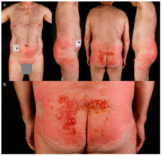
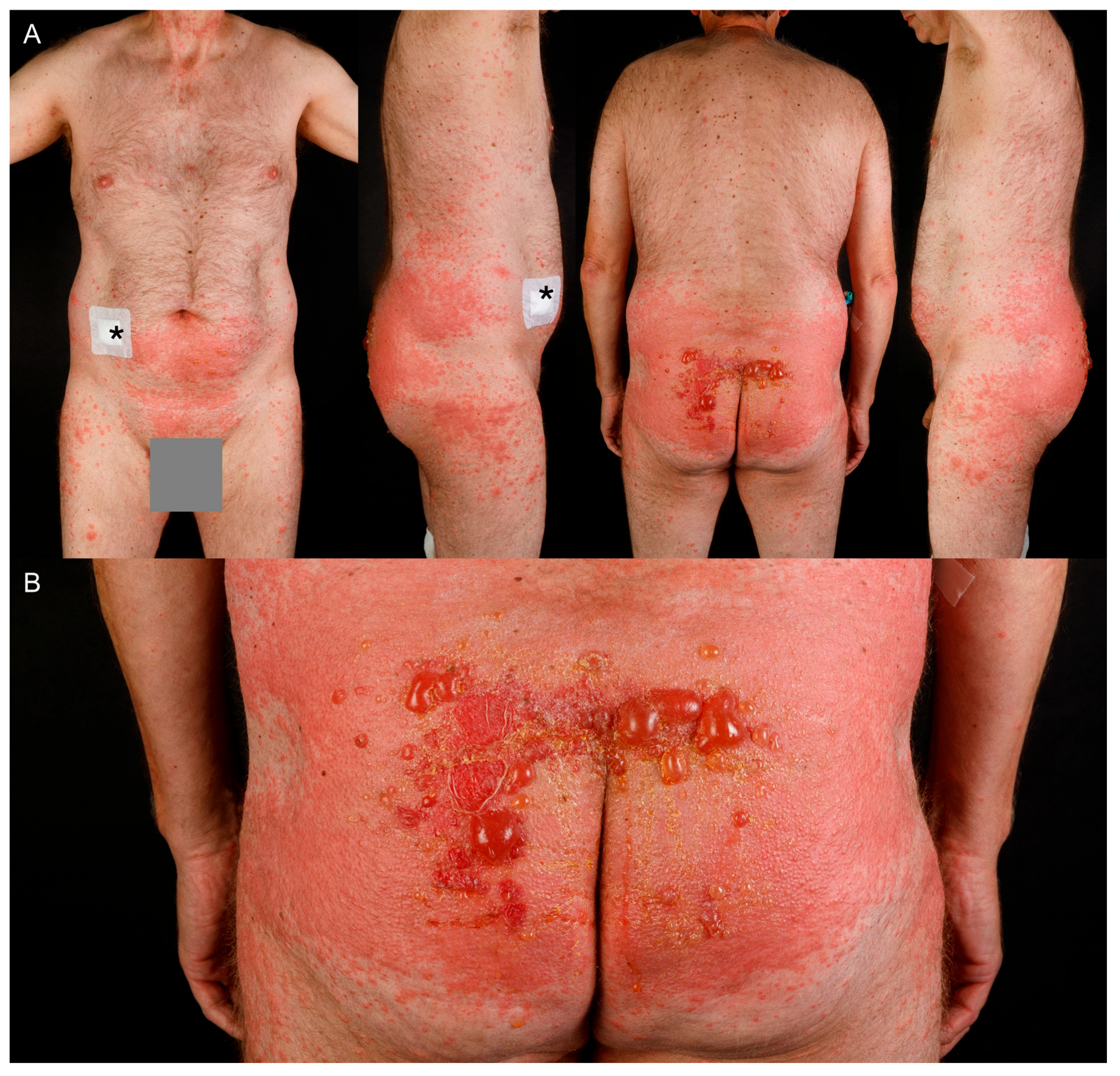
On admission, examination revealed a widespread maculopapular exanthem involving the trunk and extremities, with particularly pronounced involvement of the gluteal region. The buttocks showed confluent erythema and edema with multiple tense, fluid-filled bullae measuring up to approximately 4 cm in diameter (Figure 1). Shallow erosions were additionally present on the buccal mucosa, which had developed a few days after the onset of the skin manifestations.
Figure 1.
Clinical presentation of EPPER. (A) Overview of the cutaneous eruption involving the trunk, shown from the anterior, right lateral, posterior, and left lateral views. The asterisk indicates the site of skin biopsy on the right lower abdomen. (B) Close-up view of the gluteal region showing confluent erythema with multiple tense bullae.
The patient denied any recently introduced systemic medications or dietary supplements, use of new topical agents, relevant UV exposure, or other recent environmental exposures.
Laboratory investigations demonstrated mild leukocytosis (11.29 × 10³/µL; reference range, 4–10 × 103/µL) without blood hypereosinophilia, and an elevated CRP level (21.1 mg/L; reference range, <5 mg/L). A bacterial swab obtained from the eroded skin lesions yielded Cutibacterium acnes and mixed saprophytic Gram-positive flora, consistent with secondary colonization rather than a primary infectious etiology. Anti-BP180 and anti-BP230 antibody levels were within normal limits. Indirect immunofluorescence on monkey esophagus and human salt-split skin was negative for circulating autoantibodies. A skin biopsy was obtained from a representative lesion for histopathological evaluation and clinicopathological correlation.
2.1. Histopathological Findings
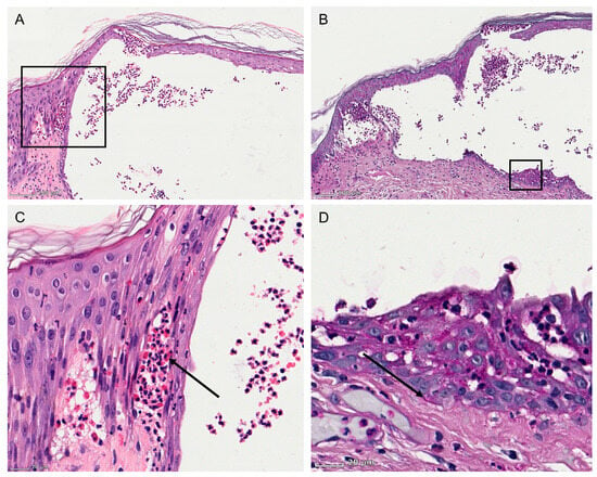
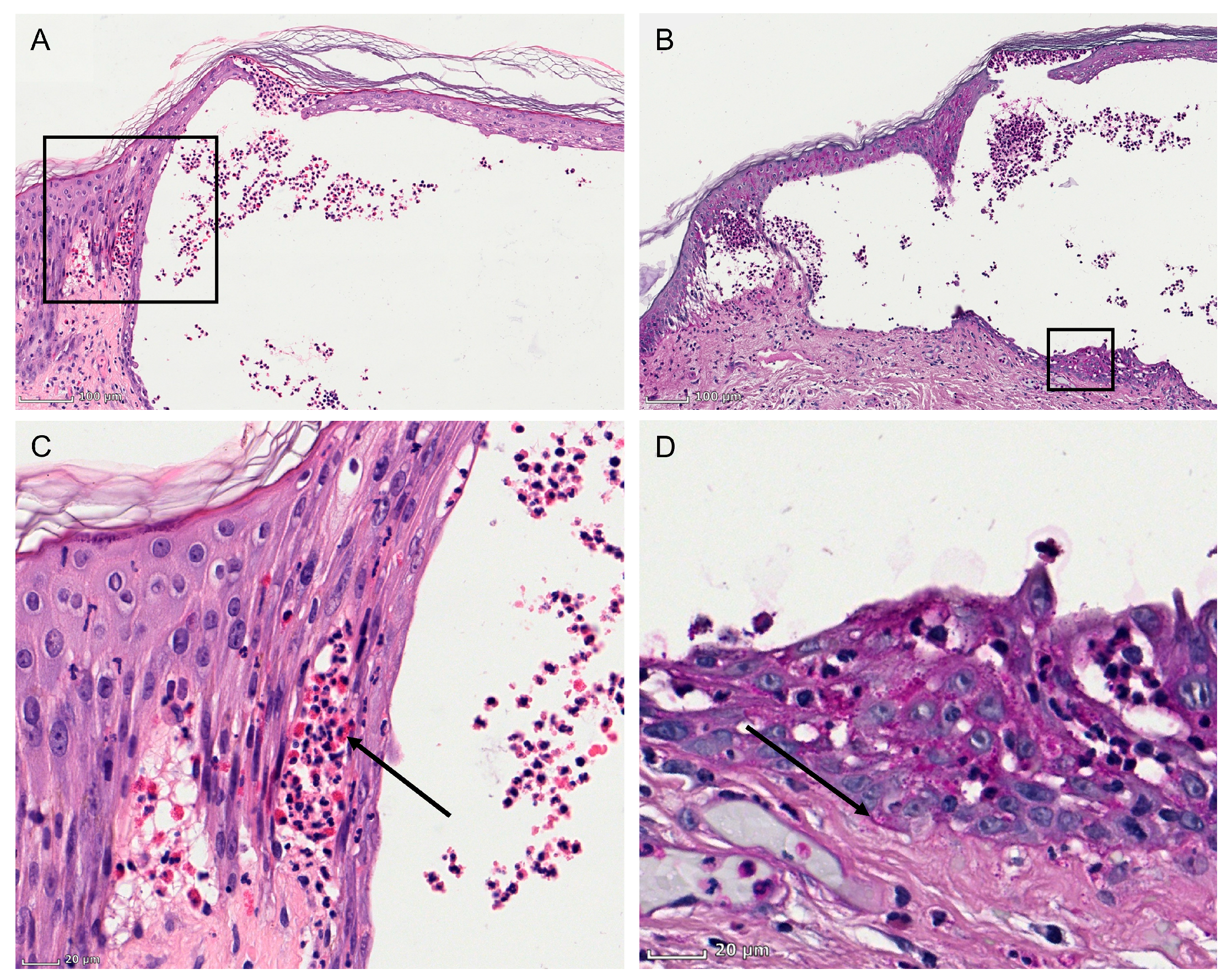
Histopathological examination revealed an eosinophil-rich bullous spongiotic dermatitis (Figure 2). The epidermis showed marked eosinophilic spongiosis with intraepidermal vesicle formation containing numerous eosinophils and scattered neutrophils. The superficial to mid-dermis showed a mild perivascular and interstitial inflammatory infiltrate, predominantly composed of lymphocytes with numerous admixed eosinophils, accompanied by mild papillary dermal edema. In view of the clinical presentation and the temporal association with radiation therapy, the histopathological findings were considered consistent with EPPER.
Figure 2.
Histopathological findings. (A) Low-magnification overview of the skin biopsy stained with hematoxylin and eosin (H&E). Scale bar = 100 µm. The black annotation indicates the area shown at higher magnification in panel (C). (B) Low-magnification overview of the skin biopsy stained with periodic acid–Schiff (PAS). Scale bar = 100 µm. The black annotation indicates the area shown at higher magnification in panel (D). (C) Higher-magnification H&E view demonstrating eosinophilic spongiosis with intraepidermal vesiculation. Scale bar = 20 µm. The arrow indicates intraepidermal eosinophils. (D) Higher-magnification PAS view of the blister floor highlighting the basement membrane (arrow), with multiple layers of epidermal keratinocytes remaining above it, supporting an intraepidermal level of blister formation. Scale bar = 20 µm.
2.2. Clinical Course and Treatment
Given the extent and severity of the eruption and the systemic inflammatory response, the patient was admitted for inpatient management. Intravenous corticosteroid therapy with prednisolone 80 mg daily (1 mg/kg body weight) was initiated. However, despite initial treatment, the eruption continued to progress with formation of new bullae and extension to the thighs. The prednisolone dose was therefore increased to 100 mg daily. Given the elevated inflammatory markers and disruption of the cutaneous and mucosal barriers, intravenous cefuroxime (1.5 g three times daily) was initiated empirically to cover a possible secondary bacterial infection.
Topical treatment consisted of a mometasone furoate–polyhexanide ointment applied to the affected areas. The large bullae were aseptically aspirated using sterile technique to reduce tension and minimize the risk of spontaneous rupture. The bullae sites and eroded areas were subsequently dressed with a non-adherent, lipidocolloid wound-contact layer and a non-adherent wound dressing with a metallized wound-contact surface to maintain a moist wound environment, promote healing, and minimize infection risk.
2.3. Clinical Outcome
Over the course of hospitalization, the eruption gradually faded under higher-dose corticosteroid therapy. No new bullae developed after dose escalation, and the patient showed gradual clinical improvement.
On hospital day 9, the patient was transferred to the radiation oncology clinic to resume his scheduled pelvic radiation therapy while receiving systemic corticosteroid prophylaxis. However, upon re-evaluation at the radiation clinic, the persistent extent and severity of the cutaneous eruption prompted the radiation team to temporarily suspend further radiation therapy. Radiotherapy was subsequently paused for two weeks and then resumed following replanning of the planning target volume to improve skin sparing, while maintaining the originally prescribed radiation dose. Approximately two to three weeks after resumption, the patient developed mild cutaneous erythema, without any new bullae formation. During the remainder of radiotherapy, the patient continued to receive topical corticosteroids, while systemic corticosteroids were gradually tapered and continued until one week after completion of radiotherapy. Under this regimen, the remaining fractions were administered over approximately four weeks, allowing the full prescribed course of 28 fractions to be completed without further complications. At follow-up approximately two and a half months after completion of treatment, the patient reported only residual hyperpigmentation at the previously affected sites and persistent mild pruritus, with no recurrence of bullae or blistering.
3. Discussion
EPPER is a rare and probably underrecognized complication characterized by a temporal association with radiotherapy, considerable clinical variability, and eosinophil-rich histopathological changes ranging from perivascular and interstitial infiltrates to spongiotic and vesiculobullous patterns. Since its initial description in 32 patients by Rueda et al. in 1999, EPPER has been reported predominantly in patients treated for breast and gynecological malignancies, but also in association with lymphoma, Merkel cell carcinoma, and prostate cancer [1,2,3,4,5,6,7,8,9,10]. EPPER may also affect nonirradiated skin, including sites distant from the radiation field [5,8]. The latency is highly variable, with eruptions occurring during irradiation as well as weeks to months after treatment completion. In our patient, pruritus and dysesthesia developed approximately two weeks after initiation of pelvic radiotherapy, followed by rapid progression and generalization of the eruption. This close temporal relationship to irradiation represented a key diagnostic clue. Eosinophils are a consistent histopathological feature of EPPER but are not themselves diagnostically specific. Rather, the combination of an eosinophil-rich reaction pattern with the appropriate clinical morphology, distribution, and temporal relationship to radiotherapy is essential for establishing the diagnosis. This is particularly relevant for dermatopathologists, as interpretation of the biopsy without knowledge of the oncological treatment may favor more common diagnoses such as eczematous or drug-induced dermatitis or an autoimmune bullous disorder.
The pathogenesis of EPPER remains unresolved. A direct role of ionizing radiation is supported by an experimental porcine model in which irradiation induced an eosinophil-rich cutaneous reaction within the treated fields [11]. Immunologically, both an IgE-mediated hypersensitivity response and a delayed T-cell-mediated reaction with aberrant Th2 polarization have been proposed [1,2]. However, the occurrence of EPPER in patients undergoing multimodal oncological treatment suggests that radiation may interact with additional host- or treatment-related factors rather than acting through a single mechanism.
Notably, our patient showed a mild systemic inflammatory response, a feature that has not been well characterized in previous reports of EPPER. In the context of extensive cutaneous involvement and marked barrier disruption, this was considered more likely to reflect the severity of the inflammatory skin process than an overt bacterial infection, supported by the absence of a defined pathogenic organism on skin swab. Nevertheless, given the extensive barrier disruption and the associated risk of secondary bacterial superinfection, empiric antimicrobial therapy was considered appropriate.
Published cases generally show a favorable response to anti-inflammatory treatment, most commonly topical or systemic corticosteroids, and antihistamines [8]. In one case, recovery was achieved through a combination of 311 nm UVB phototherapy and topical corticosteroids [4]. Recognition of EPPER is clinically relevant because correct classification of the eruption may prevent unnecessary changes or interruption of oncological therapy. Data specifically addressing the risk of recurrence of EPPER upon re-exposure to radiotherapy are limited. Barbu et al. reported complete resolution of EPPER under corticosteroid therapy in both of their prostate cancer patients, without need for permanent discontinuation of oncological treatment, suggesting a generally favorable prognosis when the condition is recognized early and managed appropriately [8]. This is consistent with our case, in which radiotherapy could be resumed and completed under corticosteroid treatment, with only a mild, self-limited flare and no recurrence of bullous lesions.
4. Conclusions
EPPER should be considered in patients who develop pruritic, polymorphic eruptions during or after radiotherapy, particularly when eosinophil-rich spongiotic or vesiculobullous changes are present histologically. Because these findings are not specific in isolation, relevant clinical information, especially the history and timing of radiotherapy, should be communicated to the dermatopathologist. In diagnostically challenging cases, direct clinicopathological discussion may be crucial for establishing the correct diagnosis.
Author Contributions
Investigation, B.K., C.B., C.E.-B., E.A.T.K., R.K., S.S. and W.S.; writing—original draft preparation, B.K. and E.A.T.K.; writing—review and editing, C.B., C.E.-B., S.S. and W.S.; visualization, E.A.T.K. All authors have read and agreed to the published version of the manuscript.
Funding
This research received no external funding.
Institutional Review Board Statement
Ethical review and approval were not required for this case report in accordance with local institutional requirements.
Informed Consent Statement
Written informed consent was obtained from the patient for publication of this case report and the accompanying images.
Data Availability Statement
The original contributions presented in this case report are included in the article. Further inquiries can be directed to the corresponding author.
Acknowledgments
The authors would like to thank the photographic team of the Department of Dermatology, Uniklinikum Erlangen, for their assistance with the clinical image documentation. We are grateful to the patient for consenting to the publication of this case and the accompanying images. We also thank all colleagues and staff members who contributed to the clinical care, diagnostic work-up, and documentation of the case but who do not meet the criteria for authorship.
Conflicts of Interest
The authors declare no conflicts of interest regarding this report. Outside the submitted work, W.S. received travel support for participation in conferences and/or (speaker) honoraria as well as research grants from medi GmbH Bayreuth, AbbVie, Almirall, Amgen, Boehringer Ingelheim, Bristol-Myers Squibb, GSK, Johnson & Johnson, LEO Pharma, Lilly, Novartis, Pfizer, Sanofi, UCB, and Takeda outside the submitted work. C.B. has received honoraria for consulting and/or oral presentations from Almirall Hermal, BMS, Delcath, Immunocore, Incyte, MSD, Novartis, Pierre Fabre, Regeneron, and SunPharma, outside the submitted work.
Abbreviations
The following abbreviations are used in this manuscript:
| EPPER | Eosinophilic polymorphic pruritic eruption associated with radiotherapy |
| CRP | C-reactive protein |
| IgE | Immunoglobulin E |
| Th2 | T helper 2 |
| UV | Ultraviolet |
| UVB | Ultraviolet B |
References
- Rueda, R.A.; Valencia, I.C.; Covelli, C.; Escobar, C.; Alzate, A.; Saldarriaga, B.; Sanclemente, G.; Blank, A.; Falabella, R. Eosinophilic, polymorphic, and pruritic eruption associated with radiotherapy. Arch. Dermatol. 1999, 135, 804–810. [Google Scholar] [CrossRef] [Scilit] [PubMed]
- Werchniak, A.E.; Perry, A.E.; Dinulos, J.G. Eosinophilic, polymorphic, and pruritic eruption associated with radiotherapy (EPPER) in a patient with breast cancer. J. Am. Acad. Dermatol. 2006, 54, 728–729. [Google Scholar] [CrossRef] [Scilit] [PubMed]
- Lee, D.J.; Kim, Y.C. A pustular form of eosinophilic, polymorphic, and pruritic eruption associated with radiotherapy. J. Am. Acad. Dermatol. 2011, 65, e51–e53. [Google Scholar] [CrossRef] [Scilit] [PubMed]
- Lobelenz, L.; Schliep, S.; Worl, P.; Uslu, U. Eosinophilic polymorphic and pruritic eruption associated with radiotherapy: Case report and overview of disease characteristics. Clin. Exp. Dermatol. 2019, 44, 567–569. [Google Scholar] [CrossRef] [Scilit] [PubMed]
- Garcia-Donoso, C.; Tardio, J.C.; Arias, D.; Romero, A.; Borbujo, J.M. Eosinophilic, polymorphic and pruritic eruption associated with radiotherapy (EPPER) in two patients with breast tumour. J. Eur. Acad. Dermatol. Venereol. 2007, 21, 1102–1104. [Google Scholar] [CrossRef] [Scilit] [PubMed]
- Masuno, Y.; Matsumura, Y.; Katoh, M.; Arakawa, A.; Kore-Eda, S.; Ishikawa, T.; Miyachi, Y. Eosinophilic, polymorphic, and pruritic eruption associated with radiotherapy (EPPER) mimicking bullous pemphigoid in a patient with anaplastic large cell lymphoma. Eur. J. Dermatol. 2011, 21, 421–422. [Google Scholar] [CrossRef] [Scilit] [PubMed]
- Lee, J.E.; Kim, S.C. Eosinophilic, polymorphic, and pruritic eruption associated with radiotherapy in a patient with breast cancer. J. Am. Acad. Dermatol. 2007, 56, S60–61. [Google Scholar] [CrossRef] [Scilit] [PubMed]
- Barbu, M.A.; Anghel, R.M.; Ricu, G.; Tebeica, T.; Patru, R.; Michire, A. Eosinophilic, polymorphic, and pruritic eruption associated with radiation therapy in two patients diagnosed with prostate cancer. Clin. Case. Rep. 2023, 11, e7266. [Google Scholar] [CrossRef] [Scilit] [PubMed]
- Abdelrahman, W.; Shum, L.; McManus, D.; Walsh, M.Y.; Dolan, O. Eosinophilic polymorphic and pruritic eruption associated with radiotherapy in a patient with primary nodal Merkel cell carcinoma. Int. J. Dermatol. 2018, 57, 864–866. [Google Scholar] [CrossRef] [Scilit] [PubMed]
- D’Ali, L.; Fagotto, L.; Stinco, G.; Pileri, A.; Zengarini, C.; Guglielmo, A. Eosinophilic Polymorphous Pruritic Eruption Associated With Radiotherapy: Clinical, Dermoscopic, and Histological Aspects. Dermatol. Pract. Concept 2024, 14, e2024272. [Google Scholar] [CrossRef] [Scilit] [PubMed]
- Kim, J.S.; Jang, W.S.; Bae, M.J.; Park, S.; Lee, S.S. Radiation-induced eosinophilic, polymorphic, and pruritic eruption in a pig skin model. Lab. Anim. Res. 2015, 31, 204–208. [Google Scholar] [CrossRef] [Scilit] [PubMed]
Disclaimer/Publisher’s Note: The statements, opinions and data contained in all publications are solely those of the individual author(s) and contributor(s) and not of MDPI and/or the editor(s). MDPI and/or the editor(s) disclaim responsibility for any injury to people or property resulting from any ideas, methods, instructions or products referred to in the content. |
© 2026 by the authors. Published by MDPI on behalf of the European Society of Dermatopathology. Licensee MDPI, Basel, Switzerland. This article is an open access article distributed under the terms and conditions of the Creative Commons Attribution (CC BY) license.