


Abstract
Reservoirs containing cemented natural fractures are widely developed in deep subsalt formations and play an important role in controlling rock mechanical behavior and failure evolution. However, the influence of fracture cementation on strength degradation, deformation response, and failure mode remains insufficiently understood, limiting the mechanical basis for evaluating hydraulic fracturing behavior in fractured dolomite reservoirs. A laboratory experimental framework is established to investigate the mechanical effects of cemented natural fractures in Cambrian subsalt dolomite from the Tarim Basin. CT scanning is used to characterize pore structure, fracture distribution, and cementation features of the samples. Based on these observations, Brazilian splitting tests and triaxial compression tests under a confining pressure of 40 MPa are conducted to analyze tensile strength, compressive strength, deformation characteristics, pore structure evolution, and failure modes under different cementation conditions. The results show that cemented natural fractures significantly weaken the tensile resistance of dolomite, and failure during Brazilian splitting mainly occurs along pre-existing fracture interfaces. Under triaxial compression, well-cemented samples exhibit limited porosity increase and relatively stable mechanical responses, whereas poorly cemented or heterogeneously cemented samples show more pronounced pore structure evolution, stronger nonlinear deformation, and unstable failure behavior. Cementation condition appears to influence fracture activation and failure path within the tested specimens. In the tested specimens, weaker cementation was associated with preferential opening and propagation along natural fractures, whereas stronger cementation was associated with relatively enhanced structural integrity and reduced fracture coalescence. These findings clarify the mechanical role of cemented natural fractures in dolomite failure and provide mechanical insights into the potential response of cemented natural fractures during hydraulic fracturing.
1. Introduction
Natural fractures are widely developed in unconventional oil and gas reservoirs, and their spatial distribution characteristics and mechanical properties are among the key factors controlling the effectiveness of hydraulic fracturing [1,2,3]. Unlike ideal homogeneous media, these natural fractures are often filled and cemented by minerals such as carbonates, forming cemented fractures with certain mechanical strength. Such fractures differ from conventional open or frictional fractures in that they possess measurable tensile strength and fracture toughness, enabling the fracture interfaces to bear and transmit part of the stress during loading [4,5,6]. When hydraulic fractures propagate toward cemented natural fractures, their propagation behavior typically exhibits multiple modes, including crossing, deflection, or arrest [7,8,9], thereby directly influencing the spatial geometry and connectivity of the fracture network, and further controlling the stimulated reservoir volume and fluid flow capacity [10,11]. Therefore, a thorough understanding of the mechanisms by which cemented natural fractures affect mechanical failure, fracture activation, and potential hydraulic fracture propagation behavior is essential for optimizing fracture network geometry and improving fracturing performance.
Related geomechanical and reservoir responses have also been investigated in other subsurface energy systems, including enhanced geothermal reservoirs [12], naturally fractured oil shale reservoirs [13], and gas hydrate reservoirs under different temperature–pressure conditions [14]. These studies show that reservoir response is closely related to both geological structure and operating conditions.
At present, extensive experimental studies have been conducted on the mechanisms of fracture intersection and propagation. Bahorich et al. [15] revealed the influence of cemented natural fractures on the capture and redirection of hydraulic fractures through gypsum block experiments. Wang et al. [16] systematically analyzed the variation in fracture propagation patterns under different approach angles. Yoshioka et al. [17] investigated, at the microscale, the role of grain boundary discontinuities in inducing fracture deflection. In addition, Wang et al. [18] demonstrated that increasing the injection rate helps overcome interfacial shielding effects and promotes fracture crossing. These studies have elucidated, from different perspectives, the controlling role of natural fractures on hydraulic fracture propagation paths.
However, existing experimental studies still have certain limitations. On the one hand, due to constraints in experimental materials and observation techniques, it remains difficult to achieve continuous and clear dynamic observation and quantitative characterization of fracture initiation and propagation within interfaces featuring mineral cementation. On the other hand, during experimental processes, factors such as the degree of cementation, cementation strength, and geometric characteristics of fractures are often difficult to control independently. The coupled effects of multiple factors make it challenging to accurately distinguish the specific influence of each parameter on fracture propagation behavior. Therefore, it is necessary to employ more refined experimental approaches to systematically investigate the structural characteristics and mechanical responses of cemented natural fractures, thereby providing reliable experimental evidence for elucidating fracture propagation mechanisms.
Therefore, this study takes reservoirs containing cemented natural fractures as the research object and conducts a systematic series of laboratory physical experiments. First, CT scanning is employed to perform three-dimensional characterization of the internal fracture structures and filling features of the rock samples, and to obtain key structural parameters such as porosity. On this basis, Brazilian splitting tests and triaxial compression tests are carried out to determine the mechanical response characteristics of the rocks under tensile and confining pressure conditions, respectively, thereby enabling a quantitative analysis of the effects of different cementation states on fracture initiation and propagation behavior. Through comprehensive analysis of the experimental results, the influence of fracture cementation on mechanical failure, fracture activation, and pore structure evolution is clarified, and its implications for potential hydraulic fracture propagation in fractured dolomite reservoirs are discussed.
2. Materials and Experimental Methods
To investigate the mechanisms by which cemented natural fractures influence fracture propagation behavior, this study primarily employs laboratory physical experiments. First, CT scanning is conducted on rock samples containing naturally cemented fractures to obtain fundamental petrophysical parameters, including porosity and fracture filling degree. On this basis, Brazilian splitting tests and triaxial compression tests are subsequently performed to characterize the mechanical responses of the rocks under tensile loading and confining pressure conditions, respectively. Finally, comparative CT analyses are carried out on the rock samples before and after testing to quantitatively characterize fracture structure evolution and failure features.
2.1. Rock Samples and CT-Based Structural Characterization
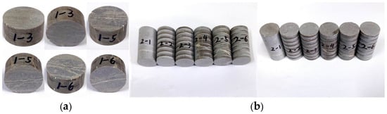
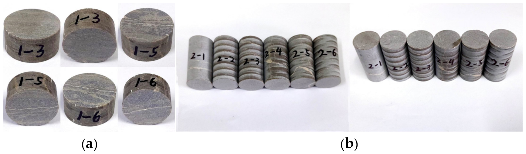
The rock samples used in this study were collected from outcrops of Cambrian subsalt dolomite in the Tarim Basin, China. Natural fractures are widely developed in these outcrops and are filled with cementing materials, exhibiting clear geometric characteristics, which provide an ideal basis for analyzing the influence of fracture cementation on the mechanical behavior of rocks. To ensure comparability among different experimental samples, all specimens were prepared using a standardized processing procedure, with consistent geometric dimensions. To reduce the influence of fracture orientation on the mechanical response, specimens were selected to maintain a consistent relative orientation between the principal natural fracture and the loading direction. The orientation of the principal natural fracture was initially identified by visual inspection during specimen selection and was subsequently verified using pre-test CT images. Specimens whose fracture orientations did not satisfy the required orientation were excluded from the test series. A total of three cylindrical specimens with dimensions of Φ25 mm × 12.5 mm were prepared for the Brazilian splitting tests (Figure 1a), and six cylindrical specimens with dimensions of Φ25 mm × 50 mm were prepared for the triaxial compression tests (Figure 1b).
Figure 1.
Pre-test photographs of experimental rock samples. (a) Brazilian splitting test samples 1-3, 1-5, and 1-6; (b) triaxial compression test samples 2-1–2-6.
To characterize the internal pore structures and fracture filling features of the rock samples, CT scanning was conducted prior to the experiments using a GE Phoenix Nanotom S scanner (GE Inspection Technologies, Wunstorf, Germany). Based on the CT scanning data, three-dimensional reconstructions of the internal pore and fracture networks were performed using phoenix datos|x 2 software (GE Inspection Technologies, Wunstorf, Germany). The reconstructed models were further analyzed and visualized using Avizo 2020.1 (Thermo Fisher Scientific, Waltham, MA, USA) and VGStudio MAX 3.4 (Volume Graphics GmbH, Heidelberg, Germany).
2.2. Experimental Setup and Testing Conditions
This study conducts Brazilian splitting tests and triaxial compression tests to systematically characterize the mechanical responses and failure behaviors of rock samples containing natural fractures with different cementation states. The two types of experiments were carried out using a rock mechanical testing machine equipped with an acoustic emission monitoring system and a high-temperature, high-pressure triaxial rock mechanics testing system, respectively.
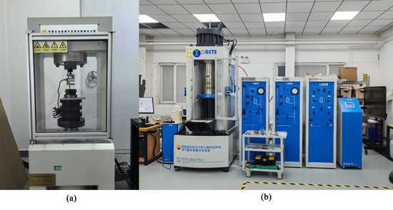
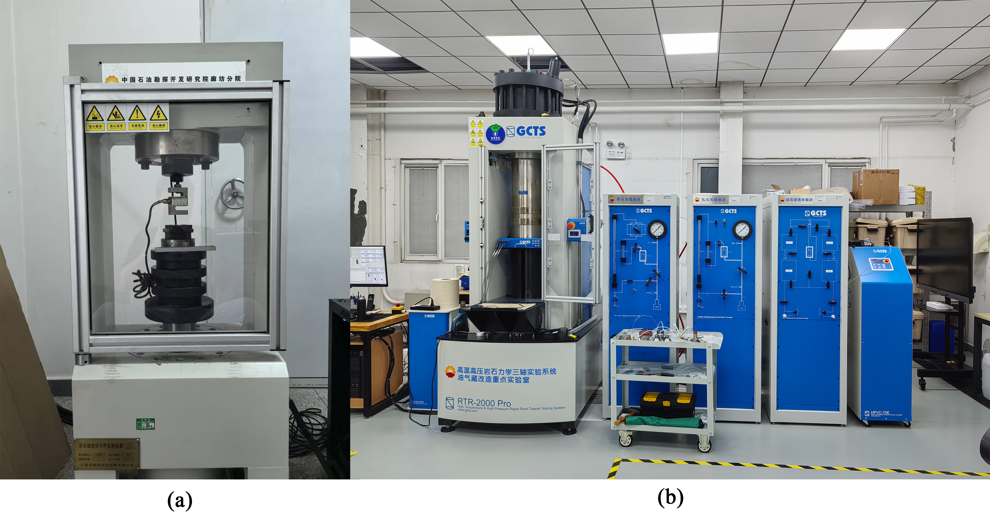
The rock mechanical testing machine equipped with an acoustic emission system (Figure 2a) was used to determine the tensile strength and failure load of both the rock matrix and cemented natural fractures. Brazilian splitting tests were conducted under load-controlled conditions with a constant loading rate of 0.1 kN/min until splitting failure occurred. During the loading process, load and displacement data were continuously recorded. Combined with post-failure macroscopic observations, the fracture initiation locations and propagation paths were identified. To minimize the influence of end effects on the experimental results, flexible pads were placed between the specimen and the loading platens during loading to ensure uniform load transmission along the diameter.
Figure 2.
Equipment used in this experiment: (a) Rock Mechanical Testing Machine; (b) RTR-1500 High-Temperature and High-Pressure Rock Mechanics Testing System.
The triaxial compression tests were conducted using an RTR-1500 high-temperature and high-pressure rock mechanics testing system (Figure 2b) manufactured by GCTS (Tempe, AZ, USA). This system is a closed-loop digitally servo-controlled apparatus and is widely used in rock mechanics research. In this study, it was employed to obtain key mechanical parameters of the rock, including compressive strength, Young’s modulus, and Poisson’s ratio, and to analyze the failure characteristics of the specimens.
The triaxial compression tests were performed under ambient temperature conditions. A confining pressure of 40 MPa was first applied, followed by axial loading under displacement-controlled conditions. During the experiments, axial stress, axial strain, and confining pressure data were continuously recorded to analyze the deformation and failure behavior of the specimens under triaxial stress conditions. Post-failure macroscopic observations were conducted to further identify the influence of natural fractures on the failure modes and fracture evolution processes of the rock samples.
2.3. Mechanical Testing Procedures
2.3.1. Brazilian Tensile Splitting Test
Brazilian splitting tests were conducted on rock samples containing natural fractures with different cementation states to obtain the failure load and tensile strength under tensile stress, and to further analyze the influence of fracture cementation on fracture initiation and propagation behavior. The experimental procedure is as follows.
- (1)
- Prior to testing, all specimens were uniformly labeled and their geometric dimensions were rechecked. The upper and lower end faces of the specimens were carefully polished to ensure that the flatness and parallelism met the testing standards. Subsequently, the loading system and data acquisition system were calibrated to ensure the accuracy and reliability of load application and data recording during the experiments.
- (2)
- The specimen was positioned at the center of the loading apparatus according to the predetermined orientation, such that the loading direction was aligned with the radial direction of the natural fractures. Load-controlled loading was applied at a constant rate of 0.1 kN/min until typical splitting failure occurred. During the loading process, the load–displacement relationship was recorded in real time, and acoustic emission signals were simultaneously monitored to capture the fracture evolution process.
- (3)
- After the test, the failed specimens were macroscopically examined and documented to identify the fracture initiation locations, propagation paths, and final failure patterns. Combined with the load–displacement curves, the tensile failure characteristics of specimens with different cementation states of natural fractures were comparatively analyzed and discussed.
2.3.2. Triaxial Compression Test
To systematically investigate the influence of natural fractures with different cementation states on the mechanical properties of rocks under in-situ stress conditions, triaxial compression tests were conducted under a confining pressure of 40 MPa. The experimental procedure is summarized as follows:
- (1)
- Based on the CT scanning results, rock samples containing natural fractures with different cementation states and meeting the dimensional requirements of the testing standards were selected. The dimensions and mass of each specimen were measured and recorded, and the testing system was calibrated prior to the experiments.
- (2)
- Each specimen was encased in a rubber jacket to prevent the confining medium from penetrating into the sample. The specimen was then placed between the upper and lower loading platens, ensuring strict axial alignment to avoid eccentric loading.
- (3)
- A confining pressure of 40 MPa was applied around the specimen. Under constant confining pressure, axial stress was gradually increased until failure occurred. During the loading process, axial stress, axial strain, and lateral strain were continuously recorded.
- (4)
- Based on the obtained stress–strain curves, the Young’s modulus and Poisson’s ratio of the specimens were calculated, and the compressive strength was determined.
3. Experimental Results
3.1. Tensile Failure Behavior of Cemented Naturally Fractured Dolomite
3.1.1. Pre-Test Fracture and Pore Structure Characterization
For ease of description and differentiation, specimen 1-3 is designated as specimen 1-a, specimen 1-5 as specimen 1-b, and specimen 1-6 as specimen 1-c.
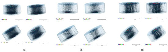
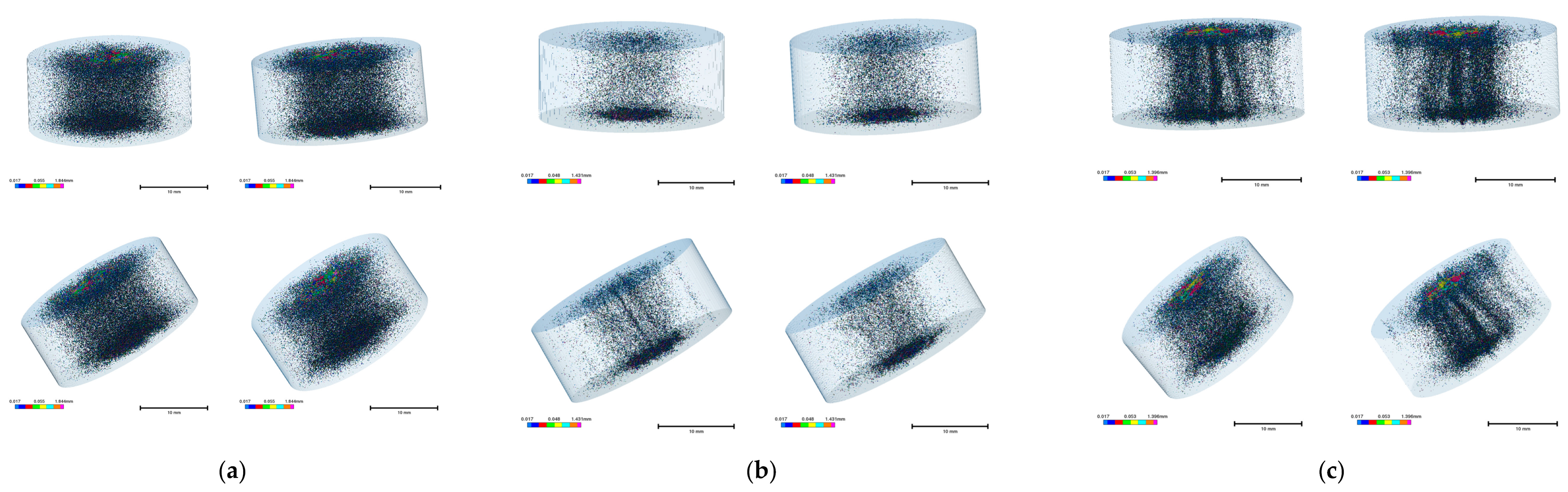
CT scanning results indicate that specimen 1-a exhibits pore enrichment only in the central region (Figure 3a), with no obvious fractures observed; a through-going fracture has developed at the center of specimen 1-b (Figure 3b), and specimen 1-c shows a more well-developed fracture system (Figure 3c).
Figure 3.
Pore distribution of core samples obtained by CT scanning for the Brazilian splitting experiment. (a) Sample 1-a; (b) Sample 1-b; (c) Sample 1-c.
A comparison of the pore statistics in Table 1 shows that the matrix pores of the three rock samples are highly consistent in terms of average equivalent diameter and volume, effectively eliminating the influence of matrix differences on the mechanical properties. Meanwhile, the aspect ratios of pores in all samples are consistently within the range of 1.45–1.49, indicating that the pore morphology is approximately ellipsoidal, and this geometric characteristic remains highly consistent among different specimens.
Table 1.
Pore Structure Data of Rock Samples.
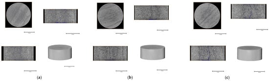
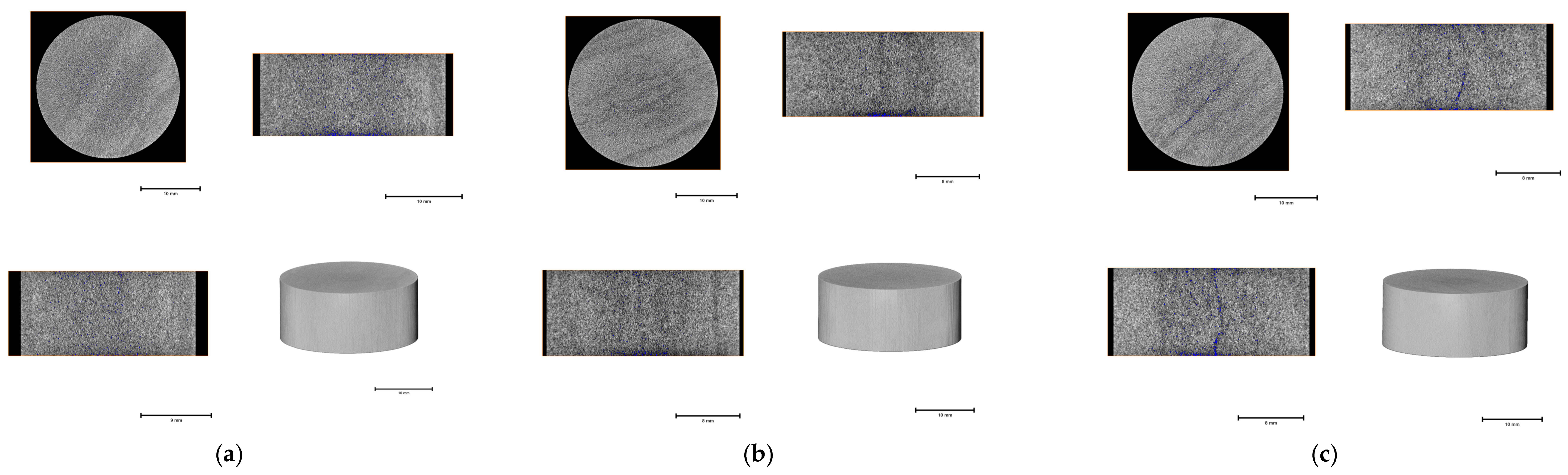
Since the fractures are mainly located near the central region of the specimens, the 550th slices in the xy, xz, and yz directions, corresponding approximately to the mid-sections of the samples, were selected for analysis. The results show that pores in specimen 1-a are uniformly distributed without preferential orientation (Figure 4a), whereas pores in specimens 1-b (Figure 4b) and 1-c exhibit pronounced linear concentration, particularly in specimen 1-c (Figure 4c), directly confirming the presence and extension of fractures.
Figure 4.
Pore slices and 3D reconstruction of rock samples. (a) Sample 1-a; (b) Sample 1-b; (c) Sample 1-c.
3.1.2. Tensile Strength and Fracture-Controlled Failure
The CT-scanned specimens were subsequently loaded, and the full load–displacement curves were obtained via sensors, thereby determining the failure load and tensile strength of the rock samples.
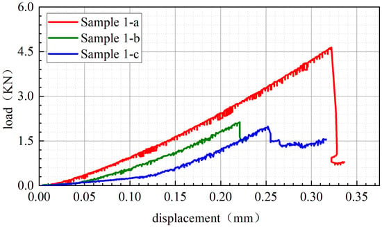
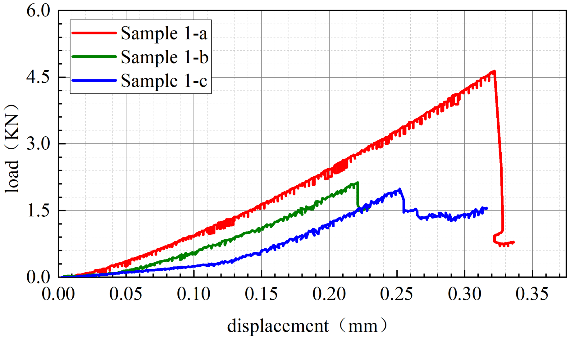
The experimentally measured failure loads and tensile strengths are listed in Table 2, and the corresponding load–displacement curves are shown in Figure 5. As illustrated in Figure 5, the load–displacement curves of all specimens exhibit typical elastic–brittle failure characteristics, with the load dropping rapidly after reaching the peak value, indicating that no significant plastic deformation occurred prior to failure.
Table 2.
Failure Load and Tensile Strength Results of Core Samples.
Figure 5.
Load–Displacement Curves of Rock Samples in the Brazilian Splitting Test.
Among them, the matrix specimen 1-a requires a larger loading displacement before reaching the peak load, with a failure load of 4640 N and a corresponding tensile strength of 9.4 MPa, reflecting the high tensile strength and strong resistance to failure of the intact matrix rock. In contrast, the curves of specimens 1-b and 1-c containing natural fractures exhibit an overall “low–gentle–long” trend, with failure loads of 2170 N and 1990 N and tensile strengths of 4.4 MPa and 4.1 MPa, respectively. This indicates that the presence of fractures significantly weakens the overall tensile performance of the specimens, although the cement within the fractures still provides a certain level of tensile strength.
Further analysis of the fractured specimens reveals noticeable differences among them. Specimen 1-b (2170 N) exhibits a relatively pronounced plateau stage prior to the peak load, which may be associated with progressive deformation or sliding along the pre-existing interface and local microcracking before final failure. Since the fracture-filling material was not independently characterized in terms of mineralogical composition and thickness, the origin of this plateau cannot be determined from the present results alone.
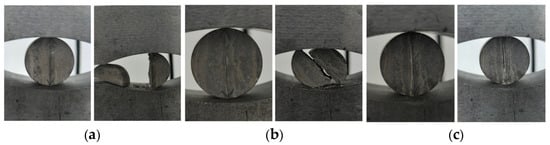
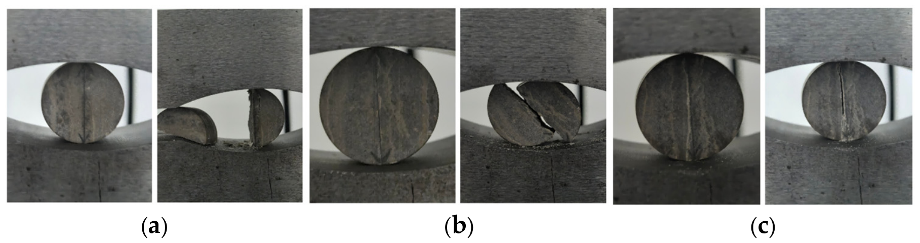
Figure 6 presents a comparison of the specimens before and after loading. As shown in the figure, specimen 1-a undergoes typical tensile brittle failure along the loading diameter plane, with a relatively intact splitting surface, indicating relatively well-cemented. Under radial loading, the specimens containing radial fractures (1-b and 1-c) both fail along the pre-existing fractures.
Figure 6.
Comparison of rock samples before and after failure. (a) Sample 1-a; (b) Sample 1-b; (c) Sample 1-c.
In terms of tensile strength, the fractured group shows an overall reduction of approximately 50–65% compared with the matrix specimen. The observed strength differences are associated with variations in fracture continuity and filling characteristics. However, because the specimens contain naturally occurring fractures, the effects of cementation cannot be completely separated from those of fracture geometry and spatial distribution. In particular, for specimens 1-b and 1-c, the fractures have already penetrated the sample center as observed in CT scans, and their mechanical responses and failure modes are characterized by instability along the fracture interfaces, exhibiting pronounced structural control. This “strong matrix–weak interface” combination not only significantly reduces the overall tensile capacity of the rock but also indicates that fractures preferentially open under loading and dominate the failure evolution process.
3.2. Mechanical Response and Damage Evolution Under Triaxial Compression
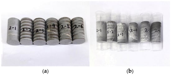
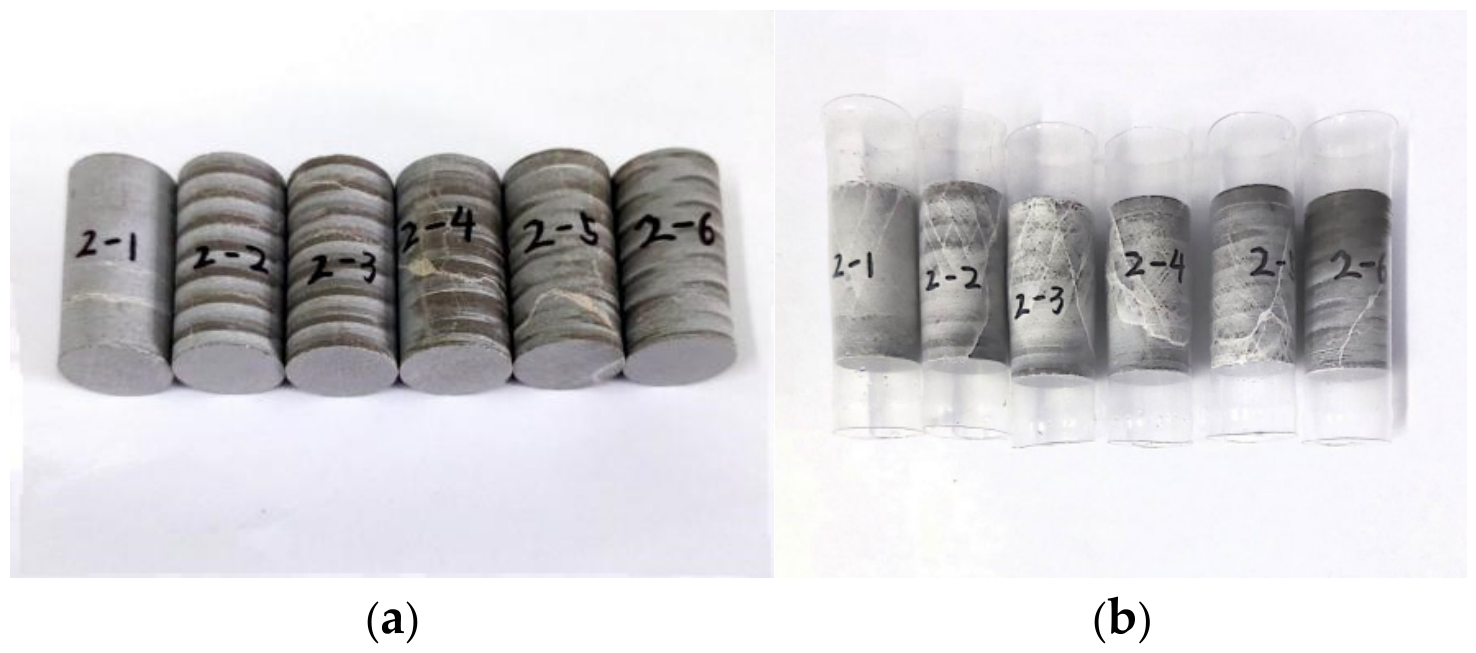
The rock samples before and after the triaxial compression tests are shown in Figure 7. CT scanning was performed both before and after the experiments, and the three-dimensional spatial distribution of fractures was compared in situ.
Figure 7.
Comparison of rock samples before and after the triaxial compression experiment. (a) Physical image of the rock sample before the experiment; (b) physical image of the rock sample after the experiment.
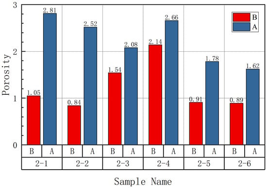
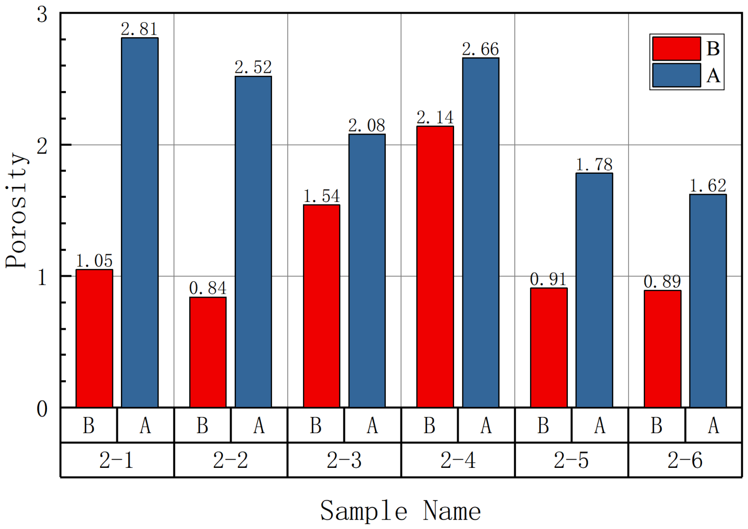
Based on the CT scanning results, the porosity distributions were extracted to obtain the porosity variation before and after triaxial compression (Figure 8). It can be observed that the porosity of all specimens increases after triaxial compression, although the magnitude of increase varies significantly. The porosity increases in specimens 2-1 and 2-2 are 168% and 200%, respectively; those of specimens 2-3 and 2-4 are 35% and 24%, respectively; and those of specimens 2-5 and 2-6 are 95% and 82%, respectively.
Figure 8.
Porosity Variation Before and After Triaxial Compression.
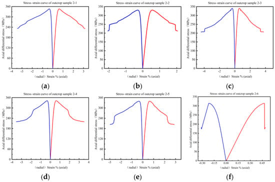
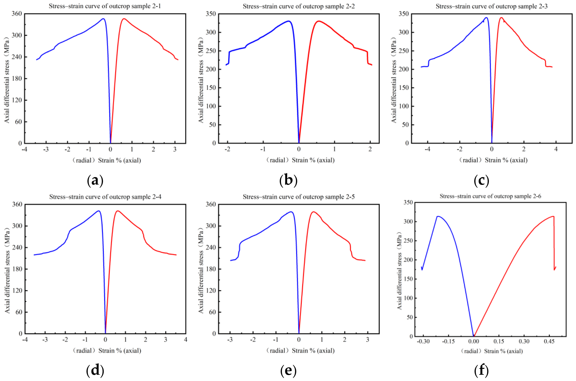
The triaxial compression tests obtained key mechanical parameters of the specimens, including Young’s modulus, Poisson’s ratio, and compressive strength (Table 3), and the stress–strain curves during the failure process were fully recorded (Figure 9).
Table 3.
Results of Triaxial Compression Tests.
Figure 9.
Stress–strain curves of all rock samples in the triaxial compression experiment. (a) Sample 2-1; (b) Sample 2-2; (c) Sample 2-3; (d) Sample 2-4; (e) Sample 2-5; (f) Sample 2-6.
Based on the pre-test CT images, the six specimens were qualitatively grouped according to the observed continuity and filling characteristics of the natural fractures. Specimens 2-1 and 2-2 were classified as poorly cemented, specimens 2-3 and 2-4 as well-cemented, and specimens 2-5 and 2-6 as heterogeneously cemented. The classification was based solely on the pre-test CT observations; post-test porosity changes were analyzed separately to characterize the structural response to loading.
3.2.1. Mechanical Response of Poorly Cemented Specimens
The poorly cemented group exhibits the most significant changes in pore structure before and after triaxial compression. The porosity of specimen 2-1 increases from 1.05% to 2.81%, corresponding to an increase of 168%, while the porosity of specimen 2-2 increases from 0.84% to 2.52%, with an increase of 200%. Both the number and volume of pores increase markedly, indicating that fractures rapidly propagate and become interconnected during loading.
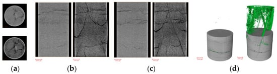
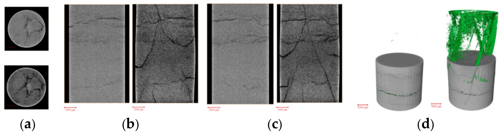
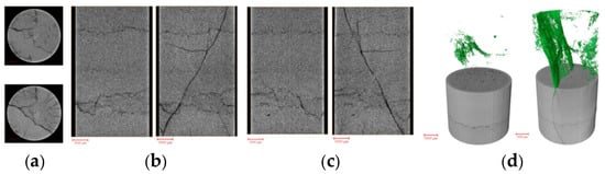
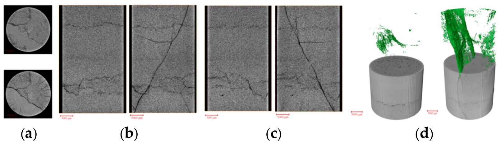
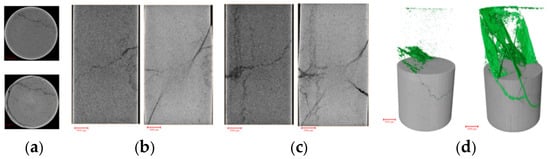
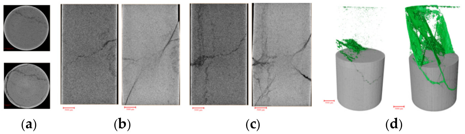
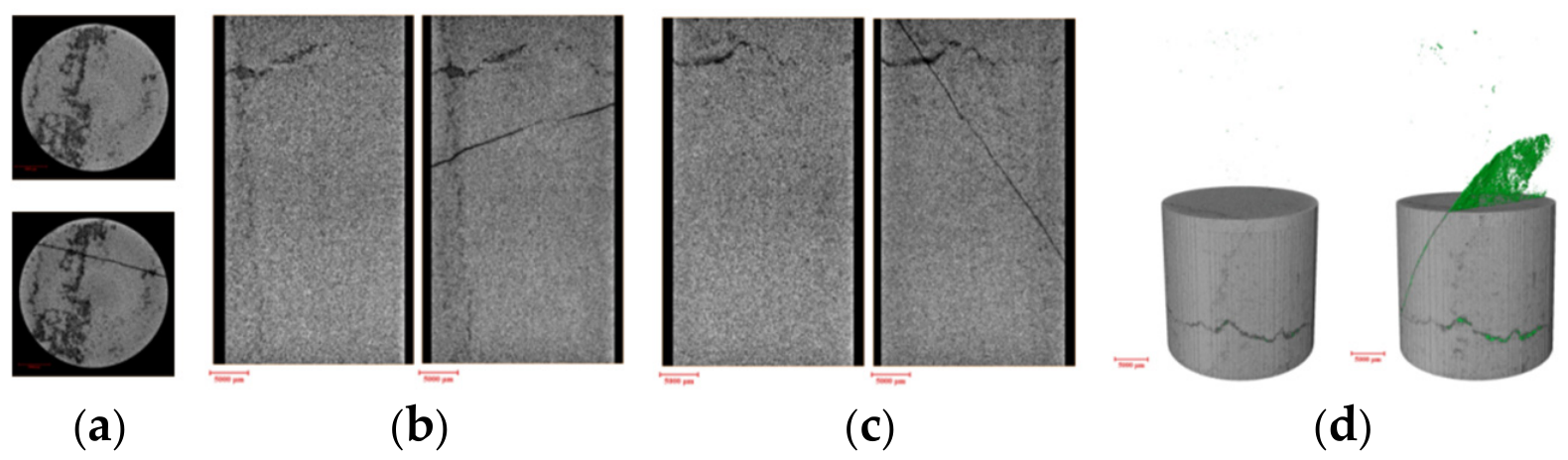
In terms of mechanical response, the compressive strengths of specimens 2-1 and 2-2 are 386.1 MPa and 370.4 MPa, respectively. After the peak stress, specimens 2-1 and 2-2 show stress reductions of 39.86% and 42.76%, respectively, indicating a substantial loss of load-bearing capacity. Together with the fluctuations in the stress–strain curves, these reductions reflect pronounced damage development during failure. Although the poorly cemented group still maintains a certain level of compressive load-bearing capacity, the increase in porosity and fluctuations in the stress–strain curves indicate that internal fractures undergo significant propagation during loading. Among them, specimen 2-2 exhibits a Young’s modulus of 92,395 MPa, which is the highest among all specimens in this study. This result may be related to a relatively dense local structure or limited deformation during the initial loading stage, leading to a higher apparent stiffness. However, noticeable fluctuations appear near the peak of its stress–strain curve, reflecting non-uniform internal stress distribution, where interfaces with weaker cementation fail preferentially. Combined with CT scanning and experimental results, it can be concluded that although such specimens possess relatively strong overall load-bearing capacity, their poor internal cementation conditions result in pronounced localized fracture propagation. The CT comparisons before and after the experiment for specimens 2-1 and 2-2 are illustrated in Figure 10 and Figure 11, respectively.
Figure 10.
Comparison of filtered images in different directions before and after the experiment for rock sample 2-1. (a) xy direction; (b) xz direction; (c) yz direction; (d) comparison of fracture models before and after the experiment.
Figure 11.
Comparison of filtered images in different directions before and after the experiment for rock sample 2-2. (a) xy direction; (b) xz direction; (c) yz direction; (d) comparison of fracture models before and after the experiment.
3.2.2. Mechanical Response of Well-Cemented Specimens
The well-cemented specimens exhibit relatively small changes in pore structure before and after triaxial compression. The porosity of specimen 2-3 increases from 1.54% before loading to 2.08%, corresponding to an increase of approximately 35%, while the porosity of specimen 2-4 increases from 2.14% to 2.66%, with an increase of about 24%, both significantly lower than those of other specimens. The increases in pore number and pore volume are also limited, with newly generated pores mainly manifested as local enlargement of pre-existing pores, without forming obvious connected pores or through-going fractures.
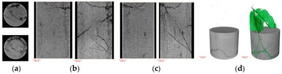
In terms of mechanical response, the compressive strengths of specimens 2-3 and 2-4 are 380.0 MPa and 381.8 MPa, respectively, while their Young’s moduli are 88,475 MPa and 89,041 MPa, indicating relatively consistent mechanical responses between the two specimens. The stress–strain curves show that stable elastic deformation dominates prior to the peak, with relatively limited fluctuations compared with the other tested specimens. Following the peak stress, specimens 2-3 and 2-4 both exhibited stress drops of 45.72%, indicating a pronounced loss of load-bearing capacity after peak failure. Therefore, although the two well-cemented specimens showed relatively stable pre-peak responses, substantial post-peak weakening still occurred. Combined with the CT observations, these results suggest that the relatively continuous cementation of the pre-existing fractures may help maintain structural integrity and constrain fracture activation before peak failure, but does not prevent pronounced strength degradation once macroscopic failure occurs. These observations are consistent with the limited structural changes and fracture comparisons for specimens 2-3 and 2-4, as shown in Figure 12 and Figure 13.
Figure 12.
Comparison of filtered images in different directions before and after the experiment for rock sample 2-3. (a) xy direction; (b) xz direction; (c) yz direction; (d) comparison of fracture models before and after the experiment.
Figure 13.
Comparison of filtered images in different directions before and after the experiment for rock sample 2-4. (a) xy direction; (b) xz direction; (c) yz direction; (d) comparison of fracture models before and after the experiment.
3.2.3. Mechanical Response of Heterogeneously Cemented Specimens
The heterogeneously cemented specimens exhibit pronounced heterogeneity in pore structure evolution after triaxial compression. The porosity of specimen 2-5 increases from 0.91% to 1.78%, corresponding to an increase of 95%. Although the average pore radius slightly decreases from 57.96 μm to 57.76 μm, the obvious increase in porosity indicates that loading mainly induces the generation of new pores, local fracture propagation, and enhanced pore connectivity rather than uniform enlargement of individual pores. The porosity of specimen 2-6 increases from 0.89% to 1.62%, with an increase of 82%. Both the number and volume of pores increase more significantly than those of the well-cemented specimens, suggesting that fractures undergo rapid propagation in localized regions.
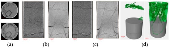
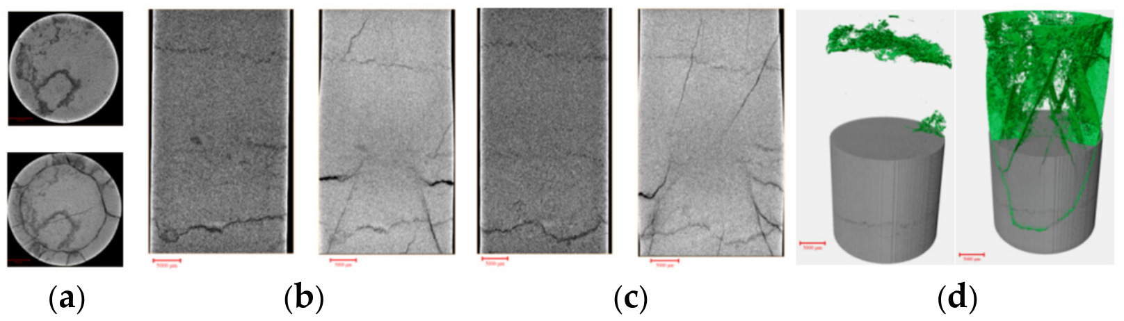
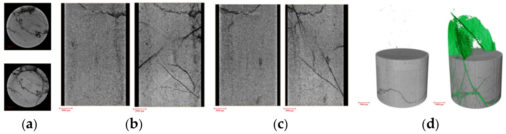
In terms of mechanical response, the compressive strengths of this group are relatively low. In particular, specimen 2-6 exhibits a compressive strength of only 353.6 MPa, the lowest among all specimens, while specimen 2-5 has a Young’s modulus of 81,449 MPa, also at a relatively low level. The stress–strain curves show evident nonlinear deformation prior to the peak, followed by a sharp post-peak stress drop. The stress reductions in specimens 2-5 and 2-6 are 42.56% and 49.86%, respectively, with specimen 2-6 showing the largest reduction among the six specimens. This pronounced decrease reflects a substantial loss of load-bearing capacity after peak failure. These results suggest that the internal fractures in this group are heterogeneously cemented, with locally weakly cemented interfaces and poor interfacial constraint. The significant localized fracture activation in these specimens is further confirmed by the CT comparisons before and after the experiment, as illustrated in Figure 14 and Figure 15.
Figure 14.
Comparison of filtered images in different directions before and after the experiment for rock sample 2-5. (a) xy direction; (b) xz direction; (c) yz direction; (d) comparison of fracture models before and after the experiment.
Figure 15.
Comparison of filtered images in different directions before and after the experiment for rock sample 2-6. (a) xy direction; (b) xz direction; (c) yz direction; (d) comparison of fracture models before and after the experiment.
4. Discussion
4.1. Cementation-Controlled Mechanical Weakening and Failure
The experimental results indicate that cemented natural fractures play a significant role in controlling the mechanical failure behavior of dolomite. In the Brazilian splitting tests, the intact matrix specimen exhibits a tensile strength of 9.4 MPa, whereas the fractured specimens show much lower tensile strengths of 4.4 MPa and 4.1 MPa. This reduction demonstrates that cemented natural fractures significantly weaken the tensile resistance of dolomite.
Post-failure observations further show that the intact matrix specimen mainly fails along the loading diameter, while the fractured specimens preferentially fail along pre-existing natural fracture interfaces. This suggests that, even when natural fractures are filled and cemented, the fracture interfaces remain mechanically weaker than the surrounding matrix. Therefore, the tensile failure of cemented naturally fractured dolomite is mainly controlled by a “strong matrix–weak interface” mechanism.
However, the fractured specimens still retain measurable tensile strength, indicating that cementing materials within the fracture interfaces provide a certain degree of bonding and load-bearing capacity. Thus, cemented natural fractures should not be regarded as completely open or cohesionless fractures. Instead, their mechanical behavior depends strongly on the cementation state of the fracture interface.
4.2. Cementation-Dependent Damage and Pore Structure Evolution
The CT scanning results before and after triaxial compression show that different cementation states lead to distinct pore structure evolution. Poorly cemented specimens show the largest porosity increases after loading, with increases of 168% and 200%, respectively. This indicates that weakly cemented fracture interfaces are easily activated under stress, resulting in pore expansion, fracture propagation, and enhanced connectivity.
In contrast, well-cemented specimens show much smaller porosity increases of 35% and 24%. The limited pore structure change suggests that strong cementation can effectively constrain fracture opening and coalescence, thereby maintaining better structural integrity during triaxial compression. Heterogeneously cemented specimens show intermediate porosity increases of approximately 95% and 82%, reflecting localized fracture activation in weakly cemented regions.
Although the compressive strengths of the six specimens are relatively close, their deformation behavior and post-loading pore structure evolution differ significantly. This indicates that peak compressive strength alone is insufficient to evaluate the mechanical influence of cemented natural fractures. Instead, stress–strain response, CT-based pore structure evolution, and failure morphology should be jointly considered when assessing the damage behavior of cemented naturally fractured dolomite.
4.3. Implications for Hydraulic Fracture–Natural Fracture Interaction
The results provide useful implications for hydraulic fracturing in cemented naturally fractured dolomite reservoirs. Poorly cemented natural fractures may be more easily reactivated under fluid pressure, which could promote fracture branching and increase fracture network complexity. However, excessive activation of weak interfaces may also cause energy dissipation, fluid leakoff, and premature fracture deflection, thereby limiting the stable extension of the main fracture.
Well-cemented natural fractures may provide stronger mechanical constraint and better structural integrity. In such reservoirs, hydraulic fractures may have a greater tendency to maintain stable propagation, but the activation of natural fractures and the complexity of the fracture network may be limited. For heterogeneously cemented reservoirs, fracture propagation may become more spatially non-uniform because local weakly cemented regions are preferentially activated.
It should be noted that this study mainly focuses on mechanical failure under Brazilian splitting and triaxial compression conditions, rather than directly simulating fluid-driven hydraulic fracture propagation. The present experiments were limited to a relatively small number of specimens, and the triaxial tests were conducted at a single confining pressure of 40 MPa under ambient temperature without pore pressure. Because temperature and pore pressure can affect fracture closure and interfacial deformation, the observed behavior may change under deep reservoir conditions. Further work should therefore examine a wider range of confining pressures, temperatures, and pore pressures and incorporate fluid injection to evaluate fracture initiation and propagation under more representative reservoir conditions. Future studies should combine hydraulic fracturing experiments, acoustic emission monitoring, and real-time CT observation under different confining pressures, temperatures, pore pressures, and injection conditions to further clarify the fracture initiation and propagation mechanisms in cemented naturally fractured dolomite.
5. Conclusions
Based on CT scanning, Brazilian splitting tests, and triaxial compression tests, this study investigated the mechanical response, pore structure evolution, and failure behavior of dolomite samples containing cemented natural fractures. The main conclusions are as follows.
(1) CT scanning results show that the tested dolomite samples exhibit distinct pore and fracture structure characteristics. The intact matrix specimen exhibits relatively uniform pore distribution without obvious through-going fractures, whereas the fractured specimens show linear pore concentration and fracture development in the central region. These structural characteristics provide a basis for interpreting the variations in mechanical behavior and failure modes among the tested specimens.
(2) Brazilian splitting tests show that the presence of cemented natural fractures reduces the apparent tensile resistance of the tested dolomite specimens. The apparent splitting strength decreases from 9.4 MPa for the intact matrix specimen to 4.4 MPa and 4.1 MPa for the fractured specimens. The fractured specimens preferentially fail along pre-existing natural fracture interfaces, indicating that these interfaces provide preferential paths for tensile failure under the tested conditions.
(3) Despite the reduction in apparent splitting strength, the fractured specimens retain measurable load-bearing capacity, indicating that the cemented natural fractures do not behave as completely open or cohesionless discontinuities. Nevertheless, preferential failure along the pre-existing fracture interfaces suggests that these interfaces exhibit lower mechanical resistance than the surrounding dolomite matrix under the tested tensile loading conditions.
(4) Triaxial compression tests show that variations in cementation state are associated with differences in pore structure evolution and internal damage development under the tested confining pressure. Poorly cemented specimens exhibit the largest porosity increases of 168% and 200%, indicating significant fracture activation and pore connectivity. Well-cemented specimens show much smaller porosity increases of 35% and 24%, suggesting that fracture propagation is suggesting relatively limited fracture propagation compared with the other tested specimens. Heterogeneously cemented specimens show intermediate but localized damage evolution, with porosity increases of approximately 95% and 82%.
(5) The mechanical response and failure behavior of the tested cemented naturally fractured dolomite specimens can be interpreted through a “strong matrix–weak interface” mechanism. Within the tested specimens, stronger cementation was associated with relatively higher structural integrity and reduced fracture coalescence, whereas weaker or heterogeneous cementation was associated with localized fracture activation, pore expansion, and unstable failure behavior. These findings provide a mechanical basis for evaluating fracture activation and failure evolution in cemented naturally fractured dolomite reservoirs and offer useful implications for hydraulic fracturing design in deep carbonate reservoirs.
These conclusions are limited to the tested natural dolomite specimens and the present laboratory conditions, particularly the small specimen set and the single confining pressure of 40 MPa. Further tests covering a larger number of specimens and different confining pressures, temperatures, pore pressures, and fluid-injection conditions are needed to determine whether the observed trends remain valid under a wider range of conditions.
Author Contributions
Conceptualization, G.S., Z.Y. and F.Y.; methodology, Z.Y., S.L. and J.L.; validation, Z.Y.; writing—original draft preparation, F.Y. and S.L.; writing—review and editing, L.Z. and S.S. All authors have read and agreed to the published version of the manuscript.
Funding
This research was funded by China Petroleum Institute of Science and Technology Co., Ltd., grant number 2025JS174. This work is part of the project “Fracture Network Fracturing Technology Analysis for the BZ19-6 Fractured Buried Hill Reservoir”.
Data Availability Statement
The original contributions presented in this study are included in the article. Further inquiries can be directed to the corresponding author.
Conflicts of Interest
Guiqi SUN, Shaofeng LUO, Shuaishuai SUN, and Fei YAN were employed by CNOOC China Limited Tianjin Branch. Zhanwei YANG was employed by PetroChina Research Institute of Petroleum Exploration & Development. Jiawei LIU and Liuyuan ZHANG were employed by China University of Geosciences (Beijing). The remaining authors declare that the research was conducted in the absence of any commercial or financial relationships that could be construed as a potential conflict of interest.
References
- Gale, W.F.J.; Reed, M.R.; Holder, J. Natural fractures in the Barnett Shale and their importance for hydraulic fracture treatments. AAPG Bull. 2007, 91, 603–622. [Google Scholar] [CrossRef] [Scilit]
- Zhang, Z.; Hou, B.; Weng, D.; Cai, B.; Yang, Z.; Zhao, J. Application and Improvement of Ultradeep Fractured Reservoir Effective Stimulation Reservoir Volume Technology. ACS Omega 2025, 10, 61192–61201. [Google Scholar] [CrossRef] [Scilit] [PubMed]
- Wang, J.; Xie, H.-P.; Matthai, S.K.; Hu, J.-J.; Li, C.-B. The role of natural fracture activation in hydraulic fracturing for deep unconventional geo-energy reservoir stimulation. Pet. Sci. 2023, 20, 2141–2164. [Google Scholar] [CrossRef] [Scilit]
- Wang, H.; Yang, S.; Zhou, D.; Wang, Q. Influence of non-intersecting cemented natural fractures on hydraulic fracture propagation behavior. J. Struct. Geol. 2024, 181, 105111. [Google Scholar] [CrossRef] [Scilit]
- Chen, Z.; Yang, Z.; Wang, M. Hydro-mechanical coupled mechanisms of hydraulic fracture propagation in rocks with cemented natural fractures. J. Pet. Sci. Eng. 2018, 163, 421–434. [Google Scholar] [CrossRef] [Scilit]
- Xu, W.; Li, Y.; Zhao, J.; Chen, X.; Rahman, S.S. Simulation of a hydraulic fracture interacting with a cemented natural fracture using displacement discontinuity method and finite volume method. Rock Mech. Rock Eng. 2020, 53, 3373–3382. [Google Scholar] [CrossRef] [Scilit]
- Kolawole, O.; Ispas, I. Interaction between hydraulic fractures and natural fractures: Current status and prospective directions. J. Pet. Explor. Prod. Technol. 2020, 10, 1613–1634. [Google Scholar] [CrossRef] [Scilit]
- Hu, Y.; Gan, Q.; Hurst, A.; Elsworth, D. Investigation of coupled hydro-mechanical modelling of hydraulic fracture propagation and interaction with natural fractures. Int. J. Rock Mech. Min. Sci. 2023, 169, 105418. [Google Scholar] [CrossRef] [Scilit]
- Wang, H.; Xiao, P.; Zhou, D.; Wang, Q. Numerical modelling of interaction of cemented natural fractures and hydraulic fractures. Sci. Rep. 2024, 14, 32050. [Google Scholar] [CrossRef] [Scilit] [PubMed]
- Mayerhofer, M.J.; Lolon, E.P.P.; Warpinski, N.R.; Cipolla, C.L.; Walser, D.; Rightmire, C.M. What is stimulated reservoir volume? SPE Prod. Oper. 2010, 25, 89–98. [Google Scholar] [CrossRef] [Scilit]
- Zou, J.; Jiao, Y.; Tan, F.; Lv, J.; Zhang, Q. Complex hydraulic-fracture-network propagation in a naturally fractured reservoir. Comput. Geotech. 2021, 135, 104165. [Google Scholar] [CrossRef] [Scilit]
- Li, Q.; Wang, F.; Wu, J.; Li, Q.; Zhang, G. Multivariate Coupling Model and Reservoir Characteristics of Enhanced Geothermal Reservoirs. Energies 2026, 19, 3180. [Google Scholar] [CrossRef] [Scilit]
- Feng, A.; Li, Q.; Li, Q.; Wu, J.; Wang, F.; Yan, C. Numerical Simulation Investigation of Fracture Propagation Behavior Patterns and Sensitivity Factors of Oil Shale Reservoirs in the Xunyi Region Considering the Influence of Natural Fracture. Geofluids 2025, 2025, 2762142. [Google Scholar] [CrossRef] [Scilit]
- Wu, J.; Li, Q.; Li, Q.; Wang, F.; Cheng, Y.; Yan, C. Design and Optimization of Bottom-Hole Temperature–Pressure Combinations in Gas Production from Gas Hydrates via Carbon Dioxide Replacement Strategy. Energies 2026, 19, 3536. [Google Scholar] [CrossRef] [Scilit]
- Bahorich, B.; Olson, J.E.; Holder, J. Examining the effect of cemented natural fractures on hydraulic fracture propagation in Hydrostone block experiments. In SPE Annual Technical Conference and Exhibition; Society of Petroleum Engineers: San Antonio, TX, USA, 2012. [Google Scholar]
- Wang, W.; Olson, J.E.; ProdanoviĆ, M.; Schultz, R.A. Interaction between cemented natural fractures and hydraulic fractures assessed by experiments and numerical simulations. J. Pet. Sci. Eng. 2018, 167, 506–516. [Google Scholar] [CrossRef] [Scilit]
- Yoshioka, K.; Katou, M.; Tamura, K.; Arima, Y.; Ito, Y.; Chen, Y.; Ishida, T. Interactions of hydraulic fractures with grain boundary discontinuities in the near wellbore region. J. Geophys. Res. Solid Earth 2023, 128, e2022JB024509. [Google Scholar] [CrossRef] [Scilit]
- Wang, Y.; Li, X.; Tang, C. Effect of injection rate on hydraulic fracturing in naturally fractured shale formations: A numerical study. Environ. Earth Sci. 2016, 75, 935. [Google Scholar] [CrossRef] [Scilit]

Disclaimer/Publisher’s Note: The statements, opinions and data contained in all publications are solely those of the individual author(s) and contributor(s) and not of MDPI and/or the editor(s). MDPI and/or the editor(s) disclaim responsibility for any injury to people or property resulting from any ideas, methods, instructions or products referred to in the content. |
© 2026 by the authors. Licensee MDPI, Basel, Switzerland. This article is an open access article distributed under the terms and conditions of the Creative Commons Attribution (CC BY) license.