


Abstract
Tarragon (Artemisia dracunculus) essential oil contains bioactive phytochemicals that may interact with hepatic transporters involved in drug disposition. This study used molecular docking and interaction analysis to evaluate the binding potential of compounds identified in tarragon essential oil samples from the Romanian market toward organic anion transporters OATP1B1 and OATP1B3, using multiple cryo-EM structures representing distinct conformational states. All investigated compounds were predicted to bind within the cavities of OATP1B1 and OATP1B3, exhibiting moderate predicted binding scores ranging from –3.956 to –6.583 kcal/mol, whereas the reference ligands resolved in the experimental structures showed binding scores ranging from –7.152 to –11.212 kcal/mol. Eugenol and its oxygenated derivatives exhibited relatively higher scores, likely due to their ability to form both hydrophobic and hydrogen-bonding interactions, whereas monoterpene hydrocarbons relied mainly on hydrophobic contacts. Interaction profiling predicted for both transporters binding environments dominated by aromatic and hydrophobic residues, alongside key polar residues contributing to hydrogen bonding. Binding patterns varied across OATP1B1 conformations, indicating state-dependent ligand recognition. Overall, the results suggest that tarragon essential oil constituents may interact with OATP1B1 and OATP1B3. Experimental studies are required to confirm the functional and clinical relevance of these findings.
1. Introduction
Membrane transporter proteins are crucial for the movement of endogenous substances and xenobiotics across biological membranes. Organic anion transporting polypeptides (OATPs) constitute a family of transporters within the solute carrier organic anion superfamily [1]. OATPs mediate the cellular uptake of a wide variety of endogenous compounds and xenobiotics in multiple tissues. They are broadly expressed in the gastrointestinal tract, liver, kidney, lung, brain, testis, placenta, and ciliary body, where they facilitate the sodium-independent transport of structurally diverse substrates. Among human OATPs, OATP1B1 and OATP1B3 are predominantly expressed in the liver and therefore play a critical role in hepatic drug uptake and clearance. Evidence indicates that inhibition of OATP1B1- or OATP1B3-mediated transport may result in clinically significant drug–drug interactions, while genetic polymorphisms in the corresponding genes can contribute to interindividual variability in drug disposition and therapeutic outcomes [2].
OATP1B1 is a 691-amino-acid transporter exclusively expressed in the liver and localized to the basolateral (sinusoidal) membrane of hepatocytes [3]. It shares approximately 80% amino acid sequence identity with OATP1B3. OATP1B3 comprises 702 amino acids and, like OATP1B1, is localized to the basolateral membrane of hepatocytes; however, its hepatic expression is considerably lower than that of OATP1B1. Both transporters primarily mediate hepatic uptake and function as gatekeepers, facilitating the transfer of endogenous compounds and xenobiotics from the bloodstream into hepatocytes for metabolism and elimination. They are involved in the uptake of a broad range of substrates, including large organic anions, drugs, bile acids returning through the portal circulation, bilirubin, bilirubin conjugates, steroid conjugates, and numerous other endogenous metabolites [4].
OATP1B1 and OATP1B3 transport a wide range of endogenous compounds and xenobiotics, with substantial overlap in substrate specificity. Endogenous substrates include bilirubin and its glucuronide conjugates, bile acids, peptides, eicosanoids, hormones, and their conjugated metabolites [5]. Clinically relevant drug substrates include statins, antihypertensive agents, chemotherapeutics, antidiabetic drugs, antibiotics, and various pharmacological modulators [4].
OATP1B1 and OATP1B3 are inhibited by a wide range of drugs, endogenous compounds, and dietary constituents. Such inhibition can significantly reduce hepatic drug uptake, resulting in increased systemic drug exposure and clinically relevant drug–drug interactions. Several compounds that are also substrates of OATP1B1 and OATP1B3, including statins and macrolide antibiotics, can act as competitive inhibitors. In addition, endogenous substances such as bile acids and bilirubin, as well as dietary flavonoids, have been shown to inhibit OATP-mediated transport [6]. Because OATP1B1 and OATP1B3 play a critical role in the hepatic uptake and clearance of numerous drugs, their inhibition represents an important mechanism underlying altered pharmacokinetics and transporter-mediated drug–drug interactions. Consequently, inhibition of OATP1B1 and/or OATP1B3 may increase plasma concentrations of their substrates, potentially leading to adverse drug reactions and toxicity.
Tarragon (Artemisia dracunculus), a perennial herb belonging to the Asteraceae family, is widely used as a culinary spice and has a long history of traditional medicinal use. Its essential oil exhibits diverse biological activities, including anticoagulant, antiepileptic, anti-inflammatory, antihyperglycemic, antimicrobial, antifungal, antioxidant, and antispasmodic effects [7]. These culinary and medicinal applications have contributed to growing commercial interest, with the global tarragon market projected to increase by USD 127.4 million by 2028 [8]. Although tarragon is generally regarded as safe, concerns have been raised regarding the safety of its essential oil. Animal studies have suggested potential mutagenic and carcinogenic effects, which have been largely attributed to estragole, one of its major constituents [9]. In addition, computational toxicological analyses have indicated that certain tarragon oil components may be associated with skin sensitization, respiratory toxicity, or inhibition of organic anion-transporting polypeptides (OATPs) [10]. Furthermore, as a member of the Asteraceae family, tarragon may elicit allergic reactions in susceptible individuals [11].
This study focuses on tarragon essential oil because it is widely used in food preparation and traditional medicine, is commercially available in Romania, and contains bioactive compounds with known pharmacological and toxicological properties. However, its potential to interfere with clinically relevant drug transporters such as OATP1B1 and OATP1B3 remains poorly characterized. To the best of our knowledge, no experimental studies have directly investigated the effects of compounds present in tarragon essential oil on organic anion transporting polypeptides. Nevertheless, previous reports have shown that several natural dietary and herbal compounds can interact with renal and hepatic organic anion transporters and modulate their transport activity [12,13]. Therefore, the present study aimed to evaluate the potential interactions of the major constituents of tarragon essential oil with OATP1B1 and OATP1B3 using a molecular docking approach.
2. Materials and Methods
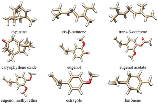
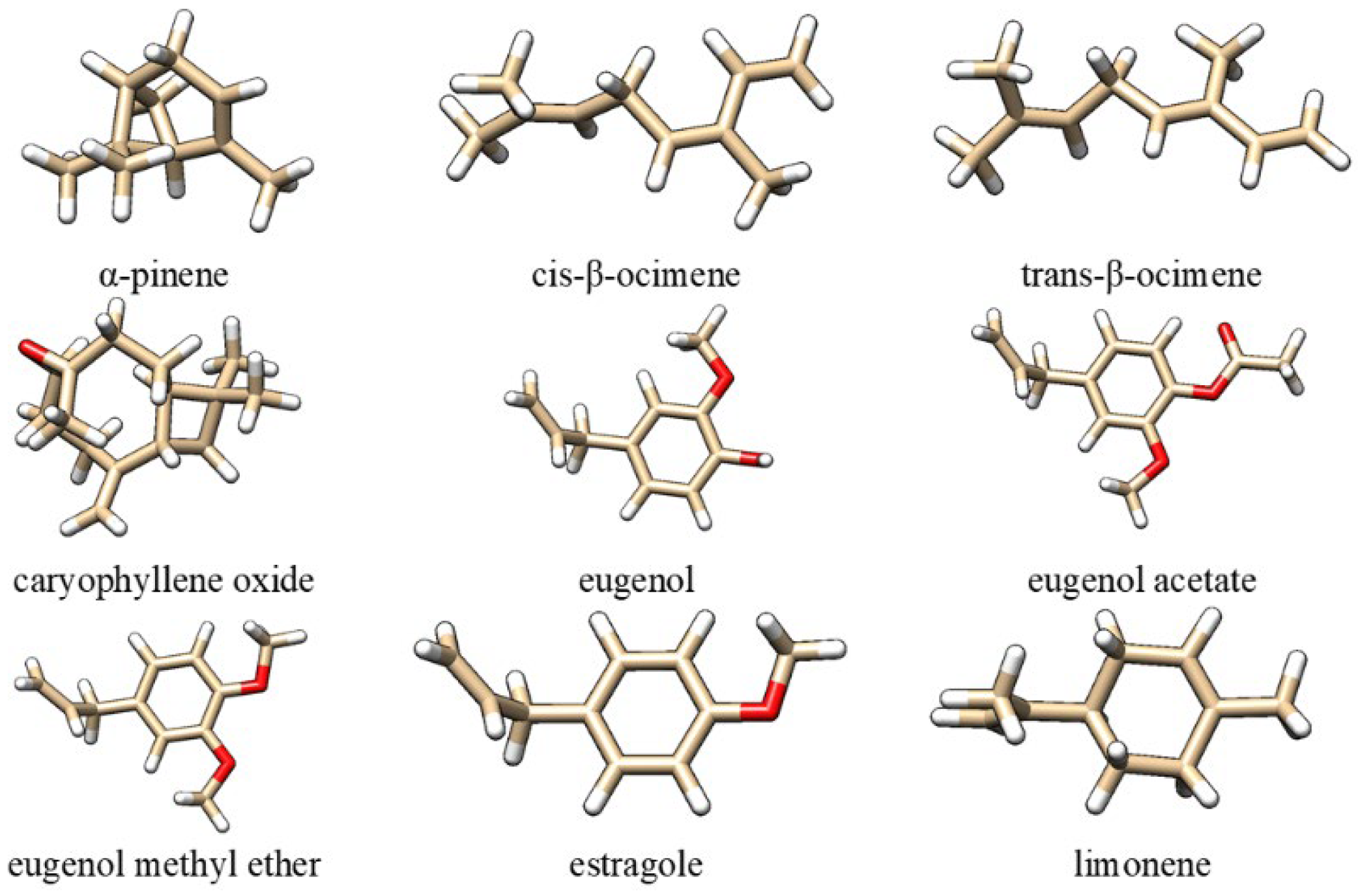
Based on previously published compositional analyses of tarragon essential oil samples from the Romanian market [10], the constituents present at concentrations above 1% were α-pinene, cis-β-ocimene, trans-β-ocimene, caryophyllene oxide, eugenol, eugenol acetate, eugenol methyl ether, estragole, and limonene (Figure 1). Supplementary Table S1 presents the reported percentages for selected compounds of the selected compounds as previously published [10]. Of these, estragole was the major component, representing approximately 80% of the essential oil. Further investigation was warranted for these compounds due to their status as major constituents of Romanian tarragon essential oil, their documented pharmacological and toxicological profiles, and their prior prediction as inhibitors of OATP1B1 and OATP1B3 [10].
Figure 1.
The chemical structures of the main compounds identified in tarragon essential oils found on the Romanian market: oxygen atoms—red; hydrogen atoms—white, carbon atoms—brown.
Consequently, in the present study, these compounds are considered for molecular docking, in association with structures of wild-type human OATP1B1 and OATP1B3 in distinct functional states, obtained using electron microscopy (cryo-EM), as presented in Table 1.
Table 1.
PDB structures of human OATP1B1 and OATP1B3 deposited in Protein Data Bank (PDB).
While multiple cryo-EM structures are available for OATP1B1, only a single cryo-EM structure has been reported for OATP1B3 bound to synthetic Fab fragments [16]. The structural models were superimposed using Chimera 1.19 software [17], and the corresponding root-mean-square deviation (RMSD) values were calculated. RMSD measures the average distance between equivalent Cα atoms following optimal structural alignment. Superposition of the OATP1B1 and OATP1B3 structures available in the Protein Data Bank (PDB) yielded the RMSD values presented in Table 2, together with the corresponding numbers of equivalent Cα atom pairs. As shown in Table 2, the analyzed structures exhibited low RMSD values, indicating a high degree of structural similarity and only minor global conformational differences. The observed deviations are likely attributable to local structural variations, including small rigid-body shifts between domains, flexible loop regions, and side-chain rearrangements. PDB structure 8PG0, the only one corresponding to OATP1B3, is the most consistently divergent structure in the matrix, with RMSDs around 1.0–1.18 Å to every other structure. However, the RMSD values suggest that it presents the same overall fold but a distinct conformational substate rather than a fundamentally different architecture.
Table 2.
Root-mean-square deviation (RMSD) values obtained from the superposition of available structures of OATP1B1 and OATP1B3.
Structural analysis suggests that many of the cavity-forming residues are conserved between OATP1B1 and OATP1B3, which explains why both transporters handle numerous common substrates such as statins, bile acids, and steroid conjugates [16]. In comparisons between apo and ligand-bound structures, the relatively low RMSD values suggest that ligand binding is associated with subtle conformational adaptations rather than major global structural rearrangements. Although the overall transporter architecture remains highly conserved, even minor local changes may influence ligand accommodation by fine-tuning the geometry and physicochemical properties of the binding cavity. Such variations can affect ligand recognition and binding without substantially altering the overall protein fold. Therefore, all structural models were considered suitable for subsequent molecular docking studies, allowing for the comparison of binding scores and interaction patterns across different conformational states.
Molecular docking simulations were performed using the SwissDock web server (2024 version), which is based on the AutoDock Vina v1.2.5 docking engine [18]. The three-dimensional structures of the target proteins were obtained from the Protein Data Bank (Table 1) and inspected prior to docking. Polar hydrogen atoms were added prior to docking, and all heteroatoms present in the experimental structures were removed. For structures containing sybodies and/or Fab fragments, these components were also removed before docking. Ligand SMILES strings were retrieved from Protein Data Bank [19]. Ligand protonation and tautomeric states were those provided in the input structures submitted to SwissDock. Because SwissDock 2024 performs internal ligand processing and does not expose user control over conformer generation, charge assignment or other parameters, these settings were maintained at their default values [18]. The search space was defined graphically to encompass the binding pocket based on the position of the ligand present in the cryo-EM structure, ensuring accurate targeting of the active site. For the apo OATP1B1 structure (PDB: 8HNB), which lacks a co-crystallized ligand, the docking search space was defined using the same box center coordinates and dimensions as those employed for the simeprevir-bound structure (PDB: 8HNH). This choice was supported by structural superposition of the available OATP1B1 structures, which showed that 8HNB and 8HNH exhibited the smallest RMSD upon alignment (Table 2), indicating a high degree of structural similarity and conservation of the binding-site region. Therefore, the binding site identified in 8HNH was considered representative of the corresponding region in 8HNB, and the same docking grid was applied to ensure consistency and comparability between the docking calculations. The box center coordinates and dimensions used for all structures are provided in Supplementary materials, Table S2. Sampling exhaustivity was set to 20. SwissDock automatically processed ligand and receptor files, assigned force-field parameters and atomic charges, and generated docking poses within the selected binding regions. The generated binding poses were ranked according to their Vina score. Because the docked compounds were not identical to the ligand resolved in the cryo-EM structure, pose selection was guided by the ability of the docked ligand to occupy the same substrate-binding cavity and establish interactions with residues involved in binding the experimentally observed ligand. The highest-ranked pose fulfilling these criteria was selected for further analysis. Visualization and analysis of docking results were performed using Chimera 1.19. To validate the docking protocol, the ligands present in the cryo-EM structures were re-docked into their respective experimental structures. The predicted binding poses (Supplementary Figure S1) showed limited agreement with the experimental binding modes, as the RMSD values between the experimental and re-docked ligand poses exceeded the commonly accepted threshold of 2 Å for successful pose reproduction (Supplementary Table S2). The RMSD values ranged from 3.617 Å for estrone-3-sulfate re-docked into the 8HND structure to 4.612 Å for simeprevir re-docked into the 8HNH structure. In the case of bilirubin re-docked into the 8HNC structure, the RMSD value reached 6.836 Å. These validation results reveal uncertainty in the predicted binding orientations. Consequently, any pose-level mechanistic interpretations must be regarded as tentative. Nevertheless, all re-docked ligands remained within the same substrate-binding cavity identified in the corresponding cryo-EM structures and interacted with key residues observed experimentally (see further in Section 3). Given the large size and conformational plasticity of the OATP1B1 binding pocket, multiple energetically favorable binding modes may coexist, leading to alternative docking solutions. This effect is expected to be particularly pronounced for large and flexible ligands such as bilirubin, which possess numerous rotatable bonds and can adopt multiple conformations within the binding cavity. Furthermore, although the overall cryo-EM structures are well resolved, local resolution may vary substantially across different regions of the map, with flexible regions often exhibiting lower local resolution than the protein core [20]. Such local variations in map quality may contribute to uncertainty in the precise placement of bound ligands. Accordingly, the docking results were used primarily to support assessment of binding-site localization and comparative binding behavior, rather than to draw detailed conclusions regarding specific binding poses.
Protein–ligand interactions were analyzed using the Protein–Ligand Interaction Profiler (PLIP; 2025 release), a fully automated computational tool for the detection and characterization of non-covalent interactions within three-dimensional protein–ligand complexes. PLIP identifies a comprehensive set of interaction types directly from input structures in PDB format, including hydrogen bonds, hydrophobic contacts, π-stacking, π-cation interactions, salt bridges, water bridges, and halogen bonds. The tool automatically detects relevant ligands and analyzes atom-level contacts without requiring manual preprocessing of the structures [21]. PLIP was used to identify and compare interaction profiles of ligands present in the cryo-EM structure, re-docked ligands present in the cryo-EM structure in their respective experimental structures, and docked tarragon compounds. The resulting interaction profiles were analyzed to elucidate the binding characteristics of each ligand within the predicted protein binding site.
3. Results and Discussion
The molecular docking study revealed that the major constituents of tarragon essential oil identified in samples from the Romanian market are predicted to bind to both OATP1B1 and OATP1B3 and may interact with the binding cavity of these transporters. The predicted binding scores are moderate (Table 3) relative to those of the reference ligands: −7.152 kcal/mol for bilirubin binding to OATP1B1 (8HNC PDB structure), −11.212 kcal/mol for estrone-3-sulfate binding to OATP1B1 (8HND PDB structure), −7.106 kcal/mol for simeprevir binding to OATP1B1 (8HNH PDB structure), −10.209 kcal/mol for 2′,7′-dichlorofluorescein binding to OATP1B1 (8K6L PDB structure), −10.099 kcal/mol for estrone-3-sulfate binding to OATP1B1 (8PHW PDB structure), and −7.025 kcal/mol for atorvastatin binding to OATP1B1 (9CY3 PDB structure). Nevertheless, even relatively weak or moderate interactions may be relevant, as inhibition of OATP1B1 and/or OATP1B3 can alter the hepatic uptake and clearance of substrate drugs. Consequently, the effects of high concentrations of tarragon essential oil constituents on drug transporters remain uncertain and should be investigated through experimental validation.
Table 3.
Predicted binding scores between the compounds identified in tarragon essential oil and OATP1B1 and OATP1B3.
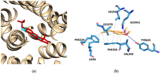
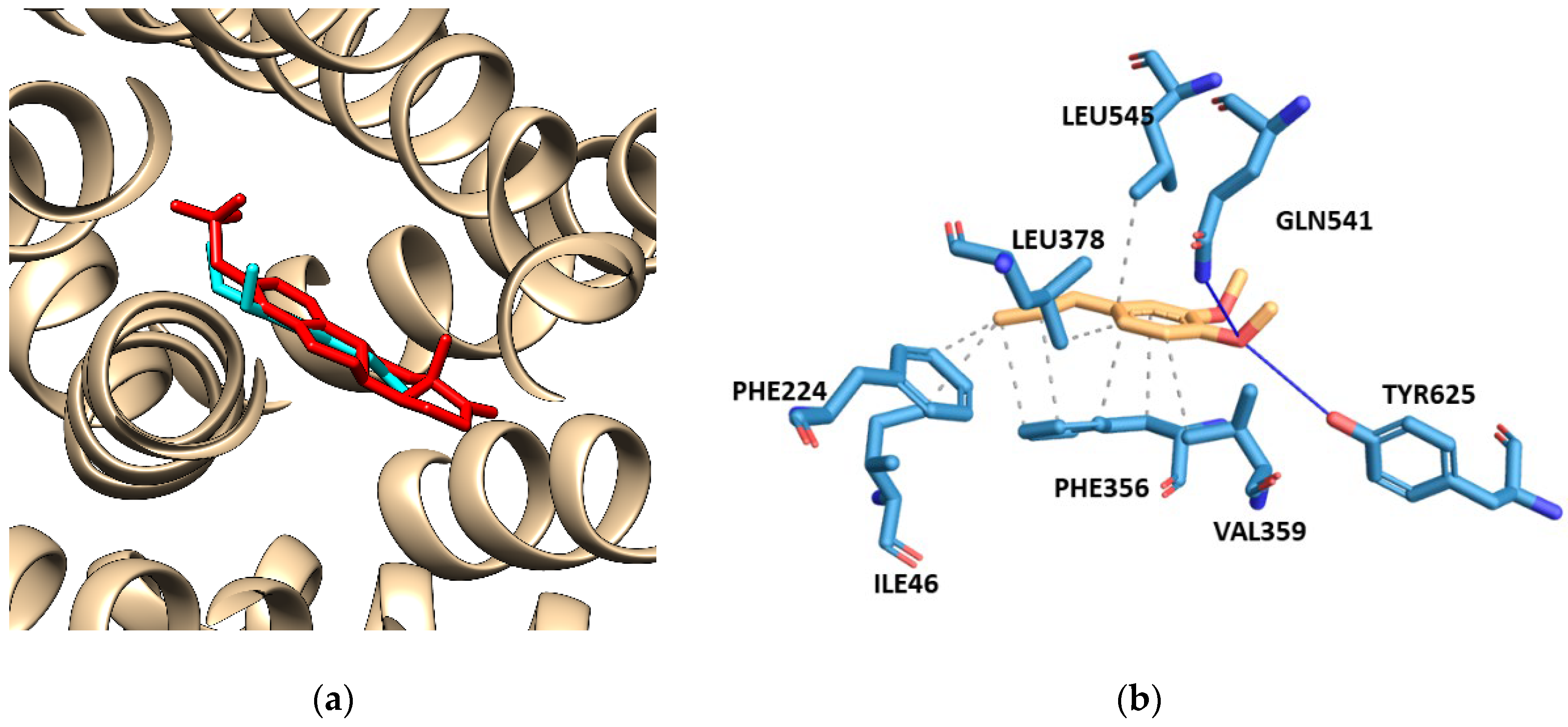
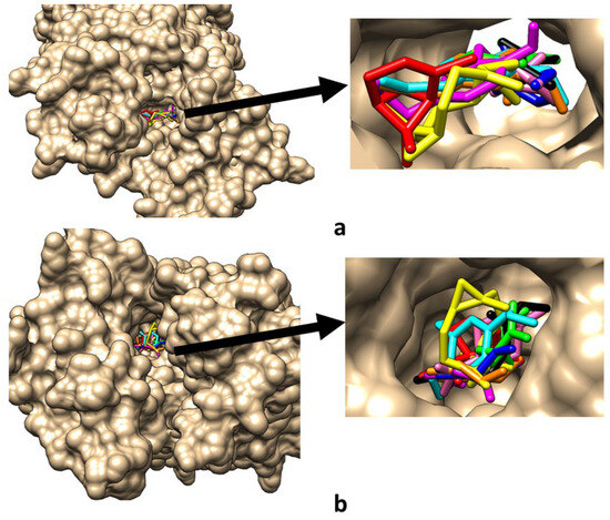
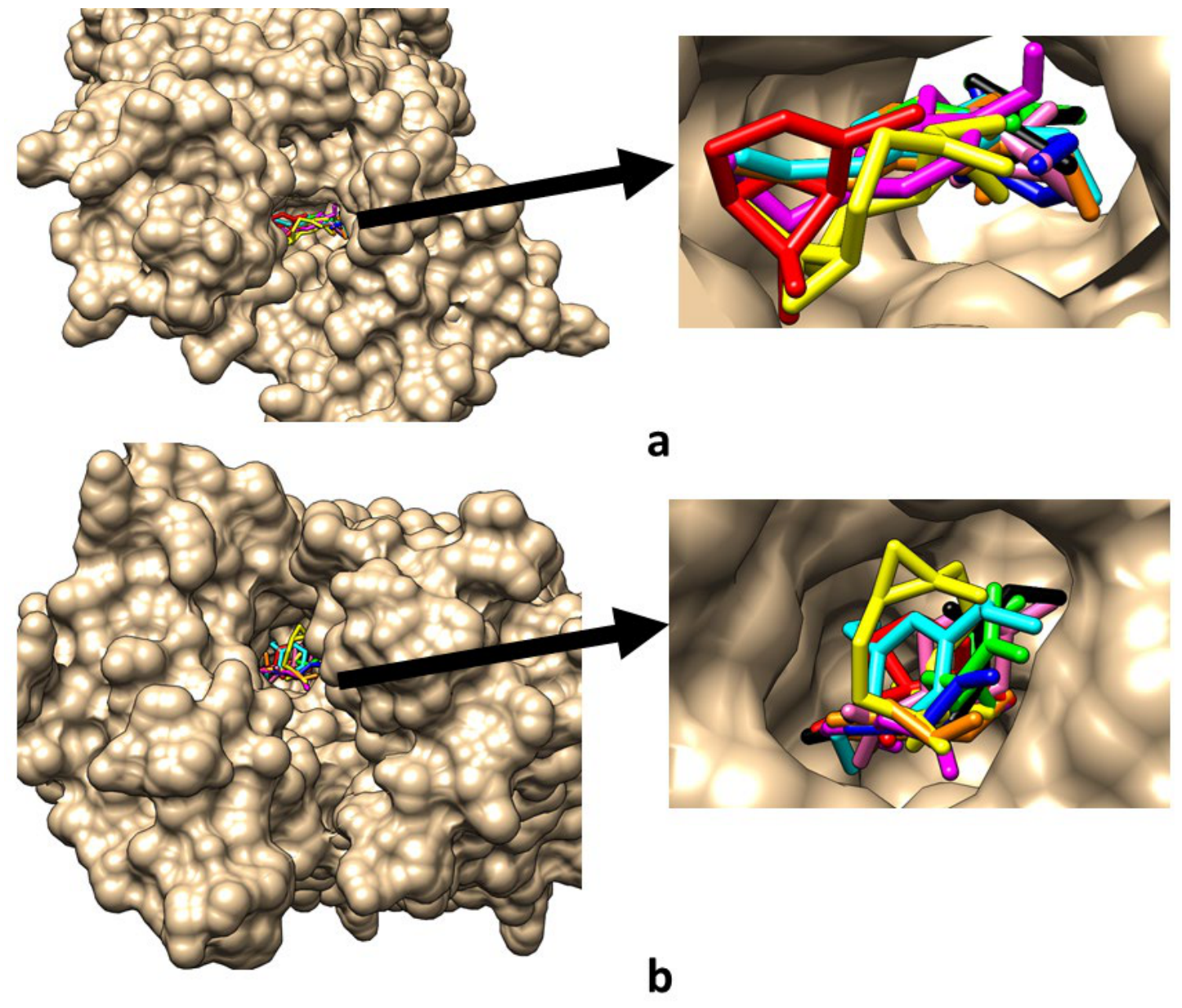
The data presented in Table 3 indicate that, among the investigated compounds, eugenol and its derivatives exhibit the highest binding scores, likely because their functional groups (–OH, –OCH3, and acetate) enable a wider range of interactions and may enhance binding affinity. It is important to note that eugenol methyl ether exhibited the highest binding score toward structure 8HND (−7.102 kcal/mol), comparable to that of the reference ligand atorvastatin (−7.025 kcal/mol, structure 9CY3). Figure 2a shows the predicted optimal binding pose of eugenol methyl ether within OATP1B1 based on the 8HND structure, while Figure 2b illustrates the corresponding protein–ligand interactions identified using PLIP. Supplementary Figures S2–S10 present the protein–ligand complexes corresponding to the highest binding scores of all investigated compounds across the analyzed OATP1B1 and OATP1B3 structural models, together with the protein–ligand interactions identified using PLIP. Furthermore, Figure 3 shows the superimposed binding poses of the investigated compounds within the OATP1B1 8HND structure and within OATP1B3 structure 8PG0, demonstrating that all ligands are predicted to occupy the central transporter cavity despite differences in their size, shape, and orientation. In parallel, Supplementary Tables S3–S12 summarize the interactions of the co-resolved ligands, re-docked reference ligands, and tarragon-derived compounds with the analyzed protein structures, as determined using PLIP.
Figure 2.
The mest binding mode of eugenol methyl ether (cyan sticks) in OATP1B1 based on the PDB structure 8HND, which contains the endogenous metabolite estrone-3-sulfate (red sticks) (a), and a detailed view of the interactions between eugenol methyl ether (orange sticks) and residues of OATP1B1 (blue sticks) (b).
Figure 3.
Superposition of the predicted binding poses of the investigated compounds within the OATP1B1 8HND PDB structure (a) and within OATP1B3 8PG0 PDB structure (b). The docked ligands are shown as stick representations and are displayed simultaneously within the central transporter cavity: α-pinene—red sticks, caryophyllene oxide—yellow sticks, cis-β-ocimene—green sticks, estragole—blue sticks, eugenol—magenta sticks, eugenol acetate—orange sticks, eugenol methyl ether—cyan sticks, limonene—pink sticks, and trans-β-ocimene—black sticks.
The data presented in Table 3 also emphasize that, despite the relatively small global structural differences between the OATP1B1 structures (between 0.4 and 1.2 Å, Table 2), the docking results revealed conformation-dependent variations in predicted binding scores. For example, the structure 8HND consistently produced the most favorable docking scores for all tested ligands (e.g., −7.102 kcal/mol for eugenol methyl ether and −6.600 kcal/mol for estragole), whereas other conformations generally yielded less favorable values.
Depending on the ligand, the difference between the most and least favorable conformations reached approximately 1–2 kcal/mol. The use of multiple cryo-EM conformations therefore enabled us to account for the intrinsic structural flexibility of OATP1B1 and to evaluate the robustness of the docking predictions. While the overall predicted binding region remained consistent across structures, the observed differences in docking scores demonstrate that local conformational variations can significantly influence ligand binding.
Although the structure of OATP1B3 (PDB ID 8PG0) is structurally similar to the other OATP1B structures (RMSDs ≈ 1 Å), it consistently yields weaker docking scores for the tarragon constituents than 8HND. Among all structures tested, 8PG0 represents a less permissive binding state for the predominantly hydrophobic phytochemicals found in tarragon essential oil. These findings indicate that the binding of tarragon essential oil constituents to OATP1B3 is predicted to be weaker than their binding to OATP1B1, and that even subtle conformational changes can influence the geometry and physicochemical properties of the binding cavity, thereby affecting ligand accommodation and the resulting binding scores. However, even if interaction patterns change, the investigated compounds are predicted to occupy the same transporter cavity (Supplementary Tables S3–S12).
The results obtained using PLIP software and presented in Supplementary Tables S3–S12 provide an exploratory overview of possible interaction patterns between compounds identified in tarragon essential oil and OATP1B1 and OATP1B3. Because the docking poses are computational predictions and have not been experimentally validated, the residue-level interactions described below should be interpreted with caution. Across both transporters, many predicted poses positioned the investigated compounds near PHE224, PHE356, VAL359, PHE386, and LEU545, suggesting the potential involvement of hydrophobic contacts, whereas oxygenated compounds were frequently predicted to localize near TYR352, TYR425, GLN541, ASN544, and TYR625, where hydrogen-bonding interactions may be possible. These observations may indicate that the predicted binding cavities possess a predominantly aromatic and hydrophobic character, although the exact residue contributions remain uncertain. In the predicted poses, monoterpene hydrocarbons, including limonene, α-pinene, cis-β-ocimene, and trans-β-ocimene, were associated mainly with hydrophobic contacts and showed limited opportunities for hydrogen bonding. In contrast, oxygenated derivatives such as eugenol and its analogs frequently displayed additional putative hydrogen-bonding interactions involving polar residues, suggesting a broader range of possible interaction types. However, these differences should not be interpreted as evidence of distinct binding mechanisms or binding specificity, as they are dependent on uncertain docking geometries. Some variability in the predicted interaction patterns was observed among the different transporter conformations, which may reflect the sensitivity of docking results to the selected protein structures and the conformational flexibility of OATP1B1 and OATP1B3. Finally, comparison with ligands present in the available cryo-EM structures (estrone-3-sulfate, bilirubin, atorvastatin, and simeprevir) suggests that these reference ligands engage in more extensive polar interactions, whereas the investigated compounds were predicted to exhibit comparatively fewer hydrogen-bonding contacts and a greater contribution from hydrophobic interactions.
Overall, these findings should be considered preliminary and hypothesis-generating, and their biological and clinical relevance will require confirmation through experimental transporter assays and pharmacokinetic studies. The toxicological relevance of these findings should also be considered in the context of the known safety concerns associated with estragole-containing tarragon essential oil. Estragole has been extensively investigated because of its potential genotoxic and carcinogenic effects following metabolic activation in the liver [22]. Although the present docking results do not demonstrate transporter inhibition or substrate transport, they suggest that estragole may be capable of interacting with the OATP1B1 and OATP1B3. The potential biological significance of these interactions remains unclear, particularly given the limitations of in silico approaches in general, and those of this study in particular. While such interactions could be of interest in the context of hepatic drug disposition, especially following exposure to concentrated estragole-containing products, their relevance under physiological conditions has yet to be established. Further experimental studies are needed to clarify whether estragole interacts with OATP1B1 and/or OATP1B3 as a substrate, inhibitor, or modulator and to determine the extent to which such interactions may influence its pharmacokinetic or toxicological profile.
A limitation of the present study is that the assessment of possible interactions of OATP1B1 and OATP1B3 with investigated compounds was based primarily on molecular docking scores. While docking can provide valuable information regarding potential binding modes and relative binding scores, it represents a static description of protein–ligand interactions and does not account for the dynamic nature of transporter function. OATP1B1 and OATP1B3 transporters undergo substantial conformational changes during the transport cycle, transitioning between distinct structural states that may differentially influence ligand binding, substrate translocation, and inhibitor recognition. Consequently, docking scores alone cannot fully capture the complex mechanisms underlying transporter inhibition. Furthermore, docking scoring functions provide only approximate estimates of binding affinity and generally neglect important factors such as protein flexibility, membrane environment, solvent effects, ligand-induced conformational changes, and kinetic aspects of ligand binding. Therefore, compounds exhibiting favorable docking scores may not necessarily act as effective inhibitors in vitro or in vivo, whereas weaker-scoring ligands may still modulate transporter activity through mechanisms not adequately represented by static docking models. To partially address protein flexibility, multiple cryo-EM conformations of OATP1B1 were included in the present study.
Another limitation of the docking protocol is that water molecules were removed during receptor preparation. Although this is standard practice in many docking workflows, conserved water molecules can contribute to ligand recognition through water-mediated hydrogen bonds. As these interactions are not explicitly considered by the docking calculations, the predicted hydrogen-bonding patterns should be interpreted with caution.
Nevertheless, the results should be interpreted as indicative of potential ligand–transporter interactions rather than definitive evidence of transporter inhibition. Experimental transport assays and advanced computational approaches, such as molecular dynamics simulations, would be required to further characterize the binding mechanisms of the investigated compounds.
4. Conclusions
The molecular docking and interaction analysis performed using PLIP suggests that the compounds identified in tarragon essential oil could interact with the binding sites of OATP1B1 and OATP1B3 through a combination of non-covalent interactions, including hydrophobic contacts, hydrogen bonding, and other stabilizing interactions. Among the investigated compounds, oxygenated derivatives of eugenol display a slightly more varied contact profile through potential hydrogen bonding, whereas monoterpene hydrocarbons primarily rely on hydrophobic interactions. Although the predicted binding scores are generally moderate and must be treated with caution, these preliminary trends point toward a possible orientation within key binding regions. Overall, these findings offer a tentative model for how dietary phytochemicals might generally occupy space within hepatic uptake transporters. However, the results should be interpreted as computational estimates that reflect potential binding tendencies rather than experimentally confirmed inhibitory activity. Direct experimental data are needed to confirm that the compounds actually inhibit transport activity in biological systems, at what concentrations, and with what clinical relevance.
Supplementary Materials
The following supporting information can be downloaded at: https://www.mdpi.com/article/10.3390/pr14132207/s1, Figure S1. Comparison of the binding modes of ligands present in the PDB structures (red sticks) and re-docked ligands present in the PDB structures (blue sticks); Figure S2. Best binding pose of α-pinene (magenta sticks) in OATP1B1 based on the PDB structure 8PHW, containing the endogenous metabolite estrone-3-sulfate (green sticks) (a), and in OATP1B3 based on the PDB structure 8PG0 (b). Corresponding OATP1B1–α-pinene (c) and OATP1B3–α-pinene (d) interaction profiles were generated using PLIP; Figure S3. Best binding pose of caryophyllene oxide (magenta sticks) in OATP1B1 based on the PDB structure 8HND, containing the endogenous metabolite estrone-3-sulfate (green sticks) (a), and in OATP1B3 based on the PDB structure 8PG0 (b). Corresponding OATP1B1–caryophyllene oxide (c) and OATP1B3–caryophyllene oxide (d) interaction profiles were generated using PLIP; Figure S4. Best binding pose of cis-β-ocimene (magenta sticks) in OATP1B1 based on the PDB structure 8HNC, containing the endogenous metabolite bilirubin (green sticks) (a), and in OATP1B3 based on the PDB structure 8PG0 (b). Corresponding OATP1B1–cis-β-ocimene (c) and OATP1B3–cis-β-ocimene (d) interaction profiles were generated using PLIP; Figure S5. Best binding pose of trans-β-ocimene (magenta sticks) in OATP1B1 based on the PDB structure 8HND, containing the endogenous metabolite estrone-3-sulfate (green sticks) (a), and in OATP1B3 based on the PDB structure 8PG0 (b). Corresponding OATP1B1–trans-β-ocimene (c) and OATP1B3–trans-β-ocimene (d) interaction profiles were generated using PLIP; Figure S6. Best binding pose of estragole (magenta sticks) in OATP1B1 based on the PDB structure 8HND, containing the endogenous metabolite estrone-3-sulfate (green sticks) (a), and in OATP1B3 based on the PDB structure 8PG0 (b). Corresponding OATP1B1–estragole (c) and OATP1B3–estragole (d) interaction profiles were generated using PLIP; Figure S7. Best binding pose of eugenol (magenta sticks) in OATP1B1 based on the PDB structure 8HND, containing the endogenous metabolite estrone-3-sulfate (green sticks) (a), and in OATP1B3 based on the PDB structure 8PG0 (b). Corresponding OATP1B1–eugenol (c) and OATP1B3–eugenol (d) interaction profiles were generated using PLIP; Figure S8. Best binding pose of eugenol acetate (magenta sticks) in OATP1B1 based on the PDB structure 8HND, containing the endogenous metabolite estrone-3-sulfate (green sticks) (a), and in OATP1B3 based on the PDB structure 8PG0 (b). Corresponding OATP1B1–eugenol acetate (c) and OATP1B3–eugenol acetate (d) interaction profiles were generated using PLIP; Figure S9. Best binding pose of eugenol methyl ether (magenta sticks) in OATP1B1 based on the PDB structure 8HND, containing the endogenous metabolite estrone-3-sulfate (green sticks) (a), and in OATP1B3 based on the PDB structure 8PG0 (b). Corresponding OATP1B1–eugenol methyl ether (c) and OATP1B3–eugenol methyl ether (d) interaction profiles were generated using PLIP.; Figure S10. Best binding pose of limonene (magenta sticks) in OATP1B1 based on the PDB structure 8HND, containing the endogenous metabolite estrone-3-sulfate (green sticks) (a), and in OATP1B3 based on the PDB structure 8PG0 (b). Corresponding OATP1B1–limonene (c) and OATP1B3–limonene (d) interaction profiles were generated using PLIP; Supplementary Table S1. The reported percentages of investigated constituents of tarragon essential oils found on the Romanian market: eot-c—commercial tarragon essential oil, eot-mw—essential oil obtained after microwave hexane extraction of shredded dried tarragon; Table S2. The box centers and box dimensions used for docking for each PDB structure and RMSD values between experimental and re-docked ligand poses for each structure; Table S3: Interactions between the compounds found in tarragon essential oil and OATP1B1 based on the PDB structure 8HNB. Numbers in parentheses indicate how many interactions occur for each amino acid; Table S4: Interactions between the compounds found in tarragon essential oil and OATP1B1 based on the PDB structure 8HNC. Numbers in parentheses indicate how many interactions occur for each amino acid; Table S5: Interactions between the compounds found in tarragon essential oil and OATP1B1 based on the PDB structure 8HND. Numbers in parentheses indicate how many interactions occur for each amino acid; Table S6: Interactions between the compounds found in tarragon essential oil and OATP1B1 based on the PDB structure 8HNH. Numbers in parentheses indicate how many interactions occur for each amino acid; Table S7: Interactions between the compounds found in tarragon essential oil and OATP1B1 based on the PDB structure 8K6L. Numbers in parentheses indicate how many interactions occur for each amino acid; Table S8. Interactions between the compounds found in tarragon essential oil and OATP1B1 based on the PDB structure 9CY1. Numbers in parentheses indicate how many interactions occur for each amino acid; Table S9. Interactions between the compounds found in tarragon essential oil and OATP1B1 based on the PDB structure 9CY3. Numbers in parentheses indicate how many interactions occur for each amino acid; Table S10: Interactions between the compounds found in tarragon essential oil and OATP1B1 based on the PDB structure 9CY4. Numbers in parentheses indicate how many interactions occur for each amino acid: Table S11: Interactions between the compounds found in tarragon essential oil and OATP1B1 based on the PDB structure 8PHW. Numbers in parentheses indicate how many interactions occur for each amino acid; Table S12: Interactions between the compounds found in tarragon essential oil and OATP1B3 based on the PDB structure 8PG0. Numbers in parentheses indicate how many interactions occur for each amino acid.
Author Contributions
Conceptualization, A.I.; methodology, A.I.; validation, A.I.; formal analysis, A.P.; investigation, A.P. and D.-L.R.; data curation, D.-L.R.; writing—original draft preparation, A.P.; writing—review and editing, A.I.; visualization, A.I.; supervision, A.I. All authors have read and agreed to the published version of the manuscript.
Funding
This research was funded by West University of Timișoara. The APC was funded by the Romanian Ministry of Research, Innovation and Digitalization via Romania’s National Recovery and Resilience Plan, Call No. PNRR-III-C9-2022-I10.
Data Availability Statement
All data are presented within the article and Supplementary materials.
Acknowledgments
We gratefully acknowledge the support provided by the West University of Timișoara for covering the Open Access publication fee through the grant ”Research Career Guidance and Counseling Center – Western Region”, funded by the Romanian Ministry of Research, Innovation and Digitalization via Romania’s National Recovery and Resilience Plan, Call No. PNRR-III-C9-2022-I10.
Conflicts of Interest
The authors declare no conflicts of interest.
Abbreviations
The following abbreviations are used in this manuscript:
| OATP1B1 | Organic Anion Transporter Polypeptide 1B1 |
| OATP1B3 | Organic Anion Transporter Polypeptide 1B3 |
| PLIP | Protein Ligand Interaction Profile |
| PDB | Protein Data Bank |
References
- Hagenbuch, B.; Meier, P.J. Organic anion transporting polypeptides of the OATP/SLC21 family: Phylogenetic classification as OATP/SLCO superfamily, new nomenclature and molecular/functional properties. Pflug. Arch. 2004, 447, 653–665. [Google Scholar] [CrossRef] [Scilit] [PubMed]
- Abe, T.; Kakyo, M.; Tokui, T.; Nakagomi, R.; Nishio, T.; Nakai, D.; Nomura, H.; Unno, M.; Suzuki, M.; Naitoh, T.; et al. Identification of a novel gene family encoding human liver-specific organic anion transporter LST-1. J. Biol. Chem. 1999, 274, 17159–17163. [Google Scholar] [CrossRef] [Scilit] [PubMed]
- König, J.; Cui, Y.; Nies, A.T.; Keppler, D. A novel human organic anion transporting polypeptide localized to the basolateral hepatocyte membrane. Am. J. Physiol. Gastrointest. Liver Physiol. 2000, 278, G156–G164. [Google Scholar] [CrossRef] [Scilit] [PubMed]
- Alam, K.; Crowe, A.; Wang, X.; Zhang, P.; Ding, K.; Li, L.; Yue, W. Regulation of Organic Anion Transporting Polypeptides (OATP) 1B1- and OATP1B3-Mediated Transport: An Updated Review in the Context of OATP-Mediated Drug-Drug Interactions. Int. J. Mol. Sci. 2018, 19, 855. [Google Scholar] [CrossRef] [Scilit] [PubMed]
- Hagenbuch, B.; Stieger, B. The SLCO (former SLC21) superfamily of transporters. Mol. Asp. Med. 2013, 34, 396–412. [Google Scholar] [CrossRef] [Scilit] [PubMed]
- Kotsampasakou, E.; Brenner, S.; Jäger, W.; Ecker, G.F. Identification of Novel Inhibitors of Organic Anion Transporting Polypeptides 1B1 and 1B3 (OATP1B1 and OATP1B3) Using a Consensus Vote of Six Classification Models. Mol. Pharm. 2015, 12, 4395–4404. [Google Scholar] [CrossRef] [Scilit] [PubMed]
- Ekiert, H.; Świątkowska, J.; Knut, E.; Klin, P.; Rzepiela, A.; Tomczyk, M.; Szopa, A. Artemisia dracunculus (Tarragon): A Review of Its Traditional Uses, Phytochemistry and Pharmacology. Front. Pharmacol. 2021, 12, 653993. [Google Scholar] [CrossRef] [Scilit] [PubMed]
- Tarragon Market Size 2024–2028. Available online: https://www.technavio.com/report/tarragon-market-industry-analysis (accessed on 26 January 2026).
- Kalantari, H.; Galehdari, H.; Zaree, Z.; Gesztelyi, R.; Varga, B.; Haines, D.; Bombicz, M.; Tosaki, A.; Juhasz, B. Toxicological and Mutagenic Analysis of Artemisia Dracunculus (Tarragon) Extract. Food Chem. Toxicol. 2013, 51, 26–32. [Google Scholar] [CrossRef] [Scilit] [PubMed]
- Pujicic, A.; Popescu, I.; Dascalu, D.; Petreuș, D.E.; Isvoran, A. Predictions of the Biological Effects of the Main Components of Tarragon Essential Oil. Int. J. Mol. Sci. 2025, 26, 1860. [Google Scholar] [CrossRef] [Scilit] [PubMed]
- Denisow-Pietrzyk, M.; Pietrzyk, Ł.; Denisow, B. Asteraceae Species as Potential Environmental Factors of Allergy. Environ. Sci. Pollut. Res. 2019, 26, 6290–6300. [Google Scholar] [CrossRef] [Scilit] [PubMed]
- Wang, L.; Sweet, D.H. Interaction of Natural Dietary and Herbal Anionic Compounds and Flavonoids with Human Organic Anion Transporters 1 (SLC22A6), 3 (SLC22A8), and 4 (SLC22A11). Evid. Based Complement. Altern. Med. 2013, 2013, 612527. [Google Scholar] [CrossRef] [Scilit] [PubMed]
- Kang, Y.J.; Lee, C.H.; Park, S.J.; Lee, H.S.; Choi, M.K.; Song, I.S. Involvement of Organic Anion Transporters in the Pharmacokinetics and Drug Interaction of Rosmarinic Acid. Pharmaceutics 2021, 13, 83. [Google Scholar] [CrossRef] [Scilit] [PubMed]
- Shan, Z.; Yang, X.; Liu, H.; Yuan, Y.; Xiao, Y.; Nan, J.; Zhang, W.; Song, W.; Wang, J.; Wei, F.; et al. Cryo-EM structures of human organic anion transporting polypeptide OATP1B1. Cell Res. 2023, 33, 940–951. [Google Scholar] [CrossRef] [Scilit] [PubMed]
- Sung, M.W.; Hu, K.; Hurlimann, L.M.; Lees, J.A.; Fennell, K.F.; West, M.A.; Costales, C.; Rodrigues, A.D.; Zimmermann, I.; Dawson, R.J.P.; et al. Cyclosporine A sterically inhibits statin transport by solute carrier OATP1B1. J. Biol. Chem. 2025, 301, 108484. [Google Scholar] [CrossRef] [Scilit] [PubMed]
- Ciută, A.D.; Nosol, K.; Kowal, J.; Mukherjee, S.; Ramírez, A.S.; Stieger, B.; Kossiakoff, A.A.; Locher, K.P. Structure of human drug transporters OATP1B1 and OATP1B3. Nat. Commun. 2023, 14, 5774. [Google Scholar] [CrossRef] [Scilit] [PubMed]
- Pettersen, E.F.; Goddard, T.D.; Huang, C.C.; Couch, G.S.; Greenblatt, D.M.; Meng, E.C.; Ferrin, T.E. UCSF Chimera—A visualization system for exploratory research and analysis. J. Comput. Chem. 2004, 25, 1605–1612. [Google Scholar] [CrossRef] [Scilit] [PubMed]
- Bugnon, M.; Röhrig, U.F.; Goullieux, M.; Perez, M.A.S.; Daina, A.; Michielin, O.; Zoete, V. SwissDock 2024: Major enhancements for small-molecule docking with Attracting Cavities and AutoDock Vina. Nucleic Acids Res. 2024, 52, W324–W332. [Google Scholar] [CrossRef] [Scilit] [PubMed]
- Berman, H.M.; Westbrook, J.; Feng, Z.; Gilliland, G.; Bhat, T.N.; Weissig, H.; Shindyalov, I.N.; Bourne, P.E. The Protein Data Bank. Nucleic Acids Res. 2000, 28, 235–242. [Google Scholar] [CrossRef] [Scilit] [PubMed]
- García-Nafría, J.; Tate, C.G. Structure determination of GPCRs: Cryo-EM compared with X-ray crystallography. Biochem. Soc. Trans. 2021, 49, 2345–2355. [Google Scholar] [CrossRef] [Scilit] [PubMed]
- Schake, P.; Bolz, S.N.; Linnemann, K.; Schroeder, M. PLIP 2025: Introducing protein-protein interactions to the protein-ligand interaction profiler. Nucleic Acids Res. 2025, 53, W463–W465. [Google Scholar] [CrossRef] [Scilit] [PubMed]
- Ackermann, G.; Peil, M.; Quarz, C.; Schmidt, A.; Halaczkiewicz, M.; Thomas, A.D.; Stegmüller, S.; Richling, E.; Manolikakes, G.; Christmann, M.; et al. Molecular dosimetry of estragole and 1′-hydroxyestragole-induced DNA adduct formation, clastogenicity and cytotoxicity in human liver cell models. Arch. Toxicol. 2025, 99, 3769–3785. [Google Scholar] [CrossRef] [Scilit] [PubMed]

Disclaimer/Publisher’s Note: The statements, opinions and data contained in all publications are solely those of the individual author(s) and contributor(s) and not of MDPI and/or the editor(s). MDPI and/or the editor(s) disclaim responsibility for any injury to people or property resulting from any ideas, methods, instructions or products referred to in the content. |
© 2026 by the authors. Licensee MDPI, Basel, Switzerland. This article is an open access article distributed under the terms and conditions of the Creative Commons Attribution (CC BY) license.