


Abstract
Background: Sirtuins are recognized as critical regulators of various physiological processes, including aging, metabolic homeostasis, oxidative stress, and inflammatory responses. This study aimed to investigate whether sirtuin expression is associated with the pathogenesis and clinical manifestations of chronic otitis media (COM). Methods: Middle ear inflammatory tissues were obtained from 27 patients who underwent COM surgery. The mRNA expression levels of SIRT1–7 were measured using quantitative real-time polymerase chain reaction (qRT-PCR). The expression patterns of SIRT1–7 were comparatively analyzed according to bacterial culture results, the presence of otorrhea, and the type of hearing loss (conductive or sensorineural). Results: The study included 10 males and 17 females, with a mean age of 60.52 ± 9.79 years. Otorrhea was present in 51.9% of patients, whereas the bacterial culture-positive rate was 29.6%. Conductive hearing loss was more prevalent than sensorineural hearing loss. SIRT1–7 mRNA expression was detected in all inflamed COM tissues. Among the seven members, SIRT3 exhibited the highest expression level, followed in descending order by SIRT6, SIRT4, SIRT7, SIRT1, SIRT5, and SIRT2. Notably, the expression levels of SIRT1–4 were significantly higher in the culture-negative group compared to the culture-positive group (p < 0.05). However, no significant differences in SIRT1–7 mRNA expression were observed according to the presence of otorrhea or the type of hearing loss (p > 0.05). Conclusion: All seven sirtuin members (SIRT1–7) were expressed in inflamed COM mucosa, suggesting that the sirtuin family may be transcriptionally active in the inflamed middle ear microenvironment. Specifically, SIRT1–4 mRNA expression differed significantly according to bacterial culture status, suggesting a potential association between SIRT1–4 mRNA expression and local inflammatory processes in COM.
1. Introduction
Chronic otitis media (COM) is a prevalent inflammatory disease characterized by irreversible pathological changes within the Eustachian tube, tympanic cavity, and mastoid air cells. Clinically, it is one of the most frequent conditions encountered in otorhinolaryngology outpatient settings, primarily presenting with persistent otorrhea and hearing impairment. The pathogenesis of COM is highly multifactorial, driven by a complex interplay of anatomical and functional dysfunctions of the Eustachian tube, genetic predisposition, and environmental factors. Furthermore, persistent microbial infections—particularly by pathogenic bacteria—play a critical role in exacerbating chronic mucosal inflammation and recurrent otorrhea [1,2].
In chronic suppurative otitis media, host defense against microbial invasion is mediated by an early innate immune response and a subsequent adaptive immune response. The primary defense mechanism, innate immunity, activates within 12 h of infection and consists of physical barriers—driven by the mucociliary blanket, ciliated cells, and secretory cells (mucociliary clearance)—as well as biological defenses involving the secretion of antimicrobial enzymes. Subsequently, the adaptive immune system acts as a secondary defense mechanism, typically activating within 1 to 5 days post-infection. The entire Eustachian tube and the adjacent middle ear mucosa orchestrate both these secretory and mucociliary defense systems; thus, impairment in any of these components significantly elevates the risk of developing otitis media. Ultimately, the onset of COM is fundamentally determined by the intricate balance between invading pathogens and the host’s immune defense mechanisms. Therefore, a comprehensive understanding of both normal mucosal defense mechanisms and pathogen infectivity is essential to elucidate the core pathogenesis of the disease [3,4].
In mammals, seven members of the sirtuin family (SIRT1–7) have been identified. They are broadly recognized as critical regulators in diverse physiological and pathological processes, including cellular metabolism, aging, oxidative stress, mitochondrial homeostasis, apoptosis, and tumor dynamics. Most notably, mammalian sirtuins play a pivotal role in modulating inflammatory responses and immune regulation [5,6]. Furthermore, recent studies have highlighted that sirtuins are deeply involved in host–pathogen interactions during microbial infections, suggesting their crucial defensive role against pathogenic invasion [7]. Given these multifaceted roles, it is highly plausible that the expression of SIRT1–7 is closely associated with the pathogenesis and clinical manifestations of COM, a persistent inflammatory condition. However, to the best of our knowledge, the mRNA expression profiles of the sirtuin family in COM have not yet been elucidated. Therefore, the present study aimed to investigate the expression levels of SIRT1–7 mRNA in inflamed middle ear tissues of patients with COM, and to evaluate their potential associations with clinical parameters, including bacterial infection, otorrhea, and hearing loss.
2. Subjects and Methods
2.1. Subjects
This study included 27 patients who visited the Otorhinolaryngology-Head and Neck Surgery clinic at a single tertiary referral hospital. COM was defined as the presence of persistent symptoms—such as hearing loss, tympanic membrane perforation, otorrhea, tinnitus, or otalgia—lasting for more than 3 months, accompanied by middle ear and mastoid lesions confirmed via temporal bone computed tomography (TBCT). The presence of otorrhea was evaluated using otoscopy and endoscopy preoperatively. All patients received prophylactic antibiotic treatment (oral amoxicillin/clavulanate 875 mg/125 mg, twice a day) for one week before surgery to minimize acute inflammation. The presence and severity of hearing impairment were assessed using pure-tone audiometry (PTA). Furthermore, the definitive diagnosis of COM was confirmed histologically by the presence of chronic inflammation and granulation tissue in the inflamed middle ear biopsy specimens obtained during surgery. The sampling location primarily included the tympanic cavity and mastoid antrum, and while the exact tissue volume varied depending on the individual disease extent, representative inflamed mucosal tissues were carefully collected from all patients.
Patients suspected of having acute otitis media, otitis media with effusion, cholesteatomatous otitis media, tuberculous otitis media, craniofacial anomalies, systemic diseases, or congenital/acquired immunodeficiency were excluded from the study. Medical records were retrospectively reviewed to extract demographic and clinical data, including age, sex, date of onset, affected ear, bacterial culture results, underlying comorbidities, PTA results, and history of revision surgery. Written informed consent was obtained from all patients or their legal guardians after a thorough explanation of the study’s purpose. This study was approved by the Institutional Review Board of Kyung Hee University Medical Center (IRB 2023-05-016, approved on 15 May 2023), and written informed consent was obtained from all subjects.
2.2. Bacterial Culture
Microbiological evaluations were conducted strictly for patients exhibiting visible otorrhea. After thoroughly cleaning the external auditory canal to prevent cross-contamination, discharge specimens were aseptically collected intraoperatively using sterile cotton swabs (Xomed Trace Products, Jacksonville, FL, USA) under microscopic or endoscopic guidance. This intraoperative collection was performed to minimize contamination by other external microorganisms. The swabs were then immediately placed into Stuart transport medium. Subsequently, the specimens were inoculated onto solid blood agar and into liquid thioglycollate medium (Hangang, Kun-po, Gyeonggi-do, Republic of Korea) and incubated at 35 °C for 24 h. Colony-forming bacteria were then identified using Gram staining and standard biochemical tests.
2.3. Evaluation of Level of Hearing Loss
PTA was performed at the time of diagnosis to measure air conduction (AC) and bone conduction (BC) hearing thresholds. The mean hearing thresholds were calculated by averaging the thresholds at 500, 1000, and 2000 Hz, with a double weight applied to the 1000 Hz frequency. Patients were categorized according to the type of hearing impairment: sensorineural hearing loss (SNHL) was defined as a BC threshold ≥ 30 dB with an air-bone gap (ABG) < 10 dB, whereas conductive hearing loss (CHL) was defined as an ABG ≥ 10 dB with a BC threshold < 25 dB. Patients with mixed hearing loss (BC threshold ≥ 25 dB and ABG ≥ 10 dB) were classified into the SNHL group [8].
2.4. RNA Extraction and Quantitative Real-Time PCR (RT-qPCR)
Total RNA was extracted from the patient tissue samples using TRIzol reagent (Invitrogen, Carlsbad, CA, USA) according to the manufacturer’s protocol. Prior to cDNA synthesis, the concentration and purity of the extracted RNA were assessed using a NanoDrop spectrophotometer (Thermo Fisher Scientific, Waltham, MA, USA). Samples with an A260/280 absorbance ratio between 1.8 and 2.0 were considered to have acceptable RNA purity. cDNA was synthesized from 1 μg of total RNA using oligo(dT)18 primers with the TOPscript™ RT DryMIX (RT200, Enzynomics, Daejeon, Republic of Korea). The primer sequences used in this study are listed in Table 1. Quantitative real-time PCR was performed on a StepOnePlus Real-Time PCR System (Applied Biosystems, Foster City, CA, USA) utilizing the SensiFAST SYBR Hi-ROX Kit (Bioline, London, UK). The PCR was conducted with 2 μL of cDNA in a 20 μL reaction mixture containing 10 μL of SensiFAST SYBR Hi-ROX Mix, 2 μL of primers (1 μL each of forward and reverse), and 6 μL of PCR-grade water. The thermal cycling conditions consisted of an initial polymerase activation at 95 °C for 2 min, followed by 40 cycles of denaturation at 95 °C for 5 s and a combined annealing/extension step at 60 °C for 20 s. The relative mRNA expression of each target gene was normalized to that of β-actin using the 2−ΔCt method, where ΔCt = Ct(target gene) − Ct(β-actin) [9]. The mRNA expression levels of SIRT1–7 were analyzed according to bacterial culture positivity, otorrhea status, and the type of hearing loss (conductive vs. sensorineural).
Table 1.
Primer sequences for real-time RT-PCR.
2.5. Statistical Analysis
Statistical analyses were performed using IBM SPSS (version 20.0) Statistics for Windows, version 20.0 (IBM Corp., Armonk, NY, USA). Continuous variables are presented as the median with interquartile range (IQR) for skewed expression data, or as the mean ± standard deviation (SD) for normally distributed demographic data. Categorical variables are presented as frequencies and percentages. The normality of continuous variables was assessed before comparative analyses. For comparisons between two independent groups, Student’s t-test was used for normally distributed variables, whereas the Mann–Whitney U test was used for non-normally distributed variables. Differences in target-gene mRNA expression according to bacterial culture status and the presence or absence of otorrhea were assessed using the two-sided Mann–Whitney U test. Comparisons of mRNA expression among patients with conductive hearing loss, sensorineural hearing loss, and normal hearing were performed using the Kruskal–Wallis test. All statistical tests were two-sided, and a p-value < 0.05 was considered statistically significant.
3. Results
3.1. Demographics and Results of Bacterial Culture (Table 2)
A total of 27 patients with COM were enrolled. The mean age was 60.52 ± 9.79 years, comprising 10 male (37.0%) and 17 female (63.0%) patients. Otorrhea was present at the time of examination in 14 patients (51.9%), and 3 patients (11.1%) had a history of revision surgery. The mean pure-tone thresholds were 46.74 ± 25.78 dB for air conduction and 30.57 ± 19.52 dB for bone conduction. With respect to the type of hearing impairment, 12 patients (44.4%) had conductive hearing loss, 7 (25.9%) had sensorineural hearing loss, and 8 (29.6%) had normal hearing. Bacterial cultures of ear discharge were positive in 8 of the 27 patients (29.6%). Pseudomonas aeruginosa was the most frequently isolated organism, followed by methicillin-resistant Staphylococcus aureus (MRSA), coagulase-negative Staphylococcus, Staphylococcus aureus, and Achromobacter xylosoxidans.
Table 2.
Demographic and clinical characteristics of patients with chronic otitis media.
3.2. Expression of SIRT1–7 mRNA and Its Association with Bacterial Infection
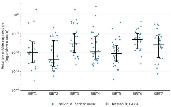
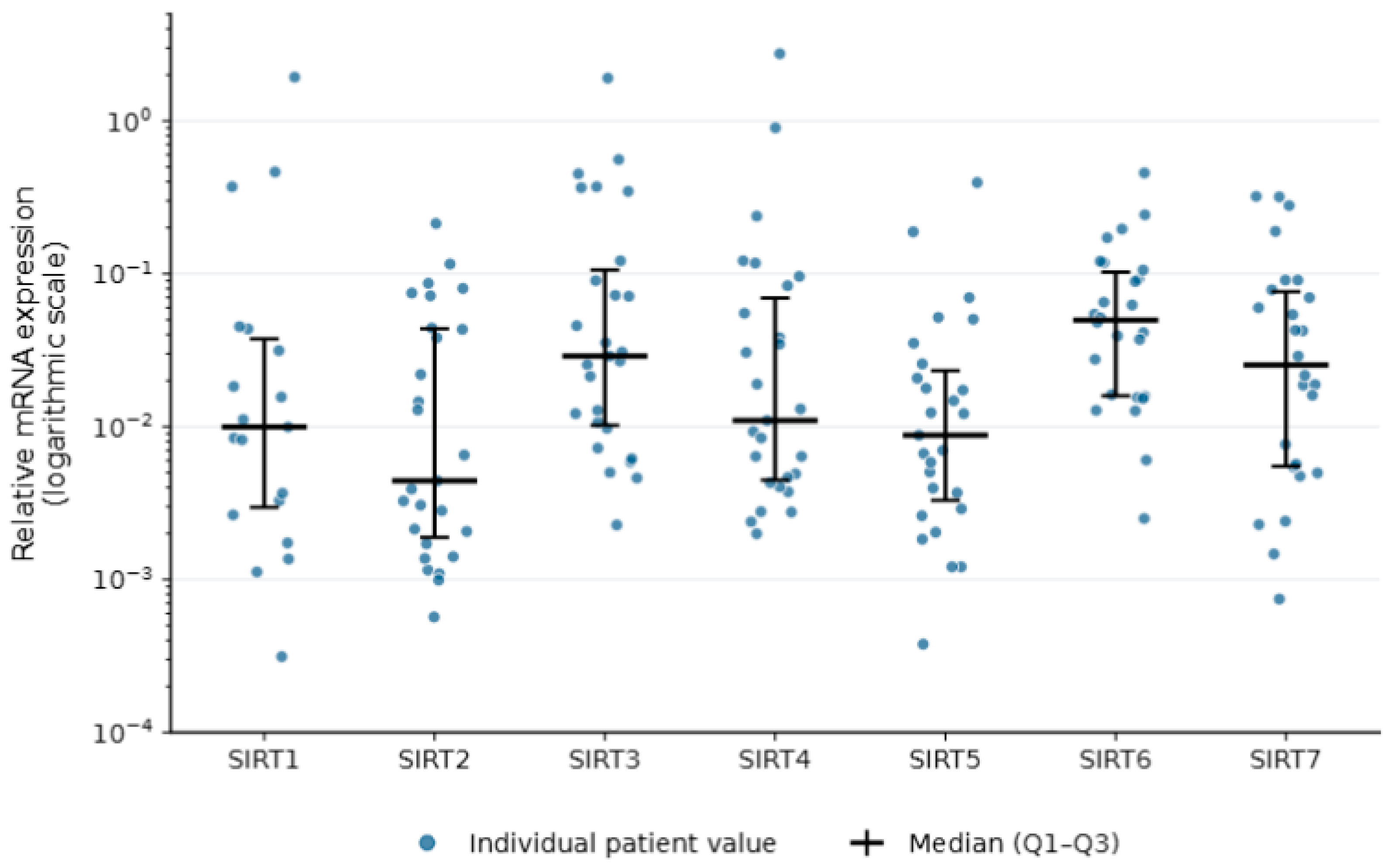
Expression of SIRT1–7 mRNA was detected in all inflamed COM tissue samples (Figure 1). Among the seven markers, SIRT3 exhibited the highest expression level, followed in descending order by SIRT6, SIRT4, SIRT7, SIRT1, SIRT5, and SIRT2. Furthermore, when the mRNA expression levels were compared according to the bacterial culture results, the expression levels of SIRT1–4 were significantly higher in the culture-negative group than in the culture-positive group (p < 0.05) (Table 3).
Figure 1.
Relative mRNA expression levels of SIRT1–7 in inflamed middle ear tissues of patients with chronic otitis media. The relative mRNA expression of SIRT1 through SIRT7 was measured using quantitative real-time PCR and normalized to the expression of the housekeeping gene β-actin using the 2−ΔCt method. Data are presented as the median with interquartile range (IQR, 25th–75th percentiles) and individual patient values (scatter dot plots) for all enrolled patients with COM (n = 27). SIRT3 exhibited the highest expression level among the sirtuin family members in the inflamed tissues.
Table 3.
Expression of mRNA encoding target genes according to a positive culture result.
3.3. Comparison of SIRT1–7 mRNA Expression According to Otorrhea and Type of Hearing Loss (Table 4 and Table 5)
No significant differences in SIRT1–7 mRNA expression levels were observed according to the presence or absence of otorrhea (p > 0.05). Similarly, no significant differences in expression profiles were found among groups classified by hearing loss type, including conductive hearing loss (CHL), sensorineural hearing loss (SNHL), and normal hearing (p > 0.05).
Table 4.
Expression of mRNA encoding target genes in the presence or absence of otorrhea.
Table 5.
Expression of mRNA encoding target genes in patients with conductive hearing loss, sensorineural hearing loss, or normal hearing.
4. Discussion
COM is characterized by recurrent or persistent inflammation within the middle ear, leading to irreversible pathological alterations in both mucosal and bony structures of the tympanic cavity and mastoid. The predominant histopathological findings include the formation of granulation tissue, osseous changes, tympanosclerosis, cholesterol granuloma, cholesteatoma, and extensive fibrosis [2,10]. This irreversible progression is widely understood as a continuum of disease driven by multifactorial etiologies, where unresolved inflammation and persistent microbial insults profoundly exacerbate mucosal destruction [11,12]. Given that these devastating pathological changes are fundamentally mediated by uncontrolled chronic inflammatory cascades, elucidating the molecular mechanisms that govern cellular defense is of paramount importance. In this context, our present study evaluated the mRNA expression profiles of the sirtuin family (SIRT1–7)—key regulators of cellular homeostasis—in inflamed middle ear tissues of patients with COM. Our principal findings demonstrated that SIRT3 exhibited the highest expression levels and that the expression levels of SIRT1–4 were significantly upregulated in patients with culture-negative results, highlighting a potential interplay between sirtuin-mediated defense mechanisms and active microbial infections.
Given the diverse and irreversible histopathological changes observed in COM, sirtuins—which are involved in cellular metabolism, inflammatory responses, and oxidative stress—may be associated with its pathogenesis [5]. While all sirtuins share a conserved catalytic core, isoform-specific structural variations dictate their distinct subcellular localizations and diverse biological functions [13,14,15,16]. Beyond their classical role in histone deacetylation, sirtuins modulate cellular proliferation, inflammatory signaling, and metabolic reprogramming under pathological conditions by targeting various non-histone proteins [17]. In the context of COM, these diverse regulatory mechanisms suggest that sirtuins may be associated with mucosal inflammatory responses and hyperproliferative changes, such as granulation tissue formation and cholesteatoma development.
In the present study, mRNA expression of all seven sirtuin members (SIRT1–7) was detected in inflamed COM tissues, suggesting that the sirtuin family may be transcriptionally active in the mucosal microenvironment under chronic inflammatory conditions. Notably, SIRT3 exhibited the highest expression level, whereas SIRT2 expression was the lowest. SIRT3 is fundamentally recognized as the primary mitochondrial deacetylase, promoting mitochondrial metabolic activity and mitigating oxidative stress by directly deacetylating key metabolic enzymes—such as acetyl-CoA synthetase 2 (ACS2) and long-chain acyl-coenzyme A dehydrogenase (LCAD)—as well as the essential antioxidant enzyme superoxide dismutase 2 (SOD2) [18,19]. Given that the inflamed COM mucosa undergoes severe metabolic and oxidative stress, the predominant SIRT3 mRNA expression observed in this study may reflect a potential compensatory response by the host cells to restore mitochondrial homeostasis and counteract excessive reactive oxygen species (ROS) production during prolonged middle ear inflammation.
Conversely, SIRT2 exhibited the lowest expression levels among the sirtuin family in our COM cohort. SIRT2 is predominantly localized in the cytoplasm, where it deacetylates α-tubulin and interacts with HDAC6; however, during mitosis, it transiently functions in the nucleus to regulate the cell cycle by promoting chromatin condensation through the specific deacetylation of histone H4 at lysine 16 (H4K16) [20]. The relatively low expression of SIRT2 in inflamed COM tissues suggests that it may be more specifically involved in subsequent phases of tissue remodeling and cell cycle regulation, rather than driving the active inflammatory cascade. Nevertheless, the current study design precludes a definitive mechanistic explanation for this polarized expression pattern—namely, the predominant upregulation of SIRT3 and the marked downregulation of SIRT2. Therefore, further longitudinal investigations are warranted to evaluate the expression dynamics of sirtuins across varying durations and severities of inflammation, and functional studies are required to verify whether these distinct mRNA transcript levels directly translate to corresponding protein expression profiles in the inflamed mucosa.
Historically, SIR2—the founding member of the sirtuin family—was initially identified during yeast mating-type studies and has been well-documented to mediate the silencing of telomeric and ribosomal DNA [21]. The closest mammalian ortholog to yeast SIR2 is SIRT1, which similarly functions as a master regulator of cellular stress and antibacterial defense. Aligning with this potential role, the present study demonstrated that the mRNA expression levels of SIRT1–4 were significantly higher in the culture-negative group compared to the culture-positive group. However, given the small sample size—particularly within the culture-positive subgroup (n = 8)—these findings should be interpreted cautiously as preliminary and hypothesis-generating. While this inverse relationship suggests a potential association between SIRT1–4 transcript levels and bacterial status in the middle ear cavity, the precise mechanisms underlying the lower expression of these sirtuins in culture-positive tissues remain unclear. Future prospective studies are necessary to determine whether this phenomenon is influenced by the stage of infection, specific pathogenic strains, or preoperative antibiotic therapy. Specifically, because all patients received prophylactic antibiotics for one week prior to surgery, this regimen may have eradicated antibiotic-susceptible bacteria—partially explaining why the culture-positivity rate (29.6%) was lower than the prevalence of visible otorrhea (51.9%)—while simultaneously altering the local inflammatory microenvironment and mucosal SIRT1–4 transcript levels.
On the other hand, no significant differences in SIRT1–7 mRNA expression were observed according to the presence of otorrhea or the type of hearing loss. These findings indicate that the molecular expression profiles of sirtuins may not directly correlate with macroscopic clinical phenotypes or functional auditory impairments in COM. Given the absence of prior literature in this domain, further prospective investigations are required to elucidate the potential clinical implications of sirtuin expression in middle ear pathologies.
The present study has several limitations that should be acknowledged. First, the molecular analysis was restricted to the mRNA level within the inflamed tissues. Subsequent validations utilizing immunohistochemistry, western blot analysis, or proteomic approaches are imperative to confirm whether these transcriptomic changes correspond to actual protein expression and enzymatic activity. Second, due to strict ethical constraints, healthy middle ear mucosal tissues could not be obtained as controls. Consequently, the baseline expression of SIRT1–7 in the normal middle ear remains unknown, precluding a direct comparative analysis between healthy and inflamed states. Third, all patients received prophylactic antibiotics for one week prior to surgery to minimize acute inflammation. This preoperative intervention might have acted as a confounding factor by influencing both microbiological culture outcomes and baseline SIRT1–7 mRNA expression profiles. Fourth, although all seven sirtuin members were expressed at varying levels in the COM tissues, the complex intracellular interactions and functional crosstalk among the distinct SIRT1–7 isoforms could not be evaluated. Fifth, the study lacked a comprehensive subgroup analysis based on critical clinical variables, including the total duration of COM, disease severity, extent of bone destruction, presence of complications, and prior treatment history. Finally, the relatively small total sample size and limited subgroup sizes limit the statistical power and generalizability of the observed associations. Importantly, the small subgroup sizes precluded reliable multivariable adjustment for potential confounding factors, such as age, sex, or antibiotic exposure. Therefore, these results should be interpreted cautiously as preliminary and hypothesis-generating, and further studies with larger cohorts are needed to confirm these findings.
5. Conclusions
The present study demonstrates that all seven sirtuin members (SIRT1–7) are expressed at the mRNA level in the inflamed mucosa of COM, suggesting that the sirtuin family may be transcriptionally active in the inflamed middle ear microenvironment. Notably, SIRT1–4 mRNA expression was significantly higher in the culture-negative group, suggesting a potential association between SIRT1–4 mRNA expression and bacterial culture status in COM. These preliminary findings provide novel insights into the transcriptional profiles of sirtuins in COM and suggest that SIRT1–4 may be associated with local inflammatory processes in the middle ear. However, further studies incorporating protein-level and functional validation are needed to clarify the clinical and pathophysiological implications of these findings.
Author Contributions
Conceptualization, J.G.D., D.C.P. and S.G.Y.; methodology, M.C., H.O.K., M.P., J.M.L. and I.U.K.; formal analysis, M.C., H.O.K., M.P. and Y.J.C.; data curation, H.O.K., J.M.L., Y.J.C., J.M.K., and S.S.K.; writing—original draft, J.G.D. and S.G.Y.; writing—review & editing, J.G.D., D.C.P. and S.G.Y.; visualization, J.G.D., J.M.L., J.M.K., and S.G.Y.; project administration, S.G.Y.; funding acquisition, S.S.K. and S.G.Y. All authors have read and agreed to the published version of the manuscript.
Funding
This work was supported by the Ministry of Education of the Republic of Korea and the National Research Foundation of Korea (NRF) grant funded by the Korean government (NRF-2025S1A5C3A03022311) (NRF-2018R1A6A1A03025124) (NRF-2022R1A2C1091779). This research was supported by a grant from the National Bio Bigdata Project, funded by four ministries (Ministry of Health and Welfare, Ministry of Science and ICT, Ministry of Trade, Industry and Resources, and Korea Disease Control and Prevention Agency) of Korea (grant number: RS-2024-00509118). The funders had no role in study design, data collection, data analysis, data interpretation, or writing of the report.
Institutional Review Board Statement
The study was conducted in accordance with the Declaration of Helsinki and approved by the Institutional Review Board of Kyung Hee University Medical Center (protocol code 2023-05-016, approved on 15 May 2023).
Informed Consent Statement
Written informed consent was obtained from all subjects involved in the study.
Data Availability Statement
The datasets presented in this article are not readily available due to patient privacy and ethical restrictions. However, the data are available from the corresponding author upon request.
Acknowledgments
The authors used generative artificial intelligence tools, including ChatGPT (version 6.0, OpenAI, San Francisco, CA, USA), solely to assist with language editing, sentence refinement, and improvement of manuscript readability. Literature searching, study selection, data extraction, reference verification, scientific interpretation, and all final revisions were performed and approved by the authors. The authors take full responsibility for the accuracy, originality, and integrity of the final manuscript.
Conflicts of Interest
The authors declare no conflicts of interest.
References
- Lee, H.A.; Jeong, J.Y.; Lee, H.N.; Byun, H.; Lee, S.H.; Chung, J.H. Microbiological culture sensitivity profile of chronic otitis media. Korean J. Otorhinolaryngol.-Head Neck Surg. 2023, 66, 805–814. [Google Scholar] [CrossRef] [Scilit]
- Park, Y.H. Chronic otitis media. In Otology; Korean Otological Society, Ed.; Seojong Medical Publishing Inc.: Seoul, Republic of Korea, 2022; pp. 469–480. [Google Scholar]
- Lee, H.W. Role of innate immunity in otitis media. Korean J. Otorhinolaryngol.-Head Neck Surg. 2016, 59, 483–489. [Google Scholar] [CrossRef] [Scilit]
- Park, K. Recent advances in pathogenesis of otitis media. J. Clin. Otolaryngol. 2009, 20, 141–148. [Google Scholar] [CrossRef] [Scilit]
- Wu, Q.J.; Zhang, T.N.; Chen, H.H.; Yu, X.F.; Lv, J.L.; Liu, Y.Y.; Liu, Y.S.; Zheng, G.; Zhao, J.Q.; Wei, Y.F.; et al. The sirtuin family in health and disease. Signal Transduct. Target. Ther. 2022, 7, 402. [Google Scholar] [CrossRef] [Scilit] [PubMed]
- Jiang, H.; Khan, S.; Wang, Y.; Charron, G.; He, B.; Sebastian, C.; Du, J.; Kim, R.; Ge, E.; Mostoslavsky, R.; et al. SIRT6 regulates TNF-α secretion through hydrolysis of long-chain fatty acyl lysine. Nature 2013, 496, 110–113. [Google Scholar] [CrossRef] [Scilit] [PubMed]
- Rack, J.G.; Morra, R.; Barkauskaite, E.; Kraehenbuehl, R.; Ariza, A.; Qu, Y.; Ortmayer, M.; Leidecker, O.; Cameron, D.R.; Matic, I.; et al. Identification of a class of protein ADP-ribosylating sirtuins in microbial pathogens. Mol. Cell 2015, 59, 309–320. [Google Scholar] [CrossRef] [Scilit] [PubMed]
- Jung, J.; Jung, S.Y.; Kim, M.G.; Kim, Y.I.; Kim, S.H.; Yeo, S.G. Comparison of autophagy mRNA expression between chronic otitis media with and without cholesteatoma. J. Audiol. Otol. 2020, 24, 191–197. [Google Scholar] [CrossRef] [Scilit] [PubMed]
- Kang, D.W.; Kim, Y.J.; Kim, S.S.; Kim, Y.I.; Doo, J.G.; Kim, S.H.; Yeo, S.G. Comparison of endoplasmic reticulum stress messenger ribonucleic acid expression between chronic otitis media with and without cholesteatoma. J. Int. Adv. Otol. 2023, 19, 271–276. [Google Scholar] [CrossRef] [Scilit] [PubMed]
- Bluestone, C.D. Epidemiology and pathogenesis of chronic suppurative otitis media: Implications for prevention and treatment. Int. J. Pediatr. Otorhinolaryngol. 1998, 42, 207–223. [Google Scholar] [CrossRef] [Scilit] [PubMed]
- Hoffman, H.J.; Daly, K.A.; Bainbridge, K.E.; Casselbrant, M.L.; Homøe, P.; Kvestad, E.; Kvaerner, K.J.; Vernacchio, L. Panel 1: Epidemiology, natural history, and risk factors. Otolaryngol.–Head Neck Surg. 2013, 148, E1–E25. [Google Scholar] [CrossRef] [Scilit] [PubMed]
- Verhoeff, M.; van der Veen, E.L.; Rovers, M.M.; Sanders, E.A.; Schilder, A.G. Chronic suppurative otitis media: A review. Int. J. Pediatr. Otorhinolaryngol. 2006, 70, 1–12. [Google Scholar] [CrossRef] [Scilit] [PubMed]
- Moniot, S.; Weyand, M.; Steegborn, C. Structures, substrates, and regulators of mammalian sirtuins—Opportunities and challenges for drug development. Front. Pharmacol. 2012, 3, 16. [Google Scholar] [CrossRef] [Scilit] [PubMed]
- North, B.J.; Marshall, B.L.; Borra, M.T.; Denu, J.M.; Verdin, E. The human Sir2 ortholog, SIRT2, is an NAD+-dependent tubulin deacetylase. Mol. Cell 2003, 11, 437–444. [Google Scholar] [CrossRef] [Scilit] [PubMed]
- Du, J.; Zhou, Y.; Su, X.; Yu, J.J.; Khan, S.; Jiang, H.; Kim, J.; Woo, J.; Kim, J.H.; Choi, B.H.; et al. Sirt5 is a NAD-dependent protein lysine demalonylase and desuccinylase. Science 2011, 334, 806–809. [Google Scholar] [CrossRef] [Scilit] [PubMed]
- Moreno-Yruela, C.; Ekundayo, B.E.; Foteva, P.N.; Ni, D.; Calvino-Sanles, E.; Stahlberg, H.; Fierz, B. Structural basis of SIRT7 nucleosome engagement and substrate specificity. Nat. Commun. 2025, 16, 1328. [Google Scholar] [CrossRef] [Scilit] [PubMed]
- Choi, H.Y.; Hah, Y.S.; Lee, S.J. Beyond histone deacetylation: Non-canonical sirtuin functions in cancer progression. J. Life Sci. 2025, 35, 481–491. [Google Scholar]
- Haigis, M.C.; Sinclair, D.A. Mammalian sirtuins: Biological insights and disease relevance. Annu. Rev. Pathol. 2010, 5, 253–295. [Google Scholar] [CrossRef] [Scilit] [PubMed]
- Hirschey, M.D.; Shimazu, T.; Goetzman, E.; Jing, E.; Schwer, B.; Lombard, D.B.; Grueter, C.A.; Harris, C.; Biddinger, S.; Ilkayeva, O.R.; et al. SIRT3 regulates mitochondrial fatty-acid oxidation by reversible enzyme deacetylation. Nature 2010, 464, 121–125. [Google Scholar] [CrossRef] [Scilit] [PubMed]
- Vaquero, A.; Scher, M.B.; Lee, D.H.; Sutton, A.; Cheng, H.L.; Alt, F.W.; Serrano, L.; Sternglanz, R.; Reinberg, D. SirT2 is a histone deacetylase with preference for histone H4 Lys 16 during mitosis. Genes Dev. 2006, 20, 1256–1261. [Google Scholar] [CrossRef] [Scilit] [PubMed]
- Bryk, M.; Banerjee, M.; Murphy, M.; Knudsen, K.E.; Garfinkel, D.J.; Curcio, M.J. Transcriptional silencing of Ty1 elements in the RDN1 locus of yeast. Genes Dev. 1997, 11, 255–269. [Google Scholar] [CrossRef] [Scilit]

Disclaimer/Publisher’s Note: The statements, opinions and data contained in all publications are solely those of the individual author(s) and contributor(s) and not of MDPI and/or the editor(s). MDPI and/or the editor(s) disclaim responsibility for any injury to people or property resulting from any ideas, methods, instructions or products referred to in the content. |
© 2026 by the authors. Licensee MDPI, Basel, Switzerland. This article is an open access article distributed under the terms and conditions of the Creative Commons Attribution (CC BY) license.