


Abstract
The treatment of burns and war injuries remains a pressing issue. Since 1993, surgeon V.A. Menzul has been using a special polyethylene film to cover wounds. This film interacts only minimally with the wound surface, so it does not interfere with regenerative processes and makes dressing changes painless. Exudate flows from the wound and is absorbed by the layers of dressing situated above the film. Over time, a membrane forms beneath the film, consisting mainly of fibrin and a large number of white blood cells (the fibrin–leukocyte membrane, FLM). The FLM and the exuding fluid perform several functions: they create a moist environment and mechanical protection; they deliver materials and immune cells to the wound site; they remove toxic breakdown products; and they exert an antimicrobial effect. The latter consists of a direct barrier effect and the recruitment of immune cells. Such an “own liquid medium” (OLM) technique with a transparent film allows the process to be observed. The results of this treatment, in comparison with the traditional dry treatment method, shorten the length of hospital stay, reduce patients’ discomfort and lead to improved cosmetic outcomes. This narrative review discusses various aspects of the OLM treatment technique. We believe that Dr Menzul’s experience should be made known to a wide range of healthcare professionals.
1. Transport Through the Vascular Wall—Inflammation
The transport function of the blood is carried out in the capillaries and post-capillary venules. There are several mechanisms by which substances and cells cross the vascular wall: diffusion through endothelial cells and through the gaps between them; active transport via vesicular transport through endothelial cells; the use of passive and active transporters and ion channels; the passage of substances via fenestrae (specific localizations); filtration through pores; and the passage of immune cells (diapedesis).
Fat-soluble substances, including oxygen and carbon dioxide molecules, alcohols and others, are transported via diffusion across the entire surface of the cells. Another form of transport is via the gaps between endothelial cells. These gaps, which vary in size, are filled with aqueous solutions. Ions and low-molecular-weight substances, such as glucose, can diffuse through these gaps. The size of these gaps (pores) is normally a few nanometers (up to 5 nm) [1].
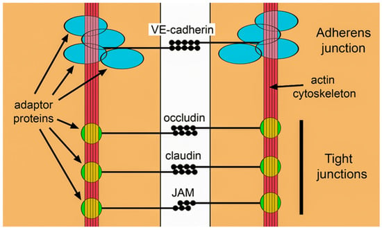
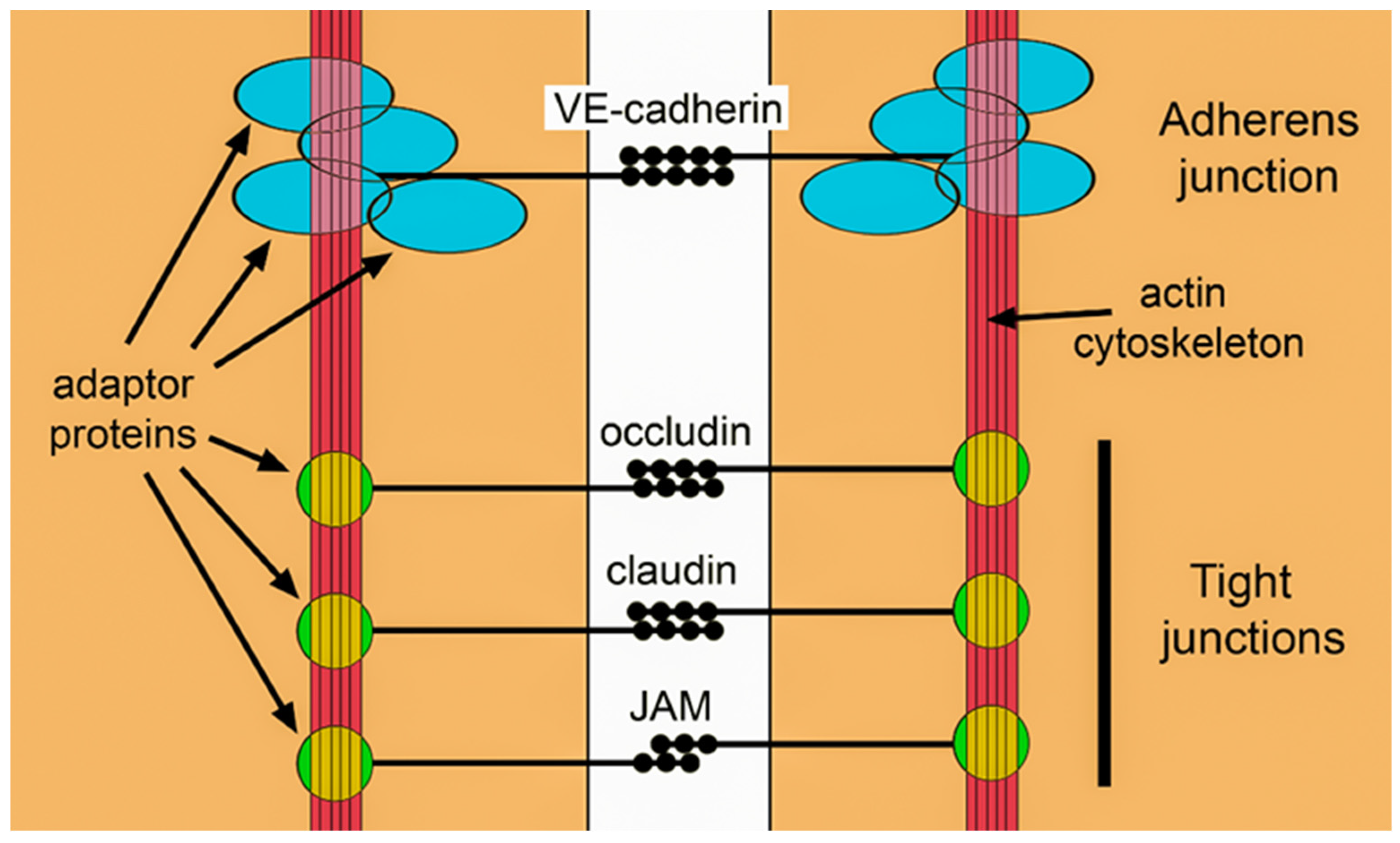
Endothelial cells are connected to one another in various ways. There are several types of binding molecules that form two types of junctions: adherent junctions and tight junctions (Figure 1).
Figure 1.
Scheme of endothelial cell junctions.
Within the first few minutes of injury, mast cells begin to release histamine and platelets release serotonin, triggering contraction of the actomyosin cytoskeleton in endothelial cells. Radial actin stress fibrils form. The gaps between cells widen, resulting in increased vascular permeability. This process is most intense in the post-capillary venules. As inflammation progresses, mechanisms for the direct suppression of adhesion complexes are activated. The pores between cells increase dramatically in size, from a few nanometers to tens or hundreds of nanometers. In typical inflammation, the average diameter of the gaps formed in post-capillary venules is around 400–500 nm. This is sufficient for large plasma proteins, such as albumin (size ~7 nm) and fibrinogen, to pass through [2].
When an injury occurs, blood vessels are damaged, the partial pressure of oxygen decreases, and the oxygen-dependent transcription regulator HIF-1 is activated, triggering adaptation processes to the reduced oxygen levels, including increased expression of VEGF. VEGF stimulates endothelial cells, increasing their proliferation, migration and viability. All of this is required for the processes of angiogenesis. The new capillaries formed in excessive numbers at the site of inflammation have imperfect intercellular junctions and exhibit increased permeability [3].
Thus, during inflammation, the vascular wall ceases to act as a filter for large molecules, and the entire contents of the plasma begin to leak into the interstitium. The wounds discussed in this review are always accompanied by damage to the epidermis, leading to a loss of its waterproofing function. Interstitial fluid begins to leak out. It contains a large amount of plasma proteins.
Fluid leakage (exudation) from the wound peaks after a few days and then gradually subsides. The amount of exudate decreases as a result of:
- A reduction in inflammation and the consequent normalization of vascular permeability,
- The formation of a superficial diffusion barrier, which subsequently, through the formation of new epidermis, becomes a waterproof layer on the surface of the wound.
2. Formation of a Temporary Barrier
The final stage of wound healing is epithelialization, but exudation decreases long before the formation of new epidermis. Connective tissue, together with new blood vessels, very quickly begins to form a new temporary barrier at the body’s boundary. After a few days, the wound begins to fill with new stroma, formed from new capillaries, fibroblasts and macrophages. So-called granulation tissue forms.
The blood vessels formed during angiogenesis are lined with fibroblasts and the extracellular matrix they synthesize. This matrix subsequently serves as the basis of the basement membrane for epidermal cells. Fibroblasts produce the matrix, whilst the matrix itself influences the fibroblasts’ ability to produce it [4,5]. Fibroblast integrin receptors, which recognize fibrin and fibronectin, determine the rate of granulation tissue formation [5,6]. Various growth factors and fibronectin are involved in the formation of granulation tissue.
Granulation tissue cannot form directly on the wound surface; all these regenerative processes must be shielded from the external environment to maintain moisture and provide protection against microbes. A temporary matrix forms directly above the granulation tissue, consisting of both products of fibroblast synthesis and remnants of the blood clot, as well as products and cells emerging from the vascular bed. It contains fibrin and fibronectin [7].
It has only recently been discovered that neutrophils initiate the formation of a temporary barrier separating the body from its environment [8]. It has been found that some neutrophils, upon entering the wound (and neutrophils are the first to arrive at the wound), begin to produce the extracellular matrix at a rapid rate. These neutrophils survive for many days. As a result of this activity by the neutrophils alone, the wound is covered by a sort of sheath which significantly slows down the penetration of Staphylococcus aureus into the deeper layers of the wound; a pathogen deliberately introduced remains in the very outermost layer (up to 75 µm). In the control group (without neutrophils), the pathogen spread over a distance of more than 200 µm. Thus, neutrophils ‘bandage’ the wound [9]. In parallel with granulation tissue formation, myofibroblasts are generated, which draw the wound edges together, reducing the overall workload involved in creating a new barrier [10].
3. Epithelialization
Epithelial cells from the wound margin or from other residual structures migrate through the temporary matrix; this is the only way regeneration can occur: if they were to remain on top of the matrix, the cells would quickly die from dehydration [11]. Around the second day, proliferation begins in the epithelial cells that follow the migrating cells.
The temporary matrix serves as a structural framework for cell migration; this applies to both underlying fibroblasts and keratinocytes [11]. Fibrin, fibronectin and hyaluronic acid—the constituent components of the temporary matrix—play a major role in the migration processes. This matrix contains a large number of blood cells, mainly neutrophils.
Cell movement through this temporary matrix requires proteolytic activity. This originates from three sources: the keratinocytes themselves (urokinase-type plasminogen activator and matrix metalloproteinases MMP-1, 2, 3); fibroblasts (the same enzymes plus MMP-13); and enzymes derived from plasma (MMP-1, 2, 3, 13 and tissue plasminogen activator) [11,12].
The temporary matrix lacks two components of the basement membrane: laminin and collagen IV. Once keratinocyte migration is complete, these components appear in the basement membrane zone, which is formed through the combined efforts of keratinocytes and fibroblasts and serves as an anchor for the keratinocytes. Epithelial tissue cannot grow on dead tissue; therefore, the spread of epithelialization removes dead tissue from the body. The path taken by migrating cells is determined by a set of integrins expressed by keratinocytes.
Once growth has ceased, the granulation tissue is gradually replaced by scar tissue. The number of capillaries and cells in general decreases. The cells undergo apoptosis. Disruption of this process can lead to the formation of keloid scars and excessive sclerosis [13,14].
4. The Special Role of Fibrinogen in Wound Healing
Fibrinogen constitutes a significant proportion of plasma proteins. Its concentration reaches 4 mg/ml, and in this respect, it is second only to albumin and immunoglobulins. It is mainly produced in the liver. Fibrinogen is a relatively heavy (340 kDa) glycoprotein consisting of three pairs of polypeptide chains linked by 29 disulphide bridges [15].
Under the action of thrombin and coagulation factor XIII, complex transformations of fibrinogen may occur. Fibrinogen breaks down into individual units (fibrin monomers), which are capable of self-assembly. Longitudinal fibrils are formed, to which lateral connections may attach. A gel of varying density (fibrin gel) is formed. The entire structure is then reinforced by covalent bonds created by activated factor XIII (transglutaminase). Next, the fibrinolysis system is triggered, which breaks down the fibrin clot. All these events are well known and are associated in our minds exclusively with hemostasis, when a thrombus forms and then dissolves. In reality, the role of fibrinogen in the wound-healing process is much broader.
Increased vascular permeability during the early days of the wound-healing process allows significant amounts of fibrinogen to reach the wound surface, where it forms a temporary matrix that accelerates the migration of epithelial cells, as well as neutrophils, monocytes, fibroblasts and endothelial cells [16]. All these cells interact directly with the fibrin gel via integrin and cadherin receptors [17].
Simultaneously with the formation of the fibrin gel, processes leading to its degradation also take place: diffusing fragments of the gel act as chemoattractants for neutrophils and monocytes. Thus, leukocytes that have left the bloodstream are directed towards the surface, where the greatest number of microbes are located [18].
Signals from the degradation products of the fibrin gel interact with signals from activated complement, altering protease activity and the expression of integrins on immune cells [19,20]. The migration of keratinocytes is determined by the specificity of the integrins on their surface; they bind to fibronectin, tenasin and vitronectin, but do not recognize the fibrin gel [21].
The provisional fibrin matrix acts as a reservoir for various growth factors necessary for migration and proliferation, whilst also providing a protective framework that enables new tissue to form. Fibrin binds various growth factors that stimulate angiogenesis (VEGF, bFGF and PDGF) [22,23,24]. Fibrin influences collagen production by fibroblasts [25]. Platelet remnants in the fibrin gel promote wound healing [26]. A fibrin degradation product (fragment E) promotes the formation of myofibroblasts [27]. Fibrin gel provides a platform for angiogenesis. Fibrin and fibrinogen bind to specific integrins and surface adhesion molecules on endothelial cells [28]. Fibrinogen fragments can both stimulate and inhibit angiogenesis [29,30,31].
5. The Role of Fibrin (Fibrinogen) in Antibacterial Defense
In addition to stopping bleeding and creating conditions conducive to healing, fibrinogen (fibrin) possesses pronounced antimicrobial properties. These properties involve creating a physical barrier against microbes, immobilizing them, and influencing immune cells by recruiting and activating them. The formation of a wound leads to a massive influx of microbes into the wound; the immune system becomes overloaded, and one solution is to attempt to immobilize the microbes in order to give the immune system more time to destroy them.
The direct barrier function of fibrin has been described. Fibrinogen (fibrin), by concentrating at the liquid–air interface, forms a specialized membrane that prevents the penetration of microbes into the blood clot [32].
Fibrin peptides (fibrinopeptide B (GHR28)) sometimes exhibit direct antimicrobial activity [33]. Fibrin acts as a ligand for certain integrins and other receptors on cells involved in inflammation. As a result, the expression of cytokines and chemokines in leukocytes is altered; degranulation and phagocytosis may be stimulated; and cell adhesion and migration are modified [34,35,36,37,38].
The formation of a fibrin network at the site of infection reduces the spread of bacteria and supports the recruitment of immune cells [39,40]. The long-term co-evolution of microbes and multicellular organisms has led to fibrin becoming a target for many bacterial products, which promote the formation or dissolution of the fibrin gel, immobilization within fibrin or escape from it, and so on.
Microbes trapped in the fibrin gel cannot spread rapidly throughout the body; however, the immune system ceases to recognize them. By stimulating fibrinolysis, the microbes regain their freedom. On the other hand, if microbes are completely surrounded by a fibrin capsule, they are capable of long-term survival [41,42]. Cross-linking of fibrin by transglutaminase contributes to the immobilization of the pathogen [43,44,45,46,47].
Pathogenic bacteria sometimes exploit fibrin for their own purposes [48,49]. But it is not all good news. The mass entry of bacteria into fibrin can protect them from phagocytosis. Cases have been reported of bacterial proliferation within a clot, leading to the formation of an abscess [50].
Staphylococcus aureus secretes two coagulases which, upon reacting with prothrombin, form the staphylothrombin complex [51]. Staphylothrombin converts fibrinogen into fibrin, which isolates the bacterium, enabling it to evade the immune response, including phagocytosis [52]. There are two forms of this fibrin: as a pseudocapsule or as a network [53]. A staphylothrombin inhibitor has been tried as an adjunct to antibiotic therapy for staphylococcal bacteraemia [54].
Certain streptococci produce streptokinase, whilst Staphylococcus aureus produces staphylokinase—both compounds are non-enzymatic activators of plasminogen. It is likely that these compounds prevent bacteria from becoming trapped in fibrin and facilitate their dissemination throughout the body. In addition, staphylokinase forms complexes with alpha-defensins from neutrophils, reducing their bactericidal activity [55,56].
Y. pestis possesses the Pla protein on its surface, which has protease activity that converts plasminogen into plasmin [57]. The expression of this protein has a very significant effect on virulence [58].
Many bacteria simply enhance the interaction of plasminogen with plasminogen activators (tissue plasminogen activator and urokinase) [59,60]. Some bacteria inhibit fibrinolysis. S. pyogenes secretes a protein called SIC (streptococcal inhibitor of complement). This promotes the survival of bacteria within fibrin clots in a wound [61,62].
Thus, a thrombin–fibrinogen axis is postulated in antimicrobial defence. And this defence may be organized differently in various parts of the body [63].
6. The Role of the Temporary Fibrin Matrix in Wound Healing
The data presented indicate that plasma and blood serum organize and regulate the wound-healing process. Serum, which is formed during blood clotting, contains a wealth of growth factors released by platelets. Plasma contains fewer growth factors, but it does contain fibrinogen, which forms a temporary matrix that protects the proliferation of endothelial cells, fibroblasts and keratinocytes—the cells responsible for regeneration.
On this basis, it can be assumed that maintaining a temporary matrix on the wound surface should have a positive effect on the wound-healing process. Indirect evidence that a temporary matrix promotes wound healing can be found in the use of fibrin and platelet-rich plasma.
The external application of fibrin and platelet-rich plasma can be used to improve healing and eliminate microbes during wound treatment. This is indeed widely practiced.
In surgery, fibrin is used as a biological adhesive and as a hemostatic agent [64,65,66]. It can serve as a drug delivery system [67,68]. Fibrin nanoparticles have recently been developed [69]. Fibrin and platelet-rich plasma are used for their antibacterial effects and to improve wound healing [70,71,72,73,74,75].
However, the use of fibrin gel and platelet-rich plasma has a number of drawbacks. These include the high cost of the preparations and the risk of contamination. Nevertheless, these preparations are very widely used in a number of applications (particularly in ophthalmology and cosmetic medicine).
7. Healing in Your “Own Liquid Medium” According to Dr. Menzul
Guidelines on the use of membrane materials for wound closure state that they should not be used to treat deep wounds containing a lot of exudate because they cannot ensure removal of excess fluid. Excess fluid is thought to promote maceration of the wound edges and increase the likelihood of infection. However, it is fairly obvious that membrane materials can be used if fluid removal is ensured. Treatment of burns and wounds in general using membranes has been known for a long time. This treatment allows painless dressings, is affordable and often results in faster healing [76,77,78].
Surgeon Dr. Menzul has been treating burns, including children’s burns, as well as gunshot and mine-explosion wounds, in various Russian hospitals for over 30 years. All this time (more than 1500 patients), he has been using membrane wound closure with the help of polyethylene film developed by him [79,80,81].
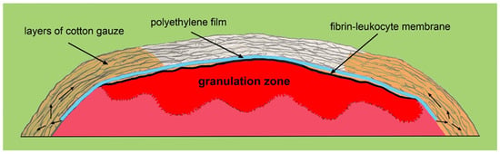
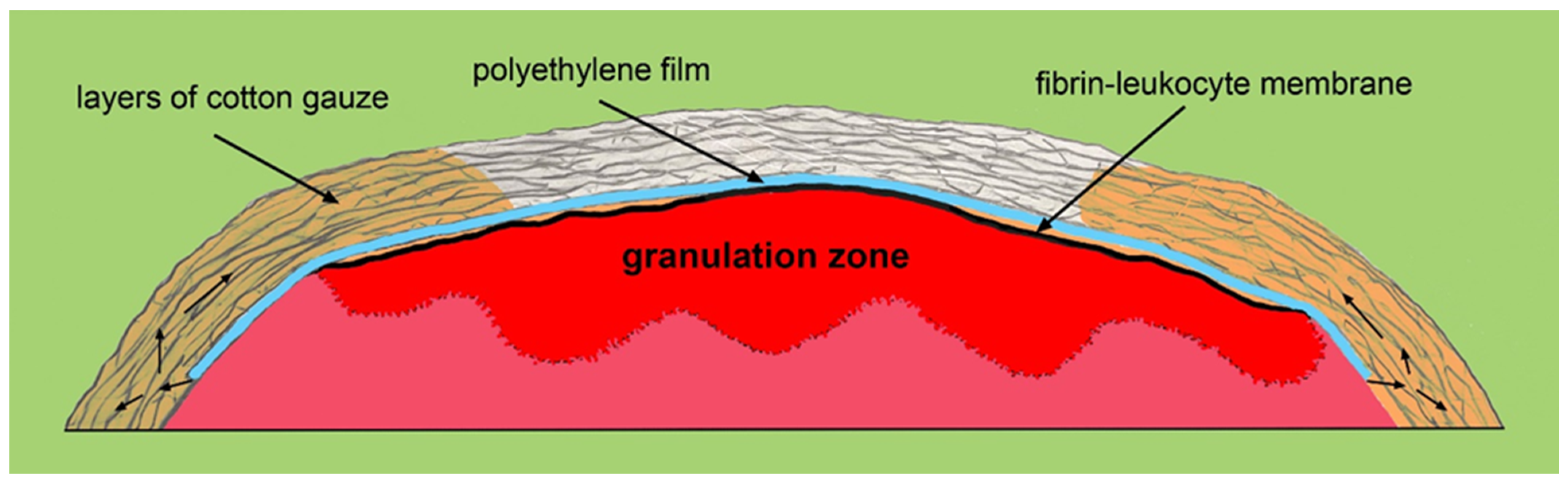
A sterile polyethylene membrane is applied to the wound and, in most cases, secured in place with a medical plaster. Next, over the membrane, bandaging is carried out with an ordinary cotton bandage. This bandage serves as a tampon for the fluid flowing out of the wound (Figure 2). Dr. Menzul called this method “wound care in its own liquid medium”.
Figure 2.
Diagram of treatment in the patient’s “own liquid medium”. The small arrows indicate the flow of fluid from beneath the film into the dressing.
This technology has been tested in several hospitals by independent surgeons and has been approved [80,81]. The use of the Menzul technique reduced the time of hospitalization, improved cosmetic results and increased the quality of life of patients.
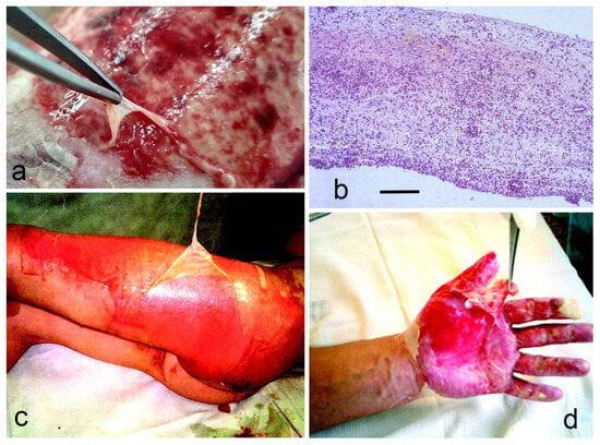
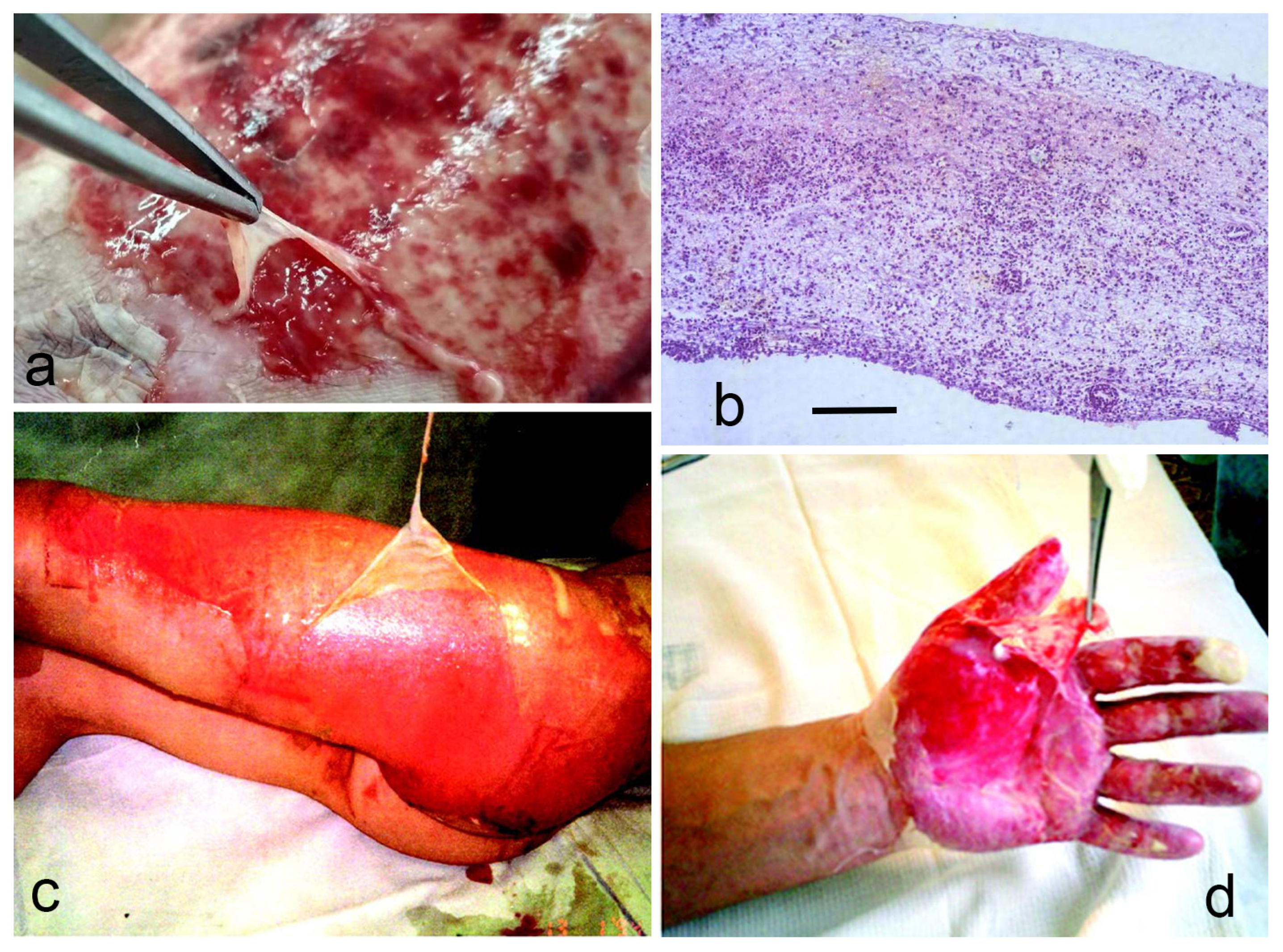
With this treatment, the temporary matrix at the wound site is well preserved and takes on the appearance of a membrane covering the wound. Dr Menzul refers to this as the fibrin–leukocyte membrane (FLM). The FLM can be carefully separated from the wound surface (though this should not be done!) (Figure 3). Over time (days), the FLM becomes firmer. Histological examination reveals tangential fibrin deposits and extensive infiltration by white blood cells, mainly neutrophils and macrophages. In essence, there is hypertrophy of the temporal matrix on the surface of the wound.
Figure 3.
Fibrin–leukocyte membrane (FLM): (a) on the donor wound; (b) histological specimen. Haematoxylin–eosin staining. Massive infiltration of the fibrin–leukocyte membrane (FLM) by blood cells is visible. Scale bar: 100 µm. (c,d) show FLM on a burn wound. Dr Menzul demonstrates that the FLM is a distinct structure that can be separated from the wound surface. The photographs in (a,b) are from ref. [82], and (c,d) are from ref. [79].
8. Example of Wound Treatment in the Patient’s Own Liquid Medium (OLM) According to Menzul
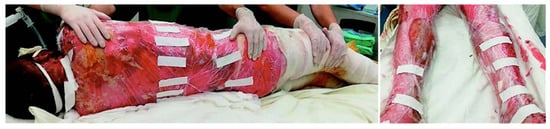
Figure 4 illustrates the treatment of a patient with burns covering 97 per cent of their body. In fact, the patient was completely wrapped in polyethylene film. The patient made a full recovery with a good cosmetic outcome [79].
Figure 4.
Patient with burns involving 97% of the total body surface area. Treatment under the OLM condition using a polyethylene film dressing [79].
9. Quantitative Characteristics of the Application of Menzul’s Wound Treatment Technology
We conducted a literature search online using the surname ‘Menzul V.A.’. We also examined the work of two hospitals where Menzul’s technology was being evaluated, namely the Central City Hospital, Ulyanovsk, Russia, and the Main Military Clinical Hospital of the Russian National Guard Troops of the Russian Federation, Balashikha, Russia. The monograph [79], which contains works by other authors relevant to Menzul’s technique, proved to be of great assistance in our search. Of all the studies, we used those in which Menzul’s technique was compared with the traditional method, without a film coating (Table 1). Virtually all of the studies were published in Russian. To enable English-speaking readers to access the articles in Russian, we have included full web links to them in the Supplementary Materials.
- Treatment under the OLM conditions minimizes trauma to the wound surface, allowing regenerative processes to proceed continuously rather than being repeatedly interrupted by dressing changes. This uninterrupted healing accelerates epithelialization and overall wound repair, shortens the length of hospitalization, and consequently reduces treatment costs.
- A clinical study [81] confirmed the acceleration of wound healing. Eighty patients were randomly assigned to two groups. Split-thickness skin grafting was performed in both groups. In the first group, the Menzul perforator and polyethylene film dressings were used, whereas the second group was treated using the Tanner perforator and atraumatic mesh dressings (the traditional dry method). The mean hospital stay was significantly shorter in the first group than in the second group (52.4 ± 3.7 vs. 83.8 ± 5.3 days, p < 0.001). The mean time to epithelialization after grafting was 13.9 ± 0.9 days (95% CI, 13.6–14.3) in the first group and 20.3 ± 1.3 days (95% CI, 19.9–20.8) in the second group (p = 0.001). Partial graft lysis occurred in one patient in the first group and in five patients in the second group (p < 0.001). A similar study [80] had previously been conducted on patients with burns. In 90 patients, a reduction in wound epithelialisation time (depending on the depth of the burn) of 3–8 days was demonstrated. A retrospective analysis of the treatment of 281 patients also showed a reduction in epithelialisation time by an average of 7 days [83]. In study [84], conducted on a large sample (n = 123), a reduction in the length of hospital stay by 10 days was demonstrated.
- Combat-related wounds are highly heterogeneous, making systematic analysis and reliable statistical comparisons difficult. In contrast, donor-site wounds created during split-thickness skin harvesting are highly reproducible and therefore well suited for controlled clinical studies. Such studies have been performed. When donor-site wounds (0.3–0.4 mm deep) were treated under the Menzul OLM protocol, epithelialisation was accelerated by 2 days [85], 3.5 days [82] or 5 days [86].
- It has long been recognized that prolonged wound healing is associated with increased scar formation [87,88]. Therefore, accelerated healing not only shortens recovery time but also reduces the incidence of scar contractures and improves cosmetic outcomes. In the study [81], hypertrophic scars were detected 3 months after surgery in 2 patients (5.7%) treated under the Menzul OLM protocol and in 5 patients (11.9%) in the control group (p < 0.001). Application of the Menzul technique in facial reconstructive surgery has produced excellent cosmetic results.
- An equally important advantage of the Menzul technique is the substantial reduction in patient discomfort. In the same study [81], it was shown that when dressings were applied, the score on the visual analogue scale for pain was 2.1 ± 0.3 in group 1 and 4.5 ± 1.1 in group 2 (p < 0.001). The Dermatology Quality of Life Index was 5.1 ± 0.3 in Group 1 and 10.2 ± 0.5 in Group 2 (p < 0.001).
- Pages 192–224 of the monograph [79] describe a histological study of burns in children during the course of treatment. Forty-one patients took part, and 83 biopsies were analysed. The authors (Shekhter, A.B. and Rudenko, T.G.) demonstrated that, when treated in an OLM condition, the processes of necrolysis and the onset of granulation are accelerated.
- One of the most important criteria for assessing the effectiveness of treatment is mortality. Following the introduction of Menzul’s technique at the paediatric burn centre in Balashikha, a long-term downward trend in mortality was observed [79]. Of the centre’s 786 patients, 14 had burns covering between 60 and 89 per cent of their body surface area; these patients survived and fully recovered their skin [79].
Table 1.
Summary of the quantitative characteristics of the application of Menzul’s wound treatment technology.
10. Hypothesis Regarding the Etiology of FLM
As the exudate contains plasma proteins, it must contain fibrinogen, fibrin monomer and plasma protransglutaminase [89]. The extravascular role of fibrin (fibrinogen) is also indicated by the fact that the fibrin degradation product D-dimer is detected not only intravascularly but also extravascularly at sites of tissue repair [90].
Fibrin (fibrinogen) and protransglutaminase are relatively large molecules with amphiphilic properties. Consequently, in aqueous solutions, these molecules exhibit positive adsorption at the phase boundary, and their concentration increases there. Consequently, particularly given the presence of protransglutaminase (a cross-linking enzyme) at the water–air interface, a fibrin membrane of some sort must form. This was explicitly demonstrated in a recent study [32].
A distinctive continuous fibrin surface film and gel-like fibrin structures beneath it were observed. All these structures are located above the hemostatic clot and, indeed, above the wound. It has been shown that this fibrin film possesses antimicrobial activity [32].
It is likely that similar phenomena occur in the wound-healing process. Over time, the exudate emerging from the wound forms a thin surface film at the interface with the air. This film is very thin and mechanically fragile. However, it is already capable of inhibiting microbial invasion and reducing fluid loss. As this film is not mechanically strong, continued exudation—or any movement by the patient—causes it to rupture from time to time, and the process repeats: exudate flows over the film and forms a second film on top. The process is repeated many times, and the resulting structure becomes stronger. Immune cells migrate into it, as microbes are trapped between the layers of fibrin. As we have already seen, neutrophils are capable not only of killing microbes but also of secreting an extracellular matrix, thereby increasing mechanical strength. A membrane (FLM) is formed from the many films (Figure 5).
Figure 5.
Diagram of the FLM in the form of a layered cake.
The mechanical strength of the fibrin film is determined by the covalent cross-links introduced by factor XIII (transglutaminase) into the already self-assembled fibrin structure. It has long been recognized that a deficiency of factor XIII adversely affects wound healing [91,92,93] and increases the length of hospital stay [94]. It is known that following surgical intervention, there is a reduction in factor XIII concentration, which adversely affects wound healing, including that of burns [95].
We know that a fibrin film forms at the exudate–air interface. But will it form at the interface between exudate and polyethylene? From study [32], we know that a fibrin film does not form at the interface between an aqueous solution and oil. Nor does it form in the presence of a detergent (Tween 20, 0.1%). This is logical; in these experiments, a fibrin clot formed instead of a film. Therefore, since polyethylene is hydrophobic, it is similar to oil and should inhibit the formation of a fibrin film whilst not affecting the formation of fibrin clots. Since we know that FLM forms, this inhibition may simply slow down the process.
This may well be the case. With any treatment method, it is impossible to prevent the wound from being exposed to air. Consequently, some sort of film will form anyway. Some air remains trapped under the polythene film when it is applied to the wound. Therefore, exposure to air continues. Thirdly, polyethylene is hydrophobic only initially. Once moistened by exudate, its hydrophobicity is significantly reduced due to the adsorption of various amphiphilic molecules. As a result, the Menzul treatment method should produce a thicker, yet looser, membrane than one formed in the air. In this case, it probably promotes regeneration more effectively, as it provides more space for it.
11. Discussion
Menzul’s method is not something entirely new. It is simply a variation on working in a moist (liquid) environment. It is known that a moist wound environment has several benefits that result in faster and better quality of healing [96].
Furthermore, there is a series of experimental studies in which wound healing was compared in dry, moist and wet (liquid) environments [97,98].
In [97], the authors compared skin repair in full-thickness skin wounds in wet (saline), moist (hydrocolloid), and dry (gauze) conditions in an established porcine wound-healing model. To create a liquid environment on the wound, a chamber was sealed over it, into which saline solution was injected. The solution was replaced every day using a syringe. Air bubbles were removed. Twelve days after wounding, 0% of dry, 20% of moist, and 86% of saline-treated wounds were completely re-epithelialized (p values 0.0046 and 0.027 for saline wounds compared with dry and moist wounds, respectively). The authors report that, after 9 days of treatment in the solution, the wounds were covered with a thin film of a fibrinous appearance. They concluded that full-thickness porcine skin wounds healed faster in a wet environment than in a moist one.
A study with a similar design [98] showed that healing in the liquid environment significantly reduced scar formation.
To replace the small chambers used for experimental work, the Platform Wound Device (PWD) was developed for the treatment of human wounds. It can be designed to enclose any kind of injury, from small chronic wounds to large burns and ocular injuries. It can even be used to cover an entire extremity [99]. The authors emphasize that such a device allows medication to be delivered directly into the wound, thereby reducing the overall dosage. Another feature of the PWD is that it becomes a negative pressure wound device once connected to a negative pressure pump.
Unlike other methods operating in a moist (liquid) environment, the Menzul method allows exudate to drain freely from the wound on a continuous basis. This reduces intoxication and oedema, and also accelerates necrolysis (Table 1). All these features are of particular importance in the case of large wounds containing areas of necrosis.
The guidelines approved by the Ministry of Health of the Russian Federation for burn specialists (https://cr.minzdrav.gov.ru/view-cr/687_3#doc_b, accessed on 30 October 2026) state that “when using moist wound therapy, particularly on areas covering more than 10 per cent of the body surface area, fever may occur, indicating increased systemic toxicity”. “The use of dressings that create a moist wound environment is not recommended for large wounds, wounds with abundant exudate, or infected wounds. In these circumstances, the dry treatment method is more appropriate”.
Dr Menzul’s experience shows that if one follows his technique (whereby the wound is continuously cleansed with a derivative of sterile blood plasma and exudate is allowed to drain freely from the wound), the wet wound care method can be used for wounds of any size.
As with any other treatment, there are contraindications for the Menzul method. It should be noted that the OLM technique is not expected to be effective for chronic wounds associated with underlying pathological conditions, such as diabetic foot syndrome, chronic venous insufficiency, and similar disorders. Other contraindications include sepsis, anaerobic infections, bleeding within the wound, low hemoglobin levels, and terminal illness.
Working in the OLM opens up new possibilities for the surgeon, and taking full advantage of these can significantly affect the speed and quality of treatment.
During debridement, a surgeon always has two options: to remove the necrotic tissue completely (but this will also damage viable tissue), or to remove the necrotic tissue partially, while preserving the living tissue almost entirely. Working in the OLM allows for subtotal necrotomy, since autolysis processes are accelerated in the OLM, and the toxic breakdown products are removed from the body without passing through (and thus without burdening) the liver and kidneys [79].
In a typical case of extensive deep wounds, Menzul promotes granulation tissue formation within the wound over a period of 18–20 days (with dressings changed every 2–3 days). After this, autodermoplasty is performed. During this period, the wound size decreases, and the extent of autodermoplasty required becomes smaller.
Menzul’s autodermoplasty is usually accompanied by resection of granulation tissue. The resection and transplantation are performed immediately one after the other, on the same day. The wound surface, which has formed over 18–20 days, is treated with a dermatome using a very thin cut (approximately 0.1 mm). The depth of resection is monitored by the appearance of blood (a “bloody dew,” as Menzul describes it). Afterward, the grafting is performed. Further treatment is carried out under a film for 8–10–12 days, depending on the rate of epithelialization in the cells [92]. Unfortunately, a detailed discussion of split-thickness skin grafting and debridement techniques is beyond the scope of the present review.
12. Conclusions and Future Directions
The available evidence indicates that the wound treatment method in own liquid medium (OLM) conditions, developed by Dr. Menzul, provides improved, in comparison with the traditional dry method, clinical outcomes, including accelerated wound healing, reduced patient discomfort, and superior cosmetic results. The technique does not require expensive wound dressings and is applicable to burns and combat-related blast injuries. Unfortunately, the incidence of such injuries is not declining; on the contrary, they have become increasingly prevalent in modern drone warfare [100].
The fundamental concept of the technique is to enhance the natural wound-healing process by promoting the formation of a well-developed biological membrane on the wound surface. This membrane is based primarily on a fibrin gel and fibrin membranes, which together with leukocytes form the fibrin–leukocyte membrane (FLM). Various wound management strategies have attempted to achieve similar effects by applying artificial gel-like coverings, including hydrogel-containing dressings, to the wound surface. Such approaches have often demonstrated beneficial clinical outcomes [66,72,101,102], although the underlying mechanisms remain incompletely understood.
Preserving the FLM on the wound surface should create new therapeutic opportunities. Because the membrane forms a biological interface between the wound and the external environment and serves as a reservoir for microorganisms, treatment can be directed primarily at the membrane itself. This makes it possible to apply potent antimicrobial agents that would otherwise be cytotoxic to living tissues.
Another promising therapeutic approach is antimicrobial photodynamic therapy. When a photosensitizer is applied to the wound surface for a short period, the relatively large photosensitizer molecules (typically porphyrin derivatives) do not have sufficient time to diffuse deeply into the wound and therefore accumulate predominantly within the FLM. As a result, illumination generates reactive oxygen species primarily within the membrane, where microorganisms are concentrated. Consequently, this approach is expected to be equally effective against both antibiotic-susceptible and antibiotic-resistant microorganisms.
To avoid dependence on oxygenation levels, both oxygen-dependent and oxygen-independent photosensitisers can be used [103]. It is possible to limit the diffusion of the photosensitiser by incorporating it into a gel or a high-molecular-weight complex [103,104]. In parallel with antimicrobial photodynamic therapy, photobiomodulation may be carried out, targeting human cells in the wound bed to stimulate regeneration [105,106].
However, this approach requires further investigation. For example, how will photosensitisers act at low concentrations that are insufficient to kill bacteria? Might this stimulate bacterial growth? How will the resulting reactive oxygen species (ROS) affect inflammation, in particular the activation of the inflammasome (ROS/NEK7-NLRP3 inflammasome pathway) [107], etc.?
Despite all the aspects of Menzul’s treatment described above, it is quite clear that this method requires further study. First and foremost, it would be important to determine the precise composition of the FLM; to carry out immunohistochemical analysis to examine more precisely the distribution of collagen, fibrinogen, fibronectin, laminin, etc.; and to identify which leukocytes are present in its composition. It is also interesting to investigate how fibrinolytics and fibrinolysis inhibitors influence the formation of the FLM.
On the other hand, the polyethylene film itself needs to be optimized: its thickness and the method used to manufacture the polyethylene (high- or low-pressure polyethylene). These parameters should influence the rate of diffusion of oxygen and carbon dioxide, which may affect the pH of the wound and the potential for an ‘oxygen burst’ in neutrophils. It is possible to produce films with substances coated onto them: vitamins, detergents, antioxidants or, conversely, oxidants and antibiotics. A method of fixing the film, incorporated into its design, could be provided.
Overall, we believe that the clinical experience accumulated by Dr. Menzul warrants broader recognition and wider clinical implementation. This review was undertaken with the aim of introducing this technique to the international wound care community and encouraging its further clinical evaluation.
Supplementary Materials
The following supporting information can be downloaded at: https://www.mdpi.com/article/10.3390/biomedicines14112527/s1.
Author Contributions
Conceptualization, Y.E.Y.; drawing, K.S.V.; formal analysis, Y.E.Y. and K.S.V.; funding acquisition, D.N.K.; project administration, Y.E.Y.; validation, Y.E.Y. and K.S.V.; writing—original draft, Y.E.Y.; writing—review and editing, Y.E.Y. and D.N.K. All authors have read and agreed to the published version of the manuscript.
Funding
The work was financially supported by the Russian Science Foundation (project 25-24-01196).
Institutional Review Board Statement
Not applicable.
Informed Consent Statement
Not applicable.
Data Availability Statement
The data are available from the corresponding author upon reasonable request.
Acknowledgments
The authors would like to thank Vasily A. Menzul.
Conflicts of Interest
The authors declare no conflicts of interest.
References
- Wakasugi, R.; Suzuki, K.; Kaneko-Kawano, T. Molecular Mechanisms Regulating Vascular Endothelial Permeability. Int. J. Mol. Sci. 2024, 25, 6415. [Google Scholar] [CrossRef] [Scilit] [PubMed]
- Wettschureck, N.; Strilic, B.; Offermanns, S. Passing the Vascular Barrier: Endothelial Signaling Processes Controlling Extravasation. Physiol. Rev. 2019, 99, 1467–1525. [Google Scholar] [CrossRef] [Scilit] [PubMed]
- Han, C.; Barakat, M.; DiPietro, L.A. Angiogenesis in Wound Repair: Too Much of a Good Thing. Cold Spring Harb. Perspect. Biol. 2022, 14, a041225. [Google Scholar] [CrossRef] [Scilit] [PubMed]
- Miron-Mendoza, M.; Lin, X.; Ma, L.; Ririe, P.; Petroll, W.M. Individual versus Collective Fibroblast Spreading and Migration: Regulation by Matrix Composition in 3-D Culture. Exp. Eye Res. 2012, 99, 36–44. [Google Scholar] [CrossRef] [Scilit] [PubMed]
- Frangogiannis, N.G. Fibroblast—Extracellular Matrix Interactions in Tissue Fibrosis. Curr. Pathobiol. Rep. 2016, 4, 11–18. [Google Scholar] [CrossRef] [Scilit] [PubMed]
- McClain, S.A.; Simon, M.; Jones, E.; Nandi, A.; Gailit, J.O.; Tonnesen, M.G.; Newman, D.; Clark, R.A. Mesenchymal cell activation is the rate-limiting step of granulation tissue induction. Am. J. Pathol. 1996, 149, 1257–1270. [Google Scholar] [PubMed]
- Clark, R.A.; Lanigan, J.M.; DellaPelle, P.; Manseau, E.; Dvorak, H.F.; Colvin, R.B. Fibronectin and fibrin provide a provisional matrix for epidermal cell migration during wound reepithelialization. J. Investig. Dermatol. 1982, 79, 264–269. [Google Scholar] [CrossRef] [Scilit] [PubMed]
- Lowery, A.; Zhu, Y.P.; Hedrick, C.C. From killers to hoplites: Neutrophils in skin barrier defense. J. Leukoc. Biol. 2025, 117, qiaf074. [Google Scholar] [CrossRef] [Scilit] [PubMed]
- Naddaf, M. Immune cells ‘bandage’ wounds with bacteria-trapping goo. Nature 2025, 639, 850. [Google Scholar] [CrossRef] [Scilit] [PubMed]
- Baum, J.; Duffy, H.S. Fibroblasts and Myofibroblasts: What Are We Talking About? J. Cardiovasc. Pharmacol. 2011, 57, 376–379. [Google Scholar] [CrossRef] [Scilit] [PubMed]
- Singer, A.J.; Clark, R.A.F. Cutaneous wound healing. N. Engl. J. Med. 1999, 341, 738–746. [Google Scholar] [CrossRef] [Scilit] [PubMed]
- Vaalamo, M.; Mattila, L.; Johansson, N.; Kariniemi, A.-L.; Karjalainen-Lindsberg, M.-L.; Kähäri, V.-M.; Saarialho-Kere, U. Distinct populations of stromal cells express collagenase-3 (MMP-13) and collagenase-1 (MMP-1) in chronic ulcers but not in normally healing wounds. J. Investig. Dermatol. 1997, 109, 96–101. [Google Scholar] [CrossRef] [Scilit] [PubMed]
- Desmouliere, A.; Redard, M.; Darby, I.; Gabbiani, G. Apoptosis mediates the decrease in cellularity during the transition between granulation tissue and scar. Am. J. Pathol. 1995, 146, 56–66. [Google Scholar] [PubMed]
- Fang, X.; Wang, Y.; Chen, H.; Yan, Z.; Jin, S.; Wu, Y.; Shu, F.; Xiao, S. Hypertrophic Scarring and Keloids: Epidemiology, Molecular Pathogenesis, and Therapeutic Interventions. MedComm 2025, 6, e70381. [Google Scholar] [CrossRef] [Scilit] [PubMed]
- Doolittle, R.F. Fibrinogen and fibrin. Annu. Rev. Biochem. 1984, 53, 195–229. [Google Scholar] [CrossRef] [PubMed]
- Laurens, N.; Koolwijk, P.; de Maat, M.P.M. Fibrin structure and wound healing. J. Thromb. Haemostasis. 2006, 4, 932–939. [Google Scholar] [CrossRef] [Scilit] [PubMed]
- Rodrigues, M.; Kosaric, N.; Bonham, C.A.; Gurtner, G.C. Wound Healing: A Cellular Perspective. Physiol. Rev. 2018, 21, 665–706. [Google Scholar] [CrossRef] [Scilit] [PubMed]
- Petri, B.; Sanz, M.-J. Neutrophil chemotaxis. Cell Tissue Res. 2018, 371, 425–436. [Google Scholar] [CrossRef] [Scilit] [PubMed]
- Liao, C.; Lv, W.; Huang, J.; Diallo, M.; Tian, Z. Fibrinogen in extracellular matrix remodeling: Functional switching, source heterogeneity, and biomaterial translation. Front. Cell Dev. Biol. 2026, 14, 1874458. [Google Scholar] [CrossRef] [Scilit] [PubMed]
- Reyhani, V.; Seddigh, P.; Guss, B.; Gustafsson, R.; Rask, L.; Rubin, K. Fibrin binds to collagen and provides a bridge for αVβ3 integrin-dependent contraction of collagen gels. Biochem. J. 2014, 462, 113–123. [Google Scholar] [CrossRef] [Scilit] [PubMed]
- Kubo, M.; Van De Water, L.; Plantefaber, L.C.; Mosesson, M.W.; Simon, M.; Tonnesen, M.G.; Taichman, L.; Clark, R.A. Fibrinogen and fibrin are anti-adhesive for keratinocytes: A mechanism for fibrin eschar slough during wound repair. J. Investig. Dermatol. 2001, 117, 1369–1381. [Google Scholar] [CrossRef] [Scilit] [PubMed]
- Feng, X.; Tonnesen, M.G.; Mousa, S.A.; Clark, R.A. Fibrin and collagen differentially but synergistically regulate sprout angiogenesis of human dermal microvascular endothelial cells in 3-dimensional matrix. Int. J. Cell Biol. 2013, 2013, 231279. [Google Scholar] [CrossRef] [Scilit] [PubMed]
- Sahni, A.; Francis, C.W. Vascular endothelial growth factor binds to fibrinogen and fibrin and stimulates endothelial cell proliferation. Blood 2000, 96, 3772–3778. [Google Scholar] [CrossRef] [Scilit]
- Martino, M.M.; Briquez, P.S.; Ranga, A.; Lutolf, M.P.; Hubbell, J.A. Heparin-binding domain of fibrin(ogen) binds growth factors and promotes tissue repair when incorporated within a synthetic matrix. Proc. Natl. Acad. Sci. USA 2013, 110, 4563–4568. [Google Scholar] [CrossRef] [Scilit] [PubMed]
- Law, J.X.; Chowdhury, S.R.; Aminuddin, B.S.; Ruszymah, B.H.I. Role of plasma-derived fibrin on keratinocyte and fibroblast wound healing. Cell Tissue Bank. 2017, 18, 585–595. [Google Scholar] [CrossRef] [Scilit] [PubMed]
- Nandi, S.; Sproul, E.P.; Nellenbach, K.; Erb, M.; Gaffney, L.; Freytes, D.O.; Brown, A.C. Platelet-like particles dynamically stiffen fibrin matrices and improve wound healing outcomes. Biomater. Sci. 2019, 7, 669–682. [Google Scholar] [CrossRef] [Scilit] [PubMed]
- Fuchs, P.O.; Calitz, C.; Pavlovic, N.; Binet, F.; Solbak, S.M.Ø.; Danielson, U.H.; Kreuger, J.; Heindryckx, F.; Gerwins, P. Fibrin fragment E potentiates TGF-beta-induced myofibroblast activation and recruitment. Cell. Signal. 2020, 72, 109661. [Google Scholar] [CrossRef] [Scilit] [PubMed]
- Sahni, A.; Francis, C.W. Stimulation of endothelial cell proliferation by FGF-2 in the presence of fibrinogen requires alphavbeta3. Blood 2004, 104, 3635–3641. [Google Scholar] [CrossRef] [Scilit] [PubMed]
- Krajewska, E.; Lewis, C.E.; Chen, Y.-Y.; Welford, A.; Tazzyman, S.; Staton, C.A. A novel fragment derived from the b chain of human fibrinogen, b43–63, is a potent inhibitor of activated endothelial cells in vitro and in vivo. Br. J. Cancer 2010, 102, 594–601. [Google Scholar] [CrossRef] [Scilit] [PubMed]
- Yakovlev, S.; Strickland, D.K.; Medved, L. Current View on the Molecular Mechanisms Underlying Fibrin(ogen)-Dependent Inflammation. Thromb. Haemost. 2022, 122, 1858–1868. [Google Scholar] [CrossRef] [Scilit] [PubMed]
- Jennewein, C.; Tran, N.; Paulus, P.; Ellinghaus, P.; Eble, J.A.; Zacharowski, K. Novel Aspects of Fibrin(ogen) Fragments during Inflammation. Mol. Med. 2011, 17, 568–573. [Google Scholar] [CrossRef] [Scilit] [PubMed]
- Macrae, F.L.; Duval, C.; Papareddy, P.; Baker, S.R.; Yuldasheva, N.; Kearney, K.J.; McPherson, H.R.; Asquith, N.; Konings, J.; Casini, A.; et al. A fibrin biofilm covers blood clots and protects from microbial invasion. J. Clin. Investig. 2018, 128, 3356–3368. [Google Scholar] [CrossRef] [Scilit] [PubMed]
- Pahlman, L.I.; Morgelin, M.; Kasetty, G.; Olin, A.I.; Schmidtchen, A.; Herwald, H. Antimicrobial activity of fibrinogen and fibrinogen-derived peptides—A novel link between coagulation and innate immunity. Thromb. Haemost. 2013, 109, 930–939. [Google Scholar] [CrossRef] [Scilit] [PubMed]
- Tuluc, F.; Garcia, A.; Bredetean, O.; Meshki, J.; Kunapuli, S.P. Primary granule release from human neutrophils is potentiated by soluble fibrinogen through a mechanism depending on multiple intracellular signaling pathways. Am. J. Physiol. Cell Physiol. 2004, 287, C1264–C1272. [Google Scholar] [CrossRef] [Scilit] [PubMed]
- Pillay, J.; Kamp, V.M.; Pennings, M.; Oudijk, E.-J.; Leenen, L.P.; Ulfman, L.H.; Koenderman, L. Acute-phase concentrations of soluble fibrinogen inhibit neutrophil adhesion under flow conditions in vitro through interactions with ICAM-1 and MAC-1 (CD11b/CD18). J. Thromb. Haemost. 2013, 11, 1172–1182. [Google Scholar] [CrossRef] [Scilit] [PubMed]
- Yakovlev, S.; Gao, Y.; Cao, C.; Chen, L.; Strickland, D.K.; Zhang, L.; Medved, L. Interaction of fibrin with VE-cadherin and anti-inflammatory effect of fibrin-derived fragments. J. Thromb. Haemost. 2011, 9, 1847–1855. [Google Scholar] [CrossRef] [Scilit] [PubMed]
- Antoniak, S. The coagulation system in host defense. Res. Pract. Thromb. Haemost. 2018, 2, 549–557. [Google Scholar] [CrossRef] [Scilit] [PubMed]
- Smiley, S.T.; King, J.A.; Hancock, W.W. Fibrinogen stimulates macrophage chemokine secretion through toll-like receptor 4. J. Immunol. 2001, 167, 2887–2894. [Google Scholar] [CrossRef] [Scilit] [PubMed]
- Mullarky, I.K.; Szaba, F.M.; Berggren, K.N.; Parent, M.A.; Kummer, L.W.; Chen, W.; Johnson, L.L.; Smiley, S.T. Infection-stimulated fibrin deposition controls hemorrhage and limits hepatic bacterial growth during listeriosis. Infect. Immun. 2005, 73, 3888–3895. [Google Scholar] [CrossRef] [Scilit] [PubMed]
- Sun, H.; Wang, X.; Degen, J.L.; Ginsburg, D. Reduced thrombin generation increases host susceptibility to group A streptococcal infection. Blood 2009, 113, 1358–1364. [Google Scholar] [CrossRef] [Scilit] [PubMed]
- van der Poll, T.; Herwald, H. The coagulation system and its function in early immune defense. Thromb. Haemost. 2014, 112, 640–648. [Google Scholar] [CrossRef] [Scilit] [PubMed]
- Sun, H.M.; Ringdahl, U.; Homeister, J.W.; Fay, W.P.; Engleberg, N.C.; Yang, A.Y.; Rozek, L.S.; Wang, X.; Sjöbring, U.; Ginsburg, D. Plasminogen is a critical host pathogenicity factor for group A streptococcal infection. Science 2004, 305, 1283–1286. [Google Scholar] [CrossRef] [Scilit] [PubMed]
- Oukrich, S.; Hong, J.; Leon-Grooters, M.; van Cappellen, W.A.; Slotman, J.A.; Koenderink, G.H.; van Wamel, W.J.B.; de Maat, M.P.M.; Kooiman, K.; Lattwein, K.R. Early fibrin biofilm development in cardiovascular infections. Biofilm 2025, 9, 100261. [Google Scholar] [CrossRef] [Scilit] [PubMed]
- Loof, T.G.; Morgelin, M.; Johansson, L.; Oehmcke, S.; Olin, A.I.; Dickneite, G.; Norrby-Teglund, A.; Theopold, U.; Herwald, H. Coagulation, an ancestral serine protease cascade, exerts a novel function in early immune defense. Blood 2011, 118, 2589–2598. [Google Scholar] [CrossRef] [Scilit] [PubMed]
- Dickneite, G.; Herwald, H.; Korte, W.; Allanore, Y.; Denton, C.P.; Matucci Cerinic, M. Coagulation factor XIII: A multifunctional transglutaminase with clinical potential in a range of conditions. Thromb. Haemost. 2015, 113, 686–697. [Google Scholar] [CrossRef] [Scilit] [PubMed]
- Deicke, C.; Chakrakodi, B.; Pils, M.C.; Dickneite, G.; Johansson, L.; Medina, E.; Loof, T.G. Local activation of coagulation factor XIII reduces systemic complications and improves the survival of mice after Streptococcus pyogenes M1 skin infection. Int. J. Med. Microbiol. 2016, 306, 572–579. [Google Scholar] [CrossRef] [Scilit] [PubMed]
- Wang, Z.; Wilhelmsson, C.; Hyrsl, P.; Loof, T.G.; Dobes, P.; Klupp, M.; Loseva, O.; Mörgelin, M.; Iklé, J.; Cripps, R.M.; et al. Pathogen entrapment by transglutaminase—A conserved early innate immune mechanism. PLoS Pathog. 2010, 6, e1000763. [Google Scholar] [CrossRef] [Scilit] [PubMed]
- Luyendyk, J.P.; Schoenecker, J.G.; Flick, M.J. The multifaceted role of fibrinogen in tissue injury and inflammation. Blood 2019, 133, 511–520. [Google Scholar] [CrossRef] [Scilit] [PubMed]
- Flick, M.J.; Du, X.; Prasad, J.M.; Raghu, H.; Palumbo, J.S.; Smeds, E.; Höök, M.; Degen, J.L. Genetic elimination of the binding motif on fibrinogen for the S. aureus virulence factor ClfA improves host survival in septicemia. Blood 2013, 121, 1783–1794. [Google Scholar] [CrossRef] [Scilit] [PubMed]
- Ahrenholz, D.H.; Simmons, R.L. Fibrin in peritonitis. I. Beneficial and adverse effects of fibrin in experimental E. coli peritonitis. Surgery 1980, 88, 41–47. [Google Scholar] [PubMed]
- Friedrich, R.; Panizzi, P.; Fuentes-Prior, P.; Richter, K.; Verhamme, I.; Anderson, P.J.; Kawabata, S.-I.; Huber, R.; Bode, W.; Bock, P.E. Staphylocoagulase is a prototype for the mechanism of cofactor-induced zymogen activation. Nature 2003, 425, 535–539. [Google Scholar] [CrossRef] [Scilit] [PubMed]
- Crosby, H.A.; Kwiecinski, J.; Horswill, A.R. Staphylococcus aureus Aggregation and Coagulation Mechanisms, and Their Function in Host-Pathogen Interactions. Adv. Appl. Microbiol. 2016, 96, 1–41. [Google Scholar] [CrossRef] [Scilit] [PubMed]
- Guggenberger, C.; Wolz, C.; Morrissey, J.A.; Heesemann, J. Two distinct coagulase-dependent barriers protect Staphylococcus aureus from neutrophils in a three-dimensional in vitro infection model. PLoS Pathol. 2012, 8, e1002434. [Google Scholar] [CrossRef] [Scilit] [PubMed]
- Peetermans, M.; Liesenborghs, L.; Peerlinck, K.; Wijngaerden, E.; Gheysens, O.; Goffin, K.E.; Hoylaerts, M.F.; Jacquemin, M.; Verhaegen, J.; Peetermans, W.E.; et al. Targeting Coagulase Activity in Staphylococcus aureus Bacteraemia: A Randomized Controlled Single-Centre Trial of Staphylothrombin Inhibition. Thromb. Haemost. 2018, 118, 818–829. [Google Scholar] [CrossRef] [Scilit] [PubMed]
- Bhattacharya, M.; Horswill, A.R. The role of human extracellular matrix proteins in defining Staphylococcus aureus biofilm infections. FEMS Microbiol. Rev. 2024, 48, fuae002. [Google Scholar] [CrossRef] [Scilit] [PubMed]
- Jin, T.; Bokarewa, M.; Foster, T.; Mitchell, J.; Higgins, J.; Tarkowski, A. Staphylococcus aureus resists human defensins by production of staphylokinase, a novel bacterial evasion mechanism. J. Immunol. 2004, 172, 1169–1176. [Google Scholar] [CrossRef] [Scilit] [PubMed]
- Lathem, W.W.; Price, P.A.; Miller, V.L.; Goldman, W.E. A plasminogen-activating protease specifically controls the development of primary pneumonic plague. Science 2007, 315, 509–513. [Google Scholar] [CrossRef] [Scilit] [PubMed]
- Sidhu, R.K.; Fiol, G.M.; Lê-Bury, P.; Demeure, C.E.; Bougit, E.; Beau, R.; Balière, C.; Kwasiborski, A.; Caro, V.; Klunk, J.; et al. Attenuation of virulence in Yersinia pestis across three plague pandemics. Science 2025, 388, eadt3880. [Google Scholar] [CrossRef] [Scilit] [PubMed]
- Pietrocola, G.; Nobile, G.; Gianotti, V.; Zapotoczna, M.; Foster, T.J.; Geoghegan, J.A.; Speziale, P. Molecular Interactions of Human Plasminogen with Fibronectin-binding Protein B (FnBPB), a Fibrinogen/Fibronectin-binding Protein from Staphylococcus aureus. J. Biol. Chem. 2016, 291, 8148–18162. [Google Scholar] [CrossRef] [Scilit] [PubMed]
- Onder, O.; Humphrey, P.T.; McOmber, B.; Korobova, F.; Francella, N.; Greenbaum, D.C.; Brisson, D. OspC is potent plasminogen receptor on surface of Borrelia burgdorferi. J. Biol. Chem. 2012, 287, 16860–16868. [Google Scholar] [CrossRef] [Scilit] [PubMed]
- Frick, I.M.; Shannon, O.; Neumann, A.; Karlsson, C.; Wikstrom, M.; Bjorck, L. Streptococcal inhibitor of complement (SIC) modulates fibrinolysis and enhances bacterial survival within fibrin clots. J. Biol. Chem. 2018, 293, 13578–13591. [Google Scholar] [CrossRef] [Scilit] [PubMed]
- Negron, O.; Flick, M.J. Does fibrinogen serve the host or the microbe in Staphylococcus infection? Curr. Opin. Hematol. 2019, 26, 343–348. [Google Scholar] [CrossRef] [Scilit] [PubMed]
- Kearney, K.J.; Ariëns, R.A.S.; Macrae, F.L. The Role of Fibrin(ogen) in Wound Healing and Infection Control. Sem. Thromb. Hemost. 2021, 48, 174–187. [Google Scholar] [CrossRef] [Scilit] [PubMed]
- Spotnitz, W.D. Fibrin Sealant: The Only Approved Hemostat, Sealant, and Adhesive—A Laboratory and Clinical Perspective. ISRN Surg. 2014, 2014, 203943. [Google Scholar] [CrossRef] [Scilit] [PubMed]
- Albala, D.M.; Lawson, J.H. Recent Clinical and Investigational Applications of Fibrin Sealant in Selected Surgical Specialties. J. Amer. Coll. Surg. 2006, 202, 685–697. [Google Scholar] [CrossRef] [Scilit] [PubMed]
- Clark, R.A. Fibrin glue for wound repair: Facts and fancy. Thromb. Haemost. 2003, 90, 1003–1006. [Google Scholar] [CrossRef] [Scilit] [PubMed]
- Whelan, D.; Caplice, N.M.; Clover, A.J. Fibrin as a delivery system in wound healing tissue engineering applications. J. Control. Release 2014, 196, 1–8. [Google Scholar] [CrossRef] [Scilit] [PubMed]
- Heher, P.; Muhleder, S.; Mittermayr, R.; Redl, H.; Slezak, P. Fibrin-based delivery strategies for acute and chronic wound healing. Adv. Drug Deliv. Rev. 2018, 129, 134–147. [Google Scholar] [CrossRef] [Scilit] [PubMed]
- Scull, G.; Aligwekwe, A.; Rey, Y.; Koch, D.; Nellenbach, K.; Sheridan, A.; Pandit, S.; Sollinger, J.; Pierce, J.G.; Flick, M.; et al. Fighting Fibrin with Fibrin: Vancomycin Delivery into Coagulase-Mediated S. aureus Biofilms via Fibrin-Based Nanoparticle Binding. J. Biomed. Mater. Res. A 2024, 112, 2071–2085. [Google Scholar] [CrossRef] [Scilit] [PubMed]
- Schuldt, L.; Bi, J.; Owen, G.; Shen, Y.; Haapasalo, M.; Häkkinen, L.; Larjava, H. Decontamination of rough implant surfaces colonized by multispecies oralbiofilm by application of leukocyte- and platelet-rich fibrin. J. Periodontol. 2021, 92, 875–885. [Google Scholar] [CrossRef] [Scilit] [PubMed]
- Miron, R.J.; Fujioka-Kobayashi, M.; Bishara, M.; Zhang, Y.; Hernandez, M.; Choukroun, J. Platelet-Rich Fibrin and Soft Tissue Wound Healing: A Systematic Review. Tissue Eng. Part B Rev. 2017, 23, 83–99. [Google Scholar] [CrossRef] [Scilit] [PubMed]
- Abo-Ghanema, I.I.; Nemettallah, B.R.; Naser, S.M.; Mutkhatri, R.A. Preparation of fibrin glue from catfish blood and its application. Damanhour J. Vet. Sci. 2019, 1, 1–4. [Google Scholar] [CrossRef] [Scilit]
- Li, W.-D.; Lin, F.; Sun, Y.; Zhu, Z.-J.; Luo, M.-L.; Zeng, Y.-Q.; Lin, Z.; Zhou, M. Effect of platelet-rich plasma and platelet-rich fibrin on healing of burn wound with dual-species biofilm. Kaohsiung J. Med. Sci. 2025, 41, e12940. [Google Scholar] [CrossRef] [Scilit] [PubMed]
- Nellenbach, K.; Nandi, S.; Peeler, C.; Kyu, A.; Brown, A.C. Neonatal Fibrin Scaffolds Promote Enhanced Cell Adhesion, Migration, and Wound Healing In Vivo Compared to Adult Fibrin Scaffolds. Cell. Mol. Bioeng. 2020, 13, 393–404. [Google Scholar] [CrossRef] [Scilit] [PubMed]
- de Carvalho, C.K.L.; Fernandes, B.L.; de Souza, M.A. Autologous Matrix of Platelet-Rich Fibrin in Wound Care Settings: A Systematic Review of Randomized Clinical Trials. J. Funct. Biomater. 2020, 11, 31. [Google Scholar] [CrossRef] [Scilit] [PubMed]
- Shi, C.; Wang, C.; Liu, H.; Li, Q.; Li, R.; Zhang, Y.; Liu, Y.; Shao, Y.; Wang, J. Selection of Appropriate Wound Dressing for Various Wounds. Front. Bioeng. Biotechnol. 2020, 8, 182. [Google Scholar] [CrossRef] [Scilit] [PubMed]
- Hodge, J.G.; Zamierowski, D.S.; Robinson, J.L.; Mellott, A.J. Evaluating polymeric biomaterials to improve next generation wound dressing design. Biomater. Res. 2022, 26, 50. [Google Scholar] [CrossRef] [Scilit] [PubMed]
- Nguyen, H.M.; Le, T.T.N.; Nguyen, A.T.; Le, H.N.T.; Pham, T.T. Biomedical materials for wound dressing: Recent advances and applications. RSC Adv. 2023, 13, 5509–5528. [Google Scholar] [CrossRef] [Scilit] [PubMed]
- Voinovsky, E.A.; Menzul, V.A.; Rudenko, T.G. System of Treatment of Burn Wounds in Its Own Liquid Medium; Editorial Board of the Journal “At the Combat Post”: Moscow, Russia, 2015; ISBN 978-5-9904117-8-4. (In Russian) [Google Scholar]
- Midlenko, V.I.; Menzul, V.A.; Kobelev, K.S. Experience in treatment of patients with first- and second-degree burns using film dressings «Menzul dressing» combined with levomekol. Ulyanovsk Med. Biol. J. 2016, 2, 72–76. (In Russian) [Google Scholar]
- Nikolaev, K.N.; Kovalev, A.S.; Fedorchenko, V.E.; Melikova, I.V.; Barkalev, M.A.; Ivchenko, D.R.; Loich, I.B.; Derkach, S.A.; Epifanov, S.A. Results of Menzul’s autologous skin grafting for combat surgical trauma. Plast. Surg. Aesthetic Med. 2026, 1, 94–102. (In Russian) [Google Scholar] [CrossRef] [Scilit]
- Mordyakov, A.E. Optimisation of the Treatment of Donor Site Wounds in Patients with Deep Burns. Ph.D. Thesis, Ulyanovsk State University, Ulyanovsk, Russia, 2019. (In Russian) [Google Scholar]
- Kobelev, K.S. Optimisation of the Wound Healing Process in Patients with Superficial and Deep-Part-Layer Burns. Ph.D. Thesis, Ulyanovsk State University, Ulyanovsk, Russia, 2021. (In Russian) [Google Scholar]
- Kovalev, A.S.; Voinovsky, A.E.; Menzul, V.A.; Loitch, I.B.; Koltovich, A.P.; Il’in, A.A.; Yudenitch, V.A.; Ekamasova, E.A.; Barkalev, M.A.; Hodov, A.N. Surgical preparation of deep thermal burns for autodermatoplasty. J. Pirogov Nat. Med. Surg. Centre. 2010, 5, 55–59. (In Russian) [Google Scholar]
- Mordyakov, A.E.; Charyshkin, A.L.; Slesareva, E.V. Evaluation of the results of local treatment of donor site wounds in patients with deep burns. Kazan. Med. J. 2018, 99, 17–23. (In Russian) [Google Scholar] [CrossRef] [Scilit]
- Mordyakov, A.E.; Charyshkin, A.L. Evaluation of local treatment of donor sites wounds in patients with deep burns. Khirurgiya. Zhurnal im. N.I. Pirogova. 2018, 11, 49–52. (In Russian) [Google Scholar] [CrossRef] [Scilit] [PubMed]
- Werdin, F.; Tenenhaus, M.; Becker, M.; Rennekampff, H.-O. Healing time correlates with the quality of scaring: Results from a prospective randomized control donor site trial. Dermatol. Surg. 2018, 44, 521–527. [Google Scholar] [CrossRef] [Scilit] [PubMed]
- Chipp, E.; Charles, L.; Thomas, C.; Whiting, K.; Moiemen, N.; Wilson, Y. A prospective study of time to healing and hypertrophic scarring in paediatric burns: Every day counts. Burn. Trauma 2017, 5, 3. [Google Scholar] [CrossRef] [Scilit] [PubMed]
- Guria, K.; Melnikov, I.; Shtelmakh, V.; Avtaeva, Y.; Okhota, S.; Saburova, O.; Kozlov, S.; Gabbasov, Z. Fibrin Monomer in Thrombosis and Haemostasis: A Clinical Biomarker and Beyond. Int. J. Mol. Sci. 2025, 26, 11822. [Google Scholar] [CrossRef] [Scilit] [PubMed]
- Tayal, D.; Jain, P.; Goswami, B. D-dimer—A multifaceted molecule. Horm. Mol. Biol. Clin. Investig. 2024, 45, 75–84. [Google Scholar] [CrossRef] [Scilit] [PubMed]
- Dull, K.; Fazekas, F.; Törocsik, D. Factor XIII-A in Diseases: Role Beyond Blood Coagulation. Int. J. Mol. Sci. 2021, 22, 1459. [Google Scholar] [CrossRef] [Scilit] [PubMed]
- Alshehri, F.S.M.; Whyte, C.S.; Mutch, N.J. Factor XIII-A: An Indispensable “Factor” in Haemostasis and Wound Healing. Int. J. Mol. Sci. 2021, 22, 3055. [Google Scholar] [CrossRef] [Scilit] [PubMed]
- Zunic, M.; Vreca, N.; Bevc, S. The role of factor XIII in patient blood management. Blood Coagul. Fibrinolysis 2024, 35, 325–333. [Google Scholar] [CrossRef] [Scilit] [PubMed]
- Burkhardt, H.; Zellner, P.R.; Moller, I. Factor XIII deficiency in burns. Chirurg 1977, 48, 520–523. [Google Scholar] [PubMed]
- Kleber, C.; Sablotzki, A.; Casu, S.; Olivieri, M.; Thoms, K.-M.; Horter, J.; Schmitt, F.C.F.; Birschmann, I.; Fries, D.; Maegele, M.; et al. The impact of acquired coagulation factor XIII deficiency in traumatic bleeding and wound healing. Crit. Care 2022, 26, 69. [Google Scholar] [CrossRef] [Scilit] [PubMed]
- Nuutila, K.; Eriksson, E. Moist Wound Healing with Commonly Available Dressings. Adv. Wound Care 2021, 10, 685–698. [Google Scholar] [CrossRef] [Scilit] [PubMed]
- Svensjo, T.; Pomahac, B.; Yao, F.; Slama, J.; Eriksson, E. Accelerated Healing of Full-Thickness Skin Wounds in a Wet Environment. Plast. Reconstr. Surg. 2000, 106, 602–612. [Google Scholar] [CrossRef] [Scilit]
- Reish, R.G.; Zuhaili, B.; Bergmann, J.; Aflaki, P.; Koyama, T.; Hackl, F.; Waisbren, E.; Canseco, J.A.; Verma, K.D.; Eriksson, E.; et al. Modulation of scarring in a liquid environment in the Yorkshire pig. Wound Rep. Reg. 2009, 17, 806–816. [Google Scholar] [CrossRef] [Scilit] [PubMed]
- Eriksson, E.; Griffith, G.L.; Nuutila, K. Topical Drug Delivery in the Treatment of Skin Wounds and Ocular Trauma Using the Platform Wound Device. Pharmaceutics 2023, 15, 1060. [Google Scholar] [CrossRef] [Scilit] [PubMed]
- Prysiazhniuk, O.; Palyvoda, R.; Chepurnyi, Y.; Pavlychuk, T.; Chernogorskyi, D.; Fedirko, I.; Sazanskyi, Y.; Kalashnikov, D.; Kopchak, A. War-related maxillofacial injuries in Ukraine: A retrospective multicenter study. Arch. Craniofac. Surg. 2025, 26, 51–58. [Google Scholar] [CrossRef] [Scilit] [PubMed]
- Durmuş, A.S.; Çeribaşı, A.O.; İşgör, E. Evaluation of the Accelerator Effects of Platelet Rich Fibrin and Fibrin Adhesive on Wound Healing: Clinical and Histological Findings. IOSR J. Agric. Vet. Sci. 2020, 13, 33–39. [Google Scholar] [CrossRef]
- Song, J.; Shah, S.; Bushold, M.; Crouch, M.S.; Sarkar, S.; Hanson, N.; Samanta, B.R.; Guan, Y.; Eggleston, M.S.; Gerecht, S. Multimodal OCT with deep learning reveals in vivo healing dynamics in hydrogel-treated wounds. Cell Biomater. 2026, 2, 100422. [Google Scholar] [CrossRef] [Scilit]
- Lyu, S.; Li, L.; Gao, J.; Liu, D.; Song, F. Metallo-supramolecular nanofibers based on type-I photosensitizer for synergistic antibacterial therapy. Smart Mol. 2024, 2, e20240037. [Google Scholar] [CrossRef] [Scilit] [PubMed]
- Kulbay, M.; Wu, K.Y.; Truong, D.; Tran, S.D. Smart molecules in ophthalmology: Hydrogels as responsive systems for ophthalmic applications. Smart Mol. 2024, 2, e20230021. [Google Scholar] [CrossRef] [Scilit] [PubMed]
- Gaumond, S.I.; Dreifus, E.M.; Mittal, A.; Eber, A.E. Red and near-infrared photobiomodulation for burn, hypertrophic, and post-surgical scars: A scoping review of clinical trials. Lasers Med. Sci. 2026, 41, 80. [Google Scholar] [CrossRef] [Scilit] [PubMed]
- Perusseau-Lambert, A.; Broome, E.; Bray, E.; Barnes, D.; Barabas, S.P.; Jain, S.; Frew, Q. Photobiomodulation Reduces the Microbial Load of Acute Burn Wounds in the Burns Outpatient Department. Lasers Surg. Med. 2026, 58, 194–204. [Google Scholar] [CrossRef] [Scilit] [PubMed]
- Zhao, Z.; Tong, X.; Sun, J.; Lu, J.; Zhang, G.; Wang, Q.; Yang, L. Curcumin ameliorates hyperuricemia and gout-induced damage via modulating the ROS-dependent NEK7-NLRP3 inflammasome activation. Smart Mol. 2025, 3, e20250002. [Google Scholar] [CrossRef] [Scilit] [PubMed]


Disclaimer/Publisher’s Note: The statements, opinions and data contained in all publications are solely those of the individual author(s) and contributor(s) and not of MDPI and/or the editor(s). MDPI and/or the editor(s) disclaim responsibility for any injury to people or property resulting from any ideas, methods, instructions or products referred to in the content. |
© 2026 by the authors. Licensee MDPI, Basel, Switzerland. This article is an open access article distributed under the terms and conditions of the Creative Commons Attribution (CC BY) license.