


Abstract
Osteosarcoma is the most frequent cause of primary malignant bone tumors in childhood and adolescence. It is aggressive and may be associated with early metastasis, making patient management difficult. In this research, modern AI models for the diagnosis and prognosis of osteosarcoma were screened and analyzed. Our review searched for articles that used AI for the diagnosis and prognosis of osteosarcoma over the past 10 years, including AI in predicting the staging of tumors, predicting chemotherapy response, identifying prognostic biomarkers and assessing risk of metastasis. The models performed well based on AUC and C-index, with considerable discriminatory power, and were superior to the classical clinical methods analyzed. Through the identification of already existing deficiencies in the literature, this review pointed out a need for future research trends to explore with respect to prospective validation, multimodal data fusion and translation of AI tools into clinical routine.
1. Introduction
Osteosarcoma remains a rare but highly aggressive malignancy that predominantly affects adolescents and young adults. A prospective study of 91 patients reported a peak incidence in the second decade of life (60.4%), with a male-to-female ratio of 1.6:1 and frequent involvement of the distal femur (39.6%) and proximal tibia (30.7%) [1]. Histopathology is still the diagnostic gold standard but requires invasive biopsies and expert interpretation; radiological modalities (X-ray, CT and MRI) aid initial assessment but lack subtype specificity [1]. These limitations motivate the exploration of AI-driven tools to augment pathologists and oncologists. In particular, radiogenomics—the integration of quantitative imaging features with genomic and transcriptomic data—has emerged as a non-invasive approach to characterizing tumor biology.
Osteosarcoma accounts for the majority of cases in primary malignant bone tumors among children and adolescents [2]. In spite of major surgical and chemotherapeutic advancements, osteosarcoma follows rapid and aggressive progression and early metastasis [3]. This often leads to late-stage diagnosis and poor prognosis, resulting in suboptimal medical care [3]. Given the growing need for accurate and efficient diagnostic, prognostic, and predictive models, notably in oncology, a major power effort has been employed in artificial intelligence (AI) projects, yielding transformative force in medical practice [4]. Various AI techniques, such as machine learning (ML) and deep learning (DL), enable practitioners and clinical researchers to derive complex trends and reach evidence-based decisions in osteosarcoma management, genomics, and histopathological analyses [5,6].
Besides traditional machine learning and deep learning techniques, several state-of-the-art AI approaches are ready to be employed for handling the heterogeneity in osteosarcoma. Graph neural networks (GNNs) generalize deep learning to non-Euclidean data and can be used to model anatomical regions or tumor subregions as nodes connected by edges according to spatial or functional relationships. As of December 2025, GNNs—including recurrent, convolutional, attention-based and spatiotemporal GNN designs—have been implemented for medical imaging tasks such as segmentation, classification, registration and multimodal fusion [7]. The review also focuses on new trends such as graph–transformer integration, self-supervised graph learning and federated GNNs, which could allow for robust multimodal information fusion without compromising patient privacy [7]. Additionally, large-scale unlabeled datasets are also starting to enrich the computational pathology presence of these pre-trained manner-based models. One example is the BEPH (BEiT-based model Pre-training on Histopathological images) foundational model, for which we apply self-supervised masked image modeling to 11 million unlabeled histopathology patches and obtain representations that are finetuned towards patch-level diagnosis, including tasks like whole-slide classification and survival prediction [8]. A very recent systematic study on multimodal foundation models demonstrated that self-supervised pre-training from multiple modalities can bring much larger downstream performance improvement (e.g., up to 439%) than that obtained with single-modality pre-training and can enhance the label efficiency and generalization capability for representations [9]. Nevertheless, the same review advised that large multimodal models are computationally expensive and privacy-invading and proposed smaller open-source models with federated or encrypted training methods to address these concerns [9]. Such next-generation architectures hint at the future of AI in osteosarcoma, which we further revisit in the discussion and conclusion.
This narrative review synthesizes the current literature on artificial intelligence (AI) diagnostic and prognostic models in osteosarcoma. Rather than conducting a systematic extraction and meta-analysis, we provide a critical overview of AI applications in tumor staging, prediction of chemotherapy response, identification of prognostic biomarkers, and metastasis risk assessment. We evaluate the performance effectiveness and limitations of AI-driven models compared to traditional clinical and statistical tools, highlighting key findings, methodological strengths, and gaps in the literature. By synthesizing the existing evidence, we aim to contextualize the current state of AI in osteosarcoma and outline future research directions, including the need for prospective validation, multimodal data integration, and clinical implementation strategies.
This review includes the literature from the past ten years on AI applications in osteosarcoma diagnosis and prognosis, encompassing diagnostic imaging (MRI, CT), histopathological image analysis, gene expression profiling, and predictive survival modeling. This review focuses on diagnostic, staging, survival prediction, and recurrence risk stratification tools, and the goal is to critically collate developments in artificial intelligence for osteosarcoma without claiming exhaustive coverage.
2. Theoretical Framework and Background
2.1. Understanding Osteosarcoma
Osteosarcoma is predominantly a pediatric malignant tumor originating from primitive mesenchymal cells, affecting the metaphyseal regions of long bones [2]. It exhibits local aggressive dissemination with a high tendency for early pulmonary metastasis and strong genomic instability affecting its management and prognostic plans [3,5]. Patients suffering from osteosarcoma commonly complain of localized pain and swelling, most often misdiagnosed as trauma or overuse injury, thus affecting the diagnosis process and, by default, delaying treatment initiation [10]. Early detection of osteosarcoma is limited due to its characteristic rapid growth, low-grade radiographic findings and low prevalence. Consequently, clinicians have limited clinical practice exposure, resulting in late recognition and subsequent complications [11].
2.2. Traditional Diagnostic and Prognostic Methods
Traditionally, patients were diagnosed with osteosarcoma if they met certain clinical criteria and had positive imaging results and confirmatory histopathology results. For initial testing, X-rays were considered as the gold standard. To assess bone details and metastasis, CT scans and PET scans were used [12]. Practitioners relied on bone biopsy to reach the final diagnosis of osteosarcoma and identify its grade and subtype [13]. As for prognostic assessment, clinicians would evaluate tumor size, presence of metastasis, histologic subtype, and particularly response to neoadjuvant chemotherapy [14]. Nonetheless, imprecision remained a major setback in the biological heterogeneity in osteosarcoma diagnosis and prognosis [15].
2.3. Introduction to AI in Medical Diagnosis
AI is designed to simulate some elements of human cognition, including learning, reasoning, and perception [16]. In relation to the use of AI in healthcare, it is important to note that machine learning (ML), a subset of AI, in statistical analysis can be used to recognize patterns in big data without explicit programming [17]. Moreover, deep learning (DL), a more sophisticated form of ML, is mainly used in handling multivariate data such as imaging and genomics [6]. In general, AI marks a major step forward in diagnostic models, especially thanks to the improvements achieved in pattern recognition, diagnosis imaging interpretation, histopathological classification, and survival prediction [18].
2.4. Historical Perspective
AI applications in oncology encountered significant hurdles in their initial stages. By using rule-based expert systems and relying on hand-coded logic, AI deployment was subject to limitations, especially in applicability to the general population [19]. Nevertheless, with increasing data sizes and AI capabilities, the systems have shown improved performance and complexity [20]. This development has enabled the proliferation of CNNs on image classification/segmentation and ensemble models for prognostic prediction based on multimodal data [13,20]. Such developments are particularly relevant for rare forms of cancer like osteosarcoma, with AI-driven approaches that can perceive patterns too subtle or convoluted to be seen or analyzed by conventional means [21].
2.5. Key Influential Works and Debates
The debate on AI in oncology has been ongoing for several years as it relates to model interpretability and prediction accuracy [22]. Although deep learning models are accurate, their decision-making processes are opaque to everyone, including the model designers. This opaqueness poses serious problems and concerns that influence the trustworthiness and accountability that such systems are supposed to provide in clinical settings [23]. These concerns are exacerbated when algorithmic decisions that need to be transparent and evidence-based serve purposes with potentially greater risk, such as those activities supported by cancer care [21,23]. Ethical concerns also contribute to this challenging landscape, with bias in the training data, lack of external validation and ambiguous legal responsibility as particular challenges for widespread use of AI models in medicine [6]. For all these reasons and many others, thorough validation and regulatory measures have to be in place for useful integration of AI tools in healthcare [24].
3. Methodology
We aimed for a transparent but flexible overview of AI research in osteosarcoma according to the instructions of narrative review methodology. Our search in PubMed, Scopus and Ovid Medline used a Boolean string detailed afterwards and included studies published from January 2015 up to January 2026. Unlike systematic reviews, we did not have a fixed protocol like that of PRISMA; instead, decisions around inclusion and exclusion were made on the basis of relevance to AI-based diagnosis or prognosis of osteosarcoma, in addition to methodological quality. To guarantee the quality of included studies, we assessed their narrative quality per SANRA (Scale for the Assessment of Narrative Review Articles), which evaluates clarity with respect to aims, explaining why this review is important, description of a literature search and inclusion boundaries, and level of evidence discussion. However, formal STROBE or PRISMA guidelines were not fit for the purpose of this narrative synthesis. We report our search strategy to increase transparency, not to state that it is exhaustive.
3.1. Study Selection and Screening Process
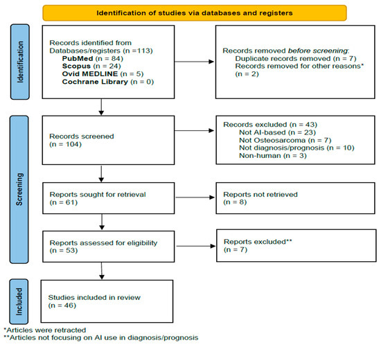
This narrative review was informed by principles of transparent reporting commonly used in structured literature reviews. A four-stage process (identification, screening, eligibility, and inclusion) was used to organize the literature search and selection (Figure 1). The flow diagram is included solely to illustrate our structured search and screening process rather than to claim PRISMA compliance.
Figure 1.
Flow diagram illustrating the literature search and study selection process for this narrative review of artificial intelligence applications in osteosarcoma diagnosis and prognosis.
3.1.1. Identification Stage
A structured literature search was conducted across the following databases: PubMed, Scopus and Ovid Medline. Our research covered the literature published between 1 January 2015 and 1 January 2026, reflecting the period of exponential advancements in modern machine learning technologies in osteosarcoma. The following Boolean search string was standardized across all the databases: “osteosarcoma” AND “artificial intelligence” AND “diagnosis” AND “prognosis”. We note that our PubMed search used “Artificial Intelligence” as a MeSH term with automatic explosion rather than a free-text keyword. In the MeSH hierarchy, machine learning and deep learning are narrower concepts nested under the artificial intelligence heading [25]. When the AI term is exploded, PubMed automatically retrieves records indexed with these narrower terms, ensuring that machine learning and deep learning studies are included even if those phrases do not appear in titles or abstracts. Consequently, we did not list “machine learning” and “deep learning” as separate MeSH terms, as they are already encompassed within the exploded AI heading. In addition to using “artificial intelligence” as an exploded MeSH term in PubMed, we adjusted our search strategy for databases without MeSH indexing. Both Scopus and Ovid rely on keyword-based queries, so we supplemented the search string with the free-text terms “machine learning” and “deep learning” alongside “artificial intelligence” to ensure that studies employing these approaches were captured. This dual strategy—MeSH explosion in PubMed and expanded keywords in Scopus and Ovid—allowed us to maximize sensitivity across different database architectures while maintaining a coherent focus on AI methods relevant to osteosarcoma. Our initial search yielded 113 articles: 84 from PubMed, 24 from Scopus, 5 from Ovid Medline, and 0 from Cochrane Library. After the removal of seven duplicate records and two retracted articles, a total of 104 articles underwent title and abstract screening.
3.1.2. Screening Stage
During this stage (at this level), titles and abstracts were screened. Inclusion and exclusion criteria (Table 1) were defined to guide the selection of studies most relevant to AI applications in osteosarcoma. Of the remaining 104 studies, 43 articles were excluded due to irrelevance to osteosarcoma, absence of AI-based methodologies, not addressing diagnostic or prognostic outcomes, and studies including non-human subjects. In addition, 8 articles were excluded because the full-text articles could not be retrieved.
Table 1.
Inclusion and exclusion criteria for selected studies.
3.1.3. Eligibility Stage
A total of 53 articles were then checked for eligibility, where we examined the summaries, methods, and goals of each. As a result, 7 articles were excluded because they did not strongly evaluate the AI-based performance in osteosarcoma.
3.1.4. Inclusion Stage
The remaining 46 articles represented the bedrock of this review and were all included in the qualitative narrative synthesis. The articles were divided into three main groups: diagnosis-related, prognosis-related, and diagnosis/prognosis-related. The diagnosis-related articles discussed radiomics as well as the genomics/transcriptomics role of AI in the diagnosis of osteosarcoma. The prognosis-related articles illustrated the role of AI using genomics, biomarkers, imaging and histopathology, metastasis prediction and risk stratification in predicting prognosis in osteosarcoma.
3.2. Quality Assessment and Risk of Bias Evaluation
The methodological features of the retrieved papers were examined with reference to domains from the QUADAS-2 tool (patient selection, index test, reference standard, and flow/timing) to highlight potential sources of bias. This approach was used to support a qualitative assessment of study robustness, rather than a formal scoring system, and to ensure that the narrative synthesis primarily drew on studies with acceptable methodological quality.
The performance of AI models described in the studies was reported using metrics such as area under the receiver operating characteristic curve (AUC), accuracy, concordance index (C-index) and others. However, due to the retrospective type of the retrieved studies, as well as the small sample size used in most of them, these metrics were interpreted carefully, and additional attention was given to the availability of external validation of results, cross-validation strategies and safeguards against overfitting.
Furthermore, when available in the original publications, comparisons between AI model performance and current clinical gold standards in osteosarcoma management (e.g., the Huvos grading system for chemotherapy response and RECIST criteria for metastatic disease) were extracted and qualitatively summarized. In studies where such benchmarking was absent, reported performance metrics were interpreted with caution and discussed as exploratory.
4. AI in the Diagnosis of Osteosarcoma
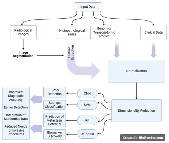
The diagnosis of osteosarcoma in patients can be facilitated by the aid that AI technology provides, mainly through radiomics, genomics and/or transcriptomics (Figure 2).
Figure 2.
Schematic representation of the artificial intelligence (AI) workflow in osteosarcoma diagnosis. Created in BioRender. Kalout, K. (2026) https://BioRender.com/bypt3a6 (accessed on 13 February 2026).
4.1. Radiomics-Based Diagnosis
First of all, the use of AI in radiomics has recently evolved, making it a promising tool in the diagnosis of diseases, especially tumors such as osteosarcoma. Radiomics, when coupled with AI tools and networks, provides a massive ability to handle data in medical imaging when compared to traditional statistical methods [26]. Zhou et al. proposed a three-step comprehensive technique that can assist doctors in the diagnosis of osteosarcoma [27]. They first pre-trained a segmentation model on unlabeled contrast-enhanced T1 and T2 MRI scans using a Multiparameter Fusion Contrast Learning (MPFCLR) algorithm, followed by constructing a Multiparameter Fusion network (MPFNet) that works on fusing complementary features from imaging data. This network shows potential advantages in that it can segment viable osteosarcoma regions from necrotic tissues and predict patient prognosis by associating with clinical characteristics (e.g., volume, spread) of the tumor. The MPFNet model achieved a mean dice coefficient of 84.19% and an F1-score of 84.56%, which proved better than other models for osteosarcoma segmentation. While the results of the MPFNet model were promising, it was applied only on 132 samples, and it was limited to MRI and did not tackle any other form of imaging, which limits its generalizability and replication attempts in areas of limited resources [27].
In another experimental study, Liu et al. proposed a medical image processing method that uses AI and works on segmenting an MRI image, thus aiding in the identification of the osteosarcoma lesion area [13]. This method is primarily based on Transformer and U-net and is called Osteosarcoma MRI Segmentation Network (OSTransnet). It mainly addresses the limitations of MRI images in diagnosing osteosarcoma by segmenting the tumor edge, making it clearer for identification, and the segmentation performance was enhanced using an edge enhancement module boundary augmentation block (BAB) [13]. More than 4000 MRI images were used, and the results showed that OSTransnet outperformed other segmentation networks in terms of intersection over union (IOU), F1 metrics and precision, and it achieved a dice score of 0.949 in the category that joins OSTransnet and BAB, which was higher than that without the edge enhancement module [13]. These results are promising because they allow clinicians to handle noisy MRI pictures, thus aiding in the diagnosis as well as the treatment plan of osteosarcoma.
AI-processed radiomics provides a significant clinical benefit in bone tumors [28]. In particular, its application has been studied in distinguishing primary from metastatic bone tumors, differentiating between bone tumor types, and subtyping osteosarcoma [28]. Large datasets were taken, and the segmentation process of osteosarcoma lesions was automated by U-Net, which is one example of convolutional neural networks (CNNs) [28]. Other machine learning models used included feature-based classifiers, such as Support Vector Machines, K-Nearest Neighbors, logistic regression and others, and deep transfer-learning models like modified SqueezeNet or honey-badger-optimized CNNs, which were mainly trained to distinguish osteosarcoma from other bone lesions [28]. These models had excellent predictive capabilities in the diagnosis and grading of osteosarcoma and were able to predict overall survival, metastasis risk, and recurrence risk related to this tumor [28]. The application of AI-assisted radiomics in osteosarcoma diagnosis may ultimately reduce demand for invasive procedures, optimize chemotherapy decision-making and significantly enhance osteosarcoma detection objectivity and accuracy [13,28]. However, these models still lack transparent reasoning behind their interpretation results, as well as validation on external independent datasets, both of which are required to be present in patient care settings.
The first imaging modality used in suspected bone pathology is usually an X-ray. Breden et al. proposed a Vision Transformer, a deep learning-based model, that can be used to screen any suspicious lesion on an X-ray for any osteosarcoma potential [29]. Most of the predictions done by the model were based on imaging anomalies that a human expert also found to be suspicious, and it achieved a sensitivity of 82.2% and specificity of 93.3%, making it a useful tool for early identification of osteosarcoma using simple imaging techniques [29].
4.2. Genomics/Transcriptomics-Based Diagnosis
In addition to radiomics, the use of AI in genomics and transcriptomics has shown to be highly successful in diagnosing osteosarcoma. AI models have been used in recent studies to extract diagnostic biomarkers that would suggest the presence of viable osteosarcoma cells. Using multiple data sources, such as GSE99671, researchers identified differentially expressed genes (DEGs) in osteosarcoma vs. normal cells [30]. Then, they did a gene set enrichment analysis (GSEA) of the hypoxia pathway in both cell lines along with weighted gene co-expression network analysis (WGCNA), which groups genes into co-expression modules and identifies the ones that correlate with osteosarcoma [30]. After intersecting hypoxia genes (from GSEA) and hub genes (from WGCNA), Least Absolute Shrinkage and Selection Operator (LASSO) was used to pick the best genes to be used in the prediction of osteosarcoma [30]. Two hypoxia-related genes, STC2 and TMEM45A, were the best biomarkers, where there was an exceptionally high discrimination between osteosarcoma and normal controls with an area under the ROC curve of ≈ 0.96 in training and test sets, which suggests a strong potential for molecular diagnosis of osteosarcoma [30]. While the results were promising, the study relied only on retrospective data, so prospective clinical validation and functional in vivo studies of the two genes are required moving forward.
Another type of biomarker reported was circulating microRNAs (miRNAs), which provide potential to diagnose and differentiate metastatic and non-metastatic osteosarcoma using machine learning [31]. Researchers started by analyzing RNA sequencing and microarray data from osteosarcoma cell lines to identify miRNAs that differ in expression in those two groups [31]. Machine learning models were utilized for the construction of diagnostic tools that could successfully identify two miRNAs (miR-34c-3p and miR-154-3p), which, in combination, transform into novel diagnostic biomarkers to detect metastatic osteosarcomas, with an accuracy of 85% (AUC ≈ 0.85) [31]. Expression levels of these miRNAs were downregulated in osteosarcoma tissue versus benign tissue. Nevertheless, the study used a small sample size, and limited generalization can be made. Moreover, the considerable reliance on RNA sequencing and microarray data could also cause biases stemming from potential technical differences in experimental protocols.
AI application in transcriptomics has also shown promising potential in the diagnosis of osteosarcoma. In one study, multi-omics (genomics, transcriptomics, methylation) data were used to identify high-risk osteosarcoma subtype samples that were shown to be driven by the MYC oncogene, which indicates the worst prognosis [32]. In order to identify this subtype, researchers used a powerful machine learning algorithm, XGBoost, combined with Bayesian optimization to build a classifier, and they integrated these multi-omics data results [33]. The final model, which used nine genes, performed extremely well in the training set with an AUC of ≈ 0.999 and an overall accuracy of ≈ 98.9%, and it was able to separate patients into high- and low-risk groups with different survival outcomes successfully. Also, the model showed that, among the nine genes, IG2BP3 was strongly linked to the MYC signaling pathway [33]. These results were externally validated using the TARGET-OS dataset, which is a public osteosarcoma database [33]. While the proposed model shows promising results with external validation, its clinical applicability is yet to be proven because transcriptomic data are not routinely used in clinical settings. In another example, Llaneza-Lago et al. used an advanced Bayesian machine learning model, which they called “Latent Process Decomposition” (LPD), for the same goal of subtyping osteosarcoma. The method they used can handle sample heterogeneity, which is the difference in cell types that the tumor contains and which is not handled in traditional classifying techniques [34]. LPD was able to identify three osteosarcoma subtypes that were classified according to their mortality risk, with LPD-1 having the worst prognosis [34]. This approach shows that the application of advanced AI and machine learning carries high potential for improving drug discovery and treatment management plans for osteosarcoma, in addition to improving the accuracy of categorical osteosarcoma diagnosis.
Adverse events can also indicate osteosarcoma as an example of such indirect aid in the diagnosis of the disease. Ref. [35] screened osteosarcoma cells for programmed cell death (PCD) genes by obtaining data from the TARGET and GEO databases. They further established a model incorporating ten machine learning approaches (such as elastic net, random survival forest (RSF), LASSO) to assess thirty prognosis-related genes of PCD in osteosarcoma [35]. The RSF algorithm had better predictive precision for prognosis than the conventional ways and found five hub genes, including MTM1, MLH1, CLTCL1, EDIL3, and SQLE [35]. Three independent GEO validation sets tested the predictive ability of the model [35]. Although the model showed robust prognostic prediction ability for osteosarcoma from a transcriptomics perspective, it might also be helpful in classifying osteosarcoma into biologically or clinically relevant subgroups and discovering potential diagnostic genes contributing to distinguishing different types of osteosarcomas.
4.3. Histopathology-Based Diagnosis
The gold standard for the definitive diagnosis of osteosarcoma requires confirmation by histopathological examination of biopsy lesions, which is done by expert pathologists [1]. Deep convolutional neural networks (DCNNs) were trained on whole-slide images (WSIs) and showed 99.8% accuracy in distinguishing normal from tumor tissues and 71.2% in distinguishing benign vs. malignant tumors with high AUC values, indicating strong discrimination for tumor detection [36]. Despite achieving a diagnostic performance that is comparable to that of senior pathologists, it remains an adjunct tool that can aid pathologists in the diagnosis but cannot replace them [36].
Multiple histological image classifiers were tested on samples in an attempt to improve the accuracy of osteosarcoma detection [37,38]. They work by denoising histological images of osteosarcoma, making them purer for classification [37,38]. The classification frameworks described were able to outperform the conventional CNNs that are currently used, as well as the deep learning approaches that are currently studied, with an average accuracy of 98% [37,38].
4.4. Radiogenomics Integration
Radiogenomics integrates radiomic features with genomics and transcriptomics to enable AI models to associate macroscopic imaging behaviors with underlying molecular states. A 2025 review of musculoskeletal oncology clarifies that inclusion of molecular data can help grade tumors, differentiate between osteosarcoma and Ewing sarcoma and predict such mutations as TP53 and RB1 [39]. Radiogenomics uses imaging phenotypes to bridge the gap between imagery and genomic signatures, reducing the dependence on invasive biopsies and offering biomarkers for diagnosis, prognosis and personalized treatment [39]. While radiogenomics has proved effective in prostate, breast and brain cancer [39], there is still limited evidence supporting its application for osteosarcoma. In the future, these will need to be prospectively integrated for multi-institutional cohorts in studies combining imaging with genomics and transcriptomics to build easily interpretable radiogenomic signatures that can predict treatment response and metastatic risk of osteosarcoma.
5. Artificial Intelligence in the Prognosis of Osteosarcoma
The osteosarcoma pattern of behavior has long been a complicated task. Clinicians often relied on certain clinical indicators, like tumor size, necrosis rates, and metastatic spread, to help aid their assessment, yet those measures only provided a brief outline. By introducing AI, clinicians can refine their clinical judgment to reach a better prognostic result. With access to a wide dataset that includes gene expression profiles, biomarkers, imaging and pathology slides, AI can help in recognizing crucial patterns that may remain undetected.
5.1. Genomics and Multi-Omics Integration
While genomic data has exploded in the past decade, making sense of it, particularly in the clinic, has been slow. That is changing. In 2024, Fan and colleagues took a machine learning approach to building a model from ceRNA networks, focusing on genes like ABCA8, CAT, and CXCL12. These seemed to act protectively, and their model demonstrated a strong ability to differentiate high-risk from low-risk patients, with an AUC of 0.90 and a C-index of 0.87. Although not established for routine clinical implementation, this suggests that one day, ceRNA patterns may help inform prognosis [40].
Another study by [5] combined transcriptomic and epigenomic characteristics to construct an AI-driven prognostic index. An interesting finding in their study is that SQLE, a gene associated with cholesterol metabolism, was highly expressed in the majority of patients with poor prognosis. The model performed well on different datasets, with an AUC > 0.80, and pinpointed metabolic liabilities for potential targeting in future therapies. This progress is not only limited to improved prediction capability; it also paves the way for promising new game-changers in cancer treatment [5].
In addition, lipid metabolism may be used to categorize osteosarcoma into different subtypes with differing outcomes. To solve this problem, Li et al. developed a Lipid Metabolism-Related Signature (LMRS) consisting of 12 genes to assess survival risk in patients with osteosarcoma [41]. They ran four machine learning algorithms—Random Survival Forest (RSF), CoxBoost, Generalized Boosted Regression Models and Survival-SVM—on a panel of 208 samples from four different datasets. They found that the LMRS model showed a stable validation performance in multiple tumor cohorts, outperforming 12 published signatures. Moreover, it also demonstrated the possibility of identifying high-risk patients at an early stage with a potential application for personalized treatment [41].
Similarly, the MLDPS, which was developed based on 66 immune-related gene pairs obtained from 10 AI algorithms, consisted of seven genes that divided patients into high- and low-risk score groups. The MLDPS showed excellent predictive value with respect to 1-, 3- and 5-year overall survival (AUC between 0.963 and 0.852) [42]. This model provides a high potential not only for predicting the prognosis of osteosarcoma but also guides therapeutic strategies as well as clinical management.
Immune biology has gained interest as a prognostic model. Qu et al. identified two genes, SERPINE2 and CPT1B, that were linked with worse survival and seemed tied to immune suppression within the tumor microenvironment [43]. This is a reminder that prognosis may be just as much about the host response as the tumor’s biology. AI helped spot these relationships, ones that might have been easily overlooked in traditional analyses.
Also, macrophage polarization-related genes in osteosarcoma were studied to construct a prognostic model. These genes influence the tumor microenvironment by either stimulating the immune response (M1 macrophages) or suppressing it (M2 macrophages) [44]. LASSO and Cox regression were used to construct an eight-gene signature model that would predict prognosis. It identified a key gene, BNIP3, which predicts a poor prognosis of osteosarcoma because it helps the tumor grow [44]. Targeting this gene through immunotherapy might improve treatment because its knockdown caused inhibition of osteosarcoma cell growth. However, in vivo validation of the model and BNIP targeting is still lacking.
5.2. Biomarkers as Prognostic Tools
In order to predict prognosis in osteosarcoma, AI has been applied to identify biomarkers that offer a wide range of information about the progression of the disease.
In Jiang et al. [45], m6A-related genes (which are implicated in RNA methylation) were analyzed, and a prognostic model for patient prognosis was constructed. LASSO regression analysis was conducted to select the most significant genes, and Multivariate Cox regression analysis was used to predict their relationship with patient survival [45]. Moreover, the study observed that low overall survival was significantly associated with two core genes: RBM15 and YTHDC1. Of interest, RBM15 was positively associated with activated natural killer (NK) immune cells and negatively correlated with gamma delta T-cells, indicating that this gene could be involved in tumor escape from immunity [45].
Another example is the use of machine learning and miRNA sequencing for prognostic biomarker prediction in osteosarcoma. Using two independent datasets, researchers inspected microRNA profiles and discovered potential microRNA signatures that could predict either better or worse survival. These signatures were then tested in several independent NCI datasets [46].
The signatures contributing to predicting the prognosis in osteosarcoma have been widely investigated by AI. Ref. [47] carried out an exploratory work with the aim of finding common gene signatures for both retinoblastoma and osteosarcoma as potential prognostic markers, using machine learning algorithms. A total of 1216 common genes were classified into modules using Weighted Gene Co-expression Network Analysis (WGCNA). The study used three machine learning models, and six hub genes with the greatest impact on prognosis were identified [47]. Then, a risk score model for predicting the overall survival in osteosarcoma patients was built with these hub genes. The model had good effectiveness and was not correlated with other clinical factors, like age or tumor type [47].
In addition, machine learning algorithms were used to generate a four-methylated lcRNA signature to predict the prognosis of osteosarcoma patients. The osteosarcoma data were downloaded from TARGET, and this signature was shown as an effective predictor for the survival prediction of osteosarcoma patients. Survival analysis (Kaplan–Meier and ROC-wide) was used to confirm the accuracy of this signature to distinguish high-risk patients from low-risk ones, independent of other clinical features, when comparing with known biomarkers in osteosarcoma [48].
In addition to the establishment of gene signature models, Liu et al. investigated using non-coding RNA pseudogenes to form a prognostic model with a deep learning algorithm [49]. The four-pseudogene signature comprised 125 pseudogenes and was developed using Cox regression analyses calculated from RNA sequencing data [49]. This signature shows a high prognostic value, with AUC reaching 0.878 and strong power to separate patients into two risk categories [49]. The study revealed the potential of AI models to predict prognosis in osteosarcoma by using junk DNA as a biomarker.
The identification of biomarkers in osteosarcoma would not only aid in predicting prognosis but also help in the management plan and provide potential drug targets. While these three studies were able to identify potential biomarkers aiding in prognosis prediction, all of them were retrospective and used small sample sizes, and they require further external validation.
5.3. Clinical Data and Survival Prediction
AI models that use basic clinical data can be really useful, especially since not every hospital has genomic labs. For example, Zhang et al. explored different survival models based on the SEER database. It was found that the advanced deep learning adaptation of the Cox model (DeepSurv) performed better than other models [23]. It efficiently discovered complex interactions among the variables that commonly used statistical methods might ignore, with an AUC of 0.78 for five-year survival and a C-index of 0.765. This is a substantial advance considering that only general information (e.g., age, tumor site, and treatment history) is used [23].
Jiang et al. employed XGBoost, which is a decision tree-based algorithm suitable for imperfect data [22]. A model derived from similar clinical data had an AUC of 0.911. This result is remarkable as no molecular information was used and clearly shows that even low-cost clinical information can deliver competitive predictive performance if being modeled wholeheartedly [22].
5.4. Imaging and Histopathology-Based Prognostic Tools
Medical imaging has always played a role in cancer care, but that role is expanding when it comes to predicting patient outcomes, and it is being driven by AI. Zhou et al. designed a deep learning model by fusing clinical information and integrated MRI images [27]. It is simple, yet it does the job. Their integrated model had a C-index of 0.806, which outperformed models built based on an individual data type, which are currently used clinically. It is important to note that the study did not report any comparison to an established clinical gold standard, such as the RECIST score. Since such types of scans and data are already being used by clinicians for osteosarcoma assessment, translation of this model into clinics seems feasible [27].
Histology is not being left behind either. Coudray and colleagues (2025) trained convolutional neural networks on digital slides, teaching their models to measure necrosis and pick up on small details related to survival [14]. What is fascinating is that the AI did not just imitate what a pathologist does; it detected some finer details—patterns like fat necrosis and changes in tissue around the tumor—that we do not yet fully understand but could end up yielding important clues about how cancer arises. The model even outperformed human judgment in survival prediction (p = 0.0028), demonstrating proof-of-concept of digital pathology. Similarly, machine learning models that were trained on osteosarcoma datasets were shown to be more effective in the detection and classification of the disease when compared to hand-crafted features, depending on the pathologist’s expertise [50].
Zhong’s group also used nnU-Net segmentation to look at tumor features before treatment started [51]. They wanted to predict how well someone would respond to chemotherapy, which is often a stand-in for survival odds. Their model scored an AUC over 0.79, meaning it can give doctors a solid idea up front about whether standard chemotherapy is likely to work, even before the first round starts [51]. These metrics were not compared to established standardized benchmarks. However, in another experiment, a prediction model that was constructed using five machine learning algorithms was tested for its potential in predicting the rate of tumor necrosis in osteosarcoma using MRI and dynamic contrast enhancement (DCE) MRI [51]. The model showed a high prediction performance, with AUC values averaging 0.86 [51]. The results were then verified postoperatively using the gold-standard Huvos grading criteria, which showed that DCE-MRI radiomics was able to predict the same necrosis categories defined by Huvos and could serve as a non-invasive surrogate marker for predicting chemotherapy response [51].
Similarly, Ho et al. studied the prediction of chemotherapy response by using AI [52]. A reproducible deep learning-based approach was proposed to estimate the necrosis ratio, i.e., the ratio of necrotic tumor to overall tumor, after chemotherapy in order to categorize the response to chemotherapy (Grade IV = 100%, III ≥ 90%, II ≥ 50% and I < 50%) [52]. A Deep Multi-Magnification Network (DMMN) was trained to calculate the necrosis ratio in multiple whole-slide images of osteosarcoma, and it showed excellent performance as the results were closely matched to those reported by pathological experts using the Huvos criteria, with low mean absolute errors in high-grade responses [52]. This approach allows patient outcome prediction and management guidance, and it implies that AI serves as an adjunct prognostic tool to standardize therapy response assessment. While the study represented the largest known digital osteosarcoma cohort studied, with 103 cases included, all the data came from a single center, which limits its generalizability. Another example was the use of machine learning in an attempt to better automate the evaluation of tumor necrosis in osteosarcoma following neoadjuvant chemotherapy. AI models were able to achieve remarkable classification of viable vs. nonviable tumors when compared to the traditional manual annotation of pathologists using the Huvos criteria [53].
5.5. Metastasis Prediction and Risk Stratification
In osteosarcoma, it is not the primary tumor that usually causes death; it is metastasis, especially to the lungs. Figuring out which patients are likely to metastasize is critical, and AI has helped in this regard as well. Su et al. developed a radiomics model using CT images and trained a random forest classifier to identify early signs of metastasis [6]. Despite its limits, it achieved an AUC of around 0.79, a solid starting point for non-invasive methods [6].
Genetic models have also revealed their worth. Liao et al. developed a 15-gene signature exclusively for the purpose of predicting metastasis [54]. The performance of their model was consistent across various datasets and achieved AUC values greater than 0.93 [54]. This cannot be easily overlooked. Concurrently, Abedi et al. investigated microRNAs and determined a pair of miRNAs that would differentiate metastatic from non-metastatic cases with an accuracy of 85% [31]. The model might eventually be included in a liquid biopsy device since these markers are potentially detectable in blood [31].
The use of AI for predicting metastasis in osteosarcoma has advanced remarkably and is providing useful new tools for prognostication at the time of diagnosis. He et al. downloaded five osteosarcoma metastasis-related datasets from the GEO database, one of which was excluded because of low quality [55]. These were identified as 64 key genes after calculating “betweenness centrality” by them in a DEG-induction meta-analysis; 353 DEGs between metastatic and nonmetastatic samples of osteosarcoma have been derived [55]. These genes were employed to build an SVM classifier (a type of supervised machine learning algorithm) optimized to discriminate between osteosarcoma non-metastatic and metastatic samples [55]. The classification performance of the AI algorithm was excellent, showing an accuracy rate of 100% on three datasets and 92.6% on one dataset [55]. Although promising results were achieved from the predictive possibility in terms of an adverse prognosis for metastasis in osteosarcoma, the work’s internal and retrospective analysis may suffer from overfitting of its model due to the limited sample size.
Early prediction of distant osteosarcoma metastasis can help clinicians during clinical decision-making. A machine learning-based prediction model that included six machine learning techniques, such as SVM, random forest (RF), XGBoost and others, was developed in correlation with clinical data like tumor size, primary surgery and age [56]. The model’s average precision (AP) ranged between 0.661 and 0.781, where RF had the best metrics in predicting distant metastasis, with AP = 0.781 and an accuracy of 71.8% [56]. The model provides a huge prospect for real-world use, where it could identify candidates for aggressive treatment based on their predicted prognosis early after diagnosis.
Similarly, the prediction of osteosarcoma metastasis specifically into the lungs was also studied because lung metastasis is the most common and deadly form. The first study used six machine learning algorithms, including logistic regression, XGBoost and others, on large datasets from the Surveillance Epidemiology and End Results (SEER), and was externally validated in four Chinese hospitals [57]. The predictors of lung metastasis in the study were sex, T stage, N stage, surgery and bone metastasis [57]. XGBoost had a better prediction accuracy and achieved an AUC of 0.738 for internal cases and 0.729 for external cases [57]. On the other hand, the other two studies used regression algorithms (LASSO and multivariate logistic regression with N stage and further laboratory markers, such as alkaline phosphatase [ALP], thyroid-stimulating hormone [TSH], free T3 as independent predictors) [58,59]. The first model showed good performance, with an AUC of 0.701 in the training cohort and 0.786 in the validation set [58], and the second model showed better performance, with an AUC of 0.777 in the training cohort and 0.742 in the validation cohort [59]. Together, these studies emphasize the importance of AI when it comes to predicting prognosis and personalized management for osteosarcoma lung metastasis in an effort to reduce both morbidity and mortality.
When comparing AI models for the prediction of metastasis in osteosarcoma, Zhou et al. used machine learning methods, such as random forest (RF), and a deep learning model implemented with TensorFlow and Keras to build metastasis-predictive models for both osteosarcoma and Ewing sarcoma [60]. The deep learning model outperformed the RF model, with a higher accuracy and lower error rate over several training rounds [60]. An interactive online nomogram was also created to make the models clinically usable, and it showed that it could accurately estimate patient survival and thus predict prognosis because the predicted and actual outcomes were highly matched [60]. Further research exploring differences and comparing AI algorithms’ potential in predicting prognosis is highly recommended, along with external validation of results across diverse populations in order to allow generalizability.
In addition to predicting metastasis, there is growing interest in predicting the risk of recurrence of osteosarcoma and its prognostic potential. A quantitative histological image classifier was constructed and used to identify tumor nuclear features for predicting recurrence and survival after standardized osteosarcoma therapy [61]. While there are no traditional benchmarks to compare with, the osteosarcoma histological image classifier was able to provide a notable performance, thus contributing to precision oncology [61].
To synthesize findings across modalities, Table 2 provides a comparative overview of key studies, summarizing cohort sizes, tasks, models, validation strategies, top metrics, risk-of-bias factors, and clinical readiness.
Table 2.
Comparative analysis of AI prognostic and diagnostic models for osteosarcoma.
6. Limitations and Future Directions
Although this seems quite promising, it is too soon to tell how AI tools will ultimately affect medical practice, especially osteosarcoma. Retrospective data from one hospital or research group mostly form the basis of models. This raises concerns about generality. A successful model in one environment might not precisely translate to another. Another issue is data quality, particularly given that AI is meant to examine flawed or unfinished records from the real world.
6.1. Limitations
One major issue to be discussed is that almost all the studies retrieved reported the favorable performance of AI-based diagnostic and prognostic models without direct benchmarking against established gold standards in osteosarcoma management. Only a few studies compared the AI models’ abilities in predicting chemotherapy response to the Huvos grading criteria, which is usually done postoperatively [66]. In addition, none of the retrieved studies used the RECIST score as a radiology evaluation benchmark when comparing the efficacy of their constructed AI models in predicting osteosarcoma metastasis. Consequently, the high metrics described in the studies do not necessarily reflect the superiority of AI and its readiness for the transition into clinical utility, but its promising potential in the medical field cannot be denied.
In addition, most of the retrieved articles reported a small dataset, often involving fewer than 200 patients [31,55]. This increases the risk of model overfitting, and the metrics described may overestimate the true clinical performance of these AI models. Also, while some studies reported external and/or cross-validation strategies, the majority did not validate their models in independent cohorts [13,28]. This limits the translation of the reported AI models into clinical practice due to their reduced performance when applied to new patient populations. In addition, a 2025 mini-review on AI-based radiomics points out that the dependence on small, single-institution series is a widespread issue that hampers robustness and leads to poor external performance [67]. The issue is also highlighted in the same review: models developed from AI trained on heterogeneous or insufficiently sourced data may learn peculiarities rather than generalizable information and, therefore, may not satisfy new populations [67]. To address these challenges, multi-institutional efforts and federated learning frameworks should be applied to gather heterogeneous imaging and molecular data while safeguarding patient privacy [68]. Standardization of imaging protocols and a central repository will also reduce heterogeneity, allowing for strong external validation. Without such controls, claimed high accuracies and AUC values could overestimate the clinical usefulness and translation to real-world applications.
Another problem is the “black box” problem. Naturally cautious, clinicians are reluctant to rely on a system whose decision-making process is not altogether evident. One field that might close that gap is explainable artificial intelligence (XAI), which is still being developed. Multimodal modeling, which integrates clinical, molecular, and imaging data into a single system, also benefits from this. Though that is most certainly the future, developing and verifying such models requires time, processing capability, and teamwork.
A significant shortcoming among the reviewed studies is that relatively few directly compare their AI models to existing clinical gold standards for osteosarcoma care. The majority of studies reported high performance measures (such as area under the curve [AUC] or concordance index) but did not cite benchmarks, such as the Huvos grading system for post-chemotherapy histologic response or Response Evaluation Criteria in Solid Tumors (RECIST) criteria for metastatic disease assessment. Only a few studies put AI models to the test against these benchmarks, and the results were mixed. For instance, an MRI radiomics model was employed in a study to predict chemotherapy response and compare its predictions with Huvos grades [1,67]. In general, from the reporting standpoint, no routine benchmarking exists to assess endless scenarios, such as whether AI truly improves upon clinical judgment. Consequently, it remains unclear whether AI models truly surpass clinician-driven assessments or simply reflect dataset-specific patterns. In future studies, standardized clinical criteria should be adapted by researchers, and they should report diagnostic accuracy, sensitivity and specificity concordance relative to Huvos and RECIST criteria. A table summarizing the presence or absence of such benchmarking in each study reviewed might help readers assess clinical significance.
6.2. Technological Innovation and Future Directions
For AI to really affect OS prognosis, we will need future validation, more data from many different places, and improved connections with clinical operations. AI will not only aid prognosis when those phases are in place; it might change how we approach therapy for every patient from day one.
However, there is a gap that exists between AI progress in oncology overall and AI progress in osteosarcoma research specifically. Most osteosarcoma studies report the application of older well-established algorithms, such as CNNs, SVMs and RF, which have been reported in other oncology domains for over a decade; however, they miss the new cutting-edge AI methods such as XAI, self-supervised learning and foundation models, graph-based learning and others [68]. The rarity of osteosarcoma overall in the population and the limited data in the literature might explain why this domain is losing the race with oncology. As a result, the absence of these new AI paradigms in the field of osteosarcoma creates a critical gap between technological advancement and current application in this field.
Recent advances in artificial intelligence point to a new generation of interpretable, multimodal and generalizable osteosarcoma models. Explainable AI (XAI) methods are essential for interpreting the rationale of model predictions and promoting clinician trust [67]. Graph neural networks (GNNs) generalize deep learning to the non-Euclidean domain and are able to model both spatial and functional interdependencies between tumor regions with respect to other modalities, thereby building models for segmentation, classification or multimodal fusion [7]. Primary models, like the BEPH model pre-trained on 11 million unlabeled histopathology images, have shown that self-supervised learning can produce generalizable representations that outperform when provided with few annotations and adjusted to multiple diagnostic and prognostic tasks [8]. In addition, multimodal backbone models with text and imaging and/or genomic data also demonstrate the significant enhancement in label efficiency and generalizability [9], while, at the same time, they bring computation resources, privacy issues for data, and “hallucination” problems to us [9]. Finally, radiogenomics (the combination of imaging phenotypes with genomic signatures) provides a pathway for accurate risk stratification and treatment planning [69]. Further innovation in these directions, and the pursuit of federated learning approaches to extract value from multi-institutional data archiving whilst respecting patient privacy [67], will be essential for driving AI-driven care for osteosarcoma forward.
Finally, it is important to address the heterogeneity in osteosarcoma and the importance of integrating multiple AI modalities together [68]. For example, linking imaging features with molecular and genomic studies is where the true technological frontier lies, and this allows clinicians to view the full picture of the disease to make interpretable predictions accordingly. Thus, multimodal fusion strategies, such as the use of radiogenomics, represent a critical intersection between medicine and technology.
7. Conclusions and Perspective
Artificial Intelligence has achieved significant advances in assisting osteosarcoma diagnosis and prognosis, with the ability to automate tumor segmentation, critical prognostic molecular signature recognition and metastatic risk prediction. However, the majority of published models are based on small retrospective collections and present no comparisons with clinical gold standards. Radiogenomics, graph neural networks and foundation models all demonstrate that leveraging multimodal integration and self-supervised learning can improve generalizability, as well as decrease the need for annotation [7,8]. The clinical implementation of such approaches will necessitate robust external validation, explainable AI strategies to create confidence among clinicians and the employment of multi-institutional federated learning frameworks that can combine heterogeneous datasets without compromising patient privacy [9,67]. Prospective radiogenomics studies, integration of imaging with molecular and clinical data, and standardized benchmarking against established diagnostic criteria should be focus areas for future research. Through these developments, AI may be able to progress beyond proof-of-concept and become a trusted tool in the treatment of osteosarcoma.
Author Contributions
Conceptualization, R.A.G., K.K. and J.E.; methodology, F.H.; investigation, R.A.G., K.K. and J.E.; resources, R.A.G., K.K. and J.E.; writing—original draft preparation, R.A.G., K.K., J.E. and F.H.; writing—review and editing, R.A.G., K.K., J.E., M.A., H.E.G., S.A. and F.H.; visualization, R.A.G., K.K. and J.E.; supervision and project administration, F.H. All authors have read and agreed to the published version of the manuscript.
Funding
This research received no external funding.
Data Availability Statement
No new data were created or analyzed in this study. Data sharing is not applicable to this article.
Conflicts of Interest
The authors declare that they have no competing interests related to this work. No author has any financial or personal relationships that could inappropriately influence or bias the content of the manuscript.
References
- Bhatt, P.S.; Maru, A.M.; Makwana, H. Clinicopathological and Radiological Correlation in Osteosarcoma: A Prospective Study of 91 Cases. J. Pharm. Bioallied Sci. 2025, 17, S2751–S2753. [Google Scholar] [CrossRef] [Scilit]
- Yu, S.; Yao, X. Advances on immunotherapy for osteosarcoma. Mol. Cancer 2024, 23, 192. [Google Scholar] [CrossRef] [Scilit] [PubMed]
- Rathore, R.; Van Tine, B.A. Pathogenesis and Current Treatment of Osteosarcoma: Perspectives for Future Therapies. J. Clin. Med. 2021, 10, 1182. [Google Scholar] [CrossRef] [Scilit] [PubMed]
- Flora, D.B. The Top 10 AI in Precision Oncology Stories of 2024: A Year of Breakthrough Moments. AI Precis. Oncol. 2025, 2, 1–5. [Google Scholar] [CrossRef] [Scilit]
- Wang, Y.; Ma, X.; Xu, E.; Huang, Z.; Yang, C.; Zhu, K.; Dong, Y.; Zhang, C. Identifying squalene epoxidase as a metabolic vulnerability in high-risk osteosarcoma using an artificial intelligence-derived prognostic index. Clin. Transl. Med. 2024, 14, e1586. [Google Scholar] [CrossRef] [Scilit]
- Su, Z.; Nong, Z.; Huang, F.; Zhou, C.; Yu, C. Anemia Risk Prediction Model for Osteosarcoma Patients Post-Chemotherapy Using Artificial Intelligence. Cancer Med. 2024, 13, e70427. [Google Scholar] [CrossRef] [Scilit]
- Mienye, I.D.; Viriri, S. Graph Neural Networks in Medical Imaging: Methods, Applications and Future Directions. Information 2025, 16, 1051. [Google Scholar] [CrossRef] [Scilit]
- Yang, Z.; Wei, T.; Liang, Y.; Yuan, X.; Gao, R.; Xia, Y.; Zhou, J.; Zhang, Y.; Yu, Z. A foundation model for generalizable cancer diagnosis and survival prediction from histopathological images. Nat. Commun. 2025, 16, 2366. [Google Scholar] [CrossRef] [Scilit]
- Huang, S.-C.; Jensen, M.; Yeung-Levy, S.; Lungren, M.P.; Poon, H.; Chaudhari, A.S. A Systematic Review and Implementation Guidelines of Multimodal Foundation Models in Medical Imaging. Res. Sq. 2025. [Google Scholar] [CrossRef] [Scilit]
- Goedhart, L.M.; Gerbers, J.G.; Ploegmakers, J.J.W.; Jutte, P.C. Delay in Diagnosis and Its Effect on Clinical Outcome in High-grade Sarcoma of Bone: A Referral Oncological Centre Study. Orthop. Surg. 2016, 8, 122–128. [Google Scholar] [CrossRef] [Scilit]
- Brar, G.S.; Schmidt, A.A.; Willams, L.R.; Wakefield, M.R.; Fang, Y. Osteosarcoma: Current insights and advances. Explor. Target. Anti-tumor Ther. 2025, 6, 1002324. [Google Scholar] [CrossRef] [Scilit]
- Cè, M.; Cellina, M.; Ueanukul, T.; Carrafiello, G.; Manatrakul, R.; Tangkittithaworn, P.; Jaovisidha, S.; Fuangfa, P.; Resnick, D. Multimodal Imaging of Osteosarcoma: From First Diagnosis to Radiomics. Cancers 2025, 17, 599. [Google Scholar] [CrossRef] [Scilit]
- Liu, F.; Zhu, J.; Lv, B.; Yang, L.; Sun, W.; Dai, Z.; Gou, F.; Wu, J. Auxiliary Segmentation Method of Osteosarcoma MRI Image Based on Transformer and U-Net. Comput. Intell. Neurosci. 2022, 2022, 1–16. [Google Scholar] [CrossRef] [Scilit]
- Coudray, N.; Occidental, M.A.; Mantilla, J.G.; Quiros, A.C.; Yuan, K.; Balko, J.; Tsirigos, A.; Jour, G. Quantitative and Morphology-Based Deep Convolutional Neural Network Approaches for Osteosarcoma Survival Prediction in the Neoadjuvant and Metastatic Settings. Clin. Cancer Res. 2024, 31, 365–375. [Google Scholar] [CrossRef] [Scilit] [PubMed]
- Bishop, M.W.; Janeway, K.A.; Gorlick, R. Future directions in the treatment of osteosarcoma. Curr. Opin. Pediatr. 2016, 28, 26–33. [Google Scholar] [CrossRef] [Scilit] [PubMed]
- Mogi, K. Artificial intelligence, human cognition, and conscious supremacy. Front. Psychol. 2024, 15, 1364714. [Google Scholar] [CrossRef] [Scilit] [PubMed]
- Habehh, H.; Gohel, S. Machine Learning in Healthcare. Curr. Genom. 2021, 22, 291–300. [Google Scholar] [CrossRef] [Scilit]
- Luchini, C.; Pea, A.; Scarpa, A. Artificial intelligence in oncology: Current applications and future perspectives. Br. J. Cancer 2022, 126, 4–9. [Google Scholar] [CrossRef] [Scilit]
- Shamshiri, M.A.; Krzyżak, A.; Kowal, M.; Korbicz, J. Compatible-domain Transfer Learning for Breast Cancer Classification with Limited Annotated Data. Comput. Biol. Med. 2023, 154, 106575. [Google Scholar] [CrossRef] [Scilit]
- Najafi, N.N.; Hajihassani, H.; Irani, M.A. The Impact of Artificial Intelligence on Cancer Diagnosis and Treatment: A Review. Cancer Inform. 2025, 24. [Google Scholar] [CrossRef] [Scilit]
- Cheng, C.H.; Shi, S.-S. Artificial intelligence in cancer: Applications, challenges, and future perspectives. Mol. Cancer 2025, 24, 274. [Google Scholar] [CrossRef] [Scilit] [PubMed]
- Jiang, J.; Pan, H.; Li, M.; Qian, B.; Lin, X.; Fan, S. Predictive model for the 5-year survival status of osteosarcoma patients based on the SEER database and XGBoost algorithm. Sci. Rep. 2021, 11, 5542. [Google Scholar] [CrossRef] [Scilit] [PubMed]
- Zhang, N.; Haizhen, Z.; Zhang, R.; Li, X. Machine learning-based selection of immune cell markers in osteosarcoma: Prognostic determination and validation of CLK1 in disease progression. Front. Immunol. 2024, 15, 1468875. [Google Scholar] [CrossRef] [Scilit] [PubMed]
- Niazi, S.K. Regulatory Perspectives for AI/ML Implementation in Pharmaceutical GMP Environments. Pharmaceuticals 2025, 18, 901. [Google Scholar] [CrossRef] [Scilit]
- Machine Learning MeSH Descriptor Data 2026 (2019) U.S. National Library of Medicine. Available online: https://meshb.nlm.nih.gov/record/ui?name=Machine%20Learning#:~:text=MeSH%20Heading%20Machine%20Learning%20Tree,Revision%20Date%202019/04/29 (accessed on 13 February 2026).
- Kocak, B.; Durmaz, E.S.; Ates, E.; Kilickesmez, O. Radiomics with artificial intelligence: A practical guide for beginners. Diagn. Interv. Radiol. 2019, 25, 485–495. [Google Scholar] [CrossRef] [Scilit]
- Zhou, Z.; Xie, P.; Dai, Z.; Wu, J. Self-supervised tumor segmentation and prognosis prediction in osteosarcoma using multiparametric MRI and clinical characteristics. Comput. Methods Programs Biomed. 2024, 244, 107974. [Google Scholar] [CrossRef] [Scilit]
- Meng, Y.; Yang, Y.; Hu, M.; Zhang, Z.; Zhou, X. Artificial intelligence-based radiomics in bone tumors: Technical advances and clinical application. Semin. Cancer Biol. 2023, 95, 75–87. [Google Scholar] [CrossRef] [Scilit]
- Breden, S.; Hinterwimmer, F.; Consalvo, S.; Neumann, J.; Knebel, C.; von Eisenhart-Rothe, R.; Burgkart, R.H.; Lenze, U. Deep Learning-Based Detection of Bone Tumors around the Knee in X-rays of Children. J. Clin. Med. 2023, 12, 5960. [Google Scholar] [CrossRef] [Scilit]
- Wen, B.; Chen, J.; Ding, T.; Mao, Z.; Jin, R.; Wang, Y.; Shi, M.; Zhao, L.; Yang, A.; Qin, X.; et al. Development and experimental validation of hypoxia-related gene signatures for osteosarcoma diagnosis and prognosis based on WGCNA and machine learning. Sci. Rep. 2024, 14, 18734. [Google Scholar] [CrossRef] [Scilit]
- Abedi, S.; Behmanesh, A.; Mazhar, F.N.; Bagherifard, A.; Sami, S.H.; Heidari, N.; Hossein-Khannazer, N.; Namazifard, S.; Arki, M.K.; Shams, R.; et al. Machine learning and experimental analyses identified miRNA expression models associated with metastatic osteosarcoma. Biochim. et Biophys. Acta (BBA)-Mol. Basis Dis. 2024, 1870, 167357. [Google Scholar] [CrossRef] [Scilit]
- Jiang, Y.; Wang, J.; Sun, M.; Zuo, D.; Wang, H.; Shen, J.; Jiang, W.; Mu, H.; Ma, X.; Yin, F.; et al. Multi-omics analysis identifies osteosarcoma subtypes with distinct prognosis indicating stratified treatment. Nat. Commun. 2022, 13, 7207. [Google Scholar] [CrossRef] [Scilit]
- Zhao, W.; Meng, H.; Dai, Z.; Zhang, L.; Cheng, Z.; Song, Y.; Xu, W.; Wang, Z.; Tian, K.; Jiang, Y.; et al. Prediction of Patients With High-Risk Osteosarcoma on the Basis of XGBoost Algorithm Using Transcriptome and Methylation Data From SGH-OS Cohort. JCO Precis. Oncol. 2025, 9, e2400732. [Google Scholar] [CrossRef] [Scilit] [PubMed]
- Llaneza-Lago, S.; Fraser, W.D.; Green, D. Bayesian unsupervised clustering identifies clinically relevant osteosarcoma subtypes. Brief. Bioinform. 2024, 26, bbae665. [Google Scholar] [CrossRef] [Scilit] [PubMed]
- Jiang, Y.-C.; Xu, Q.-T.; Wang, H.-B.; Ren, S.-Y.; Zhang, Y. A novel prognostic signature related to programmed cell death in osteosarcoma. Front. Immunol. 2024, 15, 1427661. [Google Scholar] [CrossRef] [Scilit] [PubMed]
- Deng, S.; Huang, Y.; Li, C.; Qian, J.; Wang, X. Auxiliary diagnosis of primary bone tumors based on Machine learning model. J. Bone Oncol. 2024, 49, 100648. [Google Scholar] [CrossRef] [Scilit]
- Pan, L.; Wang, H.; Wang, L.; Ji, B.; Liu, M.; Chongcheawchamnan, M.; Yuan, J.; Peng, S. Noise-reducing attention cross fusion learning transformer for histological image classification of osteosarcoma. Biomed. Signal Process. Control 2022, 77, 103824. [Google Scholar] [CrossRef] [Scilit]
- Hadi, M.R.; Hassan, A.R.; Mohammed, I.H.; Alazzai, W.K.; Alzubaidi, L.H.; Sadi, H.I.A. Integrated Design of Artificial Neural Network with Bald Eagle Search Optimization for Osteosarcoma Classification. In Proceedings of the 2023 6th International Conference on Engineering Technology and its Applications (IICETA), Al-Najaf, Iraq, 4–5 June 2023; pp. 552–558. [Google Scholar]
- Kumar, R.; Sporn, K.; Khanna, A.; Paladugu, P.; Gowda, C.; Ngo, A.; Jagadeesan, R.; Zaman, N.; Tavakkoli, A. Integrating Radiogenomics and Machine Learning in Musculoskeletal Oncology Care. Diagnostics 2025, 15, 1377. [Google Scholar] [CrossRef] [Scilit]
- Fan, J.; Liao, J.; Huang, Y. Combined bioinformatics and machine learning methodologies reveal prognosis-related ceRNA network and propose ABCA8, CAT, and CXCL12 as independent protective factors against osteosarcoma. Adv. Clin. Exp. Med. 2024, 33, 857–868. [Google Scholar] [CrossRef] [Scilit]
- Li, S.; Zheng, Z.; Wang, B. Machine learning survival prediction using tumor lipid metabolism genes for osteosarcoma. Sci. Rep. 2024, 14, 12934. [Google Scholar] [CrossRef] [Scilit]
- Zhao, Q.; Hu, W.; Xia, Y.; Dai, S.; Wu, X.; Chen, J.; Yuan, X.; Zhong, T.; Xi, X.; Wang, Q. Feasibility of machine learning–based modeling and prediction to assess osteosarcoma outcomes. Sci. Rep. 2025, 15, 17386. [Google Scholar] [CrossRef] [Scilit]
- Qu, H.; Jiang, J.; Zhan, X.; Liang, Y.; Guo, Q.; Liu, P.; Lu, L.; Yang, Y.; Xu, W.; Zhang, Y.; et al. Integrating artificial intelligence in osteosarcoma prognosis: The prognostic significance of SERPINE2 and CPT1B biomarkers. Sci. Rep. 2024, 14, 4318. [Google Scholar] [CrossRef] [Scilit] [PubMed]
- Zeng, J.; Wang, D.; Tong, Z.; Li, Z.; Wang, G.; Du, Y.; Li, J.; Miao, J.; Chen, S. Development of a prognostic model for osteosarcoma based on macrophage polarization-related genes using machine learning: Implications for personalized therapy. Clin. Exp. Med. 2025, 25, 146. [Google Scholar] [CrossRef] [Scilit] [PubMed]
- Jiang, J.; Qu, H.; Zhan, X.; Liu, D.; Liang, T.; Chen, L.; Huang, S.; Sun, X.; Chen, J.; Chen, T.; et al. Identification of osteosarcoma m6A-related prognostic biomarkers using artificial intelligence: RBM15. Sci. Rep. 2023, 13, 5255. [Google Scholar] [CrossRef] [Scilit] [PubMed]
- Lietz, C.E.; Garbutt, C.; Barry, W.T.; Deshpande, V.; Chen, Y.-L.; Lozano-Calderon, S.A.; Wang, Y.; Lawney, B.; Ebb, D.; Cote, G.M.; et al. MicroRNA-mRNA networks define translatable molecular outcome phenotypes in osteosarcoma. Sci. Rep. 2020, 10, 4409. [Google Scholar] [CrossRef] [Scilit]
- Ye, R.; Yuan, Q.; You, W.; Huang, Y.; Lin, Z.; Tang, H.; Zeng, R. Identification of the shared gene signatures in retinoblastoma and osteosarcoma by machine learning. Sci. Rep. 2024, 14, 31355. [Google Scholar] [CrossRef] [Scilit]
- Deng, Y.; Yuan, W.; Ren, E.; Wu, Z.; Zhang, G.; Xie, Q. A four-methylated LncRNA signature predicts survival of osteosarcoma patients based on machine learning. Genomics 2021, 113, 785–794. [Google Scholar] [CrossRef] [Scilit]
- Liu, F.; Xing, L.; Zhang, X.; Zhang, X. A Four-Pseudogene Classifier Identified by Machine Learning Serves as a Novel Prognostic Marker for Survival of Osteosarcoma. Genes 2019, 10, 414. [Google Scholar] [CrossRef] [Scilit]
- Gyasi-Agyei, A. A comparative assessment of machine learning models and algorithms for osteosarcoma cancer detection and classification. Health Anal. 2025, 7, 100380. [Google Scholar] [CrossRef] [Scilit]
- Zhong, J.; Zhang, C.; Hu, Y.; Zhang, J.; Liu, Y.; Si, L.; Xing, Y.; Ding, D.; Geng, J.; Jiao, Q.; et al. Automated prediction of the neoadjuvant chemotherapy response in osteosarcoma with deep learning and an MRI-based radiomics nomogram. Eur. Radiol. 2022, 32, 6196–6206. [Google Scholar] [CrossRef] [Scilit]
- Ho, D.J.; Agaram, N.P.; Jean, M.-H.; Suser, S.D.; Chu, C.; Vanderbilt, C.M.; Meyers, P.A.; Wexler, L.H.; Healey, J.H.; Fuchs, T.J.; et al. Deep Learning–Based Objective and Reproducible Osteosarcoma Chemotherapy Response Assessment and Outcome Prediction. Am. J. Pathol. 2023, 193, 341–349. [Google Scholar] [CrossRef] [Scilit]
- Shankar, N.; Kreiger, P.A.; Oldridge, D.A.; Picarsic, J.; Arnold, M.A. Digital Pathology and Artificial Intelligence in Pediatric Pathology. Surg. Pathol. Clin. 2025, 18, 397–406. [Google Scholar] [CrossRef] [Scilit]
- Liao, Y.; Liu, Q.; Xiao, C.; Zhou, J. Machine learning and experimental validation to construct a metastasis-related gene signature and ceRNA network for predicting osteosarcoma prognosis. J. Orthop. Surg. Res. 2022, 17, 516. [Google Scholar] [CrossRef] [Scilit] [PubMed]
- He, Y.; Ma, J.; Ye, X. A support vector machine classifier for the prediction of osteosarcoma metastasis with high accuracy. Int. J. Mol. Med. 2017, 40, 1357–1364. [Google Scholar] [CrossRef] [Scilit] [PubMed]
- Bai, B.; Wu, Z.; Weng, S.; Yang, Q. Application of interpretable machine learning algorithms to predict distant metastasis in osteosarcoma. Cancer Med. 2023, 12, 5025–5034. [Google Scholar] [CrossRef] [Scilit] [PubMed]
- Li, W.; Liu, W.; Memon, F.H.; Wang, B.; Xu, C.; Dong, S.; Wang, H.; Hu, Z.; Quan, X.; Deng, Y.; et al. An External-Validated Prediction Model to Predict Lung Metastasis among Osteosarcoma: A Multicenter Analysis Based on Machine Learning. Comput. Intell. Neurosci. 2022, 2022, 1–10. [Google Scholar] [CrossRef] [Scilit]
- Su, Z.; Huang, F.; Yin, C.; Yu, Y.; Yu, C. Clinical model of pulmonary metastasis in patients with osteosarcoma: A new multiple machine learning-based risk prediction. J. Orthop. Surg. 2023, 31. [Google Scholar] [CrossRef] [Scilit]
- Zhong, G.; Wang, W.; Aimaiti, A.; Wang, Y.; Xie, X.; Zou, C.; Yin, J.; Shen, J.; Huang, G.; Zhao, Z. A prediction model of pulmonary metastasis risk in pediatric patients with stage IIB osteosarcoma in the long bone of extremities. Eur. J. Surg. Oncol. (EJSO) 2025, 51, 110509. [Google Scholar] [CrossRef] [Scilit]
- Zhou, C.; Li, H.; Zeng, H.; Wang, P. Incidence trends, overall survival, and metastasis prediction using multiple machine learning and deep learning techniques in pediatric and adolescent population with osteosarcoma and Ewing’s sarcoma: Nomogram and webpage. Clin. Transl. Oncol. 2024, 27, 2327–2338. [Google Scholar] [CrossRef] [Scilit]
- Wang, Z.; Lu, H.; Wu, Y.; Ren, S.; Diaty, D.M.; Fu, Y.; Zou, Y.; Zhang, L.; Wang, Z.; Wang, F.; et al. Predicting recurrence in osteosarcoma via a quantitative histological image classifier derived from tumour nuclear morphological features. CAAI Trans. Intell. Technol. 2023, 8, 836–848. [Google Scholar] [CrossRef] [Scilit]
- Cao, P.; Dun, Y.; Xiang, X.; Wang, D.; Cheng, W.; Yan, L.; Li, H. Machine learning-based individualized survival prediction model for prognosis in osteosarcoma: Data from the SEER database. Medicine 2024, 103, e39582. [Google Scholar] [CrossRef] [Scilit]
- Hao, Y.; Liang, D.; Zhang, S.; Wu, S.; Li, D.; Wang, Y.; Shi, M.; He, Y. Machine learning for predicting the survival in osteosarcoma patients: Analysis based on American and Hebei Province cohort. Biomol. Biomed. 2023, 23, 883–893. [Google Scholar] [CrossRef] [Scilit] [PubMed]
- Chen, J.; Chen, F.; Zhang, H.; Hu, Y.; Zhou, X.; Weng, X.; Bao, G.; Ding, X. Multi-omics-based construction of a palmitoylation-driven prognostic model reveals tumor immune phenotypes in osteosarcoma. Discov. Oncol. 2025, 16, 1544. [Google Scholar] [CrossRef] [Scilit]
- Cui, L.; Zhao, S.; Teng, H.L.; Yang, B.; Liu, Q.; Qin, A. Integrins identified as potential prognostic markers in osteosarcoma through multi-omics and multi-dataset analysis. npj Precis. Oncol. 2025, 9, 19. [Google Scholar] [CrossRef] [Scilit]
- Liu, D.; Zhou, X.; Liu, Y.; Wang, H.; Liu, J.; Li, Z.; Liu, Y.; Hou, X.; Hao, Y. An MRI radiomics approach to predict the efficacy of chemotherapy for osteosarcoma. Pathol.-Res. Pract. 2026, 277, 156279. [Google Scholar] [CrossRef] [Scilit]
- Xu, Y.; Li, Y.; Wang, F.; Zhang, Y.; Huang, D. Addressing the current challenges in the clinical application of AI-based Radiomics for cancer imaging. Front. Med. 2025, 12, 1674397. [Google Scholar] [CrossRef] [Scilit]
- Govindarajan, V.; Kumar, S.; Kumar, P.; Kumar, D.; Devi, H.; Shiwlani, A. Early Detection and Risk Stratification of Osteosarcoma, A Rare Tumor, Using Artificial Intelligence: A Systematic Review. Adv. Artif. Intell. Mach. Learn. 2025, 5, 3900–3922. [Google Scholar] [CrossRef] [Scilit]
- Saxena, S.; Jena, B.; Gupta, N.; Das, S.; Sarmah, D.; Bhattacharya, P.; Nath, T.; Paul, S.; Fouda, M.M.; Kalra, M.; et al. Role of Artificial Intelligence in Radiogenomics for Cancers in the Era of Precision Medicine. Cancers 2022, 14, 2860. [Google Scholar] [CrossRef] [Scilit]

Disclaimer/Publisher’s Note: The statements, opinions and data contained in all publications are solely those of the individual author(s) and contributor(s) and not of MDPI and/or the editor(s). MDPI and/or the editor(s) disclaim responsibility for any injury to people or property resulting from any ideas, methods, instructions or products referred to in the content. |
© 2026 by the authors. Licensee MDPI, Basel, Switzerland. This article is an open access article distributed under the terms and conditions of the Creative Commons Attribution (CC BY) license.