


Abstract
Yahom Navakot (NV) is a traditional Thai polyherbal formulation with a long history of use in traditional medicine. However, its hepatoprotective potential and chemical characteristics remain incompletely characterized. This study investigated the biological activities and chemical profiles of aqueous and methanolic NV extracts using complementary in vitro and in silico approaches. The aqueous and methanolic extracts maintained human hepatocellular carcinoma (HepG2) cell viability above 70% at 25–400 µg/mL. In a hydrogen peroxide (H2O2)-induced oxidative stress model, the methanolic extract at 25 µg/mL, which was selected based on its cytoprotective effect in the H2O2-induced cytotoxicity model, significantly reduced intracellular DCF fluorescence, an indirect indicator of reactive oxygen species (ROS) levels, corresponding to a 37.53% reduction in the H2O2-induced increase in fluorescence (MFI = 8970.00). In lipopolysaccharide (LPS)-stimulated RAW 264.7 murine macrophages, both extracts inhibited nitric oxide (NO) production in a concentration-dependent manner. Gas chromatography–mass spectrometry (GC-MS) tentatively identified 11 compounds meeting the predefined spectral similarity criterion (SI ≥ 90%) across both extracts. These compounds were further assessed using Lipinski’s Rule of Five; in silico absorption, distribution, metabolism, excretion, and toxicity (ADMET) prediction, and molecular docking against Kelch-like ECH-associated protein 1 (Keap1) and inducible nitric oxide synthase (iNOS). γ-sitosterol, β-eudesmol, and macelignan showed relatively favorable predicted binding energies toward both targets. In summary, this study provides an integrated insight into the chemical characteristics of NV extracts alongside their cytoprotective and NO-inhibitory activities.
1. Introduction
Liver disease is recognized as a major global health burden with rising incidence and mortality, driven primarily by risk factors including alcohol use, metabolic disorders, and drug-induced hepatotoxicity [1]. Worldwide, liver-related mortality is responsible for about 2 million deaths annually due to cirrhosis, viral hepatitis, and hepatocellular carcinoma [2]. At the molecular level, the liver injury pathogenesis is closely driven by an imbalance in redox homeostasis. Reactive oxygen and nitrogen species (ROS/RNS) are normal byproducts of hepatic metabolism. However, when they accumulate in excess, the body’s antioxidant defenses are overwhelmed, resulting in oxidative stress and subsequent hepatocyte damage [3]. Oxidative stress can promote inflammation by stimulating Kupffer cells via the nuclear factor kappa-light-chain-enhancer of activated B cells (NF-κB) signaling pathway. The activation can upregulate inducible nitric oxide synthase (iNOS), leading to increased production of nitric oxide (NO). The excess NO then reacts with superoxide anions (O2•−) to form peroxynitrite (ONOO−), contributing to intracellular oxidative and nitrosative stress [4]. In response to oxidative stress, nuclear factor erythroid 2-related factor 2 (Nrf2) is stabilized and accumulates as Keap1-mediated degradation is reduced. Nrf2 then translocates to the nucleus, where it promotes the expression of antioxidant genes, specifically heme oxygenase-1 (HO-1) and NAD(P)H quinone dehydrogenase 1 (NQO1), thereby contributing to the maintenance of cellular redox homeostasis [5]. However, chronic or overwhelming stress compromises this regulatory mechanism, impairing cellular defense [6]. Thus, therapeutic approaches that concurrently reduce ROS accumulation and inhibit iNOS expression may represent a potential research direction for addressing oxidative stress and inflammatory responses involved in liver injury. Although substantial progress has been made in chronic liver disease therapy over the past few decades, existing treatments remain limited and are often accompanied by severe chemical-induced side effects. Therefore, developing novel drugs to prevent and manage chronic liver diseases is of paramount importance [7].
Herbal medicines have long been used to manage various health conditions. Globally, diverse cultures have relied on traditional medicinal practices involving plants for health conditions, including those affecting the liver, such as hepatocellular carcinoma, cirrhosis, and hepatitis. Preclinical studies, including in vitro and in vivo investigations, have explored the potential hepatoprotective effects of medicinal plants. These plants contain bioactive constituents with reported antioxidant, anti-inflammatory, and anti-fibrotic activities, as well as potential effects on lipid and glucose metabolism, which may influence multiple processes involved in liver injury [8,9].
Notably, Thailand possesses high biodiversity and an abundance of medicinal plants, complemented by centuries of Thai traditional medicine wisdom. Among these formulas, Yahom Navakot (NV) is a traditional formulation prepared according to Thai traditional medicine (TTM) principles by blending 54 dried herbal raw materials ground into a fine powder along with natural borneol camphor (Table S1) [10,11]. NV is included in the National List of Essential Medicines of Thailand (NLEM) and authorized for the management of dizziness, nausea, vomiting, and fatigue [10,11,12]. A previous study demonstrated that the aqueous extract of NV can prevent orthostatic hypotension in rats in a dose-dependent manner [13]. Furthermore, prior research indicated that NV enhanced sleep quality, specifically improving sleep latency, duration, efficiency, and reducing daytime dysfunction in the treatment of patients with insomnia [14]. While specific, direct studies on its hepatoprotective effects are sparse, similar polyherbal formulations with antioxidant, anti-inflammatory, and, in some cases, anti-fibrotic properties often demonstrate potential for liver protection. The major herbal components of NV are medicinal plants collectively known as Phikud Navakot (PN) [12,13]. An earlier study investigated the protective effects of PN extract against hydrogen peroxide (H2O2)-induced oxidative stress in HepG2 cells. The study reported that PN extract reduced intracellular ROS generation and significantly increased the activities of antioxidant enzymes, including catalase (CAT), glutathione peroxidase (GPx), glutathione reductase (GR), and superoxide dismutase (SOD), as well as total glutathione (total GSH) levels. These findings indicate the protective effects of PN extract against H2O2-induced oxidative stress specifically in the HepG2 cell model [15]. These findings suggest potential protective effects of PN extract against H2O2-induced oxidative stress in the experimental model. However, as PN represents only one component of NV, these findings cannot be directly extrapolated to the complete formulation. Although PN has shown hepatoprotective potential in experimental models, the biological activity of the complete NV formulation remains to be fully elucidated. Consequently, studying traditional Thai formulas, especially NV, provides preliminary scientific insights into their potential biological activities and potential hepatoprotective effects, while highlighting the need for further experimental validation, including appropriate in vivo studies. Traditionally, NV is administered by dissolving the herbal powder (½ teaspoon; roughly 1–2 g) in warm water (half cup; about 100–150 mL) or preparing it as a water decoction [10,11]. Aqueous and methanol extracts are selected as primary solvents for studying NV’s hepatoprotective effects by replicating traditional consumption and broadening phytochemical solubility, respectively [16]. Furthermore, the NV formulation contains multiple aromatic medicinal plants and spices, which may contribute volatile and semi-volatile constituents, including monoterpenes and sesquiterpenes [17]. Gas chromatography–mass spectrometry (GC-MS) is widely used for the chemical characterization and profiling of volatile and semi-volatile constituents in medicinal and aromatic plants and herbal formulations [18,19]. To investigate the potential hepatoprotective effects of NV, the present study coupled comprehensive in vitro experiments with GC-MS to profile the phytochemicals within the NV formulation. This chemical profiling provided a basis for subsequent computational analyses to explore potential interactions between selected compounds and molecular targets, generating hypotheses for further experimental investigation.
2. Results
2.1. Effect of NV Extract on HepG2 Cell Viability
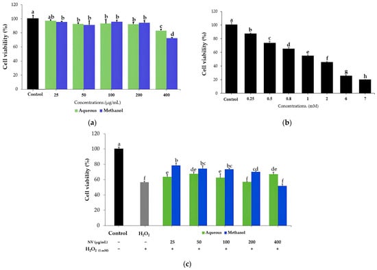
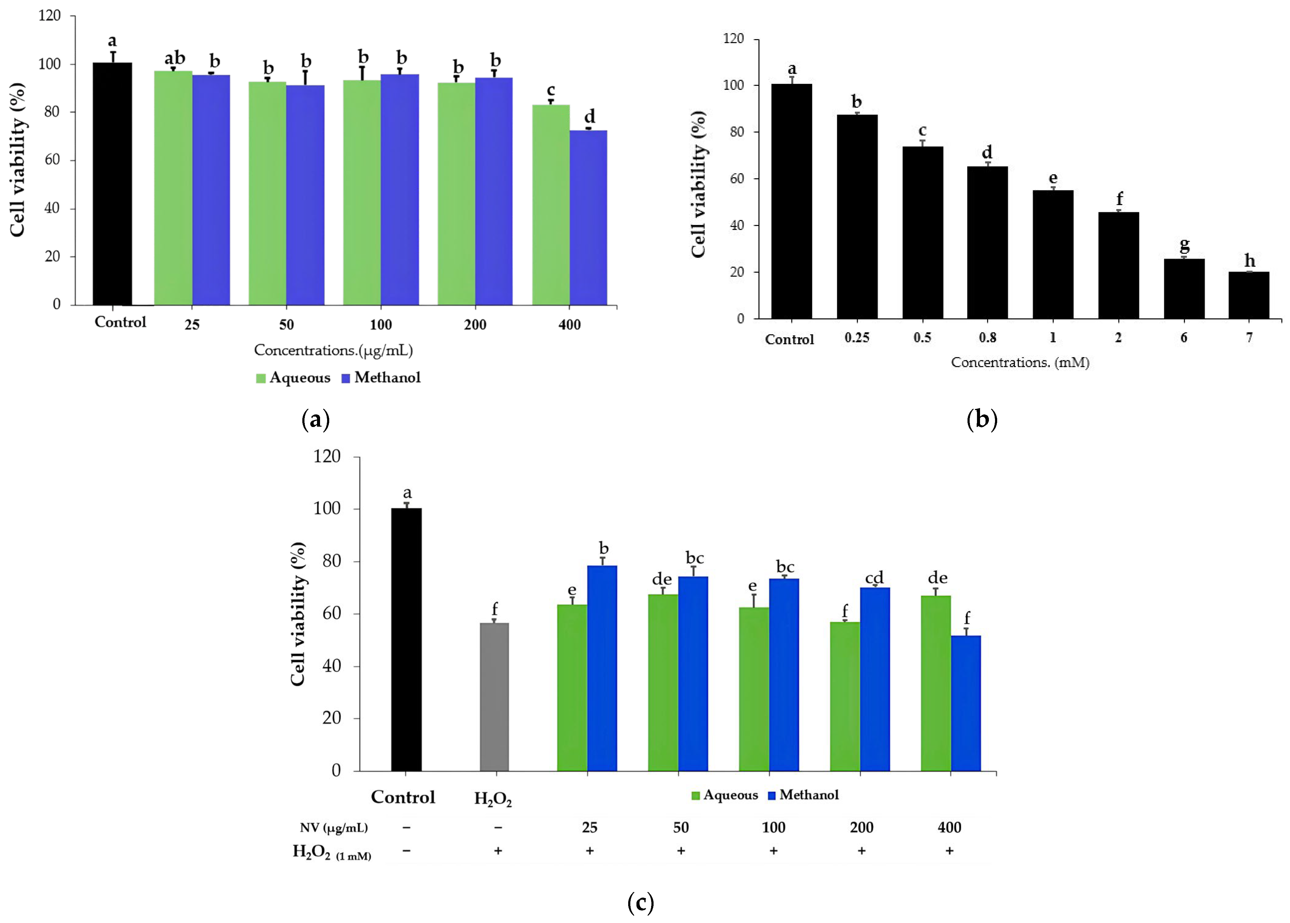
The MTT assay showed that both aqueous and methanol extracts of NV at 25, 50, 100, 200, and 400 µg/mL resulted in cell viability from 70% to 100%, decreasing with increasing concentration (Figure 1a). The aqueous extract of NV maintained HepG2 cell viability above 70% at all tested concentrations [20]. The methanolic extract also maintained cell viability above 70% at all tested concentrations, although viability decreased to 72.70 ± 0.81% at 400 µg/mL. To evaluate oxidative stress, we randomly divided cells into eight groups: a control group and seven groups treated with varying concentrations of H2O2 (0.25, 0.5, 0.8, 1, 2, 6, and 7 mM) for 24 h (Figure 1b). Since a 1 mM concentration of H2O2 has been shown to reduce cell viability by approximately 50%, this concentration was chosen to induce oxidative stress in the present study [21].
Figure 1.
MTT cell viability assay on HepG2 cells. (a) Effect of NV extract on HepG2 cell viability. (b) Cytotoxicity of H2O2 on HepG2 cells at various concentrations. (c) Cytoprotective effect of NV extracts against H2O2-induced cytotoxicity in HepG2 cells using a pretreatment model. The symbols (+) and (−) indicate the presence and absence, respectively, of the respective treatments (NV extract or H2O2). Data are expressed as mean ± SD (n = 3). Different letters (a–h) above the bars indicate significant differences among treatment groups, calculated by one-way ANOVA followed by Duncan’s multiple range test (p ≤ 0.05).
To investigate the cytoprotective effect of NV extracts, HepG2 cells were pretreated with the extracts at 25 to 400 µg/mL for 24 h before exposure to 1 mM H2O2. The H2O2-treated group exhibited a significant reduction in cell viability to 57.06 ± 1.08% compared with the control group (p < 0.05). However, pretreatment with the aqueous extract at concentrations ranging from 25 to 400 µg/mL did not significantly differ from the H2O2-treated group. In contrast, pretreatment with the methanol extract at 25 µg/mL significantly increased cell viability to 78.61 ± 2.80% compared with the H2O2-treated group (p < 0.05). These findings indicate that under the experimental conditions used, NV methanol extract at 25 µg/mL exhibited a protective effect against H2O2-induced cytotoxicity in HepG2 cells (Figure 1c).
2.2. Reactive Oxygen Species Detection
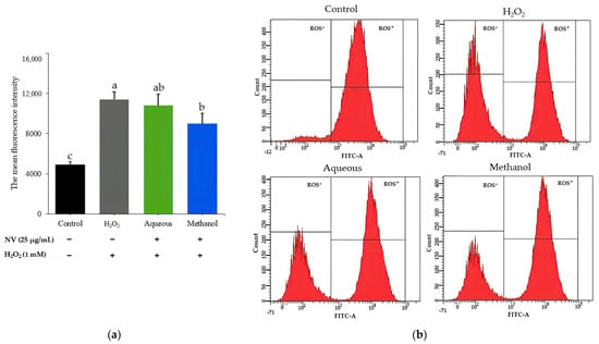
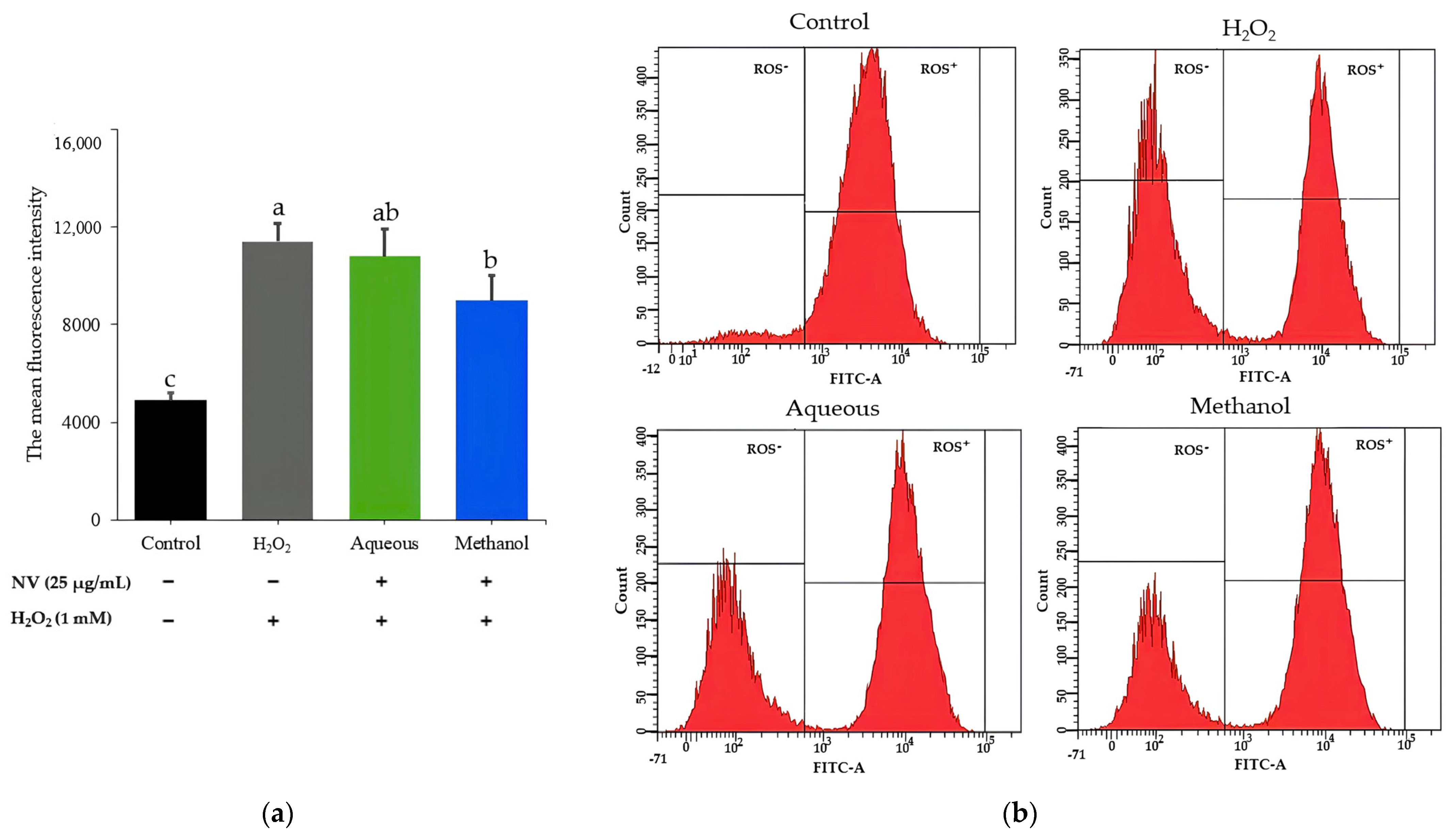
To evaluate the protective effect of NV extracts (25 µg/mL) on H2O2-induced changes in intracellular DCF fluorescence, flow cytometry was performed using the fluorescent probe 2′,7′-dichlorofluorescein diacetate (DCFH-DA), which is oxidized intracellularly to the fluorescent compound 2′,7′-dichlorofluorescein (DCF) [22]. Untreated control HepG2 cells exhibited a baseline level of DCF fluorescence, with a mean fluorescence intensity (MFI) of approximately 4909. Upon exposure to H2O2, the MFI significantly increased to 11,409.33 (Figure 2a), indicating an increase in DCF fluorescence intensity. This finding is consistent with previous research reporting increased DCF fluorescence intensity following H2O2 exposure in HepG2 cells, which was interpreted as an indication of elevated intracellular ROS levels [23].
Figure 2.
Effects of NV extracts on intracellular ROS levels in H2O2-induced oxidative stress in HepG2 cells. (a) MFI quantified via flow cytometry using DCFH-DA. (b) Representative flow cytometry histograms showing the distribution of intracellular ROS levels based on FITC fluorescence intensity. Data are expressed as mean ± SD (n = 3). Different letters (a–c) above the bars indicate significant differences among treatment groups, as determined by one-way ANOVA followed by Duncan’s multiple range test (p ≤ 0.05).
Pretreatment with the methanol extract of NV significantly attenuated H2O2-induced intracellular DCF fluorescence, reducing the MFI to 8970.00 (Figure 2a). In contrast, the aqueous extract produced only a slight, non-significant reduction in DCF fluorescence compared with the H2O2-treated group (MFI = 10,783.00). Furthermore, flow cytometric histograms showed the distribution of ROS-negative (ROS−) and ROS-positive (ROS+) cell populations classified according to fluorescein isothiocyanate (FITC) fluorescence intensity, which reflects DCF fluorescence. Compared with the H2O2-treated group, pretreatment with both NV extracts was associated with a descriptive decrease in the proportion of ROS− cells (Figure 2b). However, these changes in cell population proportions were descriptive observations from the histograms and were not independently subjected to statistical analysis.
2.3. Inhibitory Effect of NV Against Lipopolysaccharide (LPS)-Induced NO Production
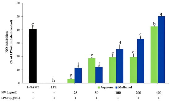
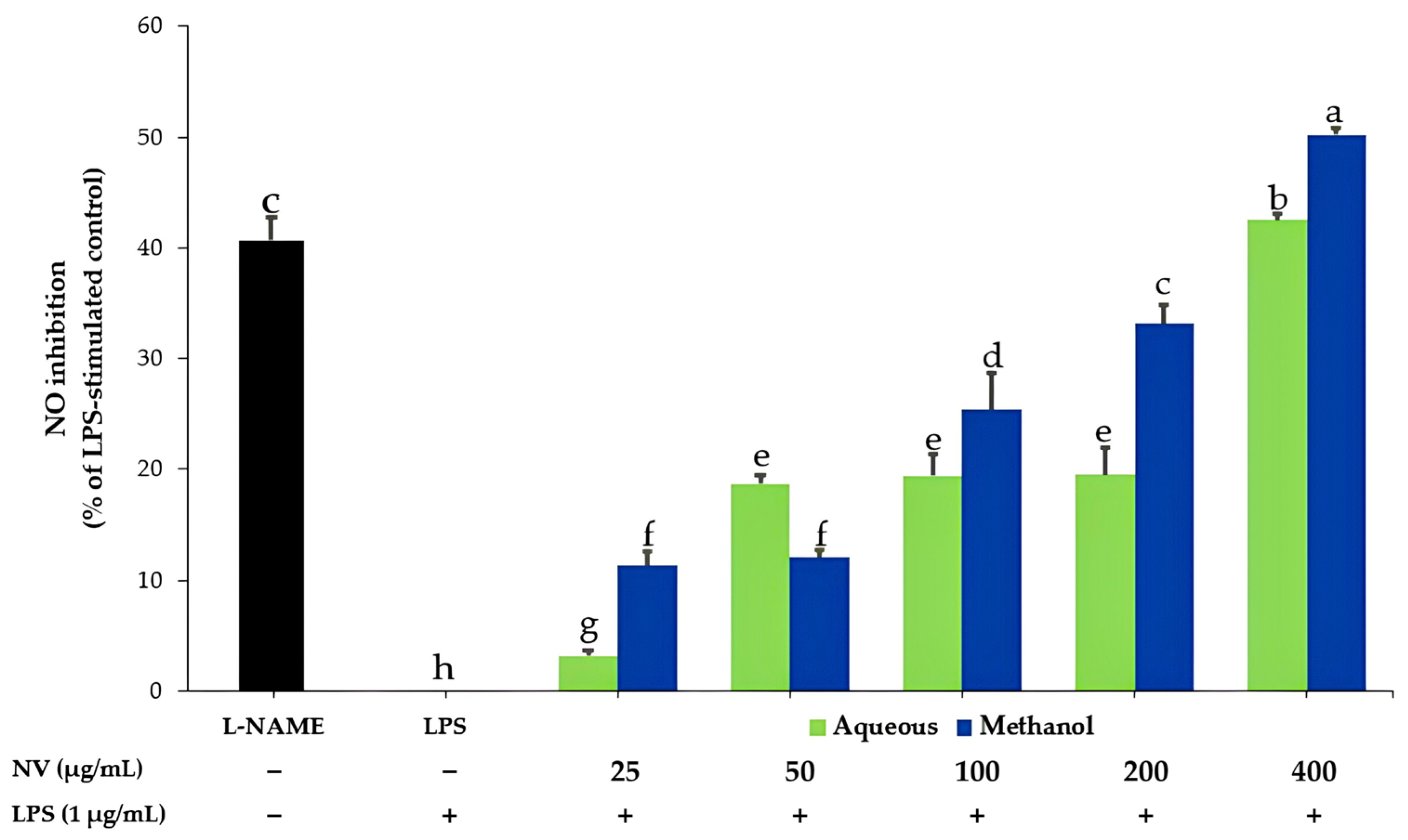
We evaluated the effects of aqueous and methanol extracts of NV on the cell viability of RAW 264.7 murine macrophages. Following a 24 h exposure to concentrations ranging from 25 to 400 µg/mL, both extracts maintained cell viability above 90% (Figure S1), indicating no apparent cytotoxicity under the tested conditions. Furthermore, the inhibitory effects of these extracts on LPS-induced NO production were investigated [24]. Cells treated with 1 µg/mL LPS alone showed no NO inhibition, as this group served as the reference for calculating the percentage inhibition, whereas the positive control group treated with Nω-Nitro-L-arginine methyl ester (L-NAME; 200 µM) showed 34.97 ± 1.78% inhibition (Figure 3). Co-treatment with either the aqueous or methanol extract resulted in a concentration-dependent reduction in LPS-induced NO production. Both NV extracts at all tested concentrations (25–400 µg/mL) significantly reduced NO production compared with the LPS-treated group (p < 0.05). At the highest concentration tested (400 µg/mL), the methanol extract exhibited 50.18 ± 0.60% inhibition, which was significantly higher than that of the aqueous extract (42.49 ± 0.52%; p < 0.05). Both extracts also showed significantly higher than those of the L-NAME-treated group (34.97 ± 1.78%; p < 0.05). These findings indicate that both aqueous and methanol extracts of NV inhibited LPS-induced NO production in RAW 264.7 macrophages under the tested conditions [25].
Figure 3.
Effects of NV on NO production in LPS-stimulated RAW264.7 cells. The symbols (+) and (−) indicate the presence and absence, respectively, of LPS (1 μg/mL). A (−) in the NV row indicates that no NV extract was added. Data are expressed as mean ± SD (n = 3). Different letters (a–h) above the bars indicate significant differences among treatment groups calculated via one-way ANOVA followed by Duncan’s multiple range test (p ≤ 0.05).
2.4. Phytochemical Profiling of NV via Gas Chromatography–Mass Spectrometry (GC-MS)
The detected compounds were tentatively identified by comparing their mass spectra with reference spectra in the National Institute of Standards and Technology (NIST) mass spectral library. A similarity index (SI) ≥ 90% was used as a selection criterion to prioritize compounds showing a high degree of spectral similarity to the reference spectra [26,27]. Compounds meeting this criterion were tentatively based on their spectral similarity to the reference spectra. Relative peak areas (%) were used as semi-quantitative indicators of chromatographic abundance rather than actual compound concentrations.
Three compounds were detected in the aqueous extract (Figure S2), and only hydroxyacetone met the SI ≥ 90% criterion. The remaining two compounds had SI values below 90% and were not considered for further analysis. Hydroxyacetone exhibited a relative peak area of 52.02% (Figure S3). For the methanolic extract, a total of 50 compounds were detected (Figure S4). Of these, 10 compounds met the SI ≥ 90% criterion, whereas the remaining 40 compounds had SI values below 90% and were not considered for further analysis. The 10 selected compounds collectively accounted for 50.69% of the total chromatographic peak area (Figure S5).
Overall, the compounds meeting the SI ≥ 90% criterion accounted for approximately half of the total chromatographic peak area in both extracts. These compounds were selected for further analysis based on their high spectral similarity to reference spectra and their relative peak areas provide a preliminary profile of the detected constituents. Table 1 summarizes the retention times (RTs), compound names, molecular formulas, and relative peak areas (%) of the 11 selected compounds. Thus, the GC-MS results provide a preliminary profile of the compounds detected in the NV extracts under the analytical conditions used in this study.
Table 1.
Tentatively identified compounds in the aqueous and methanolic extracts of NV by GC-MS analysis.
2.5. Drug-Likeness and Pharmacokinetic Prediction
The drug-likeness-related properties of the 11 tentatively identified phytochemical compounds were evaluated using Lipinski’s Rule of Five (Ro5), as implemented in the SwissADME web tool [28]. Lipinski’s Ro5 is an empirical guideline commonly used to assess physicochemical properties associated with oral drug-likeness. The criteria include a molecular weight (MW) ≤ 500 Da, an octanol–water partition coefficient (log P) ≤ 5, hydrogen bond donors (HBDs) ≤ 5, and hydrogen bond acceptors (HBAs) ≤ 10. The compounds were evaluated based on the number of violations of these criteria, with no more than one violation considered consistent with Lipinski’s Ro5 [28]. All 11 compounds were therefore classified as Lipinski-compliant, with six compounds showing no violations and Methyl palmitate, methyl linoleate, methyl petroselinate, bornyl-laurate, and γ-sitosterol each showing one violation. Overall, the selected compounds exhibited physicochemical properties generally compatible with drug-likeness according to the Lipinski Ro5 framework. The results are summarized in Table 2.
Table 2.
Lipinski’s Rule of Five analysis of the compounds identified in NV.
2.6. Molecular Docking Study
After assessing drug-likeness using Ro5, we selected the 11 compounds for molecular docking simulations to investigate potential molecular interactions relevant to hepatoprotection [29]. We evaluated the molecular interactions against key proteins associated with oxidative stress and inflammation, including Keap1 (PDB ID: 4L7B) [30,31] and iNOS (PDB ID: 1QW5) [32]. Trolox and aspirin were used as reference compounds for the oxidative stress and inflammation-related targets, respectively.
For Keap1, γ-sitosterol exhibited the strongest predicted binding affinity, with the most negative binding energy (−6.23 kcal/mol), followed by β-eudesmol (−5.80 kcal/mol) and macelignan (−5.32 kcal/mol). For iNOS, 10 of the 11 tested compounds exhibited more negative predicted binding energy values than the aspirin reference compound (−4.78 kcal/mol), indicating more favorable predicted binding interactions. However, these scores alone do not establish greater biological activity than aspirin. Among the tested compounds, γ-sitosterol showed the strongest predicted binding affinity (−9.89 kcal/mol), followed by macelignan (−8.90 kcal/mol) and β-eudesmol (−7.58 kcal/mol) (Table 3).
Table 3.
Molecular docking of the identified compounds from NV with Keap1 and iNOS.
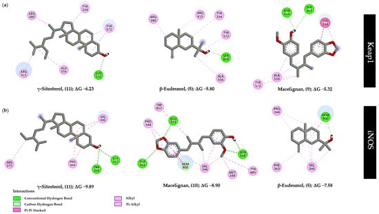
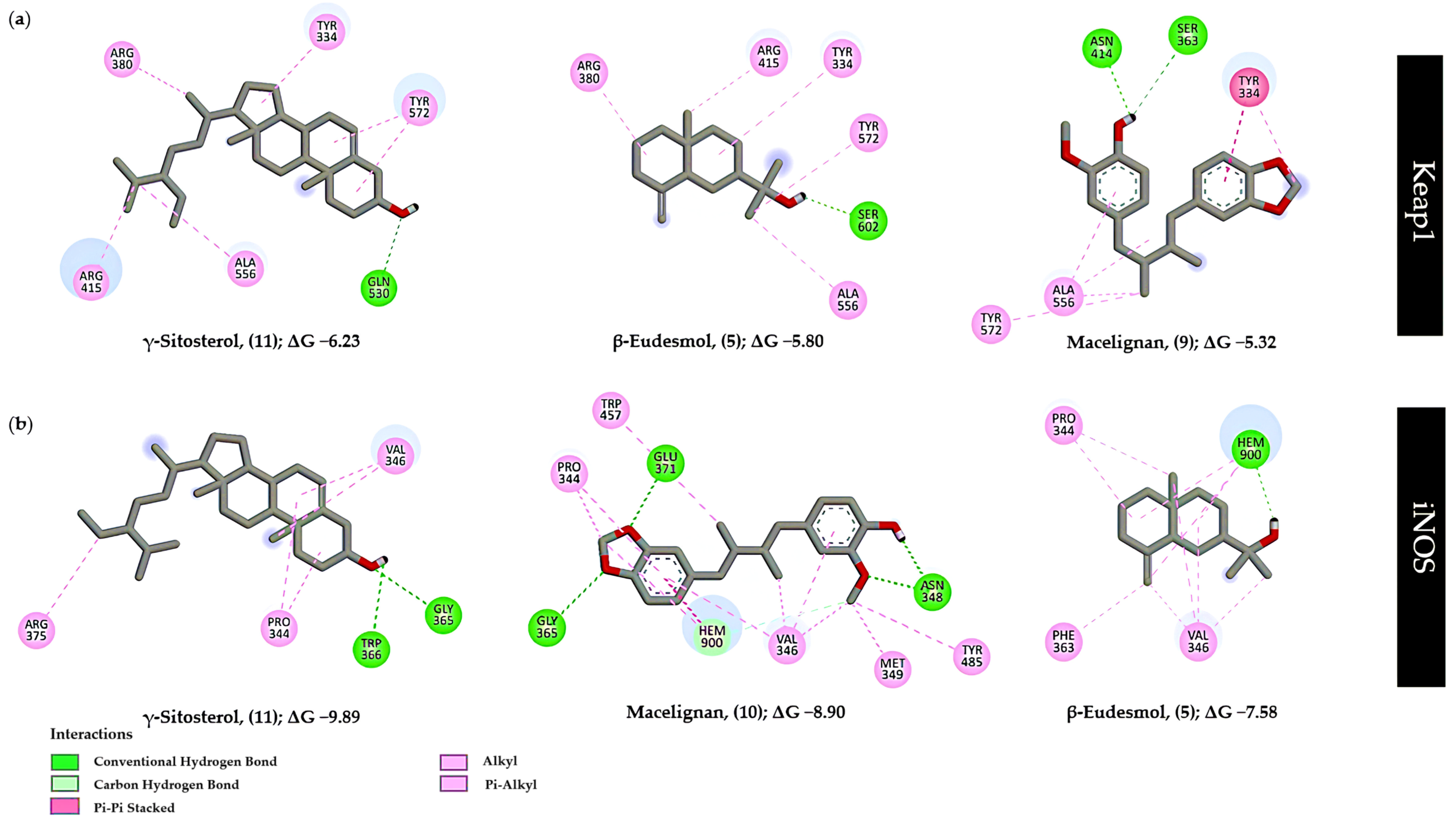
Within the Keap1 binding pocket, γ-sitosterol formed a hydrogen bond with GLN530 and exhibited hydrophobic interactions with several residues, including TYR334, ARG380, ARG415, ALA556, and TYR572. Similarly, β-eudesmol formed a hydrogen bond with SER602 and exhibited hydrophobic interactions with TYR334, ARG380, ARG415, ALA556, and TYR572. Both γ-sitosterol and β-eudesmol interacted with the ARG415 residue, which has been reported as a key residue within the Keap1 binding pocket [31]. Macelignan formed two hydrogen bonds with SER363 and ASN414 and exhibited hydrophobic interactions with TYR334, ALA556, and TYR572 (Figure 4 and Table S2).
Figure 4.
Two-dimensional structural interactions between the top 3 compounds with (a) Keap1 and (b) iNOS.
For the inflammation-related target iNOS, TRP366 and GLU371 have been reported as key residues within the iNOS binding site [32]. The docking analysis revealed that γ-sitosterol exhibited the strongest predicted binding affinity among the compounds evaluated in this study, with a binding energy of −9.89 kcal/mol. It formed two hydrogen bonds with TRP366 and GLY365, together with hydrophobic interactions involving PRO344, VAL364, and ARG375. Macelignan formed three hydrogen bonds with ASN348, GLY365, and GLU371, along with hydrophobic interactions with PRO344, VAL346, MET349, TRP457, and TRP485, as well as the heme cofactor (HEM900). β-eudesmol formed a hydrogen-bond interaction with the heme cofactor (HEM900) and exhibited hydrophobic interactions with PRO344, VAL346, and PHE363 (Figure 4 and Table S3).
2.7. Predicted ADMET Properties and Pharmacokinetic Profiles of the Identified Compounds
To further support pharmacokinetic and safety characterization of the 11 tentatively identified compounds selected for molecular docking, we evaluated their predicted absorption, distribution, metabolism, excretion, and toxicity (ADMET) profiles using the pkCSM online tool. All 11 compounds showed high predicted intestinal absorption, ranging from 92.99% to 100%. All compounds were predicted to be P-glycoprotein substrates, while P-glycoprotein inhibition was predicted for macelignan, bornyl-laurate, and γ-sitosterol. Regarding metabolism-related properties, none of the compounds were predicted to be a CYP2D6 substrate, whereas CYP3A4 substrate activity was predicted for macelignan and γ-sitosterol. Several compounds were predicted to inhibit CYP1A2, CYP2C19, or CYP2C9, while no CYP2D6 or CYP3A4 inhibition was predicted. For toxicity-related endpoints, none of the selected compounds were predicted to be AMES positive or hepatotoxic. No hERG I inhibition was predicted; however, hERG II inhibition was predicted for methyl petroselinate, macelignan, bornyl-laurate, and γ-sitosterol. Collectively, these in silico predictions provide additional information on the pharmacokinetic and toxicity-related profiles of the selected compounds and complement this study’s biological and molecular docking findings. However, these predicted properties are preliminary and require experimental validation (Table 4).
Table 4.
In silico-predicted pharmacokinetic properties (ADMET) of the identified compounds from NV.
3. Discussion
Liver disease is driven by various risk factors, including alcohol consumption, metabolic disorders, drug-induced liver injury, and infections with hepatitis B virus (HBV) and hepatitis C virus (HCV) [1,2]. Oxidative stress plays an important role in both acute liver injury and the pathogenesis of chronic liver disease, with ROS contributing to the progression of liver damage [33]. Specifically, H2O2 can generate highly reactive hydroxyl radicals (•OH) through the Fenton reaction, thereby inducing oxidative damage and cytotoxicity [34]. Therefore, this study employed a pretreatment approach in an H2O2-induced oxidative stress model to investigate the effects of NV extracts on cell viability and intracellular DCF fluorescence, which serves as an indicator of ROS-related oxidative activity, in HepG2 cells [23]. At a concentration of 25 µg/mL, the methanolic extract significantly reduced ROS levels compared with the H2O2-induced control and the aqueous extract. This finding is consistent with previous research indicating that the choice of extraction solvent can influence the chemical composition and biological activity of herbal extracts. In particular, a previous study of PN, one of the herbal ingredients of NV, reported that a hydroethanolic extract provided greater protection against H2O2-induced oxidative stress than an aqueous extract [35]. Although the previous study examined an individual herbal ingredient rather than the complete NV formulation, its findings are consistent with the solvent-dependent differences in ROS-related effects observed between the methanolic and aqueous NV extracts in the present study.
Additionally, oxidative stress and inflammation are closely interconnected processes involved in the pathogenesis of various liver diseases [36]. Growing evidence indicates that these mechanisms often co-occur and can mutually exacerbate liver injury across diverse liver pathologies, while persistent oxidative stress may contribute to chronic inflammation [37]. This inflammatory response involves enzymes such as inducible iNOS, which is responsible for the production of large amounts of NO during inflammatory activation [38]. Upon immune stimulation, iNOS produces high levels of NO, which can react with O2•− to form ONOO−, a highly reactive nitrogen species that can induce cellular oxidative and nitrosative damage [39]. Excessive NO and ONOO− formation has been associated with mitochondrial dysfunction, cell death, and activation of hepatic stellate cells, processes that may contribute to the progression of chronic liver injury and fibrosis [40].
To investigate the anti-inflammatory potential of the NV formulation by inhibiting NO production, we used RAW 264.7 murine macrophages as an in vitro model of inflammatory activation because macrophages can produce NO through iNOS induction following stimulation with LPS. In the present study, both the aqueous and methanolic extracts of the NV formulation significantly inhibited NO production in LPS-stimulated RAW 264.7 macrophages in a concentration-dependent manner over the range of 25–400 µg/mL. The concentration-dependent inhibition of NO production observed with both NV extracts is consistent with previous studies of herbal preparations using LPS-stimulated cells and the Griess assay [41,42]. These studies provide relevant comparisons in terms of the experimental model and NO measurement approach. The observed concentration-dependent inhibition cannot be attributed to any single compound and may instead reflect the combined contributions of multiple constituents in the formulation [43].
Natural products remain an important source for drug discovery and development. Phytochemical groups derived from plants, including flavonoids, phenolic compounds, terpenoids, polysaccharides, and alkaloids, have been widely reported to exhibit diverse pharmacological activities, particularly antioxidant and anti-inflammatory effects, which may help attenuate cellular damage and protect the liver [8,9]. In the present study, the aqueous extract was evaluated in the biological assays to approximate the traditional preparation and consumption of the TTM formulation, which commonly involves preparing powdered formulations with warm water. The methanolic extract was additionally evaluated to provide broader coverage of chemical constituents with different solubility characteristics that may not be fully recovered by aqueous extraction.
To characterize the chemical composition of the extracts used in the biological assays, both the aqueous and methanolic extracts of the NV formulation were analyzed by GC-MS. This approach provided complementary chemical information on GC-MS-detectable constituents in the two extracts [44]. GC-MS has been widely applied for the analysis and chemical profiling of herbal extracts and polyherbal formulations, particularly those containing multiple aromatic medicinal plants, with compound assignments commonly based on comparison of mass spectra with reference spectra in spectral libraries such as the NIST database and relative peak areas used to describe chromatographic profiles [45]. In the present study, compounds showing an SI ≥ 90% against the NIST mass spectral library were selected to prioritize constituents with a high degree of spectral similarity to the reference spectra [46]. Accordingly, 11 tentatively identified compounds were selected for further interpretation: D-camphor, isoborneol, and borneol, classified as monoterpenoids; β-eudesmol, an oxygenated sesquiterpenoid; bornyl-laurate, a monoterpenoid ester; methyl palmitate, methyl linoleate, and methyl petroselinate, classified as fatty acid methyl esters; macelignan, a lignan; γ-sitosterol, a phytosterol; and hydroxyacetone, a small aliphatic ketone. The compounds identified in this study were limited to those detectable under the GC-MS conditions used. Relative peak areas were used to describe the chromatographic profiles rather than to quantify individual compounds.
Terpenoids have been reported to exhibit antioxidant activity through direct radical-scavenging mechanisms and interactions with endogenous antioxidant systems [46]. They also possess anti-inflammatory properties [47] and have shown potential hepatoprotective effects in non-alcoholic fatty liver disease (NAFLD), involving mechanisms related to the regulation of oxidative stress and inflammation [48]. In addition, terpenoid and phenylpropanoid lignan constituents have been reported to protect QSG-7701 cells against D-galactosamine (D-GalN)-induced injury [49]. Fatty acids and phytosterols have also been reported to exhibit antioxidant and anti-inflammatory activities [50]. Taken together, the chemical profiles obtained in the present study, together with the in vitro biological findings, suggest that the diverse chemical constituents detected in the NV formulation may contribute to its observed biological activities, particularly those related to the regulation of oxidative stress and inflammatory responses. However, because the compounds were tentatively identified based on mass spectral library matching, the present data cannot establish the contribution of individual constituents to the observed activities. These findings may reflect the potential contribution of multiple chemical constituents within the multi-component formulation, consistent with the holistic nature of traditional herbal formulations used in the management of liver-related conditions [51].
To explore the potential interactions of NV phytochemical constituents with key proteins associated with oxidative stress and inflammation, Keap1 and iNOS were selected as representative molecular targets for molecular docking analysis. Keap1 was selected because it is a key negative regulator and redox sensor in the Keap1-Nrf2 pathway, an important cellular defense system against oxidative and electrophilic stress. Modulation of the Keap1-Nrf2 axis has been implicated in regulating antioxidant and cytoprotective responses and in the attenuation of liver injury [52,53,54]. In parallel, we selected iNOS because excessive iNOS-derived NO production has been associated with inflammatory and oxidative processes in several liver injury settings. However, the biological effects of NO in the liver are context-dependent and either protective or detrimental, depending on the cellular and pathological conditions [55].
In this study, Trolox and aspirin served as reference compounds for the Keap1 and iNOS docking analyses, respectively. Trolox was selected as a reference antioxidant compound for Keap1 [56], whereas aspirin was selected as a reference anti-inflammatory compound for iNOS [57,58]. Molecular docking analysis indicated that γ-sitosterol, β-eudesmol, and macelignan exhibited relatively favorable predicted binding energies toward the selected targets compared with the other compounds evaluated. These findings represent predicted ligand–target interactions and should not be interpreted as direct evidence of target inhibition. Among the tested compounds, γ-sitosterol showed favorable predicted binding energies toward both Keap1 (−6.23 kcal/mol) and iNOS (−9.89 kcal/mol). For Keap1, the hydroxyl group of γ-sitosterol was predicted to form a hydrogen bond with GLN530. For iNOS, the hydroxyl group was predicted to form a hydrogen bond with GLY365 and TRP366. These findings suggest that γ-sitosterol may interact with the binding regions of both targets; however, the predicted interactions do not establish direct inhibition of Keap1 or iNOS.
Supporting biological evidence for γ-sitosterol comes from a study in which isolated γ-sitosterol from Woodfordia floribunda showed anti-inflammatory activity in a carrageenan-induced paw edema model in rats [59]. Nevertheless, that study does not establish the specific molecular mechanism underlying this activity. Therefore, the present docking results provide complementary in silico evidence for a potential interaction between γ-sitosterol and iNOS. Experimental validation is required to determine whether this interaction contributes to the anti-inflammatory activity of NV. β-eudesmol, an oxygenated sesquiterpenoid reported in Atractylodes lancea (Thunb.) DC [60,61], which is one of the herbal components of NV, exhibited a predicted binding energy of −5.80 kcal/mol toward Keap1. Its hydroxyl group was predicted to form a hydrogen bond with SER602, with an additional predicted interaction involving ARG415. For iNOS, β-eudesmol showed a predicted binding energy of −7.58 kcal/mol and was predicted to interact with the heme group (HEM900) through hydrogen bonding. These results indicate potential interactions between β-eudesmol and the selected targets; however, docking data alone cannot establish antioxidant or anti-inflammatory activity mediated through Keap1 or iNOS. Similarly, macelignan, a lignan reported in Myristica fragrans Houtt. (nutmeg) [62], which is another herbal component of NV, showed a favorable predicted binding energy of −8.90 kcal/mol toward iNOS. The predicted interactions involved GLY365 and GLU371 through its methylenedioxy group and ASN348 through its hydroxyl and methoxy groups. For Keap1, macelignan ranked among the three compounds with the most favorable predicted binding energies, with a value of −5.32 kcal/mol. It was predicted to form hydrogen bonds with SER363 and ASN414.
Collectively, these docking results suggest that γ-sitosterol, β-eudesmol, and macelignan may interact with the selected Keap1 and iNOS target sites. Although some compounds exhibited more favorable predicted binding energy values than the reference compounds, this does not establish greater inhibitory activity. Because molecular docking is predictive, these findings should be considered hypothesis-generating. Further experimental validation is required before attributing specific target-mediated mechanisms to these compounds or linking their predicted interactions to the biological activities of NV extracts.
Taken together, the in vitro and molecular docking findings showed that the methanol extract of NV generally exhibited more pronounced biological effects than the aqueous extract under the conditions tested. In addition, several tentatively identified phytochemicals in NV exhibited relatively favorable predicted binding energies toward the selected molecular targets. The in vitro findings provide preliminary evidence of the cytoprotective and antioxidant-related potential of NV extracts, along with their inhibitory effects on NO production in LPS-stimulated RAW 264.7 macrophages. These findings warrant further investigation using appropriate experimental models of liver injury to determine whether the observed biological activities translate into measurable effects in more complex liver injury models. However, the present findings do not establish direct target-mediated mechanisms or demonstrate that specific phytochemicals are responsible for the observed biological effects. The main limitations of this study include the use of in vitro models and the predictive nature of molecular docking. In addition, the GC-MS results provide tentative compound identifications, and further studies are needed to clarify the contributions of individual constituents to the observed biological effects. Although liver injury involves complex interactions among multiple inflammatory mediators and signaling pathways, including tumor necrosis factor-alpha (TNF-α), interleukin-6 (IL-6), and transforming growth factor-beta (TGF-β), the present study focused on Keap1 and iNOS as representative molecular targets related to oxidative stress and inflammatory NO production, respectively. Further studies are warranted to investigate these pathways at the molecular level, particularly the Keap1/Nrf2 axis and iNOS-related signaling. Such studies should assess target- and pathway-related gene and protein expression using techniques such as RT-qPCR and Western blotting, along with evaluation of Nrf2 nuclear translocation. These approaches would help determine whether the observed biological effects are associated with modulation of the proposed molecular pathways.
In addition, in vivo studies are warranted to investigate the biological activities of NV in appropriate models of liver injury under physiological and pathological conditions. Such studies should include assessment of liver function biomarkers, such as alanine aminotransferase (ALT), aspartate aminotransferase (AST), and alkaline phosphatase (ALP), together with markers of oxidative stress and inflammation and histopathological examination of liver tissue. Further evaluation of NV safety, toxicity, and stability will also be important. Ultimately, well-designed clinical studies will be required to determine its efficacy, safety profile, and potential for further development.
4. Materials and Methods
4.1. Materials and Extraction Procedures
Thai traditional medicine formulation NV was obtained from E-sae Pharmacy Drug Store, Mueang Khon Kaen District, Khon Kaen Province, Thailand, in March 2024. The aqueous extraction procedure was adapted, with modifications, from Chanthasri et al. [63], who used hot-water extraction to mimic the traditional preparation of Thai polyherbal remedies. Briefly, 20 g of powdered NV was boiled in 200 mL of distilled water at 90–100 °C for 30 min per cycle. The extraction was repeated three times using the same powder sample, and the resulting extracts were pooled to obtain a total volume of 600 mL. The pooled extract was filtered under vacuum using a Büchner funnel fitted with Whatman No. 1 filter paper and freeze-dried (lyophilized) using a freeze-dryer (ScanVac CoolSafe 4L, LaboGene ApS, Allerød, Denmark).
For methanolic extraction, an ultrasonic bath was used as an alternative method for phytochemical profiling. The extraction conditions were based on an ultrasound-assisted extraction approach reported for aromatic medicinal plants, which involved ultrasonication for 30 min and three successive extraction cycles [64], with a modified solvent composition. Briefly, 20 g of powdered NV was extracted with 80 mL of methanol in an ultrasonic bath (WiseClean WUC-A22H, Witeg Labortechnik GmbH, Wertheim, Germany) at room temperature for 30 min per cycle. The same powder sample was extracted three times, using 80 mL of fresh methanol per cycle. The extraction vessel was covered to minimize solvent evaporation. The resulting extracts were filtered under vacuum through Whatman No. 1 filter paper and pooled to obtain a total volume of 240 mL. Methanol was removed under reduced pressure using a rotary evaporator (N-1300 series, EYELA, Tokyo Rikakikai Co., Ltd., Tokyo, Japan) at 45 °C, 150–200 mbar, and 70–80 rpm. The aqueous extract was obtained as a freeze-dried powder, whereas the methanolic extract was obtained as a crude extract residue after solvent removal. Both extracts were stored in tightly sealed containers at −20 °C until further analysis. Before further analysis and bioactivity testing, the extracts were reconstituted in appropriate solvents and filtered.
4.2. Chemical Reagents
Dulbecco’s Modified Eagle Medium (DMEM), penicillin–streptomycin, and trypan blue solution were purchased from Gibco (Grand Island, NY, USA). Fetal bovine serum (FBS) and 0.25% trypsin-EDTA (1×) were obtained from Gibco (Paisley, UK). 3-(4,5-Dimethylthiazol-2-yl)-2,5-diphenyltetrazolium bromide (MTT) was supplied by Invitrogen (Eugene, OR, USA). Dimethyl sulfoxide (DMSO) was procured from Sigma-Aldrich (Darmstadt, Germany). Lipopolysaccharide (LPS), 2′,7′-dichlorofluorescein diacetate (DCFH-DA), Nω-Nitro-L-arginine methyl ester (L-NAME), sulfanilamide, and N-(1-naphthyl) ethylenediamine dihydrochloride (NED) were purchased from Sigma-Aldrich (St. Louis, MO, USA). Hydrogen peroxide (30% w/v H2O2) was obtained from Chem-Supply Pty. Ltd. (Gillman, Australia).
4.3. Cell Culture
Human hepatoma (HepG2) cells (HB-8065, Lot No. 61983117) were sourced from the ATCC (Manassas, VA, USA), while murine macrophages (RAW 264.7) were acquired from Cytion (CLS, catalog number 400319; Eppelheim, Germany). HepG2 cells were used to evaluate oxidative stress-related effects in a liver cell model, whereas RAW 264.7 macrophages were used to assess LPS-induced NO production as an indicator of the inflammatory response. The two cell lines were maintained in Dulbecco’s Modified Eagle Medium (DMEM) supplemented with 10% fetal bovine serum (FBS) and 1% penicillin–streptomycin. Cells were cultured at 37 °C in a humidified incubator containing 5% CO2. Both cell lines were obtained from established cell repositories. Cell lines were not tested for mycoplasma contamination or authenticated for this study [65].
4.4. Evaluation of HepG2 Cell Viability and the Protective Effect of NV Against H2O2-Induced Cytotoxicity in HepG2 Cells
Cell viability was evaluated using the MTT assay, which is based on the reduction of MTT to formazan by metabolically active cells [20]. Briefly, HepG2 cells (passage 12) were seeded in 96-well plates at 2 × 10⁵ cells/well and allowed to adhere overnight. The crude NV extracts were dissolved in DMSO and further diluted in culture medium to achieve the desired concentrations (25, 50, 100, 200, and 400 µg/mL), with a final DMSO concentration maintained below 0.1% (v/v) in all treatment wells.
To assess extract cytotoxicity and the effect of H2O2 alone on cell viability, HepG2 cells were treated separately for 24 h with aqueous or methanolic NV extracts (25, 50, 100, 200, and 400 µg/mL) or H2O2 (0.25, 0.5, 0.8, 1, 2, 6, and 7 mM).
To evaluate the protective effects of NV against H2O2-induced cellular damage, HepG2 cells were pretreated with aqueous or methanolic NV extracts at different concentrations (25, 50, 100, 200, and 400 µg/mL) for 24 h, followed by exposure to 1 mM H2O2 for an additional 24 h.
After each treatment, the medium was carefully aspirated, and 0.5 mg/mL MTT solution was added to each well, followed by incubation for 2 h. The supernatant was then removed, and 100 µL of DMSO was added to each well to solubilize the formazan crystals. Absorbance was measured at 570 nm using a microplate reader (EnSight™ Multimode Plate Reader; PerkinElmer, Waltham, MA, USA). Background absorbance correction using cell-free blank wells was not performed in this study. All experiments were performed independently in triplicate (n = 3), with three biological replicates within each experiment. Cell viability was calculated as a percentage relative to the untreated control (DMEM supplemented with 10% FBS), which served as the reference for all treatment groups, including those exposed to H2O2, using Equation (1):
4.5. Reactive Oxygen Species (ROS) Detection in HepG2 Cells
Cells were plated in 6-well plates at 2 × 10⁵ cells per well and maintained at 37 °C for 24 h. Subsequently, cells were pretreated with aqueous or methanolic NV extracts (25 µg/mL containing 0.1% v/v DMSO) for an additional 24 h. The cells were then washed once with 1× PBS before exposure to 1 mM H2O2 for 24 h. Following H2O2 exposure, the cells were washed twice with 1× PBS before staining with 25 µM DCFH-DA. The cells were incubated in the dark for 30 min. The concentration of 25 µg/mL was selected based on the protective effects of NV against H2O2-induced cellular damage and used to assess DCFH-DA-derived fluorescence as an indirect indicator of intracellular oxidative status in HepG2 cells. After staining, the cells were harvested, washed with 1× PBS, and transferred to fluorescence-activated cell sorting (FACS) tubes. DCF fluorescence was analyzed via flow cytometry using a BD FACSCanto™ II system (BD Biosciences, San Jose, CA, USA). Unstained cells were used to assess cellular autofluorescence. However, fluorescence values from unstained cells were not subtracted from the experimental MFI values. Data were analyzed using BD FACSDiva™ Version 6.1.3 software. Cell populations were gated based on forward-scatter (FSC-A) and side-scatter (SSC-A) characteristics, and fluorescence intensity was assessed using the FITC-A channel. MFI was used to quantify DCF fluorescence, and MFI values were compared directly among the untreated control, H2O2-treated control, and NV extract-pretreated, H2O2-exposed groups. All experiments were performed independently in triplicate (n = 3), with three biological replicates within each experiment.
4.6. Evaluation of Raw 264.7 Cell Viability
For the cell viability assay, RAW 264.7 cells (passage 16) were seeded in 96-well plates at 2 × 10⁴ cells per well and allowed to adhere overnight. After 24 h of treatment with various concentrations of NV extracts (25–400 µg/mL; final DMSO concentration 0.1% v/v) in the absence of LPS, the culture medium was replaced with 0.5 mg/mL MTT solution and then incubated for an additional 3 h at 37 °C. Thereafter, the MTT-containing medium was carefully aspirated, and the resulting formazan crystals were solubilized by adding 100 µL of DMSO to each well. Absorbance was measured at 570 nm using an EnSight™ Multimode Plate Reader (PerkinElmer, Waltham, MA, USA) Background absorbance correction using cell-free blank wells was not performed in this study All experiments were performed independently in triplicate (n = 3), with three biological replicates within each experiment. Cell viability was calculated as a percentage relative to the untreated control (DMEM supplemented with 10% FBS).
4.7. Measurement of Nitric Oxide (NO) Levels
NO production was assessed indirectly by measuring nitrite accumulation in the culture supernatants using the Griess assay. RAW 264.7 macrophages were seeded at 2 × 10⁴ cells/well in 96-well plates and incubated overnight. The cells were then exposed to 1 µg/mL LPS and various concentrations of NV extracts (25–400 µg/mL) for 24 h, with a final DMSO concentration of 0.1% (v/v). L-NAME (200 µM) was included as a positive control. The culture supernatants were collected and reacted with 80 µL of Griess reagent for 30 min at room temperature, protected from light. Absorbance was measured at 520 nm using a microplate reader (EnSight™ Multimode Plate Reader; PerkinElmer, Waltham, MA, USA). Background absorbance correction was not performed in this study. The percentage of NO inhibition was calculated relative to the LPS-stimulated control group using Equation (2). All experiments were performed independently in triplicate (n = 3), with three biological replicates within each experiment.
4.8. Phytochemical Analysis by Gas Chromatography–Mass Spectrometry (GC-MS)
The aqueous and methanolic extracts of NV were analyzed by GC-MS using a Shimadzu QP-2020 system (Kyoto, Japan) equipped with an electron impact (EI) ionization source and a HP-5MS capillary column (30 m × 0.25 mm i.d., 0.25 µm film thickness; Agilent Technologies, J&W, SantaClara, CA, USA). Helium (purity grade 5.5) was used as the carrier gas at a flow rate of 1 mL/min. Mass spectra were recorded at an ionization energy of 70 eV. Before analysis, the extracts were dissolved in methanol at a concentration of 10 mg/mL, filtered, and directly injected into the GC-MS system. A 1 µL sample was injected in split mode (1:20), with the injector temperature maintained at 290 °C. The oven temperature was programmed to increase from 40 °C to 100 °C at 5 °C/min with a 5 min hold, followed by successive ramps to 150 °C (10 min hold), 200 °C (10 min hold), 250 °C (5 min hold), and finally 320 °C (15 min hold), all at a heating rate of 5 °C/min [66]. Chromatographic peaks were tentatively identified by comparing their mass spectra with those in the NIST17 library database. Neither retention indices nor authentic reference standards were used to confirm compound identities. Compounds with an SI of ≥90% were retained for further analysis.
4.9. Drug-Likeness and Pharmacokinetic Prediction
All compounds tentatively identified by GC-MS were screened using an SI threshold of ≥90%. Compounds with an SI below 90% were excluded from further analyses, whereas 11 compounds met this criterion and were included in the subsequent in silico analyses. The subsequent in silico predictions were performed using the proposed structures of the tentatively identified compounds. Drug-likeness was evaluated according to Ro5 using the SwissADME web server (https://www.swissadme.ch/, accessed on 22 March 2026) [28,67]. Additionally, pharmacokinetic profiles, including absorption, distribution, metabolism, excretion, and toxicity (ADMET), were predicted using the pkCSM web-based tool (http://biosig.unimelb.edu.au/pkcsm/prediction, accessed on 23 January 2026) [68]. The predicted absorption parameters included Caco-2 permeability, intestinal absorption, P-glycoprotein substrate status, and P-glycoprotein I inhibition. Distribution parameters included volume of distribution, fraction unbound, and blood–brain barrier permeability. Metabolism parameters included CYP2D6 and CYP3A4 substrate status, as well as CYP1A2, CYP2C19, CYP2C9, CYP2D6, and CYP3A4 inhibition. Excretion parameters included total clearance and renal OCT2 substrate. Toxicity parameters included AMES toxicity, maximum tolerated dose (human), hERG I and hERG II inhibition, oral rat acute toxicity (LD50), oral rat chronic toxicity (LOAEL), and hepatotoxicity.
4.10. Molecular Docking
To predict the potential binding poses and molecular interactions of NV constituents with anti-inflammatory and antioxidant targets, we performed molecular docking simulations using AutoDock Tools v.1.5.7 (MGLTools, Scripps Research Institute, https://ccsb.scripps.edu/mgltools/, accessed on 27 January 2026) [69]. We obtained the 3D crystal structures of Keap1 (PDB ID: 4L7B) [31] and iNOS (PDB ID: 1QW5) [32,70] from the RCSB PDB. Chain B of Keap1 and chain A of iNOS were used for molecular docking. Before docking, we adjusted protein protonation states to pH 7.4 using the PDB2PQR web server (https://server.poissonboltzmann.org/pdb2pqr, accessed on 30 March 2026) [71]. Protein preparation was then completed using AutoDock Tools. Water molecules and other nonessential heteroatoms were removed from the protein structures, while the heme cofactor (HEM900) was retained in the iNOS structure. Polar hydrogen atoms were added, and Kollman charges were assigned to the protein structures. The 3D structures of the ligands were retrieved from the PubChem database and processed using the Clean Geometry function in BIOVIA Discovery Studio Visualizer 2021. The ligands were subsequently prepared for docking using AutoDock Tools, including the addition of hydrogen atoms, assignment of charges, and conversion to PDBQT format. We centered a grid box on the native ligand-binding site of each target protein. We used the Lamarckian Genetic Algorithm (LGA) to evaluate receptor–ligand interactions. The LGA parameters were set as follows: 100 GA runs, a population size of 100, a maximum of 2,500,000 energy evaluations, and a maximum of 27,000 generations. We validated the docking methodology by redocking the co-crystallized native ligands into their respective receptors, with a root-mean-square deviation (RMSD) threshold of ≤2 Å considered successful [72]. For Keap1, the co-crystallized ligand was 1VV, and the grid box dimensions were set to 26 × 26 × 26 Å with a grid center of X = −3.56, Y = 2.506, and Z = 27.501 Å and a grid spacing of 0.375 Å. The resulting RMSD was 0.899 Å. For iNOS, the co-crystallized ligand was W1400, and the grid box dimensions were set to 30 × 30 × 30 Å with a grid center of X = −53.451, Y = 135.240, and Z = 43.447 Å and a grid spacing of 0.575 Å. The resulting RMSD was 1.148 Å. Redocking was performed using 100 runs. We used Trolox and aspirin as standard reference compounds. Finally, we selected the docking conformations with the lowest binding energy for interaction analysis using BIOVIA Discovery Studio Visualizer 2021.
4.11. Statistical Analysis
Statistical analyses were performed using SPSS (version 25). Data are presented as mean ± standard deviation (SD). Normality was assessed using the Shapiro–Wilk test. Homogeneity of variances was not assessed separately before one-way ANOVA. One-way ANOVA followed by Duncan’s multiple range test was used to compare the groups, with statistical significance set at p < 0.05. All experiments were performed independently in triplicate (n = 3), with three biological replicates within each experiment.
5. Conclusions
In conclusion, this study provides preliminary in vitro findings on the cytoprotective and antioxidant-related effects of the NV formulation, as well as its effects on LPS-induced NO production. The aqueous and methanolic extracts showed no apparent cytotoxicity toward HepG2 cells under the tested conditions, while the methanolic extract at 25 µg/mL significantly reduced intracellular ROS levels in the H2O2-induced oxidative stress model. Both extracts also inhibited LPS-induced NO production in RAW 264.7 macrophages in a concentration-dependent manner. GC-MS analysis tentatively identified 11 compounds, including terpenoids, fatty acid derivatives, a lignan, a phytosterol, and a small aliphatic ketone; however, the contribution of individual constituents to the observed biological activities could not be established from the present data. Molecular docking further suggested potential interactions of γ-sitosterol, β-eudesmol, and macelignan with Keap1 and iNOS based on their predicted binding energies. Overall, the integrated in vitro and in silico findings provide initial insights into the biological effects of NV, including cytoprotective and antioxidant-related effects that are associated with oxidative stress and inflammatory responses. The observed effects may involve contributions from multiple chemical constituents within the formulation; however, the specific roles of individual compounds and their interactions with molecular targets remain to be experimentally clarified. Further studies are warranted to confirm the chemical identities of the detected compounds and to experimentally validate their biological activities and proposed molecular interactions.
Supplementary Materials
The following supporting information can be downloaded at: https://www.mdpi.com/article/10.3390/plants15223571/s1, Figure S1. Cell viability of RAW 264.7 cells treated with NV was measured by the MTT assay. Figure S2. GC-MS chromatogram of compounds identified in aqueous extracts of NV. Figure S3. Mass spectra of compounds identified in aqueous extracts of NV with a similarity index (SI) ≥ 90. Figure S4. GC-MS chromatogram of compounds identified in methanolic extract of NV. Figure S5. Mass spectra of compounds identified in methanolic extract of NV with a similarity index (SI) ≥ 90. Table S1. Herbal constituents and composition of the Yahom Navakot (NV) formulation. Table S2. Molecular docking results and interaction profiles of tentatively identified compounds from NV with Keap1. Table S3. Molecular docking results and interaction profiles of tentatively identified compounds from NV with iNOS.
Author Contributions
Conceptualization, P.P. (Ploenthip Puthongking), T.P. and B.S.; methodology, B.B., P.P. (Ploenthip Puthongking), J.R., B.S. and T.P.; software, B.B., M.S., P.S., S.D. and J.R.; validation, B.B., P.P. (Ploenthip Puthongking), M.S., P.S., S.D. and J.R.; formal analysis, P.P. (Ploenthip Puthongking), B.B., M.S., S.D., C.Y., P.P. (Panyada Panyatip) and J.R.; investigation, P.P. (Ploenthip Puthongking), B.B., M.S., S.D., C.Y. and J.R.; resources, P.P. (Ploenthip Puthongking), B.S. and T.P.; data curation, B.B., C.Y. and P.P. (Ploenthip Puthongking); writing—original draft preparation, B.B., M.S., P.P. (Panyada Panyatip), T.P. and P.P. (Ploenthip Puthongking); writing—review and editing, B.B., P.P. (Ploenthip Puthongking), S.D., P.S., J.R., M.S., C.Y., P.P. (Panyada Panyatip), B.S. and T.P.; visualization, B.B., M.S. and P.S.; supervision, P.P. (Ploenthip Puthongking), T.P. and B.S.; project administration, P.P. (Ploenthip Puthongking) and T.P.; funding acquisition, P.P. (Ploenthip Puthongking) and T.P. All authors have read and agreed to the published version of the manuscript.
Funding
This work was supported by the National Science, Research and Innovation Fund (NSRF) through the Fundamental Funding fiscal year 2024 (FF2567) program at Khon Kaen University, Thailand.
Data Availability Statement
The original contributions presented in this study are included in the article/Supplementary Materials. Further inquiries can be directed to the corresponding author.
Acknowledgments
The first author, Benjamat Bangthong, expresses her gratitude to the Faculty of Pharmaceutical Sciences, Khon Kaen University, for providing a master’s degree scholarship.
Conflicts of Interest
The authors declare no conflicts of interest.
Abbreviations
| ROS | Reactive oxygen species |
| Keap1 | Kelch-like ECH-associated protein 1 |
| Nrf2 | Nuclear factor erythroid 2-related factor 2 |
| LPS | Lipopolysaccharide |
| NO | Nitric oxide |
| iNOS | Inducible nitric oxide synthase |
| H2O2 | Hydrogen peroxide |
| HepG2 | Human liver cancer cell line |
| RAW 264.7 | RAW 264.7 murine macrophage cells |
| Ro5 | Lipinski’s Rule of Five |
| MFI | Mean fluorescence intensity |
| MW | Molecular weight |
| Log Po/w | Octanol–water partition coefficient |
| MLOGP | Moriguchi octanol–water partition coefficient |
| HBD | Hydrogen bond donor |
| HBA | Hydrogen bond acceptor |
References
- Younossi, Z.M.; Koenig, A.B.; Abdelatif, D.; Fazel, Y.; Henry, L.; Wymer, M. Global Epidemiology of Nonalcoholic Fatty Liver Disease—Meta-Analytic Assessment of Prevalence, Incidence, and Outcomes. Hepatology 2016, 64, 73–84. [Google Scholar] [CrossRef] [Scilit] [PubMed]
- Asrani, S.K.; Devarbhavi, H.; Eaton, J.; Kamath, P.S. Burden of Liver Diseases in the World. J. Hepatol. 2019, 70, 151–171. [Google Scholar] [CrossRef] [Scilit] [PubMed]
- Queiroz de Andrade, K.; Moura, F.A.; dos Santos, J.M.; de Araújo, O.R.P.; dos Santos, J.C.F.; Goulart, M.O.F. Oxidative Stress and Inflammation in Hepatic Diseases: Therapeutic Possibilities of N-Acetylcysteine. Int. J. Mol. Sci. 2015, 16, 30269–30308. [Google Scholar] [CrossRef] [Scilit] [PubMed]
- Ramos-Tovar, E.; Muriel, P. Molecular Mechanisms That Link Oxidative Stress, Inflammation, and Fibrosis in the Liver. Antioxidants 2020, 9, 1279. [Google Scholar] [CrossRef] [Scilit] [PubMed]
- Magesh, S.; Chen, Y.; Hu, L. Small Molecule Modulators of Keap1-Nrf2-ARE Pathway as Potential Preventive and Therapeutic Agents. Med. Res. Rev. 2012, 32, 687–726. [Google Scholar] [CrossRef] [Scilit] [PubMed]
- Zhou, J.; Zheng, Q.; Chen, Z. The Nrf2 Pathway in Liver Diseases. Front. Cell Dev. Biol. 2022, 10, 826204. [Google Scholar] [CrossRef] [Scilit] [PubMed]
- Hong, M.; Li, S.; Tan, H.Y.; Wang, N.; Tsao, S.-W.; Feng, Y. Current Status of Herbal Medicines in Chronic Liver Disease Therapy: The Biological Effects, Molecular Targets and Future Prospects. Int. J. Mol. Sci. 2015, 16, 28705–28745. [Google Scholar] [CrossRef] [Scilit] [PubMed]
- Saraswat, I.; Goel, A.; Gupta, J. Herbal Remedies for Hepatic Diseases: A Review of Medicinal Herbs in the Treatment of Liver Disorders. Chin. Herb. Med. 2026, 18, 329–342. [Google Scholar] [CrossRef] [Scilit] [PubMed]
- Niu, Q.; Mou, Y.; Wang, K.; Dong, H.; Zeng, Z.; Li, H. Natural Medicines for Treating Liver Fibrosis by Modulating Post-Translational Modifications. Front. Pharmacol. 2025, 16, 1660908. [Google Scholar] [CrossRef] [Scilit] [PubMed]
- National Drug System Development Committee. National List of Essential Drugs A.D. 2023: Herbal Medicine Products; Herbal Medicinal Products Division, Food and Drug Administration, Ministry of Public Health: Bangkok, Thailand, 2023; Available online: https://www.ratchakitcha.soc.go.th (accessed on 20 January 2026).
- National Drug System Development Committee. National List of Essential Medicines A.D. 2020 (List of Herbal Medicine Products); Special Part 72 D; Royal Thai Government Gazette: Bangkok, Thailand, 2011; Volume 128, 57p, Available online: https://www.ratchakitcha.soc.go.th/DATA/PDF/2554/E/072/57.PDF (accessed on 20 January 2026).
- Tirawanchai, N.; Supapornhemin, S.; Somkasetrin, A.; Suktitipat, B.; Ampawong, S. Regulatory Effect of Phikud Navakot Extract on HMG-CoA Reductase and LDL-R: Potential and Alternate Agents for Lowering Blood Cholesterol. BMC Complement. Altern. Med. 2018, 18, 258. [Google Scholar] [CrossRef] [Scilit] [PubMed]
- Inchan, A.; Pathomwichaiwat, T.; Bualeong, T.; Tipratchadaporn, S.; Chootip, K. Anti-Hypotensive Effect of “Yahom Navakot” in Rats with Orthostatic Hypotension. J. Tradit. Complement. Med. 2022, 12, 180–189. [Google Scholar] [CrossRef] [Scilit] [PubMed]
- Siriyong, T.; Chanphool, H.; Jitjum, S.; Laohaprapanon, S.; Voravuthikunchai, S.P. Therapeutic Efficacy and Safety of Traditional Thai Herbal Medicines for Insomnia: A Double-Blind Randomized Controlled Trial. Adv. Integr. Med. 2025, 12, 100459. [Google Scholar] [CrossRef] [Scilit]
- Chiangsom, A.; Maniratanachote, R.; Meksuriyen, D.; Luechapudiporn, R.; Kulthong, K.; Aueviriyavit, S.; Oda, S.; Yokoi, T.; Lawanprasert, S. Protective Effect of Phikud Navakot Extract Against Hydrogen Peroxide-Induced Oxidative Stress in HepG2 Cells. Thai J. Pharm. Sci. 2019, 43, 186–194. [Google Scholar] [CrossRef] [Scilit]
- Lefebvre, T.; Destandau, E.; Lesellier, E. Selective Extraction of Bioactive Compounds from Plants Using Recent Extraction Techniques: A Review. J. Chromatogr. A 2021, 1635, 461770. [Google Scholar] [CrossRef] [Scilit] [PubMed]
- Chootip, K.; Chaiyakunapruk, N.; Soonthornchareonnon, N.; Scholfield, C.N.; Fuangchan, A. Efficacy and Safety of “Yahom” as a Traditional Thai Herbal Therapy: A Systematic review. J. Ethnopharmacol. 2017, 196, 110–123. [Google Scholar] [CrossRef] [Scilit] [PubMed]
- Pongsaree, R.; Boonjang, N.; Chokchaisiri, S. Gas Chromatography–Mass Spectrometry (GC-MS) Analysis and Antioxidant Activity of a Thai Traditional Ayuwatana Herbal Formulation. Trop. J. Nat. Prod. Res. 2026, 10, 59. [Google Scholar] [CrossRef] [Scilit]
- Łyczko, J.; Kiełtyka-Dadasiewicz, A.; Skrzyński, M.; Klisiewicz, K.; Szumny, A. Chemistry Behind Quality—The Usability of Herbs and Spices Essential Oils Analysis in Light of Sensory Studies. Food Chem. 2023, 411, 135537. [Google Scholar] [CrossRef] [Scilit] [PubMed]
- ISO 10993-5:2009(E); Biological Evaluation of Medical Devices—Part 5: Tests for In Vitro Cytotoxicity. International Organization for Standardization: Geneva, Switzerland, 2009.
- Jiang, J.; Yu, S.; Jiang, Z.; Liang, C.; Yu, W.; Li, J.; Du, X.; Wang, H.; Gao, X.; Wang, X. N-Acetyl-Serotonin Protects HepG2 Cells from Oxidative Stress Injury Induced by Hydrogen Peroxide. Oxid. Med. Cell. Longev. 2014, 2014, 310504. [Google Scholar] [CrossRef] [Scilit] [PubMed]
- de Haan, L.R.; Reiniers, M.J.; Reeskamp, L.F.; Belkouz, A.; Ao, L.; Cheng, S.; Ding, B.; van Golen, R.F.; Heger, M. Experimental Conditions That Influence the Utility of 2′,7′-Dichlorodihydrofluorescein Diacetate (DCFH2-DA) as a Fluorogenic Biosensor for Mitochondrial Redox Status. Antioxidants 2022, 11, 1424. [Google Scholar] [CrossRef] [Scilit] [PubMed]
- Siddiqui, M.A.; Ali, Z.; Chittiboyina, A.G.; Khan, I.A. Hepatoprotective Effect of Steroidal Glycosides from Dioscorea villosa on Hydrogen Peroxide-Induced Hepatotoxicity in HepG2 Cells. Front. Pharmacol. 2018, 9, 797. [Google Scholar] [CrossRef] [Scilit] [PubMed]
- Stuehr, D.J.; Marletta, M.A. Mammalian Nitrate Biosynthesis: Mouse Macrophages Produce Nitrite and Nitrate in Response to Escherichia coli Lipopolysaccharide. Proc. Natl. Acad. Sci. USA 1985, 82, 7738–7742. [Google Scholar] [CrossRef] [Scilit] [PubMed]
- Joo, T.; Sowndhararajan, K.; Hong, S.; Lee, J.; Park, S.-Y.; Kim, S.; Jhoo, J.-W. Inhibition of Nitric Oxide Production in LPS-Stimulated RAW 264.7 Cells by Stem Bark of Ulmus pumila L. Saudi J. Biol. Sci. 2014, 21, 427–435. [Google Scholar] [CrossRef] [Scilit] [PubMed]
- Nipun, T.S.; Khatib, A.; Ibrahim, Z.; Ahmed, Q.U.; Redzwan, I.E.; Primaharinastiti, R.; Saiman, M.Z.; Fairuza, R.; Widyaningsih, T.D.; AlAjmi, M.F.; et al. GC-MS- and NMR-Based Metabolomics and Molecular Docking Reveal the Potential Alpha-Glucosidase Inhibitors from Psychotria malayana Jack Leaves. Pharmaceuticals 2021, 14, 978. [Google Scholar] [CrossRef] [Scilit] [PubMed]
- González Campos, R.E.; Bautista Rodríguez, E.; Ale, C.E.; Garcés Eisele, S.J.; Hernández, L.R.; Juárez, Z.N. Phytochemical Characterization and Toxicity Activity of Tradescantia pendula Extracts. Molecules 2026, 31, 3000. [Google Scholar] [CrossRef] [Scilit] [PubMed]
- Daina, A.; Michielin, O.; Zoete, V. SwissADME: A Free Web Tool to Evaluate Pharmacokinetics, Drug-Likeness and Medicinal Chemistry Friendliness of Small Molecules. Sci. Rep. 2017, 7, 42717. [Google Scholar] [CrossRef] [Scilit] [PubMed]
- Shidiki, A.; Vyas, A. Molecular Docking and Pharmacokinetic Prediction of Phytochemicals from Syzygium cumini in Interaction with Penicillin-Binding Protein 2a and Erythromycin Ribosomal Methylase of Staphylococcus aureus. BioTechnologia 2022, 103, 5–18. [Google Scholar] [CrossRef] [Scilit] [PubMed]
- Zhou, J.; Chen, J.-Q.; Gong, S.; Ban, Y.-J.; Zhang, L.; Liu, Y.; Wu, J.-L.; Li, N. Isolation, Bioactivity, and Molecular Docking of a Rare Gastrodin Isocitrate and Diverse Parishin Derivatives from Gastrodia elata Blume. ACS Omega 2024, 9, 14520–14529. [Google Scholar] [CrossRef] [Scilit] [PubMed]
- Londhe, A.M.; Gadhe, C.G.; Lim, S.M.; Pae, A.N. Investigation of Molecular Details of Keap1-Nrf2 Inhibitors Using Molecular Dynamics and Umbrella Sampling Techniques. Molecules 2019, 24, 4085. [Google Scholar] [CrossRef] [Scilit] [PubMed]
- Macchiarini, C.; Montagnani, M.; Pucciotti, R.; Ammazzalorso, A.; De Filippis, B.; Di Matteo, M.; Di Silvestre, S.; Fantacuzzi, M.; Giampietro, L.; Potenza, M.A. Selective Acetamidine-Based Nitric Oxide Synthase Inhibitors: Synthesis, Docking, and Biological Studies. ACS Med. Chem. Lett. 2015, 6, 635–640. [Google Scholar] [CrossRef] [Scilit] [PubMed]
- Xu, J.; Zhang, Y.; Ren, G.; Yang, R.; Chen, J.; Xiang, X.; Qin, H.; Chen, J.; Qin, S. Inhibitory Effect of Delphinidin on Oxidative Stress Induced by H2O2 in HepG2 Cells. Oxid. Med. Cell. Longev. 2020, 2020, 4694760. [Google Scholar] [CrossRef] [Scilit] [PubMed]
- Lenzen, S.; Lushchak, V.I.; Scholz, F. Pro-Radical Hydrogen Peroxide as a Stable Hydroxyl Radical Distributor: Lessons from Pancreatic Beta Cells. Arch. Toxicol. 2022, 96, 1915–1920. [Google Scholar] [CrossRef] [Scilit] [PubMed]
- Nalinratana, N.; Kaewprem, W.; Tongumpai, S.; Luechapudiporn, R.; Sotanaphun, U.; Meksuriyen, D. Synergistic Antioxidant Action of Phikud Navakot Ameliorates Hydrogen Peroxide-Induced Stress in Human Endothelial Cells. Integr. Med. Res. 2014, 3, 74–82. [Google Scholar] [CrossRef] [Scilit] [PubMed]
- Hu, C.; Zhao, L.; Tao, J.; Li, L. Protective Role of Melatonin in Early-Stage and End-Stage Liver Cirrhosis. J. Cell. Mol. Med. 2019, 23, 7151–7162. [Google Scholar] [CrossRef] [Scilit] [PubMed]
- Li, S.; Hong, M.; Tan, H.-Y.; Wang, N.; Feng, Y. Insights into the Role and Interdependence of Oxidative Stress and Inflammation in Liver Diseases. Oxid. Med. Cell. Longev. 2016, 2016, 4234061. [Google Scholar] [CrossRef] [Scilit] [PubMed]
- Iwakiri, Y. Nitric Oxide in Liver Fibrosis: The Role of Inducible Nitric Oxide Synthase. Clin. Mol. Hepatol. 2015, 21, 319–325. [Google Scholar] [CrossRef] [Scilit] [PubMed]
- Förstermann, U.; Sessa, W.C. Nitric Oxide Synthases: Regulation and Function. Eur. Heart J. 2012, 33, 829–837. [Google Scholar] [CrossRef] [Scilit] [PubMed]
- Allameh, A.; Mehr, R.N.; Aliarab, A.; Sebastiani, G.; Pantopoulos, K. Oxidative Stress in Liver Pathophysiology and Disease. Antioxidants 2023, 12, 1653. [Google Scholar] [CrossRef] [Scilit] [PubMed]
- Chaloemram, C.; Rattarom, R.; Kijjoa, A.; Nualkaew, S. Integration of Network Pharmacology and Molecular Docking Together with an In Vitro Nitric Oxide Inhibition for the Insight for Antipyretic Effects of Benjalokawichian, the Thai Traditional Polyherbal Remedy. Int. J. Mol. Sci. 2026, 27, 2697. [Google Scholar] [CrossRef] [Scilit] [PubMed]
- Chen, G.; Li, K.K.; Fung, C.H.; Liu, C.L.; Wong, H.L.; Leung, P.C.; Ko, C.H.; Lau, C.B.S. Er-Miao-San, a Traditional Herbal Formula Containing Rhizoma Atractylodis and Cortex Phellodendri Inhibits Inflammatory Mediators in LPS-Stimulated RAW264.7 Macrophages Through Inhibition of NF-κB Pathway and MAPKs Activation. J. Ethnopharmacol. 2014, 154, 711–718. [Google Scholar] [CrossRef] [Scilit] [PubMed]
- Ma, Y.-M.; Zhang, X.-Z.; Su, Z.-Z.; Li, N.; Cao, L.; Ding, G.; Wang, Z.-Z.; Xiao, W. Insight into the Molecular Mechanism of a Herbal Injection by Integrating Network Pharmacology and In Vitro. J. Ethnopharmacol. 2015, 173, 91–99. [Google Scholar] [CrossRef] [Scilit] [PubMed]
- Ouandaogo, H.S.; Diallo, S.; Odari, E.; Kinyua, J. Phytochemical Screening and GC-MS Analysis of Methanolic and Aqueous Extracts of Ocimum kilimandscharicum Leaves. ACS Omega 2023, 8, 47560–47572. [Google Scholar] [CrossRef] [Scilit] [PubMed]
- Stein, S.E. Estimating Probabilities of Correct Identification from Results of Mass Spectral Library Searches. J. Am. Soc. Mass Spectrom. 1994, 5, 316–323. [Google Scholar] [CrossRef] [Scilit] [PubMed]
- Grassmann, J. Terpenoids as Plant Antioxidants. Vitam. Horm. 2005, 72, 505–535. [Google Scholar] [CrossRef] [Scilit] [PubMed]
- Devi, M.; Bamrah, P.K.; Goyal, R.; Choudhary, M.; Chopra, H. Insights on the Emerging Therapeutic Potential of Terpenoids as Anti-inflammatory Agents: A Scoping Review. J. Bio-X Res. 2024, 7, 0006. [Google Scholar] [CrossRef] [Scilit]
- Yao, P.; Liu, Y. Terpenoids: Natural Compounds for Non-Alcoholic Fatty Liver Disease (NAFLD) Therapy. Molecules 2023, 28, 272. [Google Scholar] [CrossRef] [Scilit] [PubMed]
- Wang, G.-W.; Deng, L.-Q.; Luo, Y.-P.; Liao, Z.-H.; Chen, M. Hepatoprotective Triterpenoids and Lignans from the Stems of Schisandra pubescens. Nat. Prod. Res. 2017, 31, 1855–1860. [Google Scholar] [CrossRef] [Scilit] [PubMed]
- Khan, D.-E.-N.; Shah, S.M.M.; Khan, M.M.; Zafar, R.; Jan, M.S.; Ibrar, M.; Rauf, A.; Al-Awthan, Y.S.; Bahattab, O.S.; Rahman, R. Exploring the Therapeutic Potential of Oenothera laciniata: Antioxidant, Anti-Hyperglycemic and Histopathological Activities Using In Vitro and In Vivo Approaches. Food Sci. Nutr. 2026, 14, e72095. [Google Scholar] [CrossRef] [Scilit] [PubMed]
- Zhou, X.; Seto, S.W.; Chang, D.; Kiat, H.; Razmovski-Naumovski, V.; Chan, K.; Bensoussan, A. Synergistic Effects of Chinese Herbal Medicine: A Comprehensive Review of Methodology and Current Research. Front. Pharmacol. 2016, 7, 201. [Google Scholar] [CrossRef] [Scilit] [PubMed]
- Galicia-Moreno, M.; Lucano-Landeros, S.; Monroy-Ramirez, H.C.; Silva-Gomez, J.; Gutierrez-Cuevas, J.; Santos, A.; Armendariz-Borunda, J. Roles of Nrf2 in Liver Diseases: Molecular, Pharmacological, and Epigenetic Aspects. Antioxidants 2020, 9, 980. [Google Scholar] [CrossRef] [Scilit] [PubMed]
- González-Rodríguez, Á.; Reibert, B.; Amann, T.; Constien, R.; Rondinone, C.M.; Valverde, Á.M. In Vivo siRNA Delivery of Keap1 Modulates Death and Survival Signaling Pathways and Attenuates Concanavalin-A-Induced Acute Liver Injury in Mice. Dis. Model. Mech. 2014, 7, 1093–1100. [Google Scholar] [CrossRef] [Scilit] [PubMed]
- Reisman, S.A.; Yeager, R.L.; Yamamoto, M.; Klaassen, C.D. Increased Nrf2 Activation in Livers from Keap1-Knockdown Mice Increases Expression of Cytoprotective Genes that Detoxify Electrophiles more than those that Detoxify Reactive Oxygen Species. Toxicol. Sci. 2009, 108, 35–47. [Google Scholar] [CrossRef] [Scilit] [PubMed]
- Iwakiri, Y.; Kim, M.Y. Nitric Oxide in Liver Diseases. Trends Pharmacol. Sci. 2015, 36, 524–536. [Google Scholar] [CrossRef] [Scilit] [PubMed]
- Culletta, G.; Buttari, B.; Arese, M.; Brogi, S.; Almerico, A.M.; Saso, L.; Tutone, M. Natural Products as Non-Covalent and Covalent Modulators of the KEAP1/NRF2 Pathway Exerting Antioxidant Effects. Eur. J. Med. Chem. 2024, 270, 116355. [Google Scholar] [CrossRef] [Scilit] [PubMed]
- Alım Toraman, G.Ö.; Şenol, H.; Yazıcı Tütünüş, S.; Tan, N.; Topçu, G. Phytochemical analysis and molecular docking studies of two endemic varieties of Salvia sericeotomentosa. Turk. J. Chem. 2023, 47, 1260–1270. [Google Scholar] [CrossRef] [Scilit] [PubMed]
- Elshaer, A.; Lizaola-Mayo, B.C. Evaluating the Role of Aspirin in Liver Disease: Efficacy, Safety, Potential Benefits and Risks. Life 2024, 14, 1701. [Google Scholar] [CrossRef] [Scilit] [PubMed]
- Naikwadi, P.H.; Phatangare, N.; Mane, D.V. Active Anti-Inflammatory Potency of γ-Sitosterol from Woodfordia floribunda Salisb. J. Plant Sci. Res. 2022, 38, 691–700. [Google Scholar] [CrossRef] [Scilit]
- Yu, D.Q.; Han, X.J.; Shan, T.Y.; Xu, R.; Hu, J.; Cheng, W.X.; Zha, L.P.; Peng, H.S. Microscopic Characteristic and Chemical Composition Analysis of Three Medicinal Plants and Surface Frosts. Molecules 2019, 24, 4548. [Google Scholar] [CrossRef] [Scilit] [PubMed]
- Na-Bangchang, K.; Plengsuriyakarn, T.; Karbwang, J. Research and Development of Atractylodes lancea (Thunb.) DC. as a Promising Candidate for Cholangiocarcinoma Chemotherapeutics. Evid. Based Complement. Altern. Med. 2017, 2017, 5929234. [Google Scholar] [CrossRef] [Scilit] [PubMed]
- Yanti; Rukayadi, Y.; Kim, K.-H.; Hwang, J.-K. In vitro Anti-biofilm Activity of Macelignan isolated from Myristica fragrans Houtt. Against Oral Primary Colonizer Bacteria. Phytother. Res. 2008, 22, 308–312. [Google Scholar] [CrossRef] [Scilit] [PubMed]
- Chanthasri, W.; Puangkeaw, N.; Kunworarath, N.; Jaisamut, P.; Limsuwan, S.; Maneenoon, K.; Choochana, P.; Chusri, S. Antioxidant capacities and total phenolic contents of 20 polyherbal remedies used as tonics by folk healers in Phatthalung and Songkhla provinces, Thailand. BMC Complement. Altern. Med. 2018, 18, 73. [Google Scholar] [CrossRef] [Scilit] [PubMed]
- Luca, S.V.; Zengin, G.; Sinan, K.I.; Skalicka-Woźniak, K.; Trifan, A. Post-Distillation By-Products of Aromatic Plants from Lamiaceae Family as Rich Sources of Antioxidants and Enzyme Inhibitors. Antioxidants 2023, 12, 210. [Google Scholar] [CrossRef] [Scilit] [PubMed]
- Karacosta, L.G.; Anchang, B.; Ignatiadis, N.; Kimmey, S.C.; Benson, J.A.; Shrager, J.B.; Tibshirani, R.; Bendall, S.C.; Plevritis, S.K. Mapping lung cancer epithelial-mesenchymal transition states and trajectories with single-cell resolution. Nat. Commun. 2019, 10, 5587. [Google Scholar] [CrossRef] [Scilit] [PubMed]
- Yongram, C.; Sungthong, B.; Puthongking, P.; Weerapreeyakul, N. Chemical Composition, Antioxidant and Cytotoxicity Activities of Leaves, Bark, Twigs and Oleo-Resin of Dipterocarpus alatus. Molecules 2019, 24, 3083. [Google Scholar] [CrossRef] [Scilit] [PubMed]
- Ismail, Y.; Fahmy, D.M.; Ghattas, M.H.; Ahmed, M.M.; Zehry, W.; Saleh, S.M.; Abo-elmatty, D.M. Integrating Experimental Model, LC-MS/MS Chemical Analysis, and Systems Biology Approach to Investigate the Possible Antidiabetic Effect and Mechanisms of Matricaria aurea (Golden Chamomile) in Type 2 Diabetes Mellitus. Front. Pharmacol. 2022, 13, 924478. [Google Scholar] [CrossRef] [Scilit] [PubMed]
- Pires, D.E.V.; Blundell, T.L.; Ascher, D.B. pkCSM: Predicting Small-Molecule Pharmacokinetic and Toxicity Properties Using Graph-Based Signatures. J. Med. Chem. 2015, 58, 4066–4072. [Google Scholar] [CrossRef] [Scilit] [PubMed]
- Morris, G.M.; Huey, R.; Lindstrom, W.; Sanner, M.F.; Belew, R.K.; Goodsell, D.S.; Olson, A.J. AutoDock4 and AutoDockTools4: Automated docking with selective receptor flexibility. J. Comput. Chem. 2009, 30, 2785–2791. [Google Scholar] [CrossRef] [Scilit] [PubMed]
- Njoya, E.M.; Maza, H.L.D.; Swain, S.S.; Chukwuma, C.I.; Mkounga, P.; Mba, Y.M.N.; Mashele, S.S.; Makhafola, T.J.; McGaw, L.J. Natural Compounds Isolated from African Mistletoes (Loranthaceae) Exert Anti-Inflammatory and Acetylcholinesterase Inhibitory Potentials: In vitro and studies. Appl. Sci. 2023, 13, 2606. [Google Scholar] [CrossRef] [Scilit]
- Dolinsky, T.J.; Nielsen, J.E.; McCammon, J.A.; Baker, N.A. PDB2PQR: An Automated Pipeline for the Setup of Poisson–Boltzmann Electrostatics Calculations. Nucleic Acids Res. 2004, 32, W665–W667. [Google Scholar] [CrossRef] [Scilit] [PubMed]
- Ekowati, J.; Tejo, B.A.; Maulana, S.; Kusuma, W.A.; Fatriani, R.; Ramadhanti, N.S.; Norhayati, N.; Nofianti, K.A.; Sulistyowaty, M.I.; Zubair, M.S.; et al. Potential Utilization of Phenolic Acid Compounds as Anti-Inflammatory Agents through TNF-α Convertase Inhibition Mechanisms: A Network Pharmacology, Docking, and Molecular Dynamics Approach. ACS Omega 2023, 8, 46851–46868. [Google Scholar] [CrossRef] [Scilit] [PubMed]

Disclaimer/Publisher’s Note: The statements, opinions and data contained in all publications are solely those of the individual author(s) and contributor(s) and not of MDPI and/or the editor(s). MDPI and/or the editor(s) disclaim responsibility for any injury to people or property resulting from any ideas, methods, instructions or products referred to in the content. |
© 2026 by the authors. Licensee MDPI, Basel, Switzerland. This article is an open access article distributed under the terms and conditions of the Creative Commons Attribution (CC BY) license.